Embed Size (px)
Citation preview
Proc. Natl. Acad. Sci. USAVol. 87, pp. 9333-9337, December 1990Biochemistry
Primary structure of keratinocyte transglutaminase(molecular cloning/evolution/cross-linked envelopes)
M. A. PHILLIPS*, B. E. STEWART*, Q. QIN*, R. CHAKRAVARTYt, E. E. FLOYDf, A. M. JETTENt,AND R. H. RICE*§*Department of Environmental Toxicology, University of California, Davis, CA 95616; tDepartment of Internal Medicine, University of Cincinnati MedicalCenter, Cincinnati, OH 45267; and fLaboratory of Pulmonary Pathobiology, National Institute of Environmental Health Sciences,Research Triangle Park, NC 27713
Communicated by Howard Green, September 6, 1990
ABSTRACT The nucleotide and deduced amino acid se-quences of the coding regions of human and rat keratinocytetransglutaminases (protein-glutamine: amine y-glutamyltrans-ferase; EC 2.3.2.13) have been determined. These yield pro-teins of -90 kDa that are 92% identical, indicative of theconservation of important structural features. Alignments ofamino acid sequences show substantial similarity among thekeratinocyte transglutaminase, human clotting factor XIIIcatalytic subunit, guinea pig liver tissue transglutaminase, andthe human erythrocyte band-4.2 protein. The keratinocyteenzyme is most similar to factor XIII, whereas the band-4.2protein is most similar to the tissue transglutaminase. A salientfeature of the keratinocyte transglutaminase is its 105-residueextension beyond the N terminus of the tissue transglutami-nase. This extension and the unrelated activation peptide offactor XIII (a 37-residue extension) appear to be added forspecialized functions after divergence of the tissue transglu-taminase from their common lineage.
During terminal differentiation, keratinocytes of the epider-mis and other stratified squamous epithelia synthesize anenvelope consisting of cross-linked protein beneath theplasma membrane (1). Localization of the envelope appearsdue to the presence of a membrane-bound transglutaminase(2, 3) and several of its substrate proteins at the cell periphery(4). The enzyme is anchored in the membrane by acylatedfatty acid (5) and is activated by flux of Ca2+ into thecytoplasm when cellular membranes lose their integrity dur-ing the final maturation stage (6). The biochemical eventsresulting in mature envelopes have been difficult to followdue to the intractable nature of the highly cross-linkedproduct. In view of the many proteins and amines in kerati-nocytes serving as transglutaminase substrates (4, 7), furtherstudy of the enzyme structure may help in analysis of thisprocess. In addition to acylation, for example, phosphory-lation of the membrane anchorage region has been seen,which could alter the interaction of the enzyme with potentialsubstrate proteins (8).The blood clotting factor XIII catalytic subunit (9-11) and
tissue transglutaminase (12) have recently been cloned andsequenced. These enzymes are distantly related to each otherbut display significant similarity in certain regions, especiallyaround the active site. An origin of the latter region incommon with thiol proteases has been proposed (9). Themore recent demonstration of striking similarity between theactive site and a corresponding region in the erythrocyteband-4.2 protein (13, 14), however, indicates that closerrelatives of transglutaminases exist. A cDNA clone for thekeratinocyte-specific enzyme of the rabbit was originallyidentified by using an oligonucleotide probe directed toward
the active site and was partially sequenced (15). Using thatclone as probe, we have now cloned and determined thecomplete primary structure of this third type of transglutam-inase for the human and rat.O Although many types oftransglutaminase have been reported (16), little is known oftheir interrelations. Study of more members of this familymay offer insight into their origin and relationship to otherprotein families as well. The present results reveal a salientstructural feature of the keratinocyte transglutaminase (pro-tein-glutamine: amine -y-glutamyltransferase; EC 2.3.2.13)and the evolutionary relationship of this enzyme to otherfamily members.
MATERIALS AND METHODSMaterials. A cDNA library prepared in Agtll by using
poly(A)+ RNA from cultured human epidermal cells andprimed with oligo(dT)-cellulose (catalog no. HL1045b) wasobtained from Clontech. In addition, Agtll libraries preparedby using random primers [including oligo(dT)] were providedby N. Riedel (Boston University School ofMedicine, Boston,MA). For this and other purposes, the RNA used wasprepared by CsCl step-gradient centrifugation (19) from cul-tured human epidermal (17) or rat esophageal (18) kerati-nocytes that were dissolved in 6 M guanidine thiocyanate.Cloning and Sequencing. With the use of a Sac I fragment
[2.2 kilobases (kb)] of the pTG-7 cDNA probe previouslyobtained for the rabbit enzyme (15), positive Agtll cloneswere selected from the Clontech human cDNA library de-scribed above. Few (<0.1%) of the plaques in the unscreenedlibrary were reactive with B.C1 monoclonal antibody (3) byimmunoblotting on nitrocellulose filters according to stan-dard methods (20). After two screenings with the rabbitcDNA probe, however, 4 of 16 positive clones were immu-noreactive ('1 of 6 clones were expected to be positive dueto reading frame and directional requirements). Inserts fromthe 16 positive clones were excised and sized, and the twolongest sequences (2.3 and 1.7 kb, the former displayingimmunoreactivity) were inserted in M13. These inserts werecharacterized by partial dideoxynucleotide chain-terminationsequencing (21) with a Sequenase kit (United States Bio-chemical), and then appropriate fragments were prepared byBal-31 exonuclease digestion (22) and sequenced. A probe(0.7 kb) was prepared from the 5' end of the longest clone bydigestion with EcoRI and Sma I and used to select positiveclones from a cDNA library prepared with random primers.As above, Bal-31-deleted fragments were prepared and se-quenced from the clones selected. Positive clones from arandom-primed rat keratinocyte cDNA library were selectedby using a 2.3-kb human cDNA probe, and these clones weresequenced similarly.
Abbreviation: nt, nucleotide(s).§To whom reprint requests should be addressed.$The sequences reported in this paper have been deposited in theGenBank data base (accession nos. M55183 and M57263).
9333
The publication costs of this article were defrayed in part by page chargepayment. This article must therefore be hereby marked "advertisement"in accordance with 18 U.S.C. §1734 solely to indicate this fact.
Proc. Natl. Acad. Sci. USA 87 (1990)
Primer Extension. The 32-base oligonucleotide 5'-CGTGGT AGG GGG CTG CAA GGG GTT GCC ACC CC-3' was32P-labeled at the 5' end with T4 polynucleotide kinase. Totalhuman keratinocyte RNA was primed with this oligonucle-otide, and cDNA was synthesized by using avian myeloblas-tosis virus reverse transcriptase according to standard pro-cedures. The resulting product was then sized on a sequenc-ing gel by electrophoresis in parallel with a sequencingladder. Two experiments with different enzyme/substrateratios gave equivalent results.Northern (RNA) Blotting. Samples of poly(A)+ (2 ug) or
total RNA (25 ug) were electrophoresed in agarose gels(1.0-1.3%) containing formaldehyde (23) and transferred toBiotrans nylon membrane (ICN). Blots were hybridized at650C with 2.3-kb human or 2.2-kb rat cDNA probes or the32-base oligonucleotide primer and washed at this tempera-ture. The last wash with the oligonucleotide probe was 2xSSC (1 x SSC is 0.15 M sodium chloride and 0.015 M sodiumcitrate, pH 7)/0.1% SDS and with cDNA probes was 0.5xSSC/0.1% SDS.
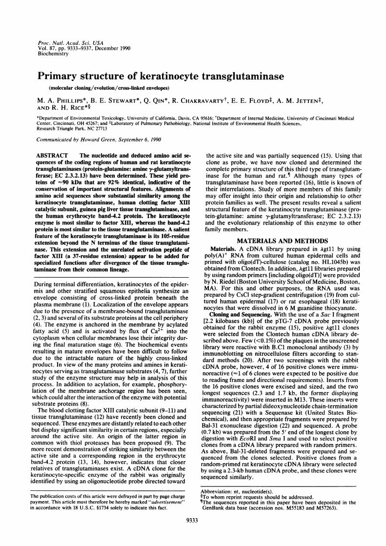
RESULTS AND DISCUSSIONThe nucleotide sequences obtained for the rat and humankeratinocyte transglutaminases were each represented in atleast two independent clones that were sequenced in oppositedirections. The subclones analyzed are presented in Fig. 1.The transglutaminase cDNA clones selected in these ex-
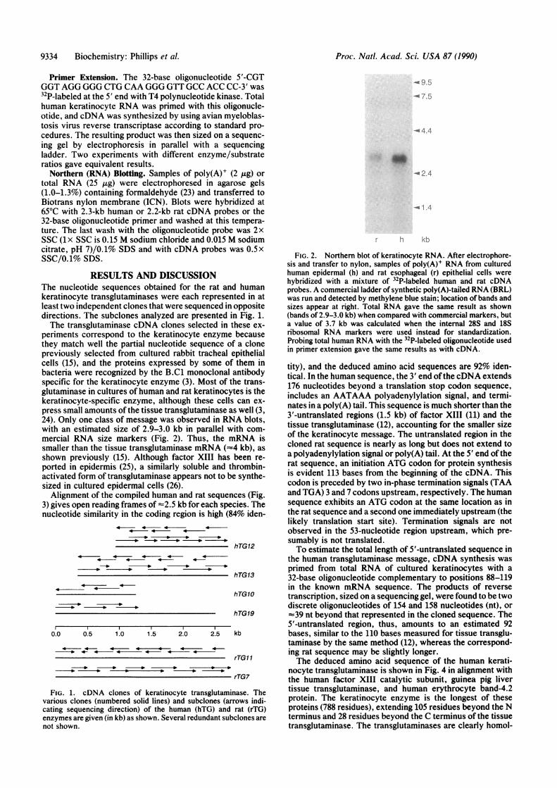
periments correspond to the keratinocyte enzyme becausethey match well the partial nucleotide sequence of a clonepreviously selected from cultured rabbit tracheal epithelialcells (15), and the proteins expressed by some of them inbacteria were recognized by the B.C1 monoclonal antibodyspecific for the keratinocyte enzyme (3). Most of the trans-glutaminase in cultures of human and rat keratinocytes is thekeratinocyte-specific enzyme, although these cells can ex-press small amounts of the tissue transglutaminase as well (3,24). Only one class of message was observed in RNA blots,with an estimated size of 2.9-3.0 kb in parallel with com-mercial RNA size markers (Fig. 2). Thus, the mRNA issmaller than the tissue transglutaminase mRNA (z4 kb), asshown previously (15). Although factor XIII has been re-ported in epidermis (25), a similarly soluble and thrombin-activated form of transglutaminase appears not to be synthe-sized in cultured epidermal cells (26).Alignment of the compiled human and rat sequences (Fig.
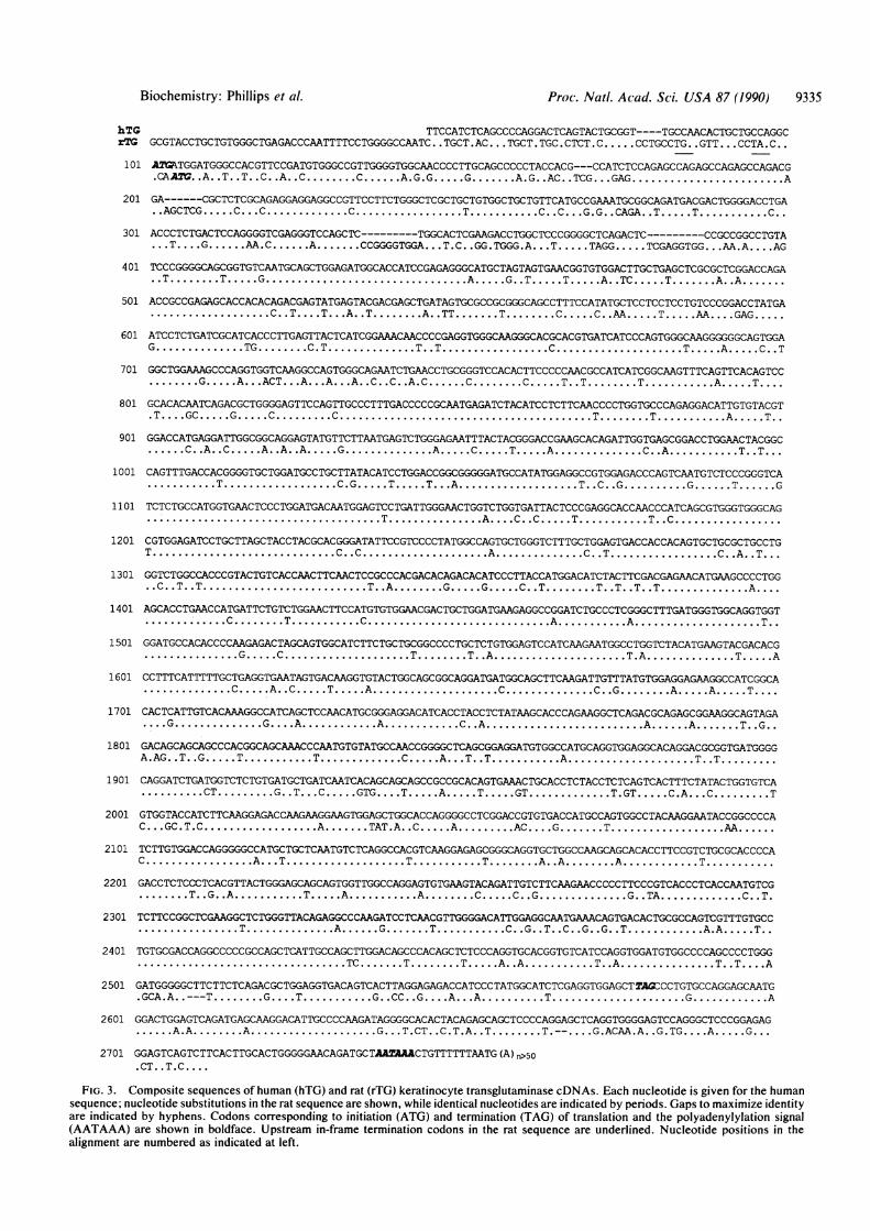
3) gives open reading frames of -2.5 kb for each species. Thenucleotide similarity in the coding region is high (84% iden-
4- 4- 4- 4
- * _ -* -*-
.4 --4 4- 4- 4
-so- -_ -*-
4 44
hTG12
hTG13
hTG10
hTG19
0.0 0.5 1.0 1.5 2.0 2.5 kb
4 4 4 44 4
rTGl 1
rTG7
FIG. 1. cDNA clones of keratinocyte transglutaminase. Thevarious clones (numbered solid lines) and subclones (arrows indi-cating sequencing direction) of the human (hTG) and rat (rTG)enzymes are given (in kb) as shown. Several redundant subclones arenot shown.
<9 5
-7.5
.44
4.2.4
~1 4
h kb
FIG. 2. Northern blot of keratinocyte RNA. After electrophore-sis and transfer to nylon, samples of poly(A)+ RNA from culturedhuman epidermal (h) and rat esophageal (r) epithelial cells werehybridized with a mixture of 32P-labeled human and rat cDNAprobes. A commercial ladder of synthetic poly(A)-tailed RNA (BRL)was run and detected by methylene blue stain; location of bands andsizes appear at right. Total RNA gave the same result as shown(bands of 2.9-3.0 kb) when compared with commercial markers, buta value of 3.7 kb was calculated when the internal 28S and 18Sribosomal RNA markers were used instead for standardization.Probing total human RNA with the 32P-labeled oligonucleotide usedin primer extension gave the same results as with cDNA.
tity), and the deduced amino acid sequences are 92% iden-tical. In the human sequence, the 3' end ofthe cDNA extends176 nucleotides beyond a translation stop codon sequence,includes an AATAAA polyadenylylation signal, and termi-nates in a poly(A) tail. This sequence is much shorter than the3'-untranslated regions (1.5 kb) of factor XIII (11) and thetissue transglutaminase (12), accounting for the smaller sizeof the keratinocyte message. The untranslated region in thecloned rat sequence is nearly as long but does not extend toa polyadenylylation signal or poly(A) tail. At the 5' end of therat sequence, an initiation ATG codon for protein synthesisis evident 113 bases from the beginning of the cDNA. Thiscodon is preceded by two in-phase termination signals (TAAand TGA) 3 and 7 codons upstream, respectively. The humansequence exhibits an ATG codon at the same location as inthe rat sequence and a second one immediately upstream (thelikely translation start site). Termination signals are notobserved in the 53-nucleotide region upstream, which pre-sumably is not translated.To estimate the total length of 5'-untranslated sequence in
the human transglutaminase message, cDNA synthesis wasprimed from total RNA of cultured keratinocytes with a32-base oligonucleotide complementary to positions 88-119in the known mRNA sequence. The products of reversetranscription, sized on a sequencing gel, were found to be twodiscrete oligonucleotides of 154 and 158 nucleotides (nt), or:39 nt beyond that represented in the cloned sequence. The5'-untranslated region, thus, amounts to an estimated 92bases, similar to the 110 bases measured for tissue transglu-taminase by the same method (12), whereas the correspond-ing rat sequence may be slightly longer.The deduced amino acid sequence of the human kerati-
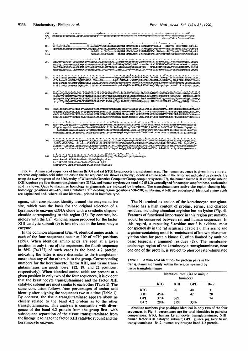
nocyte transglutaminase is shown in Fig. 4 in alignment withthe human factor XIII catalytic subunit, guinea pig livertissue transglutaminase, and human erythrocyte band-4.2protein. The keratinocyte enzyme is the longest of theseproteins (788 residues), extending 105 residues beyond the Nterminus and 28 residues beyond the C terminus of the tissuetransglutaminase. The transglutaminases are clearly homol-
9334 Biochemistry: Phillips et al.
Biochemistry: Phillips et al. Proc. Natl. Acad. Sci. USA 87 (1990) 9335
hTG TTCCATCTCAGCCCCAGGACTCAGTACTGCGGT----TGCCAACACTGCTGCCAGGCrTG GCGTACCTGCTGTGGGCTGAGACCCAATTTTCCTGGGGCCAATC. .TGCT .AC... TGCT. TGC .CTCT. C....CCTGCCTG. .GTT... CCTA. C..
.CAA2G..A..T..T..C..A..C.....C....A.G.G...G.....A.G..AC..TCG...GAG.............A
201 GA----CGCTCTCGCAGAGGAGGAGGCCGTTCCTTCTGGGCTCGCTGCTGTGGCTGCTGTTCATGCCGAAATGGGGCAGATGACGACTGGGGACCTGA..AGCTCG....C...C........C..........T.......C..C...G.G..CAGA..T....T.......C..
301 ACCCTCTGACTCCAGGGGTCGAGGGTCCAGCTC------TGGCACTCGAAGACCTGGCTCCCGGGGCTCAGACTC------CCGCCGGCCTGTA...T.....G. AA...C....A.....CCGGGGTGGA... .T.C. .G.TGGA... T.....T.TAGG...TCGAGGTGG...A.A... ..AG
501 ACCGCCGAGAGCACCACACAGACGAGTATGAGTACGACGAGCTGATAGTGCGCCGCGGGCAGCCTTTCCATATGCTCCTCCTCCTGTCCCGGACCTATGA...........C. .T. ...T. ..A.A.T.....A..TT.....T.....C....C..AA....T....AA. ... .GAG...
G........TG.....C.T.T..... .T.T..........C...........T....A.C..C.T
....G....A....ACT. . ..A.. A....A..C. .C. .A.C....C.....C.T..T.T.....T.......A....T...
.T. .....GC...G....C......C.....................T.....T.......A....T..
....C..A..C....A..A..A...G........A....C ...T....A........C..A.......T..T...
......T..........C.G....T....T....A...........T..C. .G......G....T....G
1101 TCTCTGCCATGGTGAACTCCCTGGATGACAATGGAGTCCTGATTGGGAACTGGTCTGGTGATTACTCCCGAGGCACCAACCCATCAGCGTGGGTGGGCAG
T.C.............C.C...........A.C.......C.T..........C. .A. .T...
..C..T..T..............T..A.....G...G....C..T.....T..T..T..T........A....
1401 AGCACCTGAACCATGATTCTGTCTGGAACTTCCATGTGTGGAACGACTGCTGGATGAAGAGGCCGGATCTGCCCTCGGGCTTTGATGGGTGGCAGGTGGT. ~~~C.....T.......C................A.......A...........T..
1501 GGATGCCACACCCCAAGAGACTAGCAGTGGCATCTTCTGCTGCGGCCCCTGCTCTGTGGAGTCCATCAAGAATGGCCTGGTCTACATGAAGTACGACACG........G...C...........T.....T..A............T.A........T....A
.......C....A..C....T....A...........C........C..G.....A....A....T....
1701 CACTCATTGTCACAAAGGCCATCAGCTCCAACATGCGGGAGGACATCACCTACCTCTATAAGCACCCAGAAGGCTCAGACGCAGAGCGGAAGGAGTAGA..G........G....A.......A.......C..A..............A....A.....T..G..
A.AG. .T. .G....T.......T........C....A....T..T.......A.T..........T.T.....1901 CAGGATCTGATGGTCTCTGTGATGCTGATCAATCACAGCAGCAGCCGCCGCACAGTGAAACTGCACCTCTACCTCTCAGTCACTTTCTATACTGGTGTCA
......CT......G..T...C....GTG....T....A....T....GT........T.GT....C.A...C......T
2001 GTGGTACCATCTTCAAGGAGACCAAGAAGGAAGTGGAGCTGGCACCAGGGGCCTCGGACCGTGTGACCATGCCAGTGGCCTACAAGGAATACCGGCCCCAC... .GC.T.C..........A.....TAT.A. .C....A......AC.....G.....T..........AA....
C........ .A...T...........T.......T.....A..A.....A.......T......
.....T..G..A.......T....A.......A.....C....C..G........G..TA........C..T.
....... .T........A....G.....T.......C..G..T..C..G..G..T.......A.A....T..
..................TC.....T.....T.A..A.A.T.....T.A.T.......T.T ... A
.GCA.A. .---T.G....G ... T.......G. .CC..G. .... A....A......T...........G.......A
....A.A.....A............G ...T.CT. .C.T.A. .T.....T.-......G.ACAA.A. .G..... A.....A.G...
2701 GGAGTCAGTCTTCACTTGCACTGGGGGAACAGATGCTAA2'AAACTGTTTTTTAATG (A >5o
FIG. 3. Composite sequences of human (hTG) and rat (rTG) keratinocyte transglutaminase cDNAs. Each nucleotide is given for the humansequence; nucleotide substitutions in the rat sequence are shown, while identical nucleotides are indicated by periods. Gaps to maximize identityare indicated by hyphens. Codons corresponding to initiation (ATG) and termination (TAG) of translation and the polyadenylylation signal(AATAAA) are shown in boldface. Upstream in-frame termination codons in the rat sequence are underlined. Nucleotide positions in thealignment are numbered as indicated at left.
9336 Biochemistry: Phillips et al.
rTG -.e rs.w.......... epdrss. g.r g. s .t..rgg.s.ggd.. r..rGG...
hTG mdgprsdvgrwggnplqppttpspepepepd---grsrrgggrsfwarscgccscrnaadddwgpepsdsrgrgssS--gTrRpgsrgsds---RRpxiii mS---eTsRtaf------GGRRa
e..L............. c....--n. e ...........-.A
101 VsrgsgvnAagD---------GtireLvVngVDLlssRsDqlirREE HTDeYEyde LIVRRGQPFhmlLl-SRtYEs-sDriTLE11I GnnPeVppnnsnaAedDlptvelqgvvprGvnlqEfLnVtSVhLfkeRwDtNkvD&I HTDkYEnnk LIVRlGQsFyvqidF-SRpYdPrrDlfrvEyVI GryPq
GPL MaEdilerCDLQ--levNgRDE rTadLcreRLvLERtGQPFwltLhFeg--RgYEagvDtlTfnAVTGpdPSB4 .2 MgqaLgikSCDfQaaR--NneER HTkaLssrRLfVRRGQP~tiiLyFrapvRaf lPalkkvALtAqT GeqPS
201. -t.tn.h..T. ......r.e.
vgKGmviiPvgk-GgSGgWkAqWkasgonlnLrVhTSPnAIIGKFqftVrtqsdaGefQLpfdPrneiYI LFNP WCPEDi VYvDhEdwRQEYVLNesGEnKGTyipvPIvSelqSGkWgAkiVmReDrSVrLsiqsSPkcIvGRF~mnyVaVwTpYGvlrtsrnPetdtYI LFP WCedDAVYLDNEkEReEYVLNdiG
kinrTqAtFPISS1GdrkwW.AvVeeRdaQSwTiSVtTPADAvIGhYsLlLqVS--GrkQLlLG---qFtLLFNP WnrEDAVfLkNEaqRmElfY1 LNQnG
.... ............................... . ........... S V ................. T ..... ..----.......... ....
301 rIYYGTeaqIgetWNYGQdhGVLDaaCYiLDRr-------GQpygGRGVn VSRViSAMVNsLDDnGYLiGnWsgdYSr GtnPSAWvGSVe ILLsYLvIfYGevndlktRSWsYGQVEDGILDtCLYvmDR-----AqMD1SGRlnhik VSRVgSAMVNakDDeGVLvGSWDNiYay GVpP SA8t GSVD ILLeY-fIYqGsAkfIngipMWNFGEDGILD iC~aIDtnpkf1knAGqDcSrRSrPVy VgRVVSAMVNcnDDQGVLqGrWDNnYSd GVsPmsWi GSVD ILRrWkIZYl GTAdcIqaeSWdFGQlEgdViD lsLrLLsk----dkqvekwSqWVh VaRVlgAllhfLkeQrVLptpqTqatqe Gallnkrr GSVp ILRqwL.............. ...... ............. ......................... ............. ........ ...... ....................... ................... .. ............... ........
401 RtGy-sVpYGQCWVFAGVttTVLRCLGlaTR tVNFNS&HDTDtsLtJIYFdErnnkplEhLnhDSWNFHvWNdCWMKRPDLPsGF-DGWQvVDaTPQERssenPVrYGQCWrFAGVfnTfLRLGIPAR iVTNyfSHDnDaNLqDIfleEdGnvNskLtkDSVWNyHcWNEaWttRPDLPvGF-gGWQAVDsTPQEdyGcqrVkYGWCWVFAAVACTVLGIPTRVVTNFNSAHDqnsNLLI-EYFrnEsgeiEgnksemIWNFHsllggvvddqagPgawvrGvQALDPTPQEtgrgrPVydGQaWVlAWVACTVLRCLGIPARVVTtFaS qgTggrLLIDEYynEEGlqNgegqrgrIIiFqtstECW1KR-gLPcqgyDGWQ iLhPsapn.n . Hi~k.............................................................. .....
501 tSsGifCCGPcSVesIFnG1VymKYDtPFiFEVNSDkVYWqRQdDGSfkivyVeeKaI GtLIVTKaIsSnmREDITylYKhpEGSdaERkAvETAaahGnSd~mYrCGPaSVqAIllhG HVcfqfDAPFVEAEVNSDliYitakKDOlThvvenVdathI GkLIVTqIGgDgmmDITdTYltfqXG:*EERLATTAlmyGkSeGtYCCGPVPVRAIKEGHLnvKYDAPFVFAEVNADvVnWiRQKDGSLrks-iNhlvVGlkI STKsVGrDeREDITHTYKYPEGSEERREAfvRAn---gggvlgsCdlVPVRAvKEGtLgltpavsdlPlaiNAscVvWkcceDGTIeltdsNtKyVGnn I STKgVGSDrcEDITqnYKYPEGSlqEkEvLERvekek
.. ..... ..........t ... v.T.rg............ c..y . ... ..Pt ..... -...v .......t.A ........ k.601 sKp-NvyAnrgSAEd--VAMqvE-aQdAVMGqDlmvSVmLiUAHSssRrTVkLhLylsvT YTGVsGtiFKetK-keVeLaPgasdrvmpvaYkEYrp
aKkpLNtegvmkSr--SnVdcdfE-veNAVLGkDFklSiTfrNnShnRyTitayLsAnITF YTGVpkAeFKketf-dVTLePlSfKkeAvlIqagEYmg---hLNklATkeeAqeetgVAMrirvgQNmtMGsDFdIfayiTlgtaeshecQLlLcArIVs YNGVLGPvcstndLlnLTLdPfSEnsIplhIlYekYgdmerekdngirppSlEtaSplylllkapsslpLrgDaqISVTLvNHSeqekaVOLaigvqaVh YNGVLaAklwrkKL-hLTLsanlEKiITiglffsnfer
..................... ................................. .. .. . i ............. . . -.v .. ..........
701 hLvdQgam~LjnVsGhvkESgq--VLRK~htfrLrt PdLsltlLGaAVVG~eceVqIvFKOSIFPVTLtNVVFrLE GsGLqRP-KilnVgD-IggNETVt1qLlEQaslhffVTArinEtrd--VLAK(kstvLti PEIiIKVrGtqVW~sdmTVtIqFtllL keTLrNVwvhLdGFpGvTpm-FRe-IrpNsTvqwyLtEsn--LikVrGlliEpaaNsyVIAerdiy-LenPEIkIrVLGepk~1 rkLiAeVSLKNPLPVPLIgCiFtvEGaGLTkdqKSveVpDpveagEqakvnppEnt--fLrlTAmathSesNlscfA-QediaicrPhLaIKmpekAeQypLTASVSLqNSLdaPmedCVisi1 GrGLihrerSyrFRsvwpeN-Tmca
.. . ss.gr.- ..e.v rs. n... f....801 RqsFvPvrpG pRqLIASlDSpqLsqVhGviqVdVaPAPgdggffsdaggdshlgetipmasrgga
eevcrPwvsG hMLIASmsSDsLrhVyGeldVqIqrrPsmRvd11PTeVG ThKLvVnfeCDkLkaVkGYrnViIgPAkfqFtPThVG tqrLtVevDCnmfqnltnYksVtVVapelsa
FIG. 4. Amino acid sequences of human (hTG) and rat (rTG) keratinocyte transglutaminases. The human sequence is given in its entirety,whereas only amino acid substitutions in the rat sequence are shown explicitly; identical amino acids in the latter are indicated by periods. Byusing the GAP program of the University of Wisconsin Genetics Computer Group computer system (27), the human factor XIII catalytic subunit(XIII), guinea pig liver tissue transglutaminase (GPL), and human erythrocyte band-4.2 (B4.2) were aligned for comparison; for these, each aminoacid is -shown. Gaps to maximize homology in alignments are indicated by hyphens. The transglutaminase active-site region showing highhomology (positions 410-427) and a putative Ca2+-binding region (positions 568-578; numbering at left) are underlined. Identical amino acidsare capitalized and, where all are identical, printed in boldface type.
ogous, with conspicuous identity around the enzyme activesite, which was the basis for the original selection of a
keratinocyte enzyme cDNA clone with a synthetic oligonu-cleotide corresponding to this region (15). By contrast, ho-mology with the Ca2+-binding region proposed for the factorXIII catalytic subunit (9) is less obvious in the keratinocyteenzyme.
In the common alignment (Fig. 4), identical amino acids ineach of the four sequences occur at 109 of '710 positions(15%). When identical amino acids are seen at a givenposition in only three of the sequences, the fourth sequencein 58% (74/127) of such cases is the band 4.2 protein,indicating the latter is more dissimilar to the transglutami-nases than any of the others is to the group. Correspondingnumbers for the keratinocyte, factor XIII, and tissue trans-glutaminases are much lower (12, 19, and 22 positions,respectively). When identical amino acids are present at a
given position in only two of the four sequences, it is evidentthat the keratinocyte transglutaminase and the factor XIIIcatalytic subunit are most similar to each other (Table 1). Thesame conclusion follows from percentages of amino acididentity after aligning the sequences two at a time (Table 1).
By contrast, the tissue transglutaminase appears about as
closely related to the band 4.2 protein as to the othertransglutaminases. This analysis is compatible with diver-gence of the band 4.2 protein from the group first, withsubsequent separation of the tissue transglutaminase fromthe lineage leading to the factor XIII catalytic subunit and thekeratinocyte enzyme.
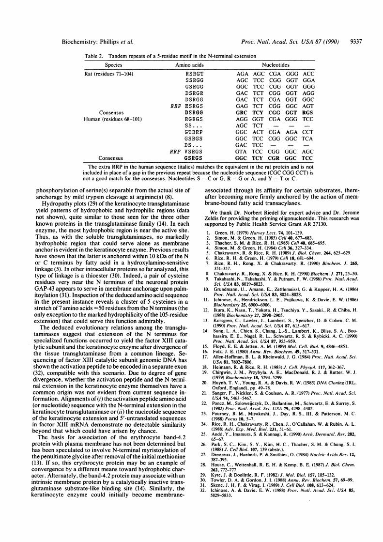
The N terminal extension of the keratinocyte transgluta-minase has a high content of proline, serine, and chargedresidues, including abundant arginine but no lysine (Fig. 4).Features of functional importance in this region presumablywould be conserved between rat and human sequences. Inthis regard, a repeating 5-residue motif is evident, mostconspicuously in the rat sequence (Table 2). This serine andarginine-containing motif is reminiscent of known phosphor-ylation sites for protein kinase C, often flanked by multiplebasic (especially arginine) residues (28). The membrane-anchorage region of the keratinocyte transglutaminase, nearone end of the protein, is subject to phorbol ester-stimulated
Table 1. Amino acid identities for protein pairs in thetransglutaminase family within the region spanned bytissue transglutaminase
Identities, total (%) or unique(number)
hTG XIII GPL B4.2
hTG 96 40 31Xlii 45% 42 28GPL 37% 36% 74B4.2 29% 25% 33%
Absolute numbers give positions identical in only two of the foursequences in Fig. 4; percentages are for total identities in pairwisecomparisons. hTG, human keratinocyte transglutaminase; XIII,human factor XIII catalytic subunit; GPL, guinea pig liver tissuetransglutaminase; B4.2, human erythrocyte band-4.2 protein.
Proc. Natl. Acad. Sci. USA 87 (1990)
Proc. Natl. Acad. Sci. USA 87 (1990) 9337
Table 2. Tandem repeats of a 5-residue motif in the N-terminal extensionSpecies
Rat (residues 71-104)
ConsensusHuman (residues 68-101)
Consensus
Amino acids
RSRGTSSRGGGSRGGDSRGRDSRGG
RRP ESRGSDSRGGRGRGSSs ....GTRRPGSRGSDS...
RRP VSRGSGSRGS
Nucleotides
AGAAGCGGCGACGACGAGGRCAGGAGCGGCGGCGACGTAGGC
AGC CGATCC CGGTCC CGGTCT CGGTCT CGATCT CUGTCY CGGGGT CGATCTACT CGATCC CGGTCCTCC CGGTCY CGR
GGGGGTGGTGGTGGTGGCGGTGGG
ACCGGAGGGAGGGGCAGTRGSTCC
AGA CCTGGC TCA
GGC AGCGGC TCC
The extra RRP in the human sequence (italics) matches the equivalent in the rat protein and is notincluded in place of a gap in the previous repeat because the nucleotide sequence (CGC CGG CCT) isnot a good match for the consensus. Nucleotides S = C or G, R = G or A, and Y = T or C.
phosphorylation of serine(s) separable from the actual site ofanchorage by mild trypsin cleavage at arginine(s) (8).Hydropathy plots (29) of the keratinocyte transglutaminase
yield patterns of hydrophobic and hydrophilic regions (datanot shown), quite similar to those seen for the three otherknown proteins in the transglutaminase family (14). In eachenzyme, the most hydrophobic region is near the active site.Thus, as with the soluble transglutaminases, no markedlyhydrophobic region that could serve alone as membraneanchor is evident in the keratinocyte enzyme. Previous resultshave shown that the latter is anchored within 10 kDa of the Nor C terminus by fatty acid in a hydroxylamine-sensitivelinkage (5). In other intracellular proteins so far analyzed, thistype of linkage is a thioester (30). Indeed, a pair of cysteineresidues very near the N terminus of the neuronal proteinGAP-43 appears to serve in membrane anchorage upon palm-itoylation (31). Inspection ofthe deduced amino acid sequencein the present instance reveals a cluster of 5 cysteines in astretch of7 amino acids z50 residues from the N terminus (theonly exception to the marked hydrophilicity ofthe 105-residueextension) that could serve this function admirably.The deduced evolutionary relations among the transglu-
taminases suggest that extension of the N terminus forspecialized functions occurred to yield the factor XIII cata-lytic subunit and the keratinocyte enzyme after divergence ofthe tissue transglutaminase from a common lineage. Se-quencing of factor XIII catalytic subunit genomic DNA hasshown the activation peptide to be encoded in a separate exon(32), compatible with this scenario. Due to degree of genedivergence, whether the activation peptide and the N-termi-nal extension in the keratinocyte enzyme themselves have acommon origin was not evident from current sequence in-formation. Alignments of(i) the activation peptide amino acid(or nucleotide) sequence with the N-terminal extension in thekeratinocyte transglutaminase or (ii) the nucleotide sequenceof the keratinocyte extension and 5'-untranslated sequencesin factor XIII mRNA demonstrate no detectable similaritybeyond that which could have arisen by chance.The basis for association of the erythrocyte band-4.2
protein with plasma membrane has not been determined buthas been speculated to involve N-terminal myristoylation ofthe penultimate glycine after removal ofthe initial methionine(13). If so, this erythrocyte protein may be an example ofconvergence by a different means toward hydrophobic char-acter. Alternately, the band-4.2 protein may associate with anintrinsic membrane protein by a catalytically inactive trans-glutaminase substrate-like binding site (14). Similarly, thekeratinocyte enzyme could initially become membrane-
associated through its affinity for certain substrates, there-after becoming more firmly anchored by the action of mem-brane-bound fatty acid transacylases.We thank Dr. Norbert Riedel for expert advice and Dr. Jerome
Zeldis for providing the priming oligonucleotide. This research wassupported by Public Health Service Grant AR 27130.1. Green, H. (1979) Harvey Lect. 74, 101-139.2. Simon, M. & Green, H. (1985) Cell 40, 677-683.3. Thacher, S. M. & Rice, R. H. (1985) Cell 40, 685-695.4. Simon, M. & Green, H. (1984) Cell 36, 327-334.5. Chakravarty, R. & Rice, R. H. (1989) J. Biol. Chem. 264, 625-629.6. Rice, R. H. & Green, H. (1979) Cell 18, 681-694.7. Rice, R. H., Rong, X. & Chakravarty, R. (1990) Biochem. J. 265,
351-357.8. Chakravarty, R., Rong, X. & Rice, R. H. (1990) Biochem. J. 271, 25-30.9. Takahashi. N., Takahashi, Y. & Putnam, F. W. (1986) Proc. Nail. ACad.
Sci. USA 83, 8019-8023.10. Grundmann, U., Amann, E., Zettlemeissl, G. & Kupper, H. A. (1986)
Proc. Nail. Acad. Sci. USA 83, 8024-8028.11. Ichinose, A., Hendrickson, L. E., Fujikawa, K. & Davie, E. W. (1986)
Biochemistry 25, 6900-6906.12. Ikura, K., Nasu, T., Yokota, H., Tsuchiya, Y., Sasaki., R. & Chiba. H.
(1988) Biochemistry 27, 2898-2905.13. Korsgren, C., Lawler, J., Lambert, S., Speicher, D. & Cohen, C. M.
(1990) Proc. Naitl. Acad. Sci. USA 87, 613-617.14. Sung, L. A., Chien, S., Chang, L.-S.. Lambert, K., Bliss, S. A., Bou-
hassira, E. E., Nagel, R. L., Schwartz, R. S. & Rybicki, A. C. (1990)Proc. Nail. Acad. Sci. USA 87, 955-959.
15. Floyd, E. E. & Jetten, A. M. (1989) Mol. Cell. Biol. 9, 4846-4851.16. Folk, 1. E. (1980) Annu. Rev. Biochem. 49, 517-531.17. Allen-Hoffman, B. L. & Rheinwald, J. G. (1984) Proc. Natl. Acad. Sci.
USA 81, 7802-7806.18. Heimann, R. & Rice, R. H. (1983) J. Cell. Physiol. 117, 362-367.19. Chirgwin, J. M., Przybyla, A. E., MacDonald, R. J. & Rutter, W. J.
(1979) Biochemistry 18, 5294-5299.20. Huynh, T. V., Young, R. A. & Davis, R. W. (1985) DNA Cloning (IRL,
Oxford, England), pp. 49-78.21. Sanger, F., Nicklen, S. & Coulson, A. R. (1977) Proc. Naitl. Acad. Sci.
USA 74, 5463-5467.22. Poncz, M., Soloweijczyk, D., Ballantine, M., Schwartz, E. & Surrey, S.
(1982) Proc. Nail. Acad. Sci. USA 79, 4298-4302.23. Fourney. R. M., Miyakoshi, J., Day, R. S., III, & Patterson, M. C.
(1988) Focus 10, 5-7.24. Rice, R. H., Chakravarty, R., Chen, J., O'Callahan, W. & Rubin, A. L.
(1988) Adv. Exp. Med. Biol. 231, 51-61.25. Ando, Y., Imamura. S. & Kannagi, R. (1990) Arch. Dermatol. Res. 282,
65-67.26. Park, S. C., Kim, S. Y., Kim, H. C., Thacher, S. M. & Chung, S. I.
(1988) J. Cell Biol. 107, 139 (abstr.).27. Devereux, J., Haeberli, P. & Smithies, 0. (1984) Nucleic Acids Res. 12,
387-395.28. House, C., Wettenhall, R. E. H. & Kemp, B. E. (1987) J. Biol. Chem.
262, 772-777.29. Kyte, J. & Doolittle, R. F. (1982) J. Mol. Biol. 157, 105-132.30. Towler, D. A. & Gordon, J. I. (1988) Annu. Rev. Biochem. 57, 69-99.31. Skene, J. H. P. & Virag, I. (1989) J. Cell Biol. 108, 613-624.32. Ichinose, A. & Davie, E. W. (1988) Proc. Nald. Acad. Sci. USA 85,
5829-5833.
Biochemistry: Phillips et al.