Embed Size (px)
Citation preview
Principles of OrthopedicsINVESTIGATIONS
Dr. Mohammed M. ZamzamAssociate Professor & Consultant
Pediatric Orthopedic Surgeon
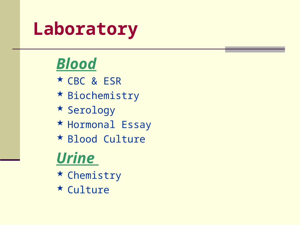
Laboratory
Blood CBC & ESR Biochemistry Serology Hormonal Essay Blood Culture
Urine Chemistry Culture
Laboratory
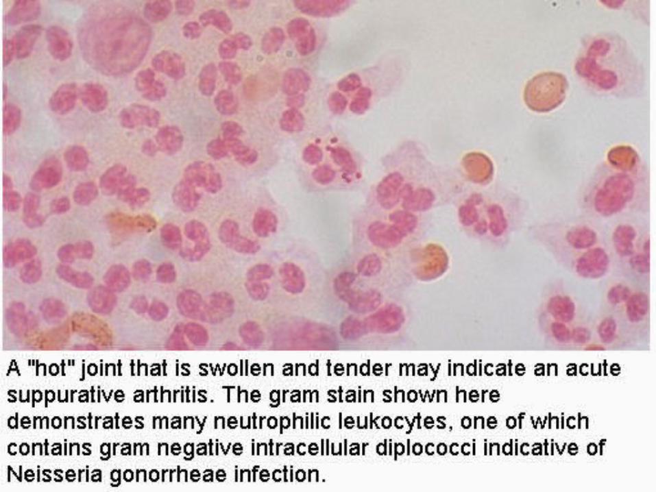
Joint Aspiration The procedure is often of a great help in
diagnosis of many effusions producing arthropathies
Diagnose or exclude septic arthritis Used routinely in haemoarthrosis A large-bore needle is used if the fluid is too
viscous The aspirate undergoes microscopic, chemical,
and gram stain examinations and culture & sensitivity test
Electro-diagnosis
Detect de-enervation and its degree Observe re-enervation before clinical signs Assess the progress of a lesion Localize the lesion
Electro-diagnosis
Elctromyography Poliomyelitis Muscle dystrophies Myelopathic diseases
It can provide useful information alongside clinical
examination and muscle biopsy
Electro-diagnosis
Nerve Conduction Study Acute compression neuropathy Chronic compression or entrapment neuropathy Peripheral neuropathies Traumatic nerve lesions
It is useful in differentiating between diseases of theaxons and of the anterior horn cells. It determines the level of interruption of a peripheral nerve lesion.
Histopathology
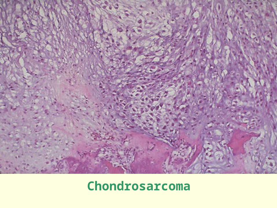
Bone Tumors Chronic Osteomyelitis Cystic lesions Osteoporosis
Chondrosarcoma
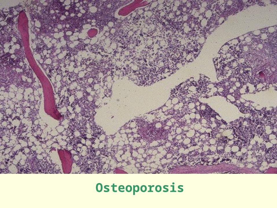
Osteoporosis
Histopathology
Soft Tissues Tumors Chronic arthritis (synovium) Myopathies (muscles) Neuropathies (nerve)
Histopathology
Needle aspiration Endoscopic biopsy Open biopsy Frozen section Excisional biopsy
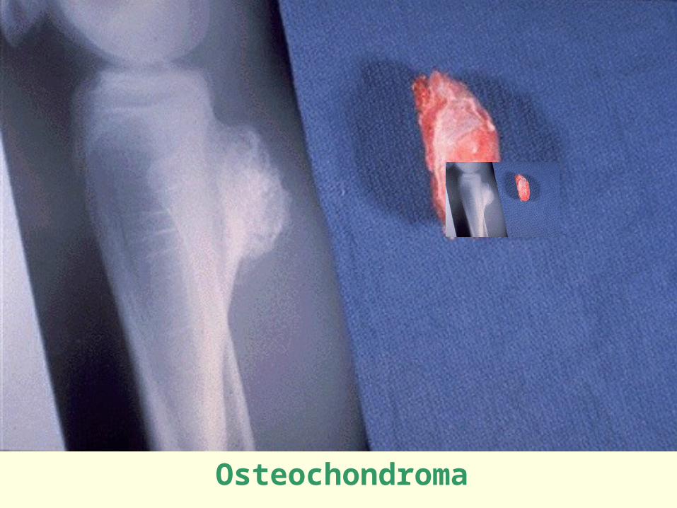
Osteochondroma
Arthroscopy
Internal visual examination of joints Confirm clinical and radiological
diagnosis Extension of the surgeon’s hand Therapeutic procedures Easy record of intra-articular lesions
Arthroscopy
Knee Loose bodies Synovial membrane Articular surfaces Medial & lateral menisci Cruciate ligaments Infrapatellar fat pad
Arthroscopy
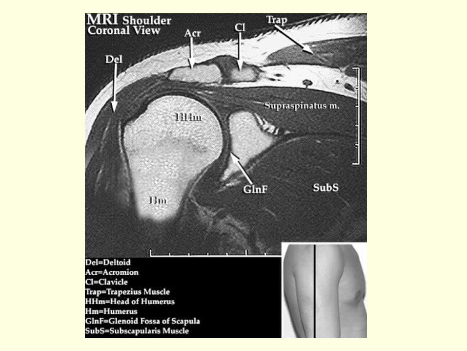
Shoulder Biceps tendon Rotator cuff Articular surfaces Glenoid labrum Capsular ligaments
Arthroscopy (rare sites)
Elbow Osteochondritis Dissecans Synovial biopsy Loose bodies
Hip Unexplained pain Loose bodies Synovial biopsy CDH
Arthroscopy (rare sites)
Ankle Difficult due to tight structures Osteochondral lesions
Wrist Still being developed Wrist pain syndrome
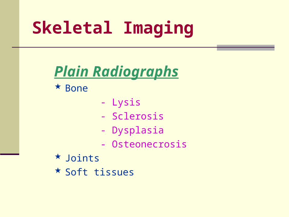
Skeletal Imaging
Plain Radiographs Bone
- Lysis
- Sclerosis
- Dysplasia
- Osteonecrosis Joints Soft tissues
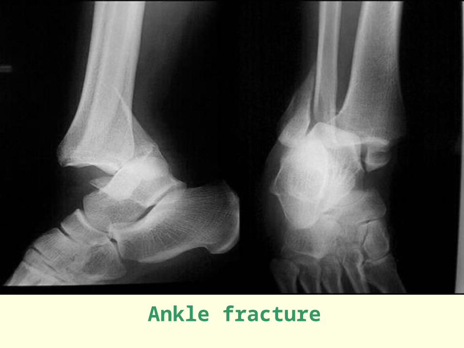
Ankle fracture
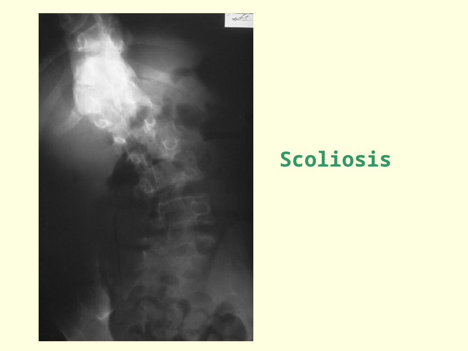
Scoliosis
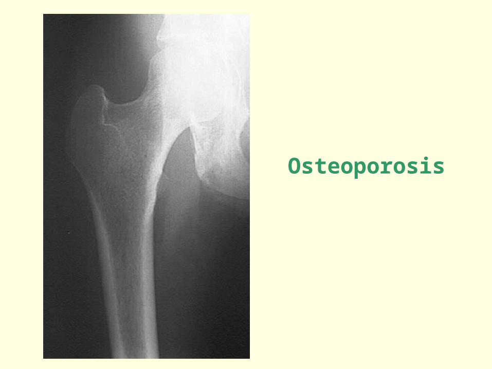
Osteoporosis
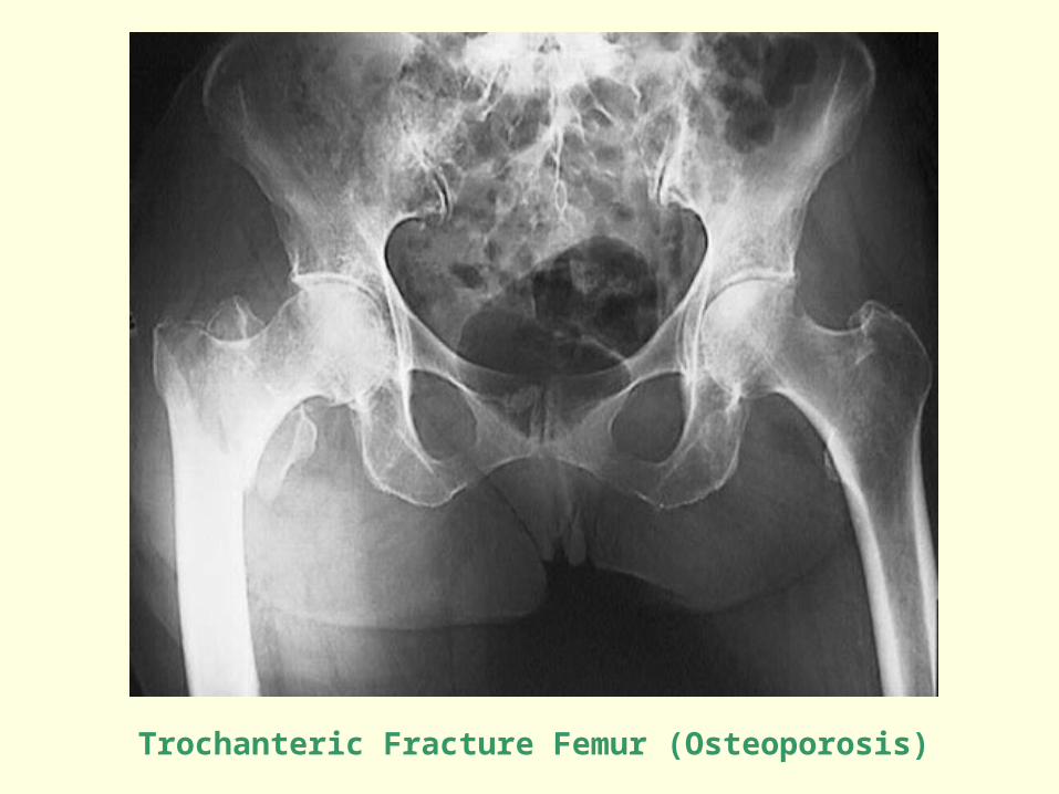
Trochanteric Fracture Femur (Osteoporosis)
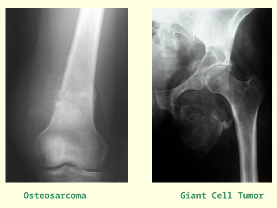
Osteosarcoma Giant Cell Tumor
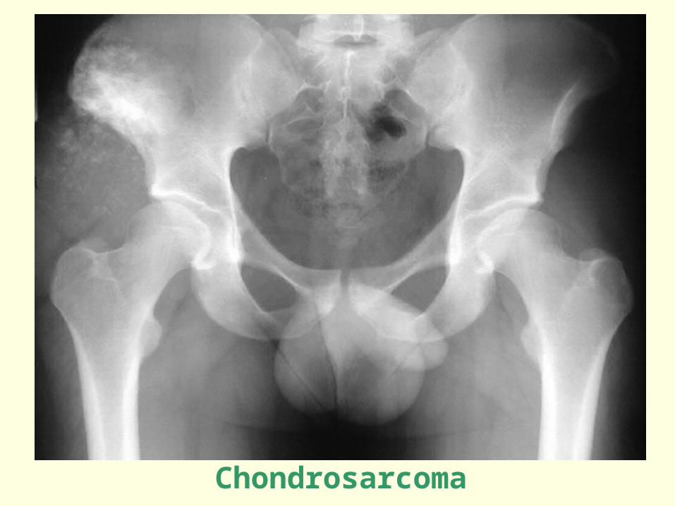
Chondrosarcoma
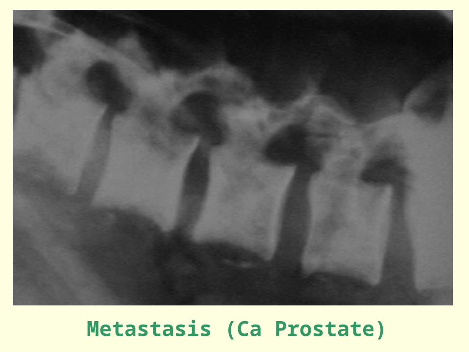
Metastasis (Ca Prostate)
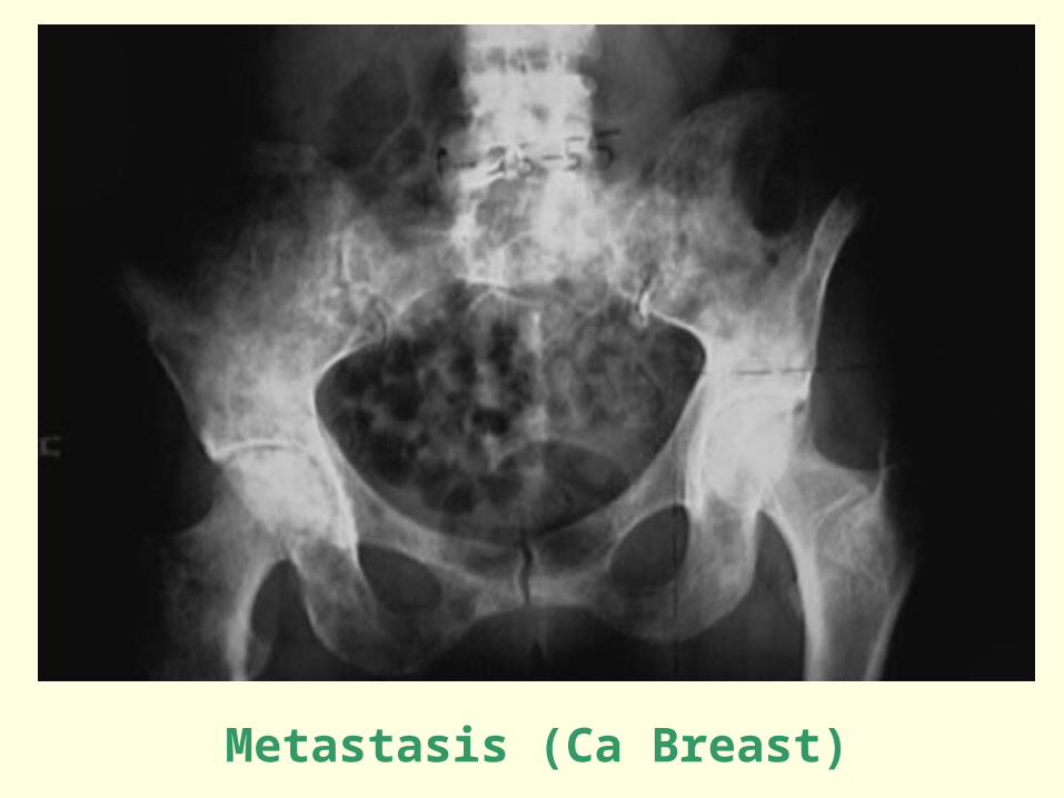
Metastasis (Ca Breast)
Skeletal Imaging
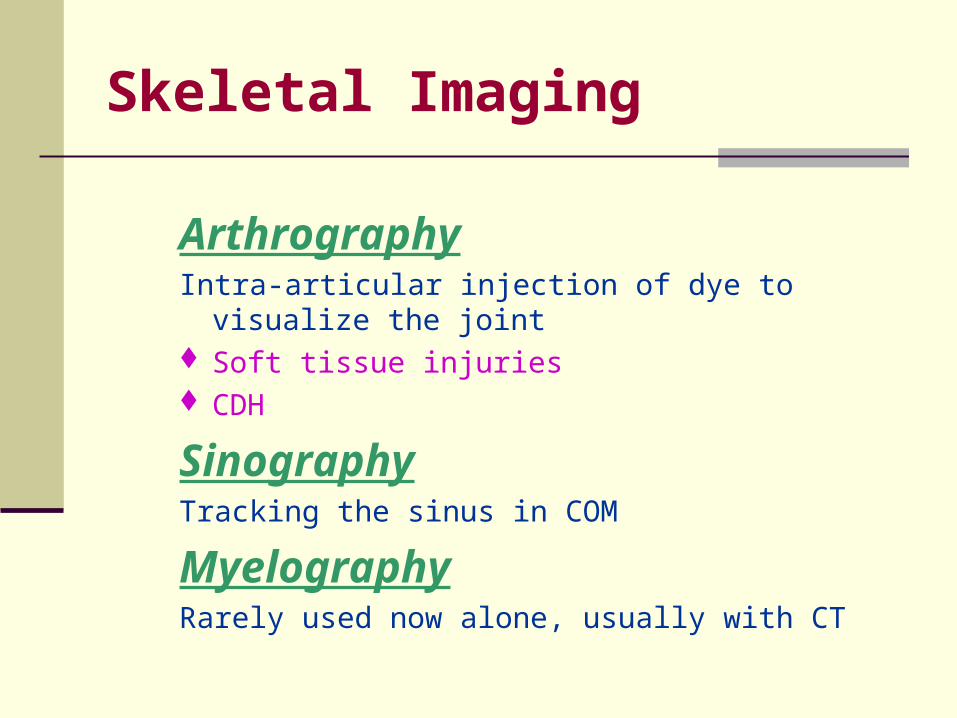
ArthrographyIntra-articular injection of dye to visualize the joint Soft tissue injuries CDH
SinographyTracking the sinus in COM
MyelographyRarely used now alone, usually with CT
Skeletal Imaging
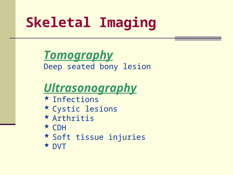
TomographyDeep seated bony lesion
Ultrasonography Infections Cystic lesions Arthritis CDH Soft tissue injuries DVT
Skeletal Imaging
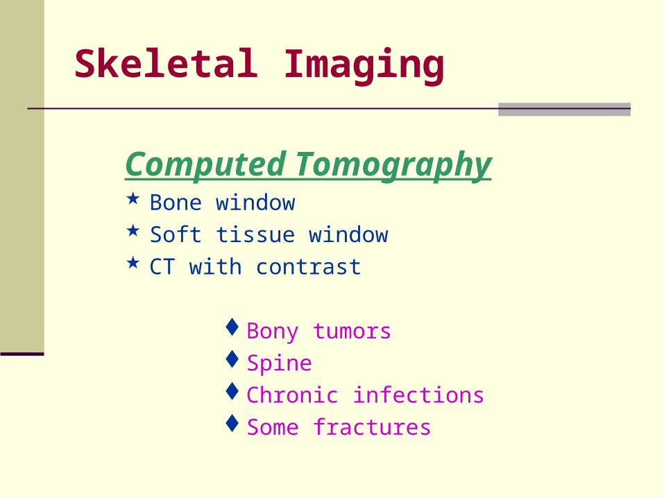
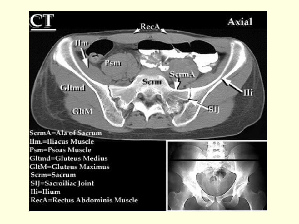
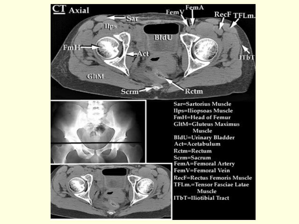
Computed Tomography Bone window Soft tissue window CT with contrast
Bony tumorsSpineChronic infectionsSome fractures
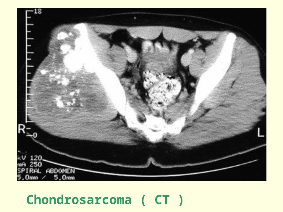
Chondrosarcoma ( CT )
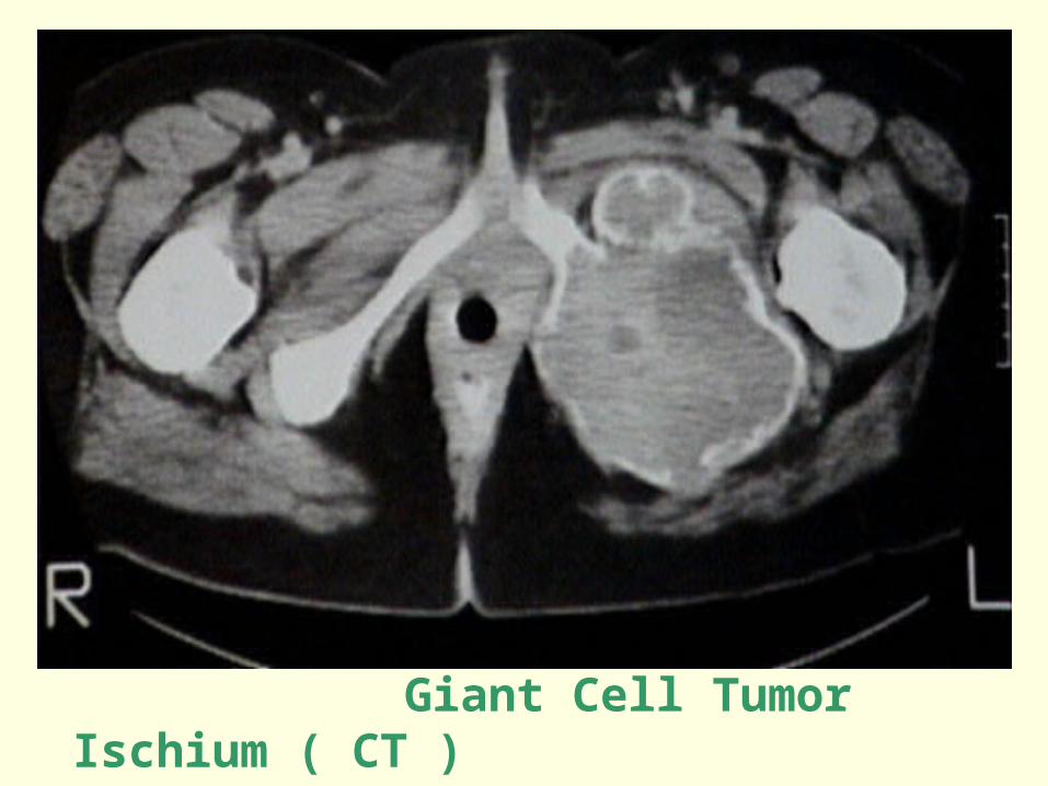
Giant Cell Tumor Ischium ( CT )
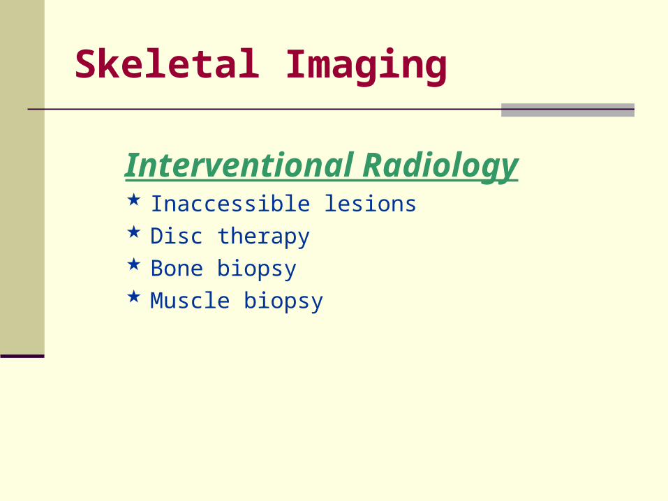
Skeletal Imaging
Interventional Radiology Inaccessible lesions Disc therapy Bone biopsy Muscle biopsy
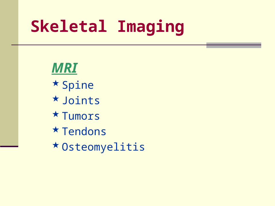
Skeletal Imaging
MRI Spine Joints Tumors Tendons Osteomyelitis
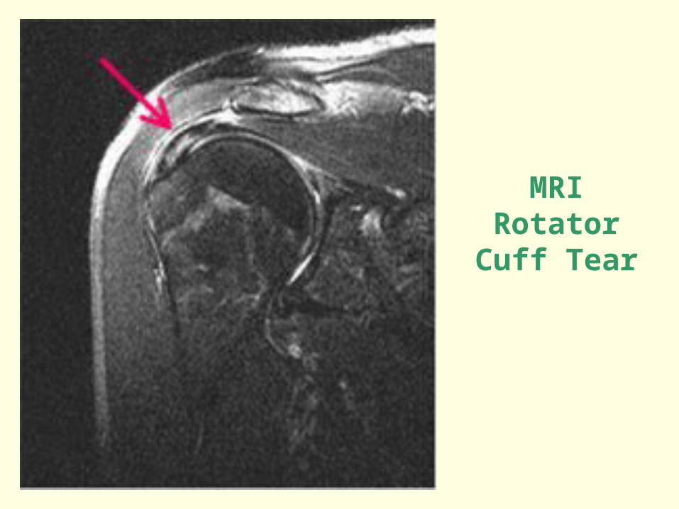
MRIRotator Cuff
Tear
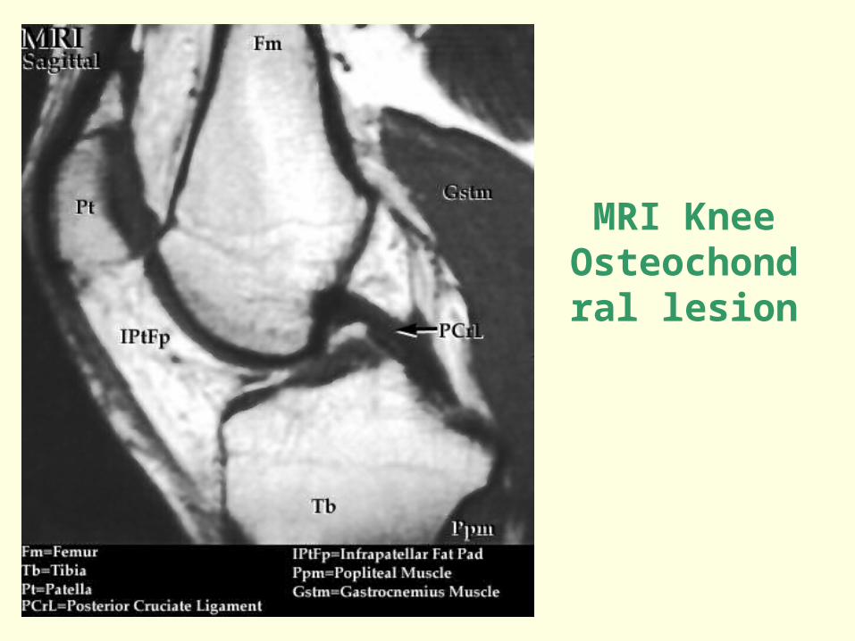
MRI KneeOsteochondral
lesion
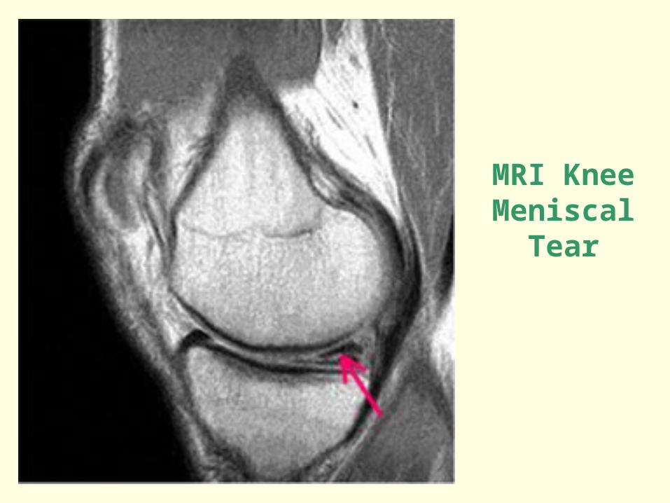
MRI KneeMeniscal Tear
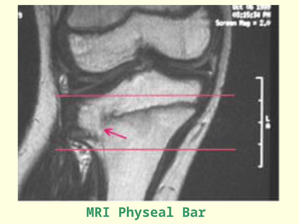
MRI Physeal Bar
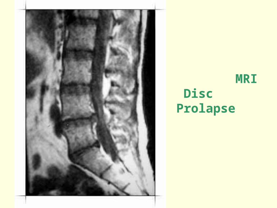
MRI Disc Prolapse
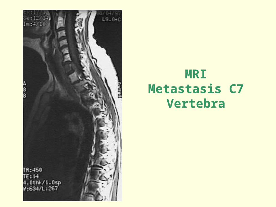
MRIMetastasis C7 Vertebra
Skeletal Imaging
Nuclear Medicine Bone and joint infections Tumors Avascular necrosis Osteoporosis
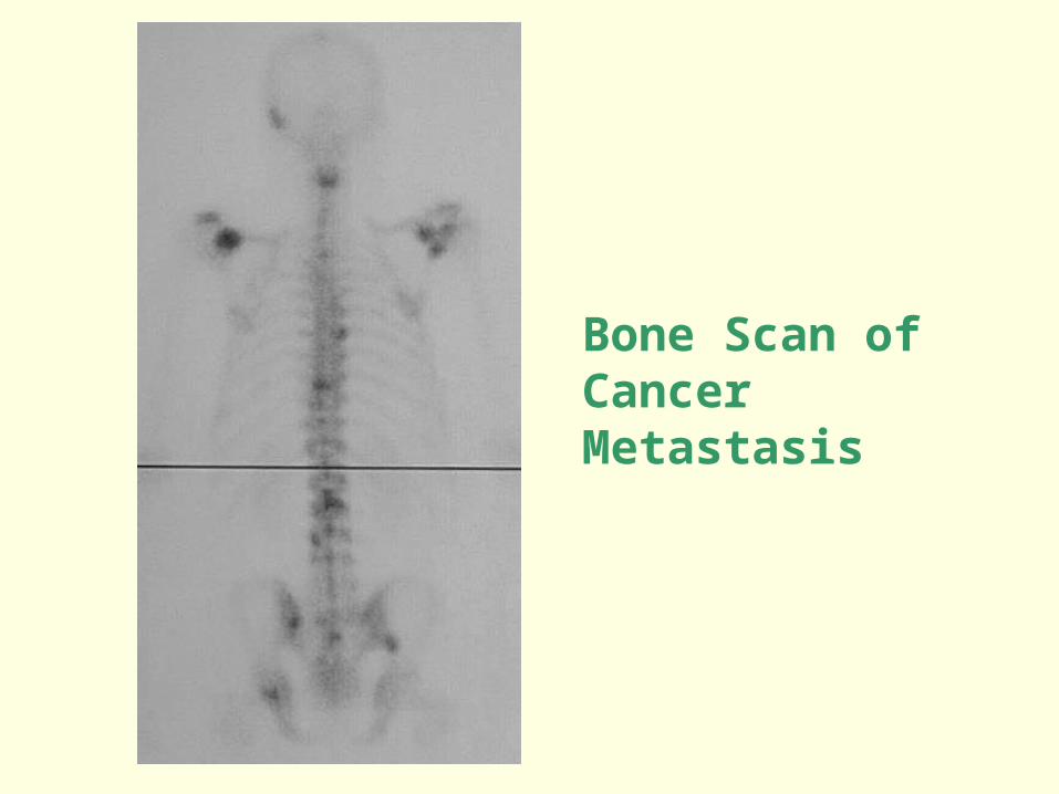
Bone Scan of Cancer Metastasis