Embed Size (px)
Citation preview
Do
Principles ofPharmacokinetics in the
Pregnant Woman and FetusRobert M. Ward, MDa,*, Michael W. Varner, MDb
KEYWORDS
� Pharmacokinetics � Perinatal pharmacology � Fetal pharmacology� Developmental pharmacology � Maternal/fetal drug transfer
KEY POINTS
� Pharmacokinetics of the maternal/placenta/fetal unit change dramatically duringpregnancy.
� Drug metabolism as well as drug transporters can alter the amount of drug reaching thefetus.
� Differences in maternal and fetal pharmacokinetics change the fetal/maternal drug con-centrations at birth.
� Many aspects of maternal and fetal drug therapy need additional study.
INTRODUCTION
In 1962, prenatal exposure to thalidomide was identified as the cause of a severecongenital multiple malformation syndrome.1–4 This was the first time that exposureto a maternal medication had been shown to directly injure the fetus. Since thattime, every effort has been made to avoid exposure of the fetus to medications, espe-cially in the first trimester during the critical period of organogenesis. Despite these ef-forts, there are occasions when fetal exposure to medications may be necessary inorder to maintain maternal health, creating a challenging risk-benefit continuum. Forexample, the anticonvulsant phenytoin is a known teratogen responsible for a clusterof structural anomalies collectively known as the fetal hydantoin syndrome.5,6 Fetalpharmacology studies subsequently confirmed that phenytoin is able to cross theplacenta to reach the fetus during the period of organogenesis, suggesting a direct
Disclosure Statement: No authors have financial interest in any products mentioned in thisarticle or with a maker of a competing product.a Pediatrics, Pediatric Clinical Pharmacology, University of Utah, University of Utah School ofMedicine, 295 Chipeta Way, Salt Lake City, UT 84108, USA; b Department of Obstetrics andGynecology, University of Utah, 30 North 1900 East, Room 2B 200, Salt Lake City, UT 84132, USA* Corresponding author.E-mail address: [email protected]
Clin Perinatol 46 (2019) 383–398https://doi.org/10.1016/j.clp.2019.02.014 perinatology.theclinics.com0095-5108/19/ª 2019 Elsevier Inc. All rights reserved.
wnloaded for Betty Burns ([email protected]) at National Certification Corporation from ClinicalKey.com by Elsevier on June 26, 2019. For personal use only. No other uses without permission. Copyright ©2019. Elsevier Inc. All rights reserved.
Ward & Varner384
Download
effect on fetal development.7–9 And yet most prenatal exposures do not result in fetalinjury. The sporadic occurrence of structural malformations associated with prenatalphenytoin exposure suggested for the first time that congenital malformation syn-dromes are more complex than simple in utero exposure. It is now clear that geneticfactors are involved; specifically, in this instance, the inheritance of a genetic variant ofepoxide hydrolase, the enzyme responsible for phenytoin metabolism, which causesthe activity of the enzyme to be reduced to less than 30% of normal.10 Not only canprenatal exposure to phenytoin (and other medications) result in structural malforma-tions, but also in neurodevelopmental delay that may only become apparent laterin life.9
Advances in perinatal diagnostics and fetal ultrasound during the 1980s allowed forthe prenatal identification of conditions previously only recognized after birth. Thisincluded not only structural malformations, but also functional disorders such as sup-raventricular tachycardia and androgenization of a female fetus because of congenitaladrenal hyperplasia. Some of these disorders can be prevented and/or treated byachieving an adequate concentration of a given drug within the fetus. As such, delib-erate exposure of the fetus to medications can also have a therapeutic objective.Our understanding of perinatal physiology has increased significantly starting in the
1990s and perinatologists have become more accepting of clinical interventions.11
There are now numerous examples of the use of maternal drug administration to treatboth fetal-related and pregnancy-related disorders, some of which are listed below.
i. Supraventricular tachycardia and other fetal dysrhythmias can be recognized andprecisely diagnosed using fetal cardiac ultrasound and can be treated with a vari-ety of anti-arrhythmic medications, usually administered to the mother but some-times administered directly to the fetus via intra-amniotic injection.12,13
ii. Women pregnant with a female fetus after having delivered a child with androge-nizing congenital adrenal hyperplasia can be treated with maternal corticosteroidsto suppress the fetal adrenal glands, thereby reducing the production of endoge-nous fetal androgens and preventing masculinization of the external genitalia.14 Inthis way, fetal treatment can either prevent the congenital syndrome altogether orat least minimize the need for extensive postnatal surgical procedures.
iii. Recent studies suggest that some cases of recurrent spontaneous preterm birth(SPTB) can be prevented with progesterone supplementation during pregnancy.15
iv. Judicious use of maternal halogenated corticosteroids can be used to reduce mor-bidities associated with prematurity, such as respiratory distress syndrome (RDS),intraventricular hemorrhage (IVH), and necrotizing enterocolitis (NEC).16
v. Bacterial infections, such as early-onset neonatal group B streptococcal infection,and vertical transmission of HIV have both been dramatically reduced through in-trapartum administration of antimicrobial medications.17
Taken together, these clinical situations created a role for drug therapy to directlybenefit the fetus and a need to better understand the pharmacokinetic changes inboth the mother and the fetus during pregnancy. Over the past few decades, the fieldof perinatal pharmacology has evolved into a clinically important discipline. It hasincreased our understanding of the dynamic changes in physiology that occur duringpregnancy and their effect on drug transport, metabolism, and degradation in themother, placenta, and fetus.18 Although the underlying physiologic changes thataccompany pregnancy remain much the same as that outlined by Mirkin in 1973,19
much has been learned about the patterns of pharmacokinetics that occur at thematernal-fetal interface, which has helped to better understand maternal-fetal drugtransfer and inform fetal drug therapy.20
ed for Betty Burns ([email protected]) at National Certification Corporation from ClinicalKey.com by Elsevier on June 26, 2019. For personal use only. No other uses without permission. Copyright ©2019. Elsevier Inc. All rights reserved.
Pharmacokinetics in Pregnancy 385
Do
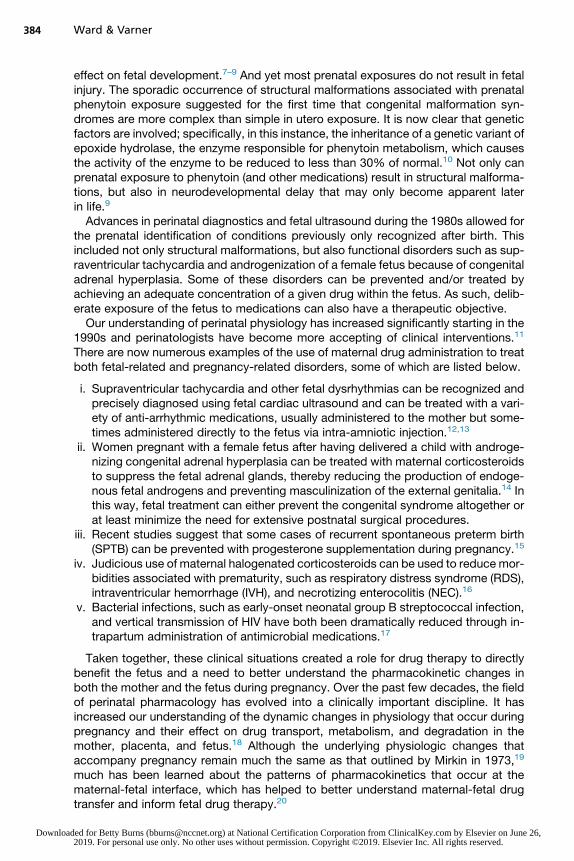
The goal of fetal drug therapy is to deliver an effective and nontoxic unbound drugconcentration to the site of action within the fetoplacental unit. To achieve this, theclinician must understand the numerous pharmacokinetic changes that accompanythe different stages of pregnancy and that may affect the drug concentration reachingthe proposed site of action. Fig. 1 lists the various steps involved in the therapeuticprocess by which medications are administered to the mother, transported acrossthe placenta, and act on the fetus. Interference or dysregulation of any one of thesesteps may adversely affect drug delivery and fetal therapy. Changes in maternalbody composition, physiology, and the activity of cytochrome P450 (CYP) enzymesthat affect pharmacokinetics in pregnancy are summarized in Tables 1 and 2 and inFig. 2 and are discussed in detail below.
CHANGES IN THE MOTHER THAT AFFECT PHARMACOKINETICSDrug Absorption
Enteral drug absorption is reduced in pregnancy because both gastric emptying andgastrointestinal motility are slowed owing to high levels of circulating progesterone.21
Both a reduction in intrinsic contractility and pressure from the enlarging uteruscontribute to the slowing of gastrointestinal transit time.
Drug Distribution
Intravascular volume begins to expand early in pregnancy and continues to do so toterm, by which time plasma volume has increased by 50%.22 This expansion of thevolume of distribution of polar drugs and those with a large molecular weight thatremain primarily within the circulation reduces circulating drug concentrations. Thisdilutional effect is partially offset by a decrease in circulating protein concentration,which increases the bioavailability of unbound (biologically active) drug. At the same
Fig. 1. Therapeutic process. Steps in the processing and transport of drugs from the motherto the proposed site of action in the fetus are shown.
wnloaded for Betty Burns ([email protected]) at National Certification Corporation from ClinicalKey.com by Elsevier on June 26, 2019. For personal use only. No other uses without permission. Copyright ©2019. Elsevier Inc. All rights reserved.
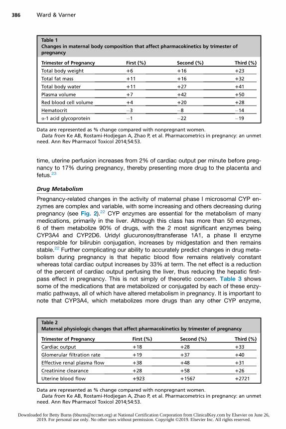
Table 1Changes in maternal body composition that affect pharmacokinetics by trimester ofpregnancy
Trimester of Pregnancy First (%) Second (%) Third (%)
Total body weight 16 116 123
Total fat mass 111 116 132
Total body water 111 127 141
Plasma volume 17 142 150
Red blood cell volume 14 120 128
Hematocrit �3 �8 �14
a-1 acid glycoprotein �1 �22 �19
Data are represented as % change compared with nonpregnant women.Data from Ke AB, Rostami-Hodjegan A, Zhao P, et al. Pharmacometrics in pregnancy: an unmet
need. Ann Rev Pharmacol Toxicol 2014;54:53.
Ward & Varner386
Download
time, uterine perfusion increases from 2% of cardiac output per minute before preg-nancy to 17% during pregnancy, thereby presenting more drug to the placenta andfetus.23
Drug Metabolism
Pregnancy-related changes in the activity of maternal phase I microsomal CYP en-zymes are complex and variable, with some increasing and others decreasing duringpregnancy (see Fig. 2).22 CYP enzymes are essential for the metabolism of manymedications, primarily in the liver. Although this class has more than 50 enzymes,6 of them metabolize 90% of drugs, with the 2 most significant enzymes beingCYP3A4 and CYP2D6. Uridyl glucuronosyltransferase 1A1, a phase II enzymeresponsible for bilirubin conjugation, increases by midgestation and then remainsstable.22 Further complicating our ability to accurately predict changes in drug meta-bolism during pregnancy is that hepatic blood flow remains relatively constantwhereas total cardiac output increases by 33% at term. The net effect is a reductionof the percent of cardiac output perfusing the liver, thus reducing the hepatic first-pass effect in pregnancy. This is not simply of theoretic concern. Table 3 showssome of the medications that are metabolized or conjugated by each of these enzy-matic pathways, all of which have altered metabolism in pregnancy. It is important tonote that CYP3A4, which metabolizes more drugs than any other CYP enzyme,
Table 2Maternal physiologic changes that affect pharmacokinetics by trimester of pregnancy
Trimester of Pregnancy First (%) Second (%) Third (%)
Cardiac output 118 128 133
Glomerular filtration rate 119 137 140
Effective renal plasma flow 138 148 131
Creatinine clearance 128 158 126
Uterine blood flow 1923 11567 12721
Data are represented as % change compared with nonpregnant women.Data from Ke AB, Rostami-Hodjegan A, Zhao P, et al. Pharmacometrics in pregnancy: an unmet
need. Ann Rev Pharmacol Toxicol 2014;54:53.
ed for Betty Burns ([email protected]) at National Certification Corporation from ClinicalKey.com by Elsevier on June 26, 2019. For personal use only. No other uses without permission. Copyright ©2019. Elsevier Inc. All rights reserved.
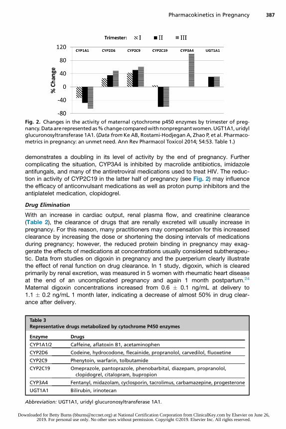
Fig. 2. Changes in the activity of maternal cytochrome p450 enzymes by trimester of preg-nancy.Dataarerepresentedas%changecomparedwithnonpregnantwomen.UGT1A1,uridylglucuronosyltransferase 1A1. (Data fromKeAB, Rostami-Hodjegan A, Zhao P, et al. Pharmaco-metrics in pregnancy: an unmet need. Ann Rev Pharmacol Toxicol 2014; 54:53. Table 1.)
Pharmacokinetics in Pregnancy 387
Do
demonstrates a doubling in its level of activity by the end of pregnancy. Furthercomplicating the situation, CYP3A4 is inhibited by macrolide antibiotics, imidazoleantifungals, and many of the antiretroviral medications used to treat HIV. The reduc-tion in activity of CYP2C19 in the latter half of pregnancy (see Fig. 2) may influencethe efficacy of anticonvulsant medications as well as proton pump inhibitors and theantiplatelet medication, clopidogrel.
Drug Elimination
With an increase in cardiac output, renal plasma flow, and creatinine clearance(Table 2), the clearance of drugs that are renally excreted will usually increase inpregnancy. For this reason, many practitioners may compensation for this increasedclearance by increasing the dose or shortening the dosing intervals of medicationsduring pregnancy; however, the reduced protein binding in pregnancy may exag-gerate the effects of medications at concentrations usually considered subtherapeu-tic. Data from studies on digoxin in pregnancy and the puerperium clearly illustratethe effect of renal function on drug clearance. In 1 study, digoxin, which is clearedprimarily by renal excretion, was measured in 5 women with rheumatic heart diseaseat the end of an uncomplicated pregnancy and again 1 month postpartum.24
Maternal digoxin concentrations increased from 0.6 � 0.1 ng/mL at delivery to1.1 � 0.2 ng/mL 1 month later, indicating a decrease of almost 50% in drug clear-ance after delivery.
Table 3Representative drugs metabolized by cytochrome P450 enzymes
Enzyme Drugs
CYP1A1/2 Caffeine, aflatoxin B1, acetaminophen
CYP2D6 Codeine, hydrocodone, flecainide, propranolol, carvedilol, fluoxetine
CYP2C9 Phenytoin, warfarin, tolbutamide
CYP2C19 Omeprazole, pantoprazole, phenobarbital, diazepam, propranolol,clopidogrel, citalopram, bupropion
CYP3A4 Fentanyl, midazolam, cyclosporin, tacrolimus, carbamazepine, progesterone
UGT1A1 Bilirubin, irinotecan
Abbreviation: UGT1A1, uridyl glucuronosyltransferase 1A1.
wnloaded for Betty Burns ([email protected]) at National Certification Corporation from ClinicalKey.com by Elsevier on June 26, 2019. For personal use only. No other uses without permission. Copyright ©2019. Elsevier Inc. All rights reserved.
Ward & Varner388
Download
CHANGES IN THE PLACENTA THAT AFFECT PHARMACOKINETICSAbsorption and Transfer of Drugs into the Fetal Circulation
Placental function is far more complex than a simple filter providing oxygen to thefetus and removing carbon dioxide and other waste products. The basic structuralunit of the placenta is the chorionic villus suspended in the intervillous space andbathed in maternal blood.25 The villi are vascular projections of fetal tissue surroundedby 2 layers of chorion: the outer syncytiotrophoblast, which is in direct contact withmaternal blood within the intervillous space, and the inner cytotrophoblast cell layer.Simple diffusion across membranes accounts for much of the transfer of drugs from
the maternal circulation to the placenta and on to the fetus. This usually occurs fromhigher to lower concentrations (usually from mother to fetus) of nonionized drugs thatare not protein bound.The anatomy and function of the placenta changes throughout gestation, and this
affects the ability of drugs to diffuse across to the fetus. Soon after the blastocyst im-plants into the uterine wall, the outer cells of the trophoblast layer immediately adja-cent to the uterine epithelium fuse into multinucleated cells, the syncytiotrophoblast,which is in direct contact with the maternal blood.25,26 Lacunae develop within thesyncytiotrophoblast that enlarge to become the intervillous spaces. Maternal spiralarteries connect to the intervillous space to establish the uteroplacental blood flow.Fetal vessels extend into the intervillous space in chorionic villi, which initially areseveral cells thick. During pregnancy, both chorionic layers thin out from a thicknessof 50 to 100 mM at 8 to 10 weeks of pregnancy to 4 to 5 mM at term. The immaturestructure of the early placenta helps to shield the fetus from potentially harmful xeno-biotics during the period of organogenesis.26 The fetoplacental circulation is reversedfrom that in newborns and adults with fetal arteries carrying deoxygenated blood fromthe fetus into the umbilical arteries, which flow into the chorionic arteries and capil-laries within the placenta. There, gas exchange occurs before the oxygenated bloodreturns to the fetus through chorionic capillaries that flow into the umbilical vein.It is important to note that only nonionized, nonprotein bound drugs diffuse
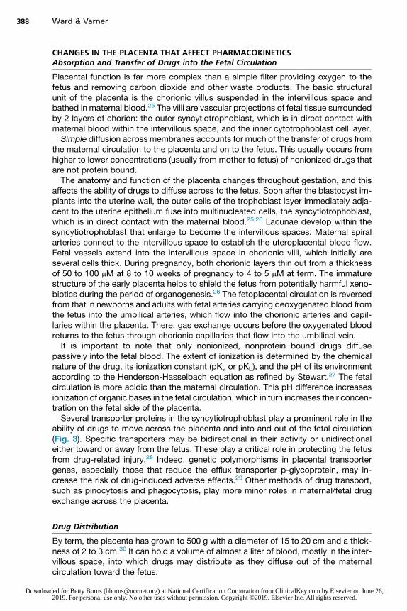
passively into the fetal blood. The extent of ionization is determined by the chemicalnature of the drug, its ionization constant (pKa or pKb), and the pH of its environmentaccording to the Henderson-Hasselbach equation as refined by Stewart.27 The fetalcirculation is more acidic than the maternal circulation. This pH difference increasesionization of organic bases in the fetal circulation, which in turn increases their concen-tration on the fetal side of the placenta.Several transporter proteins in the syncytiotrophoblast play a prominent role in the
ability of drugs to move across the placenta and into and out of the fetal circulation(Fig. 3). Specific transporters may be bidirectional in their activity or unidirectionaleither toward or away from the fetus. These play a critical role in protecting the fetusfrom drug-related injury.28 Indeed, genetic polymorphisms in placental transportergenes, especially those that reduce the efflux transporter p-glycoprotein, may in-crease the risk of drug-induced adverse effects.29 Other methods of drug transport,such as pinocytosis and phagocytosis, play more minor roles in maternal/fetal drugexchange across the placenta.
Drug Distribution
By term, the placenta has grown to 500 g with a diameter of 15 to 20 cm and a thick-ness of 2 to 3 cm.30 It can hold a volume of almost a liter of blood, mostly in the inter-villous space, into which drugs may distribute as they diffuse out of the maternalcirculation toward the fetus.
ed for Betty Burns ([email protected]) at National Certification Corporation from ClinicalKey.com by Elsevier on June 26, 2019. For personal use only. No other uses without permission. Copyright ©2019. Elsevier Inc. All rights reserved.
Fig. 3. Drug transport within the syncytiotrophoblast. The major transporter proteins withinthe syncytiotrophoblast responsible for the movement of drugs into and out of the fetal cir-culation are shown. BCRP, breast cancer resistance protein; CYP’s, cytochrome P450’s; MRP,multidrug resistance-associated protein; NET, norepinephrine transporter; OAT4, organicacid transporter 4; OATP2B1, organic anion-associated polypeptide 2B1; OATP4A1, organicanion-associated polypeptide 4A1; OCT3, organic cation transporter 3; OCTN1 and 2,organic acid/carnitine transporters; P-gp, P-glycoprotein; SERT, serotonin transporter;UGTs, uridine diphosphate glucuronosyl-transferases. (Adapted from Rubinchik-Stern M,Eyal S. Drug interactions at the human placenta: what is the evidence? Front Pharmacol2012. https://doi.org/10.3389/fphar.2012.00126.)
Pharmacokinetics in Pregnancy 389
Do
Drug Metabolism
The placenta contains several CYP enzymes that are active early in fetal development.These include CYP 1A1, 1A2, 1B1, 2C, 2D6, 2E1, 2F1, 3A4, 5, 7, and 4B1.31 By term,CYP2D6 and 1A2 are no longer detectable in the placenta. Several of these enzymesare essential in the maintenance of pregnancy by metabolizing endogenous com-pounds, such as steroids, that may otherwise activate parturition.18 Activity of mostof these CYPs are highest early in gestation and decrease toward term.32
CHANGES IN THE FETUS THAT AFFECT PHARMACOKINETICSDrug Absorption
Absorption of drugs by the fetus is usually through the blood returning from theplacenta through the umbilical vein. Transcutaneous and oral absorption by the fetusare possible, but have received limited study. With the buildup of the vernix caseosa, alipid barrier forms on the surface of the skin preventing penetration by many polarcompounds and water. Recent studies have shown that vernix is composed of 80%water, 10% proteins, and 10% lipids.33 Vernix components have been shown to beactively involved in host defense, exhibiting antifungal and antimicrobial activity.34
The lipid component of vernix are comprised of an estimated 54 different lipid medi-ators (21 oxylipins, 23 sphingolipids, and 10 endocannabinoids) all with functions yetto be fully explained.
Drug Distribution
Drug distribution within the fetus is influenced primarily by its body composition. Earlyin gestation, the fetus is 94% water with 0.5% fat.35 By full term, its water content hasdecreased to 76% and fat content has increased to 12% to 16%. Clearly, the distri-bution to tissues of polar as well as lipophilic compounds will vary significantly basedsolely on body composition related to the stage of gestation.
Drug Metabolism
Fetal drug metabolizing enzyme activity varies markedly during pregnancy. Olderstudies reported high level of activity of several enzymes early in pregnancy, including
wnloaded for Betty Burns ([email protected]) at National Certification Corporation from ClinicalKey.com by Elsevier on June 26, 2019. For personal use only. No other uses without permission. Copyright ©2019. Elsevier Inc. All rights reserved.
Ward & Varner390
Download
CYP1A1/1A2, CYP1B1, CYP2C8/2C9/2C18/2C19, CYP2D6, CYP2E1, CYP3A4,CYP3A5, and CYP3A7.36 Although these enzymes can help protect the fetus from po-tential toxins, many are also involved in the metabolism of endogenous compoundsessential for fetal development. As in older children and adults, the liver containsmost of the enzymes involved in drug metabolism. In a recent review, Hines described3 general patterns of developmental changes in fetal drug metabolizing enzymes.32
One group of enzymes demonstrate their highest activity early in gestation and eitherstay the same or decrease later in pregnancy. These include CYP3A7, flavin monoox-ygenase 1, and sulfotransferases 1A3/4 and 1E1. The second group of enzymes ex-press their activity at a relatively constant level from fetal development intoadulthood, and include CYP3A5, sulfotransferase 1A1, and CYP2C19 (although thelatter does show a moderate increase during the first year of life). The third and largestgroup of enzymes begin with little or no activity in the fetus and later increase to adultactivity. These include CYP1A2, CYP2C9, CYP2D6, CYP2E1, CYP3A4, flavin monox-ygenase3, and sulfotransferase 2A1. The time course for the increase in activity ishighly variable, ranging from 2 to 3 weeks to many years, an interval that is, particularlyrelevant to pediatric therapeutics.
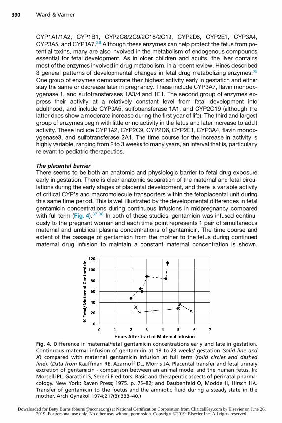
The placental barrierThere seems to be both an anatomic and physiologic barrier to fetal drug exposureearly in gestation. There is clear anatomic separation of the maternal and fetal circu-lations during the early stages of placental development, and there is variable activityof critical CYP’s and macromolecule transporters within the fetoplacental unit duringthis same time period. This is well illustrated by the developmental differences in fetalgentamicin concentrations during continuous infusions in midpregnancy comparedwith full term (Fig. 4).37,38 In both of these studies, gentamicin was infused continu-ously to the pregnant woman and each time point represents 1 pair of simultaneousmaternal and umbilical plasma concentrations of gentamicin. The time course andextent of the passage of gentamicin from the mother to the fetus during continuedmaternal drug infusion to maintain a constant maternal concentration is shown.
Fig. 4. Difference in maternal/fetal gentamicin concentrations early and late in gestation.Continuous maternal infusion of gentamicin at 18 to 23 weeks’ gestation (solid line andX) compared with maternal gentamicin infusion at full term (solid circles and dashedline). (Data from Kauffman RE, Azarnoff DL, Morris JA. Placental transfer and fetal urinaryexcretion of gentamicin - comparison between an animal model and the human fetus. In:Morselli PL, Garattini S, Sereni F, editors. Basic and therapeutic aspects of perinatal pharma-cology. New York: Raven Press; 1975. p. 75–82; and Daubenfeld O, Modde H, Hirsch HA.Transfer of gentamicin to the foetus and the amniotic fluid during a steady state in themother. Arch Gynakol 1974;217(3):333–40.)
ed for Betty Burns ([email protected]) at National Certification Corporation from ClinicalKey.com by Elsevier on June 26, 2019. For personal use only. No other uses without permission. Copyright ©2019. Elsevier Inc. All rights reserved.
Pharmacokinetics in Pregnancy 391
Do
Despite infusion for almost 6 hours, the fetal concentration at 18 to 23 weeks of gesta-tion remains less than 40% of the maternal concentration. In contrast, at term, fetalconcentrations reach 100% of maternal concentrations within 5 hours.
INTERPRETATION OF MATERNAL/UMBILICAL DRUG CONCENTRATIONS AT BIRTH
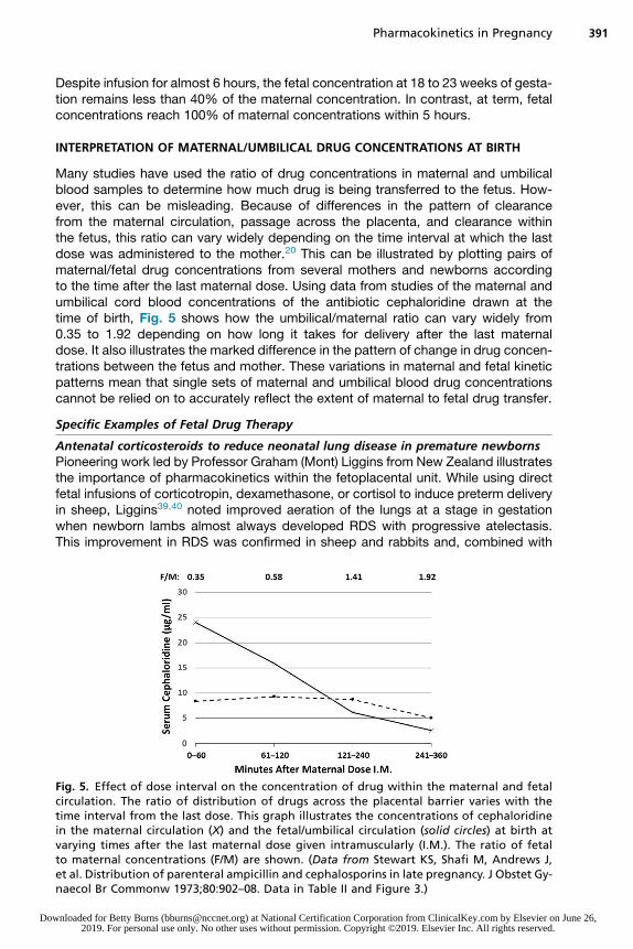
Many studies have used the ratio of drug concentrations in maternal and umbilicalblood samples to determine how much drug is being transferred to the fetus. How-ever, this can be misleading. Because of differences in the pattern of clearancefrom the maternal circulation, passage across the placenta, and clearance withinthe fetus, this ratio can vary widely depending on the time interval at which the lastdose was administered to the mother.20 This can be illustrated by plotting pairs ofmaternal/fetal drug concentrations from several mothers and newborns accordingto the time after the last maternal dose. Using data from studies of the maternal andumbilical cord blood concentrations of the antibiotic cephaloridine drawn at thetime of birth, Fig. 5 shows how the umbilical/maternal ratio can vary widely from0.35 to 1.92 depending on how long it takes for delivery after the last maternaldose. It also illustrates the marked difference in the pattern of change in drug concen-trations between the fetus and mother. These variations in maternal and fetal kineticpatterns mean that single sets of maternal and umbilical blood drug concentrationscannot be relied on to accurately reflect the extent of maternal to fetal drug transfer.
Specific Examples of Fetal Drug Therapy
Antenatal corticosteroids to reduce neonatal lung disease in premature newbornsPioneering work led by Professor Graham (Mont) Liggins from New Zealand illustratesthe importance of pharmacokinetics within the fetoplacental unit. While using directfetal infusions of corticotropin, dexamethasone, or cortisol to induce preterm deliveryin sheep, Liggins39,40 noted improved aeration of the lungs at a stage in gestationwhen newborn lambs almost always developed RDS with progressive atelectasis.This improvement in RDS was confirmed in sheep and rabbits and, combined with
Fig. 5. Effect of dose interval on the concentration of drug within the maternal and fetalcirculation. The ratio of distribution of drugs across the placental barrier varies with thetime interval from the last dose. This graph illustrates the concentrations of cephaloridinein the maternal circulation (X) and the fetal/umbilical circulation (solid circles) at birth atvarying times after the last maternal dose given intramuscularly (I.M.). The ratio of fetalto maternal concentrations (F/M) are shown. (Data from Stewart KS, Shafi M, Andrews J,et al. Distribution of parenteral ampicillin and cephalosporins in late pregnancy. J Obstet Gy-naecol Br Commonw 1973;80:902–08. Data in Table II and Figure 3.)
wnloaded for Betty Burns ([email protected]) at National Certification Corporation from ClinicalKey.com by Elsevier on June 26, 2019. For personal use only. No other uses without permission. Copyright ©2019. Elsevier Inc. All rights reserved.
Ward & Varner392
Download
the observations by Naeye and colleagues41 of smaller adrenal glands in newbornsdying from RDS, suggested that a corticosteroid deficiency or depletion may be caus-ally related to the development of RDS. As an obstetrician familiar with the clinicalsequelae of RDS, Liggins began a clinical trial of antenatal glucocorticoid treatmentof mothers in preterm labor at 24 to 36 weeks’ gestation. Maternal treatment used acompound formulation of a halogenated corticosteroid derived from prednisoloneknown as betamethasone, a stereoisomer of dexamethasone with the same structureand molecular weight but a different spatial orientation of the 16-methyl group. Thisformulation of betamethasone (Celestone) was a combination of 6 mg of the immedi-ate release phosphate salt and 6mg of the poorly soluble, slowly released acetate salt.The intramuscular (i.m.) injection was repeated once 24 hours later if the mother hadnot delivered. The success of this transplacental therapy can be explained by thepharmacokinetics of betamethasone and dexamethasone and how they cross thehuman placenta. The placenta protects the fetus from increased maternal cortisolconcentrations through the activity of 11-b hydroxysteroid dehydrogenase type 2,which inactivates cortisol to cortisone.42 In contrast, the synthetic halogenatedstructural isomers, betamethasone and dexamethasone, are minimally inactivatedby 11-b hydroxysteroid dehydrogenase type 2 and pass through the placenta largelyunchanged.The original study16 was blinded and controlled, with the control group receiving
6 mg cortisol, a minimal dosage. Of 117 betamethasone and 96 control mothersenrolled, 94 betamethasone and 78 control subjects continued pregnancy for at least24 hours, which was considered the minimal time for induction of an effect on surfac-tant release/synthesis, and which also allowed for 2 doses of steroids to be adminis-tered. In the betamethasone-treated group, the frequency of RDS progressivelydecreased with increasing time to delivery up to 7 days, from 24.0% for delivery atless than 24 hours to 10% for delivery at 24 to 48 hours and 3.6% at 2 to 7 days.Although RDS was reduced significantly overall from 24.0% to 4.3%, the significantreduction was confined to infants delivering at 26 to 32 weeks’ gestation (69.6% to11.8%). In the most premature newborns, no IVH was seen in the betamethasone-treated group, but was seen in 4 of the controls.16 Although not statistically significant,this reduction in IVH with betamethasone treatment was later confirmed in largerstudies.43,44 Despite 2 NIH Consensus Conferences that supported treatment withbetamethasone to reduce RDS,45,46 prenatal corticosteroid treatment was slow togain acceptance. This might have related to the then-recent memory of long-termproblems associated with attempts to prolong pregnancy using diethylstilbestrol lead-ing to cancer in the children and young adults exposed in utero.47,48 Such interventionhas now become “standard of care” for impending preterm birth less than 34 weeks. Arecent Cochrane review carried out a meta-analysis of 30 studies confirming that ante-natal steroids reduced perinatal death by 28%, neonatal death by 31%, RDS by 34%,moderate/severe RDS by 41%, IVH by 12%, as well as NEC, need for mechanicalventilation, and early-onset systemic infections within the first 48 hours after birth.44
Given these beneficial effects, what more do we need to know about antenatal treat-ment with corticosteroids? Here are some unanswered questions: What is the optimaldose? Is the current dosage of 2 doses of 12 mg of betamethasone acetate and phos-phate administered 24 hours apart the optimal treatment? The initial study by Ligginsand Howie16 did not include a dose-ranging study or variation of the interval betweendoses. Although 12 mg doses administered 24 hours apart is effective, some authorsrecommend completing the betamethasone treatment in a shorter time interval, suchas 12 hours.49 With the 24 hour treatment course with betamethasone, Liggins andHowie16 noted adrenal suppression in the mothers for around 72 hours and other
ed for Betty Burns ([email protected]) at National Certification Corporation from ClinicalKey.com by Elsevier on June 26, 2019. For personal use only. No other uses without permission. Copyright ©2019. Elsevier Inc. All rights reserved.
Pharmacokinetics in Pregnancy 393
Do
studies have shown a suppression of neonatal cortisol secretion for several days afterbirth.50 A lower betamethasone dosage might be equally effective as the Liggins’ pro-tocol, but produce less suppression of the hypothalamic-pituitary-adrenal axis.Studies by Jobe and colleagues51 have shown that a single dose of the slowlyreleased betamethasone acetate is more effective than 2 doses of the rapidly releasedbetamethasone phosphate. In another study in sheep, an even lower dose of betame-thasone acetate improved lung function.52 Similar studies in humans may providesimilar benefits of antenatal corticosteroids with less hypothalamic-pituitary-adrenalsuppression.Such questions are not uncommon when discussing drug treatment. If the initial
dosage tested is effective and relatively safe, additional studies may be difficult toconduct to attempt to reduce the dose or change the dosing interval. This is especiallythe case in obstetrics and pediatrics, whereby drug studies are time-consuming andexpensive with few sites prepared to carry them out. It is nonetheless important to un-derstand the extent of the initial studies of a drug and whether alternate doses, doseintervals, and formulations have been adequately explored and tested.If 1 course (2 doses totaling 24 mg) of antenatal corticosteroids is good, can
repeated courses sustain or even amplify the improvement? The effectiveness ofbetamethasone decreases in most studies by 7 days after the initiation of treatment,so some obstetricians began to repeat the 24 mg dosage on a weekly basis. If thepregnancy continued, repeated administration was continued for several weeks upto 11 times. In 1999, a retrospective review published by Banks and colleagues53
suggested that repeated courses of antenatal corticosteroids increased perinatalmortality, reduced fetal growth, and prolonged adrenal suppression. This was fol-lowed in 2000 by an NIH Consensus Conference recommending that only a singlecourse of antenatal corticosteroids be administered unless repeated courses werepart of an investigative protocol.46 Like all retrospective studies, the study by Banksand colleagues53 was susceptible to confounding effects that might not be knownand could not be controlled for. Several prospective randomized studies of repeatedcourses of antenatal corticosteroids followed. A meta-analysis in 2017 of 30 studies,including 9 with repeated courses of antenatal corticosteroids, concluded that bothsingle and repeated doses of antenatal corticosteroids reduced perinatal death,neonatal death, RDS, moderate/severe RDS, IVH, NEC, need for mechanical ventila-tion, and infections within 48 hours of birth.44 No serious adverse events werereported.Although it is clear that fetuses can respond favorably to antenatal corticosteroids,
the stages of lung development when that response occurs was not initially clear. Lig-gins and Howie found a reduction in RDS in fetuses who received antenatal cortico-steroids and delivered at 26 to 32 weeks of gestation.16 The benefit for the mostimmature preterm newborns (<26 weeks) was initially uncertain. However, morerecent studies have shown reductions in RDS and mortality also at gestations ofless than 25 weeks.54,55 The upper limit of gestational age at which fetuses couldrespond to antenatal corticosteroids has expanded as well and now includes latepreterm fetuses (gestational age 34–37 weeks), a group who are known to haveincreased respiratory morbidities compared with full term newborns. In 1 prospectiverandomized trial, antenatal corticosteroid treatment of late preterm deliveries reducedsurfactant use, transient tachypnea, and bronchopulmonary dysplasia withoutincreasing neonatal sepsis.56 The reduction was modest (14.4% versus 11.6%) andhypoglycemia was increased in the betamethasone-treated group. Other recentstudies support antenatal corticosteroid treatment before elective cesarean sectionat term to reduce respiratory morbidity.57
wnloaded for Betty Burns ([email protected]) at National Certification Corporation from ClinicalKey.com by Elsevier on June 26, 2019. For personal use only. No other uses without permission. Copyright ©2019. Elsevier Inc. All rights reserved.
Ward & Varner394
Download
Multiple studies have followed the initial 1972 report from Liggins and Howie, and allsupport single course treatment of mothers in preterm labor to improve neonatal out-comes. The importance of controlled trials in perinatal medicine to improve pregnancyoutcomes has had other far reaching and often unanticipated consequences,I including a major contribution to the development of the Cochrane systematic re-views.58–60 Questions still remain regarding antenatal corticosteroid treatment thatdeserve continued study to determine the minimal effective dosage, optimal intervalbetween doses, whether adverse effects of repeated maternal treatment harm a spe-cific population or are safe, and whether there are upper and lower limits of gestationalage at which benefits no longer occur. Long-term follow-up studies of people exposedin utero to antenatal corticosteroids to investigate growth, cardiovascular health, andneurologic outcomes should continue as this population ages.
Progesterone to prevent preterm birth, the intersection of pharmacogenomics andpharmacokineticsThe decrease in recurrent SPTB with progesterone supplementation has significantlyreduced morbidity associated with prematurity and raised several possible explana-tions. After several small studies, a large prospective, randomized trial suggestedthat 17-hydroxyprogesterone caproate (17OHPC) reduced the recurrence of SPTBin singleton pregnancies at high risk of this event by virtue of a prior SPTB over a broadrange of gestations, and resulted in a reduction in NEC, IVH, and need for supple-mental oxygen.15 The possible explanation for this effect involves pharmacokineticsand pharmacogenomics.61
One study of the pharmacokinetics of 17OHPC in singleton pregnancies wasextended after delivery with continued sampling up to 28 days postpartum.61 Womenwere treated weekly with 17OHPC 250 mg i.m. formulated in oil starting in the earlysecond trimester. At birth, the umbilical to maternal plasma concentrations averaged0.2, and the umbilical concentrations did not change with the time after the lastmaternal dose. The concentrations of 17OHPC varied inversely with body mass index,whichmay indicate a need for weight-based dosage adjustment. Clearance was fasterin African American women; unfortunately, the CYP3A4 genotype, which is respon-sible for metabolism of 17OHPC was not determined in this cohort. Interestingly,the disposition half-life of 17OHPC was 18 � 6 days, which led to progressive accu-mulation of drug with weekly injections.61
In another study, supplementation of women with twin pregnancies with 17OHPCallowed evaluation of both the pharmacokinetics and potential mechanism of action.62
Concentrations of 17OHPC were inversely correlated with gestation at delivery, withlower concentrations at more advanced gestations, the opposite of what would be ex-pected. Circulating C-reactive protein concentrations were increased in pregnancieswith the highest concentrations of 17OHPC, but it did not reach statistical significance.The authors of this study speculated that the rise in C-reactive protein was related tothe underlying mechanisms of parturition rather than to treatment with 17OHPC. Thisstudy along with many subsequent publications showed no lengthening of gestationwith 17OHPC supplementation in the setting of multiple gestations (twins or triplets)or for women with a shortened cervical length.63
Roughly two-thirds of high-risk women with singleton pregnancies who receive17OHPC supplementation do not respond and will go on to have a recurrentSPTB.64 Although a pharmacokinetic explanation is possible, Manuck and col-leagues65 used DNA from the original study by Meis and colleagues15 to look moreclosely at progesterone receptors A and B. Because of variation in allele frequencyby race, the samples were stratified by self-reported race into African American and
ed for Betty Burns ([email protected]) at National Certification Corporation from ClinicalKey.com by Elsevier on June 26, 2019. For personal use only. No other uses without permission. Copyright ©2019. Elsevier Inc. All rights reserved.
Pharmacokinetics in Pregnancy 395
Do
White/Hispanic. Several gene response interactions were observed which differed byrace/ethnicity. Some haplotypes identified women with an underlying risk of SPTB,others identified women with a favorable response to 17OHPC, whereas other geno-types were associated with an increased risk of SPTB in women who received17OHPC supplementation.65 Thus, successful prevention of SPTB with 17OHPCtreatment is not simple pharmacokinetics, but a complex interplay between pharma-cogenetics and pharmacokinetics.In women without a history of SPTB but who are found in the midtrimester to have a
short cervix on transvaginal ultrasound, supplementation with vaginal progesterone iscurrently recommended by the American College of Obstetricians and Gynecolo-gists.66 This is not a US Food and Drug Administration recommended intervention.Although some evidence exists to suggest that vaginal progesterone may be superiorto intramuscular 17OHPC,67 the American College of Obstetricians and Gynecologistshas not yet endorsed the primary use of vaginal progesterone for prevention of recur-rent SPTB. The pharmacokinetics of vaginal progesterone for SPTB prevention hasnot been adequately studied.
SUMMARY
Pregnancy profoundly alters a woman’s physiology. When combined with the pro-gressive impact of the developing fetus and placenta, these changes result in multiplealterations in drug absorption, distribution, metabolism, and elimination. As outlinedearlier in this article, these changes emphasize the pharmacologic complexity of preg-nancy. They also emphasize the dangers of extrapolating pharmacologic expectationsfrom nonpregnant populations to pregnant women and their fetuses. Although con-cerns about fetal safety have historically limited pharmacokinetic studies during preg-nancy, it is important to recognize that many medications are clinically indicated forvarious maternal or fetal conditions. Recommendations for the use of medicationsin pregnancy should be based on the prevailing evidence, including short-term andlong-term outcome data.
REFERENCES
1. Taussig HB. Thalidomide – a lesson in remote effects of drugs. Am J Dis Child1962;104:111–3.
2. Taussig HB. The thalidomide syndrome. Sci Am 1962;207:29–35.3. Taussig HB. Thalidomide and phocomelia. Pediatrics 1962;30:654–9.4. Lenz W, Knapp K. Thalidomide embryopathy. Arch Environ Health 1962;5:100–5.5. Hanson JW, Myrianthopoulos NC, Harvey MA, et al. Risks to the offspring of
women treated with hydantoin anticonvulsants, with emphasis on the fetal hydan-toin syndrome. J Pediatr 1976;89:662–8.
6. Hanson JW, Smith DW. Fetal hydantoin syndrome. Lancet 1976;1:692.7. Mirkin BL. Placental transfer and neonatal elimination of diphenylhydantoin. Am
J Obstet Gynecol 1971;109:930–3.8. Mirkin BL. Diphenylhydantoin: placental transport, fetal localization, neonatal
metabolism, and possible teratogenic effects. J Pediatr 1971;78:329–37.9. Diav-Citrin O. Prenatal exposures associated with neurodevelopmental delay and
disabilities. Dev Disabil Res Rev 2011;17:71–84.10. Buehler BA, Delimont D, van Waes M, et al. Prenatal prediction of risk of the fetal
hydantoin syndrome. N Engl J Med 1990;322:1567–72.11. Ward RM. Maternal drug therapy for fetal disorders. Semin Perinatol 1992;16:
12–20.
wnloaded for Betty Burns ([email protected]) at National Certification Corporation from ClinicalKey.com by Elsevier on June 26, 2019. For personal use only. No other uses without permission. Copyright ©2019. Elsevier Inc. All rights reserved.
Ward & Varner396
Download
12. Steinfeld L, Rappaport HL, Rossbach HC, et al. Diagnosis of fetal arrhythmias us-ing echocardiographic and Doppler techniques. J Am Coll Cardiol 1986;8:1425–33.
13. Alsaied T, Baskar S, Fares M, et al. First-line antiarrhythmic transplacental treat-ment for fetal tachyarrhythmia: a systematic review and meta-analysis. J AmHeart Assoc 2017;6 [pii:e007164].
14. Nimkarn S, New MI. Congenital adrenal hyperplasia due to 21-hydroxylase defi-ciency: a paradigm for prenatal diagnosis and treatment. Ann N YAcad Sci 2010;1192:5–11.
15. Meis PJ, Klebanoff M, Thom E, et al. Prevention of recurrent preterm delivery by17 alpha-hydroxyprogesterone caproate. N Engl J Med 2003;348:2379–85.
16. Liggins GC, Howie RN. A controlled trial of antepartum glucocorticoid treatmentfor prevention of the respiratory distress syndrome in premature infants. Pediat-rics 1972;50:515–25.
17. Benitz WE, Gould JB, Druzin ML. Antimicrobial prevention of early-onset group Bstreptococcal sepsis: estimates of risk reduction based on a critical literature re-view. Pediatrics 1999;103:e78, 71-13.
18. Mahmood I, Burckart GJ, Ward RM. Perinatal pharmacology and maternal/fetaldosing. In: Mahmood I, Burckart GJ, editors. Fundamentals of pediatric drugdosing. Cham (Switzerland): Springer International; 2016. p. 127–46.
19. Mirkin BL. Maternal and fetal distribution of drugs in pregnancy. Clin PharmacolTher 1973;14:643–7.
20. Ward RM. Pharmacological treatment of the fetus. Clinical pharmaco-kinetic con-siderations. Clin Pharmacokinet 1995;28:343–50.
21. Wald A, Van Thiel DH, Hoechstetter L, et al. Effect of pregnancy on gastrointes-tinal transit. Dig Dis Sci 1982;27:1015–8.
22. Ke AB, Rostami-Hodjegan A, Zhao P, et al. Pharmacometrics in pregnancy: anunmet need. Annu Rev Pharmacol Toxicol 2014;54:53–69.
23. Frederiksen MC. Physiologic changes in pregnancy and their effect on drugdisposition. Semin Perinatol 2001;25:120–3.
24. Rogers MC, Willerson JT, Goldblatt A, et al. Serum digoxin concentrations in thehuman fetus, neonate and infant. N Engl J Med 1972;287:1010–3.
25. Gude NM, Roberts CT, Kalionis B, et al. Growth and function of the normal humanplacenta. Thromb Res 2004;114:397–407.
26. Al-Enazy S, Ali S, Albekairi N, et al. Placental control of drug delivery. Adv DrugDeliv Rev 2017;116:63–72.
27. Kurtz I, Kraut J, Ornekian V, et al. Acid-base analysis: a critique of the Stewartand bicarbonate-centered approaches. Am J Physiol Renal Physiol 2008;294:F1009–31.
28. Iqbal M, Audette MC, Petropoulos S, et al. Placental drug transporters and theirrole in fetal protection. Placenta 2012;33:137–42.
29. Daud AN, Bergman JE, Bakker MK, et al. Pharmacogenetics of drug-inducedbirth defects: the role of polymorphisms of placental transporter proteins. Phar-macogenomics 2014;15:1029–41.
30. Rubinchik-Stern M, Eyal S. Drug interactions at the human placenta: what is theevidence? Front Pharmacol 2012;3:126.
31. Pasanen M. The expression and regulation of drug metabolism in humanplacenta. Adv Drug Deliv Rev 1999;38:81–97.
32. Hines RN. Developmental expression of drug metabolizing enzymes: impact ondisposition in neonates and young children. Int J Pharm 2013;452:3–7.
ed for Betty Burns ([email protected]) at National Certification Corporation from ClinicalKey.com by Elsevier on June 26, 2019. For personal use only. No other uses without permission. Copyright ©2019. Elsevier Inc. All rights reserved.
Pharmacokinetics in Pregnancy 397
Do
33. Checa A, Holm T, Sjodin MO, et al. Lipid mediator profile in vernix caseosa re-flects skin barrier development. Sci Rep 2015;5:15740.
34. Tollin M, Bergsson G, Kai-Larsen Y, et al. Vernix caseosa as a multi-componentdefence system based on polypeptides, lipids and their interactions. Cell MolLife Sci 2005;62:2390–9.
35. Friis-Hansen B. Body water compartments in children: changes during growthand related changes in body composition. Pediatrics 1961;28:169–81.
36. Hakkola J, Pelkonen O, Pasanen M, et al. Xenobiotic-metabolizing cytochromeP450 enzymes in the human feto-placental unit: role in intrauterine toxicity. CritRev Toxicol 1998;28:35–72.
37. Kauffman RE, Azarnoff DL, Morris JA. Placental transfer and fetal urinary excre-tion of gentamicin - comparison between an animal model and the human fetus.In: Morselli PL, Garattini S, Sereni F, editors. Basic and therapeutic aspects ofperinatal pharmacology. New York: Raven Press; 1975. p. 75–82.
38. Daubenfeld O, Modde H, Hirsch HA. Transfer of gentamicin to the foetus and theamniotic fluid during a steady state in the mother. Arch Gynakol 1974;217:333–40.
39. Liggins GC. Premature delivery of foetal lambs infused with glucocorticoids.J Endocrinol 1969;45:515–23.
40. Liggins GC. Premature parturition after infusion of corticotrophin or cortisol intofoetal lambs. J Endocrinol 1968;42:323–9.
41. Naeye RL, Harcke HT Jr, Blanc WA. Adrenal gland structure and the develop-ment of hyaline membrane disease. Pediatrics 1971;47:650–7.
42. Benediktsson R, Calder AA, Edwards CR, et al. Placental 11 beta-hydroxysteroiddehydrogenase: a key regulator of fetal glucocorticoid exposure. Clin Endocrinol(Oxf) 1997;46:161–6.
43. Canterino JC, Verma U, Visintainer PF, et al. Antenatal steroids and neonatal peri-ventricular leukomalacia. Obstet Gynecol 2001;97:135–9.
44. Roberts D, Brown J, Medley N, et al. Antenatal corticosteroids for acceleratingfetal lung maturation for women at risk of preterm birth. Cochrane DatabaseSyst Rev 2017;(3):CD004454.
45. Effect of corticosteroids for fetal maturation on perinatal outcomes. NIHconsensus development panel on the effect of corticosteroids for fetal maturationon perinatal outcomes. JAMA 1995;273:413–8.
46. Antenatal corticosteroids revisited: repeat courses - National Institutes of HealthConsensus Development Conference Statement, August 17-18, 2000. Obstet Gy-necol 2001;98:144–50.
47. Herbst AL, Ulfelder H, Poskanzer DC. Adenocarcinoma of the vagina. Associa-tion of maternal stilbestrol therapy with tumor appearance in young women.N Engl J Med 1971;284:878–81.
48. Reed CE, Fenton SE. Exposure to diethylstilbestrol during sensitive life stages: alegacy of heritable health effects. Birth Defects Res C Embryo Today 2013;99:134–46.
49. Romejko-Wolniewicz E, Teliga-Czajkowska J, Czajkowski K. Antenatal steroids:can we optimize the dose? Curr Opin Obstet Gynecol 2014;26:77–82.
50. Nykanen P, Raivio T, Heinonen K, et al. Circulating glucocorticoid bioactivity andserum cortisol concentrations in premature infants: the influence of exogenousglucocorticoids and clinical factors. Eur J Endocrinol 2007;156:577–83.
51. Jobe AH, Nitsos I, Pillow JJ, et al. Betamethasone dose and formulation forinduced lung maturation in fetal sheep. Am J Obstet Gynecol 2009;201:611.e1-7.
wnloaded for Betty Burns ([email protected]) at National Certification Corporation from ClinicalKey.com by Elsevier on June 26, 2019. For personal use only. No other uses without permission. Copyright ©2019. Elsevier Inc. All rights reserved.
Ward & Varner398
Download
52. Schmidt AF, Kemp MW, Rittenschober-Bohm J, et al. Low-dose betamethasone-acetate for fetal lung maturation in preterm sheep. Am J Obstet Gynecol 2018;218:132.e1-9.
53. Banks BA, Cnaan A, Morgan MA, et al. Multiple courses of antenatal corticoste-roids and outcome of premature neonates. North American Thyrotropin-Releasing Hormone Study Group. Am J Obstet Gynecol 1999;181:709–17.
54. Travers CP, Clark RH, Spitzer AR, et al. Exposure to any antenatal corticosteroidsand outcomes in preterm infants by gestational age: prospective cohort study.BMJ 2017;356:j1039.
55. Deshmukh M, Patole S. Antenatal corticosteroids in impending preterm deliveriesbefore 25 weeks’ gestation. Arch Dis Child Fetal Neonatal Ed 2018;103:F173–6.
56. Gyamfi-Bannerman C, Thom EA, Blackwell SC, et al. Antenatal betamethasonefor women at risk for late preterm delivery. N Engl J Med 2016;374:1311–20.
57. Nada AM, Shafeek MM, El Maraghy MA, et al. Antenatal corticosteroid adminis-tration before elective caesarean section at term to prevent neonatal respiratorymorbidity: a randomized controlled trial. Eur J Obstet Gynecol Reprod Biol 2016;199:88–91.
58. Grant A, Chalmers I. Register of randomised controlled trials in perinatal medi-cine. Lancet 1981;1:100.
59. Chalmers I. Adrian Grant’s pioneering use of evidence synthesis in perinatalmedicine, 1980-1992. Reprod Health 2018;15:79.
60. Clarke M, Chalmers I. Reflections on the history of systematic reviews. BMJ EvidBased Med 2018;23:121–2.
61. Caritis SN, Sharma S, Venkataramanan R, et al. Pharmacology and placentaltransport of 17-hydroxyprogesterone caproate in singleton gestation. Am J Ob-stet Gynecol 2012;207:398.e1-8.
62. Caritis SN, Simhan HN, Zhao Y, et al. Relationship between 17-hydroxyprogester-one caproate concentrations and gestational age at delivery in twin gestation. AmJ Obstet Gynecol 2012;207:396.e1-8.
63. Caritis SN, Feghali MN, Grobman WA, et al. What we have learned about the roleof 17-alpha-hydroxyprogesterone caproate in the prevention of preterm birth.Semin Perinatol 2016;40:273–80.
64. Manuck TA, Stoddard GJ, Fry RC, et al. Nonresponse to 17-alpha hydroxypro-gesterone caproate for recurrent spontaneous preterm birth prevention: clinicalprediction and generation of a risk scoring system. Am J Obstet Gynecol 2016;215:622.e1-8.
65. Manuck TA, Lai Y, Meis PJ, et al. Progesterone receptor polymorphisms and clin-ical response to 17-alpha-hydroxyprogesterone caproate. Am J Obstet Gynecol2011;205:135.e1-9.
66. American College of Obstetricians and Gynecologists. Practice bulletin no. 130:prediction and prevention of preterm birth. Obstet Gynecol 2012;120:964–73.
67. Maher MA, Abdelaziz A, Ellaithy M, et al. Prevention of preterm birth: a random-ized trial of vaginal compared with intramuscular progesterone. Acta Obstet Gy-necol Scand 2013;92:215–22.
ed for Betty Burns ([email protected]) at National Certification Corporation from ClinicalKey.com by Elsevier on June 26, 2019. For personal use only. No other uses without permission. Copyright ©2019. Elsevier Inc. All rights reserved.