Embed Size (px)
Citation preview
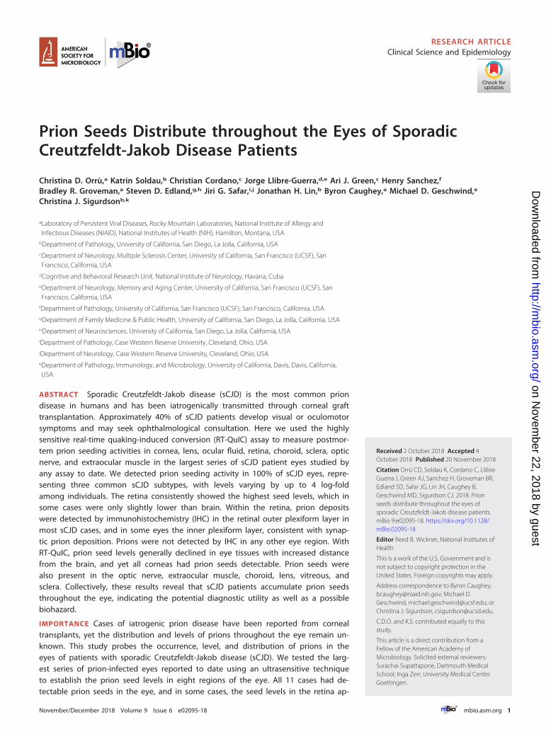
Prion Seeds Distribute throughout the Eyes of SporadicCreutzfeldt-Jakob Disease Patients
Christina D. Orrù,a Katrin Soldau,b Christian Cordano,c Jorge Llibre-Guerra,d,e Ari J. Green,c Henry Sanchez,f
Bradley R. Groveman,a Steven D. Edland,g,h Jiri G. Safar,i,j Jonathan H. Lin,b Byron Caughey,a Michael D. Geschwind,e
Christina J. Sigurdsonb,k
aLaboratory of Persistent Viral Diseases, Rocky Mountain Laboratories, National Institute of Allergy andInfectious Diseases (NIAID), National Institutes of Health (NIH), Hamilton, Montana, USA
bDepartment of Pathology, University of California, San Diego, La Jolla, California, USAcDepartment of Neurology, Multiple Sclerosis Center, University of California, San Francisco (UCSF), SanFrancisco, California, USA
dCognitive and Behavioral Research Unit, National Institute of Neurology, Havana, CubaeDepartment of Neurology, Memory and Aging Center, University of California, San Francisco (UCSF), SanFrancisco, California, USA
fDepartment of Pathology, University of California, San Francisco (UCSF), San Francisco, California, USAgDepartment of Family Medicine & Public Health, University of California, San Diego, La Jolla, California, USAhDepartment of Neurosciences, University of California, San Diego, La Jolla, California, USAiDepartment of Pathology, Case Western Reserve University, Cleveland, Ohio, USAjDepartment of Neurology, Case Western Reserve University, Cleveland, Ohio, USAkDepartment of Pathology, Immunology, and Microbiology, University of California, Davis, Davis, California,USA
ABSTRACT Sporadic Creutzfeldt-Jakob disease (sCJD) is the most common priondisease in humans and has been iatrogenically transmitted through corneal grafttransplantation. Approximately 40% of sCJD patients develop visual or oculomotorsymptoms and may seek ophthalmological consultation. Here we used the highlysensitive real-time quaking-induced conversion (RT-QuIC) assay to measure postmor-tem prion seeding activities in cornea, lens, ocular fluid, retina, choroid, sclera, opticnerve, and extraocular muscle in the largest series of sCJD patient eyes studied byany assay to date. We detected prion seeding activity in 100% of sCJD eyes, repre-senting three common sCJD subtypes, with levels varying by up to 4 log-foldamong individuals. The retina consistently showed the highest seed levels, which insome cases were only slightly lower than brain. Within the retina, prion depositswere detected by immunohistochemistry (IHC) in the retinal outer plexiform layer inmost sCJD cases, and in some eyes the inner plexiform layer, consistent with synap-tic prion deposition. Prions were not detected by IHC in any other eye region. WithRT-QuIC, prion seed levels generally declined in eye tissues with increased distancefrom the brain, and yet all corneas had prion seeds detectable. Prion seeds werealso present in the optic nerve, extraocular muscle, choroid, lens, vitreous, andsclera. Collectively, these results reveal that sCJD patients accumulate prion seedsthroughout the eye, indicating the potential diagnostic utility as well as a possiblebiohazard.
IMPORTANCE Cases of iatrogenic prion disease have been reported from cornealtransplants, yet the distribution and levels of prions throughout the eye remain un-known. This study probes the occurrence, level, and distribution of prions in theeyes of patients with sporadic Creutzfeldt-Jakob disease (sCJD). We tested the larg-est series of prion-infected eyes reported to date using an ultrasensitive techniqueto establish the prion seed levels in eight regions of the eye. All 11 cases had de-tectable prion seeds in the eye, and in some cases, the seed levels in the retina ap-
Received 2 October 2018 Accepted 4October 2018 Published 20 November 2018
Citation Orrú CD, Soldau K, Cordano C, Llibre-Guerra J, Green AJ, Sanchez H, Groveman BR,Edland SD, Safar JG, Lin JH, Caughey B,Geschwind MD, Sigurdson CJ. 2018. Prionseeds distribute throughout the eyes ofsporadic Creutzfeldt-Jakob disease patients.mBio 9:e02095-18. https://doi.org/10.1128/mBio.02095-18.
Editor Reed B. Wickner, National Institutes ofHealth
This is a work of the U.S. Government and isnot subject to copyright protection in theUnited States. Foreign copyrights may apply.
Address correspondence to Byron Caughey,[email protected]; Michael D.Geschwind, [email protected]; orChristina J. Sigurdson, [email protected].
C.D.O. and K.S. contributed equally to thisstudy.
This article is a direct contribution from aFellow of the American Academy ofMicrobiology. Solicited external reviewers:Surachai Supattapone, Dartmouth MedicalSchool; Inga Zerr, University Medical CenterGoettingen.
RESEARCH ARTICLEClinical Science and Epidemiology
crossm
November/December 2018 Volume 9 Issue 6 e02095-18 ® mbio.asm.org 1
on Novem
ber 22, 2018 by guesthttp://m
bio.asm.org/
Dow
nloaded from
proached those in brain. In most cases, prion deposits could also be seen by immu-nohistochemical staining of retinal tissue; other ocular tissues were negative. Ourresults have implications for estimating the risk for iatrogenic transmission of sCJDas well as for the development of antemortem diagnostic tests for prion diseases.
KEYWORDS Creutzfeldt-Jakob disease, RT-QuIC, eye, prion
Prion diseases are rapidly progressive neurodegenerative disorders with no availabletreatment (1–3). Creutzfeldt-Jakob disease is the most common prion disease in
humans, and is classified as sporadic, familial, or iatrogenic (4). Sporadic Creutzfeldt-Jakob disease (sCJD) has been transmitted iatrogenically from prion-contaminatedcorneal grafts, dura mater transplants, human cadaveric growth hormone, and neuro-surgical instruments (5–9). Although the underlying factors that initiate sCJD areunclear, the cause of prion disease is PrPSc, an infectious misfolded and aggregatedform of the cellular prion protein, PrPC (10, 11). PrPC is expressed ubiquitously with thehighest levels in neural tissues, including retina, although PrPC expression is higher inbrain than retina (12–15).
sCJD is often challenging to diagnose, in part due to the clinical and pathologicalheterogeneity in the disease phenotype (16–21). The disease is markedly influenced bythe PRNP prion gene sequence at polymorphic codon 129 (methionine/valine) and bythe PrPSc conformation, which is type 1 or type 2 depending on the protease-resistantaggregate core size (22, 23). sCJD has been divided into six phenotypic variants, orsubtypes, that differ by clinical presentation, rate of decline, and pathological targets inthe brain and are known as subtype MM1/MV1, VV2, MV2, MM2-cortical, MM2-thalamic,or VV1 (23–25). Approximately one-third of cases have both type 1 and type 2 PrPSc
(MM1/2, MV1/2, and VV1/2) (23).Visual disturbances are a common early presenting symptom of sCJD (approxi-
mately 10 to 20% of cases) often associated with the MM1/MV1 subtype (26, 27), andcan include diplopia, supranuclear palsies, and loss of vision (26, 28, 29). Throughoutthe disease course, more than 40% of cases have visual or oculomotor symptoms (26).In a limited number of reports, the electroretinogram sometimes shows a significantdecrease in the �-wave, potentially due to abnormalities in the outer plexiform layerwhere PrPSc has been detected (30, 31). By late-stage disease, blindness develops in 25to 42% of sCJD patients (26, 28), possibly from spongiform degeneration, neuronal loss,and PrPSc deposits in the thalamus (lateral geniculate) or primary visual cortex.
In addition to the CNS lesions that develop within the visual circuitry, prions havealso been directly detected in ocular tissues from a small number of cases, prior to thedevelopment of RT-QuIC. In two studies of variant CJD (vCJD), caused by the transmis-sion of bovine spongiform encephalopathy (BSE) to humans, PrPSc was readily detectedin the retina and optic nerve but not in the cornea, lens, vitreous fluid, or sclera byWestern blot or immunohistochemical labeling (15, 19). For sCJD, the few reportsdescribing prion detection in the eye show different results, potentially due to thesensitivity of the technique used. PrPSc was observed in the retina of two patients byimmunohistochemical labeling and Western blot (subtypes VV2 and MM1) (15, 32), butnot in a third patient studied by a highly sensitive Western blot assay (19). Notably,these studies were conducted prior to the development of the highly sensitive RT-QuICtechnique.
As the distribution and extent to which prions are established in the eyes of sCJDpatients remain unclear, the risk of iatrogenic prion transmission through ophthalmicprocedures is unknown (33, 34). For example, corneal transplantation is an increasinglycommon surgery, with 185,576 corneal transplantations performed in 116 countries in2012, more than ever reported previously (35). The United States leads in cornealprocurement and transplantation per capita, with approximately 64,000 corneal trans-plants per year (35). Corneal grafts from prion-infected patients have led to twoprobable and three possible cases of iatrogenic prion transmission, and prion infectivityhas been reported from human cornea inoculated into mice (36). The first reported case
Orrù et al. ®
November/December 2018 Volume 9 Issue 6 e02095-18 mbio.asm.org 2
on Novem
ber 22, 2018 by guesthttp://m
bio.asm.org/
Dow
nloaded from
of a corneal graft transmission of sCJD occurred in 1974, with death in the recipient27 months after transplantation. In this case, the diagnosis of CJD was confirmed atautopsy in the donor and recipient (37). Another probable transmission occurred in a45-year-old woman who developed sCJD after receiving a corneal transplant 30 yearsearlier from a donor who died from subacute spongiform encephalopathy (38). Threeadditional cases developed sCJD after corneal transplantation, with the diagnosisconfirmed at autopsy in the recipients, but with an incomplete history on the donors(39–41). These reports of iatrogenic prion spread prompted us to investigate thefrequency and level of prions in the eyes of sCJD patients to better understand thetransmission risk as well as the potential for diagnostic assay development.
Highly sensitive and specific biochemical assays have revolutionized PrPSc detectionand now enable the measurement of minute levels of PrPSc (attograms to femtograms)from tissues and body fluids, including nasal brushings and CSF (42, 43). The real-timequaking-induced conversion assay (RT-QuIC) detects the misfolding of recombinantmonomeric prion protein by prion aggregates or “seeds” within a prion-infectedsample (44, 45). Application of RT-QuIC assays to the antemortem diagnosis of sCJDusing CSF and nasal brushings has allowed provisional diagnostic sensitivities andspecificities approaching 100% (42, 43). Using this extraordinarily sensitive detectionmethod, we investigated the level and distribution of prion seeding activity in the eyetissues of eleven patients who died from sCJD.
RESULTSClinical features of sCJD patients. We analyzed the eyes from eleven cases of
pathologically confirmed sCJD representing three subtypes as well as six controlshaving nonprion diseases. All eleven patients were enrolled in the UCSF Memory andAging Center clinical prion research program in a study approved by the UCSFCommittee on Human Research. Participants were evaluated clinically for one or morevisits for neurologic deficits as well as for PRNP mutations and genotype, and diagnos-tically by CSF analysis, EEG, and brain MRI. None had mutations in PRNP or any historyof a known iatrogenic exposure to prions, and the clinical and diagnostic features wereconsistent with sporadic CJD. At PRNP polymorphic codon 129, all three genotypeswere represented: MM (n � 5), MV (n � 5), and VV (n � 1).
Three of eleven cases (27%) had visual disturbances such as transient monocularblindness and blurry or double vision (patients 1, 2, and 5), three had impairedvisuospatial skills (patients 2, 9, and 11), and no patients had visual field deficits(Table 1). Four of 11 cases had visual signs, including nystagmus, slow ocular pursuit,and increased saccade latency, but these were not considered visual symptoms. Retinalimaging using optical coherence tomography was performed on three sCJD patients(patients 6, 10, and 11) and revealed a modest decrease in peripapillary retinal nervefiber (pRNFL) layer thickness compared to 20 healthy control eyes (age-balanced
TABLE 1 Patient demographic data and clinical features
Patientno.
Age ofonset (yr)
Diseaseduration (mo) Gender
PRNP genotypeat codon 129
sCJDsubtype
Clinical signsat onset
Visualsymptomsa
Visuospatialdysfunctionb
1 60 1.5 M MM 1 Cognitive/visual Yes No2 60 20 M MV 1-2 Visuospatial Yes Yes3 69 15 M MV 2 Behavior/memory No No4 79 27 M MV 2 Cognitive No No5 55 6 F VV 2 Apraxia Yes No6 56 10 F MM 1-2 Language No No7 57 4 F MM 1 Motor No No8 55 24 F MV 1-2 Behavior No No9 63 2 F MM 1 Language No Yes10 69 6 F MV 1 Behavior/memory No No11 69 10 F MM 1-2 Cognitive/apraxia No YesaVisual symptoms noted at first through last UCSF visit.bVisuospatial dysfunction based on neuropsychological testing or neurological examination.
sCJD Prions Distribute throughout the Eye ®
November/December 2018 Volume 9 Issue 6 e02095-18 mbio.asm.org 3
on Novem
ber 22, 2018 by guesthttp://m
bio.asm.org/
Dow
nloaded from
reference population) (mean � SE: 93.8 � 2.2 �m and 102.0 � 2.3 �m, respectively;P � 0.07) (see Fig. S1 in the supplemental material).
Diffusion MRI revealed focal regions of high cortical intensity (cortical ribboning)with restricted diffusion in all cases, with three cases showing cortical ribboning in theoccipital (visual) cortex (patients 2, 4, and 11). In addition to cortical involvement, fivecases had both striatal and thalamic involvement (patients 2, 3, 6, 8, and 10), three hadstriatal involvement (patients 4, 7, and 9), and one had thalamic involvement (patient6). The diagnosis of sCJD was confirmed biochemically by PrPSc detection in the brain(Fig. S2 and S3).
PrPSc levels in six brain regions. PrPSc levels in the occipital cortex varied amongthe patients, with the type 1 cases having higher PrPSc levels than the type 2 cases(Fig. S2). We also measured PrPSc by Western blotting samples from six brain regionsfrom each patient, specifically parietal and occipital or frontal cortex, basal ganglia,thalamus, hippocampus, and cerebellum. The PrPSc levels in each brain region variedwithin a patient. The thalamus and occipital cortex commonly accumulated high levelsof PrPSc, and the cerebellum showed the lowest levels in eight of ten cases examined(Fig. S3).
Prion seeding activity measured by RT-QuIC. We next tested the PrP seedingactivity by RT-QuIC analysis in eight eye regions, which were extraocular muscle, opticnerve, cornea, lens, vitreous fluid, retina, choroid, and sclera. We found that all elevensCJD patient eyes were positive for prion seeding activity and showed the highestseeding activity in the retina (Fig. 1). Interestingly, an endpoint dilution assay revealedthat seeding activity in eye tissues varied among the patients by up to 4 log-fold(Fig. 1). Prion seed levels in the retina were significantly higher than the extraocularmuscles, optic nerve, cornea, lens, vitreous fluid, and sclera (Fig. 1 and 2) (P � 0.0001,ANOVA with Tukey’s posttest). The lens and vitreous fluid tended to show the lowestseeding activity in most patients and were negative in three patients and one patient,respectively. The high seed levels in the vitreous from two patients may have been dueto spread from the retinal layer, as both patients had very high seed levels in the retina.Similarly, the choroid consistently showed high seed levels that were significantlyhigher than cornea, lens, and vitreous fluid; prion seeds emanating from the retinallayer may have contributed to the high levels. The cornea and extraocular muscle had
FIG 1 Comparison of prion seed levels in brain (temporal cortex) and ocular compartments fromposterior to anterior eye as measured by RT-QuIC analysis. (A) Image showing the ocular tissues testedby RT-QuIC. (B) The seeding dose 50 per mg of tissue (logSD50) is shown. The average prion seed levelin eye was highest in the retina, and lowest in the vitreous and lens. One vitreous sample, 3 lens samples,and 1 muscle sample were negative and are not shown. Retinal prion seed levels were significantlyhigher than most other ocular tissues, including optic nerve, sclera, lens, cornea, vitreous, and extraocularmuscle. The prion seed level in a representative sCJD brain sample (termporal cortex) is shown forcomparison. For retina, optic nerve, sclera, cornea, vitreous, and extraocular muscle, n � 11; for choroid,n � 10; and for lens, n � 9. *, P � 0.05; **, P � 0.01; ***, P � 0.001, one-way ANOVA with Tukey’s multiplecomparison test. Graphics in panel A by Ryan Kissinger.
Orrù et al. ®
November/December 2018 Volume 9 Issue 6 e02095-18 mbio.asm.org 4
on Novem
ber 22, 2018 by guesthttp://m
bio.asm.org/
Dow
nloaded from
lower seeding activity relative to the retina (Fig. 1 and 2). None of the six control casesshowed seeding activity in any ocular tissue (Fig. 2).
Prion seeding activity in the retina ranged from levels 101-fold to 104-fold lower thansCJD brain (Fig. 1), potentially due to lower PrPC expression, and higher seeding activitydid not correlate with the presence of visual symptoms. We assessed whether theseeding activity in the retina correlated with age, gender, disease duration, PRNPgenotype, or sCJD subtypes, but found no evidence of a correlation (Fig. S4). A previousreport suggested that the sCJD subtype might influence the levels of prion deposits inretina (32). We found that the VV2 subtype showed the highest seed levels in retina,with the MM1-2 and MV1-2 mixtures also tending to show high levels. The MM/MV1cases instead showed a broad distribution of seeding activity throughout the eye.
Retinal PrPSc distribution by immunohistochemistry. To determine the distribu-
tion of prions within the retina, we immunolabeled all eyes for PrP. Nearly all sCJDpatients showed PrPSc deposits in the retina (9 of 11, 82%, of cases) with discreteaggregates visible in the plexiform layers, which consist of dense networks of neuronalsynapses. All nine positive retina cases had PrPSc deposits in the outer plexiform layer,whereas one case (VV2) showed intense staining and also developed strong PrPSc
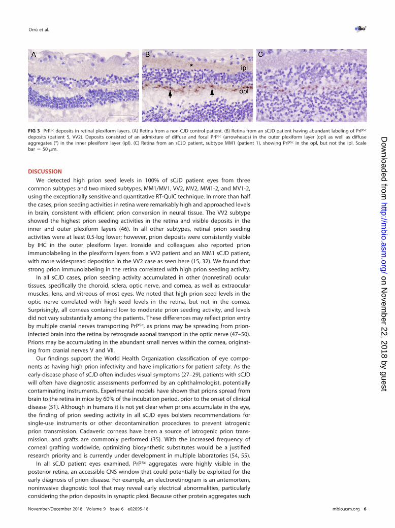
labeling in the inner plexiform layer (Fig. 3). The other subtypes primarily differed in thestaining intensity, but not the stain distribution. Deposits typically appeared as focaloval intensely stained aggregates approximately 4 to 5 �m in diameter at evenlyspaced intervals every 15 to 20 �m (Fig. 3). The VV2 case also showed fine granulardeposits throughout the plexiform layers. No PrPSc deposits were visible in other(nonretinal) ocular tissues in any case. Neither the non-CJD control eyes (Fig. 3) nor thecontrol IgG isotype-labeled sCJD eyes showed any PrPSc deposition.
FIG 2 RT-QuIC of retina, cornea, and lens from sCJD and non-sCJD patients. (A) The average prionseeding amplification kinetics are shown for retina (blue), cornea (red), and lens (orange) from sCJD(circles) and non-CJD (X’s) patients. The dotted line indicates the ThT fluorescence threshold for a positiveresult (see Materials and Methods). (B) The maximum ThT fluorescence reached within 24 hours is shown.The thin lines represent mean and standard deviation, whereas the dotted line indicates the ThTfluorescence threshold for a positive result. (C) The time to reach the threshold for positivity is shown foreach sample. The dotted line indicates the end of the 24-hour experiment. If a sample did not reach thethreshold within 24 h, it was marked as 25 h to indicate a negative result. n � 4 (lens) or 6 (retina, cornea)non-CJD control cases were analyzed.
sCJD Prions Distribute throughout the Eye ®
November/December 2018 Volume 9 Issue 6 e02095-18 mbio.asm.org 5
on Novem
ber 22, 2018 by guesthttp://m
bio.asm.org/
Dow
nloaded from
DISCUSSION
We detected high prion seed levels in 100% of sCJD patient eyes from threecommon subtypes and two mixed subtypes, MM1/MV1, VV2, MV2, MM1-2, and MV1-2,using the exceptionally sensitive and quantitative RT-QuIC technique. In more than halfthe cases, prion seeding activities in retina were remarkably high and approached levelsin brain, consistent with efficient prion conversion in neural tissue. The VV2 subtypeshowed the highest prion seeding activities in the retina and visible deposits in theinner and outer plexiform layers (46). In all other subtypes, retinal prion seedingactivities were at least 0.5-log lower; however, prion deposits were consistently visibleby IHC in the outer plexiform layer. Ironside and colleagues also reported prionimmunolabeling in the plexiform layers from a VV2 patient and an MM1 sCJD patient,with more widespread deposition in the VV2 case as seen here (15, 32). We found thatstrong prion immunolabeling in the retina correlated with high prion seeding activity.
In all sCJD cases, prion seeding activity accumulated in other (nonretinal) oculartissues, specifically the choroid, sclera, optic nerve, and cornea, as well as extraocularmuscles, lens, and vitreous of most eyes. We noted that high prion seed levels in theoptic nerve correlated with high seed levels in the retina, but not in the cornea.Surprisingly, all corneas contained low to moderate prion seeding activity, and levelsdid not vary substantially among the patients. These differences may reflect prion entryby multiple cranial nerves transporting PrPSc, as prions may be spreading from prion-infected brain into the retina by retrograde axonal transport in the optic nerve (47–50).Prions may be accumulating in the abundant small nerves within the cornea, originat-ing from cranial nerves V and VII.
Our findings support the World Health Organization classification of eye compo-nents as having high prion infectivity and have implications for patient safety. As theearly-disease phase of sCJD often includes visual symptoms (27–29), patients with sCJDwill often have diagnostic assessments performed by an ophthalmologist, potentiallycontaminating instruments. Experimental models have shown that prions spread frombrain to the retina in mice by 60% of the incubation period, prior to the onset of clinicaldisease (51). Although in humans it is not yet clear when prions accumulate in the eye,the finding of prion seeding activity in all sCJD eyes bolsters recommendations forsingle-use instruments or other decontamination procedures to prevent iatrogenicprion transmission. Cadaveric corneas have been a source of iatrogenic prion trans-mission, and grafts are commonly performed (35). With the increased frequency ofcorneal grafting worldwide, optimizing biosynthetic substitutes would be a justifiedresearch priority and is currently under development in multiple laboratories (54, 55).
In all sCJD patient eyes examined, PrPSc aggregates were highly visible in theposterior retina, an accessible CNS window that could potentially be exploited for theearly diagnosis of prion disease. For example, an electroretinogram is an antemortem,noninvasive diagnostic tool that may reveal early electrical abnormalities, particularlyconsidering the prion deposits in synaptic plexi. Because other protein aggregates such
FIG 3 PrPSc deposits in retinal plexiform layers. (A) Retina from a non-CJD control patient. (B) Retina from an sCJD patient having abundant labeling of PrPSc
deposits (patient 5, VV2). Deposits consisted of an admixture of diffuse and focal PrPSc (arrowheads) in the outer plexiform layer (opl) as well as diffuseaggregates (*) in the inner plexiform layer (ipl). (C) Retina from an sCJD patient, subtype MM1 (patient 1), showing PrPSc in the opl, but not the ipl. Scalebar � 50 �m.
Orrù et al. ®
November/December 2018 Volume 9 Issue 6 e02095-18 mbio.asm.org 6
on Novem
ber 22, 2018 by guesthttp://m
bio.asm.org/
Dow
nloaded from
as amyloid-�, �-synuclein, and tau may also spread from brain to retina, it would alsobe important to continue to evaluate eyes from patients having more commonneurodegenerative diseases, such as Alzheimer’s disease, synucleinopathies, andtauopathies, particularly in light of recent findings showing the prion-like spread ofprotein aggregates through the CNS (56–59).
MATERIALS AND METHODSPatients with sporadic Creutzfeldt-Jakob disease and controls. This prospective study of sCJD
patients was initiated in July 2015 and continued through July 2017. Patients were referred to the UC SanFrancisco (UCSF) Memory and Aging Center for rapidly progressive neurologic disease. All patients hadextensive clinical testing, including brain MRI, CSF analysis for 14-3-3, neuron-specific enolase (NSE), andtotal tau. All were classified as probable sCJD by UCSF clinical and radiological diagnostic criteria (60, 61)and were ruled out for genetic prion disease by PRNP analysis (done through the National Prion DiseasePathology Surveillance Center [NPDPSC], Case Western Reserve University, Cleveland, OH). The mean ageof sCJD patients at disease onset was 63 � 2 years (mean � SE), and consisted of seven women and fourmen. The duration of clinical neurologic signs ranged from 1.5 to 27 months (mean � SD: 11 �9 months). Control cases consisted of five men and one woman and ranged from 51 to 90 years old(mean � SD: 70 � 14 years). Two of the control cases died of neurodegenerative disease (Alzheimer’sdisease) and four controls died of nonneurologic disease (neoplasia).
Study oversight. This study was approved by the ethics committee at UC San Francisco. Informedconsent was received from all patients (IRB Study Number: 10-04905). All ocular and brain tissuesexamined at UCSD and NIAID were obtained on autopsy; this testing was therefore exempt from reviewby the NIH Office of Human Subjects Research Protections.
PRNP genotyping. The open reading frame of the PRNP gene was sequenced from all patientsamples to test for any mutations in the PrP sequence and to determine the genotype at polymorphiccodon 129 (methionine or valine; performed at the NPDPSC, Case Western Reserve University, Cleveland,OH). Genomic DNA was extracted from frozen brain tissue samples using Qiagen QIAamp DNA minikit(Qiagen, Gaithersburg, MD) according to the manufacturer’s protocol and a 760-bp fragment corre-sponding to the human PrP gene (residues 5 to 258) was PCR amplified using primers HRM-F (5=-TATGTGGACTGATGTCGGCCTCTGCAAGAAGCGC-3=) and HRM-R (5=-CCACCTCAATTGAAAGGGCTGCAGGTGGATAC-3=) with defined cycling conditions (62, 63). The Met/Val polymorphism at codon 129 and mutationof the PrP gene coding region were determined by deep (63) or direct Sanger sequencing as previouslydescribed (22, 62). Nucleotide sequences from both deep and Sanger sequencing were analyzed usingDNAStar Lasergene Software Suite v.7.1.0 (Madison, WI). There were no mutations discovered in any ofthe patients. Patients consisted of 129 MM (5), MV (5), and VV (1).
Optical coherence tomography. Antemortem retinal imaging was performed bilaterally using Spect-ralis spectral-domain OCT (Heidelberg Engineering, Heidelberg, Germany, Eye Explorer software version1.9.10.0) by two trained technicians under standard ambient conditions (illuminance level of 80 to 100 foot-candles). Peripapillary retinal nerve fiber layer (RNFL) thickness was obtained with a 360° RNFL-B circle scan(100 ART; 1,536 A scan per B scan) located at 3.4 cm from the center of the optic nerve head.
Macular volumetric scans consisting of 19 single horizontal axial B-scans (ART � 9; 1,536 A scan perB scan) were acquired in a 20- by 15-degree raster horizontal scan centered on the fovea. Intraretinallayer segmentation was executed to quantify macular RNFL, ganglion cell layer (GCL), inner nuclear layer(INL), inner plexiform layer (IPL), and outer plexiform layer (OPL) through the Viewing Module 6.0 in asemiautomatic way, with manual correction of software errors. Scans that violated international consen-sus quality control criteria (OSCAR-IB) were excluded from the analysis (64, 65). We followed the APOSTELguidelines to report OCT studies (66).
Brain and ocular tissue collection. Ocular and brain tissues were collected at autopsy from elevensCJD patients and six controls. One eye was immediately frozen and the second eye was formalin-fixed.Brain sections were collected from six to nine brain regions and frozen. The formalin-fixed eye tissueswere immersed in 98% formic acid for 1 h, and postfixed in formalin prior to paraffin-embedding andsectioning. Frozen eyes were thawed and the following tissue sections were collected for analysis byRT-QuIC using clean sterile blades to avoid contamination among the tissues: extraocular muscle, opticnerve, vitreous fluid, lens, cornea, retina, choroid, and sclera.
Immunohistochemistry for PrPSc in the eye. Four-�m sections of brain were cut onto positivelycharged silanized glass slides and stained with hematoxylin and eosin or immunostained using antibodies forPrP (12F10). For PrP staining, sections were deparaffinized and incubated for 5 min in 96% formic acid, thenwashed in water for 5 min, treated with 5 �g/ml of proteinase K for 7 min, and washed in water for 5 min.Sections were then placed in citrate buffer (pH 6) and heated in a pressure cooker for 20 min, cooled for 5min, and rinsed in distilled water. Sections were incubated with anti-PrP 12F10 (Cayman Chemical; 1:200) for45 min followed by anti-mouse IgG conjugated to biotin (Jackson Immunolabs; 1:250) for 30 min, followedby streptavidin-HRP (Jackson Immunolabs; 1:2,000) for 30 min. Sections were then incubated with DABreagent (Thermo Fisher Scientific) and counterstained with hematoxylin.
Western blot for PrPSc in the brain. Brain samples were homogenized in phosphate-buffered saline(PBS) (20% brain homogenate final [wt/vol]) using a Beadbeater tissue homogenizer. PrPSc was concen-trated from brain samples by performing sodium phosphotungstic acid (NaPTA) precipitation prior toWestern blotting (19). Briefly, 50-�l aliquots of 10% brain homogenate in an equal volume of 4% sarkosylin PBS were incubated for 30 min at room temperature, then digested with an endonuclease (Benzonase[Sigma]) and with 100 �g/ml proteinase K at 37°C for 30 min. After addition of NaPTA, MgCl2, and
sCJD Prions Distribute throughout the Eye ®
November/December 2018 Volume 9 Issue 6 e02095-18 mbio.asm.org 7
on Novem
ber 22, 2018 by guesthttp://m
bio.asm.org/
Dow
nloaded from
protease inhibitors (Complete, Roche), extracts were incubated at 37°C for 30 min and centrifuged at18,000 � g for 30 min at 37°C. Pellets were resuspended in 0.1% sarkosyl in PBS prior to electrophoresisand blotting. Membranes were incubated with monoclonal antibody POM1 (discontinuous epitope atC-terminal domain [67]) followed by incubation with an HRP-conjugated anti-mouse IgG secondaryantibody (Jackson Immunolabs). The blots were developed using a chemiluminescent substrate (ECLdetection kit, Thermo Scientific) and visualized on a Fuji LAS 4000 imager. Quantification of PrPSc signalwas performed using Multigauge V3 software (Fujifilm).
PrPSc subtyping. sCJD is classified into six main subtypes and three mixed subtypes, depending onthe codon 129 polymorphism and the electrophoretic profile of the protease-resistant PrPSc corefragment size, which is 21 kDa (type 1) or 19 kDa (type 2) (23). We assessed the PK-resistant core size andcompared the PrPSc levels in the occipital cortex among the patients. Three sCJD subtypes wererepresented, consisting of MM1/MV1 (n � 4), MV2 (n � 2), and VV2 (n � 1), as well as MM1-2 and MV1-2mixtures (n � 4).
Ocular tissue homogenate preparation for RT-QuIC analysis. Eye homogenates (10% [wt/vol])were prepared in PBS containing 2 mM CaCl2 and 0.25% (wt/vol) collagenase A (Roche). Glass homog-enization beads (1 mm; Biospec) were added to the sample in homogenization buffer and tissues werehomogenized for 1 min using a Beadbeater tissue homogenizer (Biospec). The samples were thenincubated at 37°C under shaking conditions overnight, homogenized for 1 min, and centrifuged at2,000 � g for 2 min. The supernatant was transferred to a new tube and stored at �80°C for subsequentanalysis.
RT-QuIC analysis for PrP seeding activity in eye tissues. The RT-QuIC reaction mix was composedof 10 mM phosphate buffer (pH 7.4), 300 mM NaCl, 0.1 mg/ml recombinant Syrian hamster PrP (residues90 to 230; rPrPSen; GenBank accession number K02234), 10 �M thioflavin T (ThT), 1 mM EDTA, and 0.001%SDS. Aliquots of the reaction mix (98 �l) were loaded into each well of a black 96-well plate with a clearbottom (Nunc) and seeded with 2 �l of eye tissue homogenates at different dilutions. The plate wassealed (plate sealer film, Nalgene Nunc International) and incubated at 55°C in a BMG FLUOstar Omegaplate reader at cycles of 1 min shaking (700 rpm double orbital) and 1 min rest. ThT fluorescencemeasurements (450 � 10 nm excitation and 480 � 10 nm emission; bottom read) were taken every 45min. ThT fluorescence threshold for a positive result was calculated as the mean of all values fromnegative eye tissues plus three standard deviations. For quantitation, endpoint dilution assays wereperformed using Spearman-Kärber analyses to estimate the seeding dose (� SE) giving ThT positivity in50% of technical replicate wells (SD50) (44, 68).
Statistical analysis. Data are presented as mean � SEM unless otherwise indicated with groupdifferences tested using standard parametric methods (one-way ANOVA with Tukey’s multiple compar-ison test). P values of less than 0.05 were considered statistically significant.
Data availability. The authors will make data fully available and without restriction.
SUPPLEMENTAL MATERIALSupplemental material for this article may be found at https://doi.org/10.1128/mBio
.02095-18.FIG S1, TIF file, 2.2 MB.FIG S2, TIF file, 0.6 MB.FIG S3, TIF file, 0.9 MB.FIG S4, TIF file, 0.4 MB.
ACKNOWLEDGMENTSWe thank Peter Kobalka, Don Pizzo, Taylor Winrow, and the histology team at Case
Western Reserve University for technical support. The authors thank Ryan Kissinger forgraphics assistance in Fig. 1.
Byron Caughey holds a patent on RT-QuIC technology that has been licensed byAmprion Inc. Christina Sigurdson’s involvement with Amydis, Inc., is limited to mem-bership on the Scientific Advisory Board. The terms of this arrangement have beenreviewed and approved by the University of California, San Diego in accordance withits conflict of interest policies. Michael Geschwind has consulted for Quest Diagnostics,Advanced Medical Inc., Best Doctors Inc., Grand Rounds Inc., Second Opinion Inc.,Gerson Lehrman Group Inc., Guidepoint Global LLC, MEDACorp., LCN Consulting, OptioBiopharma Solutions, Teva Pharmaceuticals, Biohaven Pharmaceuticals, Quest Diagnos-tics, and various medical-legal consulting firms. He has received speaking honoraria forvarious medical center lectures and from Oakstone Publishing. He has received pastresearch support from Alliance Biosecure, CurePSP, the Tau Consortium, Quest Diag-nostics, and NIH.
This work was supported by National Institutes of Health grants NS069566 (C.J.S.),NS076896 (C.J.S.), R01 AG031189 (M.D.G.), R01NS088485 (J.H.L.), the Michael J. HomerFamily Fund (M.D.G.), the Intramural Research Program of the NIAID (B.C.), the Equity in
Orrù et al. ®
November/December 2018 Volume 9 Issue 6 e02095-18 mbio.asm.org 8
on Novem
ber 22, 2018 by guesthttp://m
bio.asm.org/
Dow
nloaded from
Brain Health at the Global Brain Health Institute (J.L.-G.), and a gift to NIAID (B.C.) fromMary Hilderman Smith, Zoë Smith Jaye, and Jenny Smith Unruh in memory of JeffreySmith.
REFERENCES1. Prusiner SB. 1998. Prions. Proc Natl Acad Sci U S A 95:13363–13383.
https://doi.org/10.1073/pnas.95.23.13363.2. DeArmond SJ, Prusiner SB. 1995. Etiology and pathogenesis of prion
diseases. Am J Pathol 146:785– 811.3. Collinge J. 2016. Mammalian prions and their wider relevance in neuro-
degenerative diseases. Nature 539:217–226. https://doi.org/10.1038/nature20415.
4. Takada LT, Geschwind MD. 2013. Prion diseases. Semin Neurol 33:348 –356. https://doi.org/10.1055/s-0033-1359314.
5. Ironside JW, Ritchie DL, Head MW. 2017. Prion diseases. Handb Clin Neurol145:393–403. https://doi.org/10.1016/B978-0-12-802395-2.00028-6.
6. Brown P, Preece M, Brandel J-P, Sato T, McShane L, Zerr I, Fletcher A, WillRG, Pocchiari M, Cashman NR, d’Aignaux JH, Cervenakova L, Fradkin J,Schonberger LB, Collins SJ. 2000. Iatrogenic Creutzfeldt-Jakob disease atthe millennium. Neurology 55:1075–1081. https://doi.org/10.1212/WNL.55.8.1075.
7. Collinge J, Palmer MS, Dryden AJ. 1991. Genetic predisposition to iatro-genic Creutzfeldt-Jakob disease. Lancet 337:1441–1442. https://doi.org/10.1016/0140-6736(91)93128-V.
8. Will RG. 2003. Acquired prion disease: iatrogenic CJD, variant CJD, kuru,p 255–265. In Weissmann C, Aguzzi A, Dormont D, Hunter N (ed), Prionsfor physicians. Oxford University Press, Oxford, United Kingdom.
9. Hoshi K, Yoshino H, Urata J, Nakamura Y, Yanagawa H, Sato T. 2000.Creutzfeldt-Jakob disease associated with cadaveric dura mater grafts inJapan. Neurology 55:718 –721. https://doi.org/10.1212/WNL.55.5.718.
10. Prusiner SB. 1982. Novel proteinaceous infectious particles cause scrapie.Science 216:136 –144. https://doi.org/10.1126/science.6801762.
11. Prusiner SB, Scott M, Foster D, Pan KM, Groth D, Mirenda C, Torchia M,Yang SL, Serban D, Carlson GA. 1990. Transgenetic studies implicateinteractions between homologous PrP isoforms in scrapie prion replica-tion. Cell 63:673– 686. https://doi.org/10.1016/0092-8674(90)90134-Z.
12. Bendheim PE, Brown HR, Rudelli RD, Scala LJ, Goller NL, Wen GY, KascsakRJ, Cashman NR, Bolton DC. 1992. Nearly ubiquitous tissue distributionof the scrapie agent precursor protein. Neurology 42:149 –156. https://doi.org/10.1212/WNL.42.1.149.
13. Fournier JG. 2001. Nonneuronal cellular prion protein. Int Rev Cytol208:121–160. https://doi.org/10.1016/S0074-7696(01)08003-2.
14. Gong J, Jellali A, Forster V, Mutterer J, Dubus E, Altrock WD, Sahel JA,Rendon A, Picaud S. 2007. The toxicity of the PrP106-126 prion peptideon cultured photoreceptors correlates with the prion protein distribu-tion in the mammalian and human retina. Am J Pathol 170:1314 –1324.https://doi.org/10.2353/ajpath.2007.060340.
15. Head MW, Northcott V, Rennison K, Ritchie D, McCardle L, Bunn TJ,McLennan NF, Ironside JW, Tullo AB, Bonshek RE. 2003. Prion proteinaccumulation in eyes of patients with sporadic and variant Creutzfeldt-Jakob disease. Invest Ophthalmol Vis Sci 44:342–346. https://doi.org/10.1167/iovs.01-1273.
16. Parchi P, Strammiello R, Giese A, Kretzschmar H. 2011. Phenotypicvariability of sporadic human prion disease and its molecular basis: past,present, and future. Acta Neuropathol 121:91–112. https://doi.org/10.1007/s00401-010-0779-6.
17. Ironside JW, Ritchie DL, Head MW. 2005. Phenotypic variability in humanprion diseases. Neuropathol Appl Neurobiol 31:565–579. https://doi.org/10.1111/j.1365-2990.2005.00697.x.
18. Head MW, Bunn TJ, Bishop MT, McLoughlin V, Lowrie S, McKimmie CS,Williams MC, McCardle L, MacKenzie J, Knight R, Will RG, Ironside JW.2004. Prion protein heterogeneity in sporadic but not variantCreutzfeldt-Jakob disease: UK cases 1991–2002. Ann Neurol 55:851– 859.https://doi.org/10.1002/ana.20127.
19. Wadsworth JDF, Joiner S, Hill AF, Campbell TA, Desbruslais M, Luthert PJ,Collinge J. 2001. Tissue distribution of protease resistant prion protein invariant CJD using a highly sensitive immuno-blotting assay. Lancet358:171–180. https://doi.org/10.1016/S0140-6736(01)05403-4.
20. Paterson RW, Torres-Chae CC, Kuo AL, Ando T, Nguyen EA, Wong K,DeArmond SJ, Haman A, Garcia P, Johnson DY, Miller BL, Geschwind MD.
2012. Differential diagnosis of Jakob-Creutzfeldt disease. Arch Neurol69:1578 –1582. https://doi.org/10.1001/2013.jamaneurol.79.
21. Geschwind MD. 2010. Rapidly progressive dementia: prion diseases andother rapid dementias. Continuum (Minneap Minn) 16:31–56. https://doi.org/10.1212/01.CON.0000368211.79211.4c.
22. Parchi P, Zou W, Wang W, Brown P, Capellari S, Ghetti B, Kopp N,Schulz-Schaeffer WJ, Kretzschmar HA, Head MW, Ironside JW, GambettiP, Chen SG. 2000. Genetic influence on the structural variations of theabnormal prion protein. Proc Natl Acad Sci U S A 97:10168 –10172.https://doi.org/10.1073/pnas.97.18.10168.
23. Parchi P, Giese A, Capellari S, Brown P, Schulz-Schaeffer W, Windl O, ZerrI, Budka H, Kopp N, Piccardo P, Poser S, Rojiani A, Streichemberger N,Julien J, Vital C, Ghetti B, Gambetti P, Kretzschmar H. 1999. Classificationof sporadic Creutzfeldt-Jakob disease based on molecular and pheno-typic analysis of 300 subjects. Ann Neurol 46:224 –233. https://doi.org/10.1002/1531-8249(199908)46:2�224::AID-ANA12�3.0.CO;2-W.
24. Collins SJ, Sanchez-Juan P, Masters CL, Klug GM, van Duijn C, Poleggi A,Pocchiari M, Almonti S, Cuadrado-Corrales N, de Pedro-Cuesta J, BudkaH, Gelpi E, Glatzel M, Tolnay M, Hewer E, Zerr I, Heinemann U, Kretszch-mar HA, Jansen GH, Olsen E, Mitrova E, Alpérovitch A, Brandel J-P,Mackenzie J, Murray K, Will RG. 2006. Determinants of diagnostic inves-tigation sensitivities across the clinical spectrum of sporadic Creutzfeldt-Jakob disease. Brain 129:2278 –2287. https://doi.org/10.1093/brain/awl159.
25. Puoti G, Bizzi A, Forloni G, Safar JG, Tagliavini F, Gambetti P. 2012.Sporadic human prion diseases: molecular insights and diagnosis. Lan-cet Neurol 11:618 – 628. https://doi.org/10.1016/S1474-4422(12)70063-7.
26. Brown P, Gibbs CJ, Jr, Rodgers Johnson P, Asher DM, Sulima MP, BacoteA, Goldfarb LG, Gajdusek DC. 1994. Human spongiform encephalopathy:the National Institutes of Health series of 300 cases of experimentallytransmitted disease. Ann Neurol 35:513–529. https://doi.org/10.1002/ana.410350504.
27. Rabinovici GD, Wang PN, Levin J, Cook L, Pravdin M, Davis J, DeArmondSJ, Barbaro NM, Martindale J, Miller BL, Geschwind MD. 2006. Firstsymptom in sporadic Creutzfeldt-Jakob disease. Neurology 66:286 –287.https://doi.org/10.1212/01.wnl.0000196440.00297.67.
28. Armstrong RA. 2006. Creutzfeldt-Jakob disease and vision. Clin ExpOptom 89:3–9. https://doi.org/10.1111/j.1444-0938.2006.00001.x.
29. Lueck CJ, McIlwaine GG, Zeidler M. 2000. Creutzfeldt-Jakob disease andthe eye. II. Ophthalmic and neuro-ophthalmic features. Eye 14:291–301.https://doi.org/10.1038/eye.2000.76.
30. Katz BJ, Warner JE, Digre KB, Creel DJ. 2000. Selective loss of the electro-retinogram B-wave in a patient with Creutzfeldt-Jakob disease. J Neurooph-thalmol 20:116–118. https://doi.org/10.1097/00041327-200020020-00011.
31. de Seze J, Hache JC, Vermersch P, Arndt CF, Maurage CA, Pasquier F,Laplanche JL, Ruchoux MM, Leys D, Destee A, Petit H. 1998. Creutzfeldt-Jakob disease: neurophysiologic visual impairments. Neurology 51:962–967. https://doi.org/10.1212/WNL.51.4.962.
32. Head MW, Peden AH, Yull HM, Ritchie DL, Bonshek RE, Tullo AB, IronsideJW. 2005. Abnormal prion protein in the retina of the most commonlyoccurring subtype of sporadic Creutzfeldt-Jakob disease. Br J Ophthal-mol 89:1131–1133. https://doi.org/10.1136/bjo.2004.063495.
33. Armitage WJ, Tullo AB, Ironside JW. 2009. Risk of Creutzfeldt-Jakobdisease transmission by ocular surgery and tissue transplantation. Eye(Lond) 23:1926 –1930. https://doi.org/10.1038/eye.2008.381.
34. Lim R, Dhillon B, Kurian KM, Aspinall PA, Fernie K, Ironside JW. 2003.Retention of corneal epithelial cells following Goldmann tonometry:implications for CJD risk. Br J Ophthalmol 87:583–586. https://doi.org/10.1136/bjo.87.5.583.
35. Gain P, Jullienne R, He Z, Aldossary M, Acquart S, Cognasse F, Thuret G.2016. Global survey of corneal transplantation and eye banking. JAMAOphthalmol 134:167–173. https://doi.org/10.1001/jamaophthalmol.2015.4776.
36. Tateishi J. 1985. Transmission of Creutzfeldt-Jakob disease from humanblood and urine into mice. Lancet ii:1074.
37. Duffy P, Wolf J, Collins G, DeVoe AG, Streeten B, Cowen D. 1974. Possible
sCJD Prions Distribute throughout the Eye ®
November/December 2018 Volume 9 Issue 6 e02095-18 mbio.asm.org 9
on Novem
ber 22, 2018 by guesthttp://m
bio.asm.org/
Dow
nloaded from
person-to-person transmission of Creutzfeldt-Jakob disease. N Engl JMed 290:692– 693.
38. Heckmann JG, Lang CJ, Petruch F, Druschky A, Erb C, Brown P, Neun-dorfer B. 1997. Transmission of Creutzfeldt-Jakob disease via a cornealtransplant. J Neurol Neurosurg Psychiatry 63:388 –390. https://doi.org/10.1136/jnnp.63.3.388.
39. Rabinstein AA, Whiteman ML, Shebert RT. 2002. Abnormal diffusion-weighted magnetic resonance imaging in Creutzfeldt-Jakob diseasefollowing corneal transplantations. Arch Neurol 59:637– 639. https://doi.org/10.1001/archneur.59.4.637.
40. Hammersmith KM, Cohen EJ, Rapuano CJ, Laibson PR. 2004. Creutzfeldt-Jakob disease following corneal transplantation. Cornea 23:406 – 408.https://doi.org/10.1097/00003226-200405000-00019.
41. Uchiyama KIC, Yago S, Kurumaya H, Kitamoto T. 1994. An autopsy caseof Creutzfeldt-Jakob disease associated with corneal transplantation.Dementia 8:466 – 473.
42. Bongianni M, Orru C, Groveman BR, Sacchetto L, Fiorini M, Tonoli G, TrivaG, Capaldi S, Testi S, Ferrari S, Cagnin A, Ladogana A, Poleggi A, ColaizzoE, Tiple D, Vaianella L, Castriciano S, Marchioni D, Hughson AG, ImperialeD, Cattaruzza T, Fabrizi GM, Pocchiari M, Monaco S, Caughey B, ZanussoG. 2017. Diagnosis of human prion disease using real-time quaking-induced conversion testing of olfactory mucosa and cerebrospinal fluidsamples. JAMA Neurol 74:155–162. https://doi.org/10.1001/jamaneurol.2016.4614.
43. Foutz A, Appleby B, Hamlin C, Liu X, Yang S, Cohen Y, Chen W, BlevinsJ, Fausett C, Wang H, Gambetti P, Zhang S, Hughson A, Tatsuoka C,Schonberger LB, Cohen ML, Caughey B, Safar JG. 2017. Diagnostic andprognostic value of human prion detection in cerebrospinal fluid. AnnNeurol 81:79 –92. https://doi.org/10.1002/ana.24833.
44. Wilham JM, Orru CD, Bessen RA, Atarashi R, Sano K, Race B, Meade-WhiteKD, Taubner LM, Timmes A, Caughey B. 2010. Rapid end-point quanti-tation of prion seeding activity with sensitivity comparable to bioassays.PLoS Pathog 6:e1001217. https://doi.org/10.1371/journal.ppat.1001217.
45. Atarashi R, Satoh K, Sano K, Fuse T, Yamaguchi N, Ishibashi D, MatsubaraT, Nakagaki T, Yamanaka H, Shirabe S, Yamada M, Mizusawa H, KitamotoT, Klug G, McGlade A, Collins SJ, Nishida N. 2011. Ultrasensitive humanprion detection in cerebrospinal fluid by real-time quaking-inducedconversion. Nat Med 17:175–178. https://doi.org/10.1038/nm.2294.
46. Herms J, Tings T, Gall S, Madlung A, Giese A, Siebert H, Schurmann P,Windl O, Brose N, Kretzschmar H. 1999. Evidence of presynaptic locationand function of the prion protein. J Neurosci 19:8866 – 8875. https://doi.org/10.1523/JNEUROSCI.19-20-08866.1999.
47. Beekes M, McBride PA, Baldauf E. 1998. Cerebral targeting indicatesvagal spread of infection in hamsters fed with scrapie. J Gen Virol79:601– 607. https://doi.org/10.1099/0022-1317-79-3-601.
48. McBride PA, Schulz-Schaeffer WJ, Donaldson M, Bruce M, Diringer H,Kretzschmar HA, Beekes M. 2001. Early spread of scrapie from thegastrointestinal tract to the central nervous system involves autonomicfibers of the splanchnic and vagus nerves. J Virol 75:9320 –9327. https://doi.org/10.1128/JVI.75.19.9320-9327.2001.
49. Prinz M, Heikenwalder M, Junt T, Schwarz P, Glatzel M, Heppner FL, FuYX, Lipp M, Aguzzi A. 2003. Positioning of follicular dendritic cells withinthe spleen controls prion neuroinvasion. Nature 425:957–962. https://doi.org/10.1038/nature02072.
50. Glatzel M, Heppner FL, Albers KM, Aguzzi A. 2001. Sympathetic inner-vation of lymphoreticular organs is rate limiting for prion neuroinvasion.Neuron 31:25–34. https://doi.org/10.1016/S0896-6273(01)00331-2.
51. West Greenlee MH, Lind M, Kokemuller R, Mammadova N, Kondru N,Manne S, Smith J, Kanthasamy A, Greenlee J. 2016. Temporal resolutionof misfolded prion protein transport, accumulation, glial activation, andneuronal death in the retinas of mice inoculated with scrapie. Am JPathol 186:2302–2309. https://doi.org/10.1016/j.ajpath.2016.05.018.
52. Reference deleted.53. Reference deleted.
54. Brunette I, Roberts CJ, Vidal F, Harissi-Dagher M, Lachaine J, SheardownH, Durr GM, Proulx S, Griffith M. 2017. Alternatives to eye bank nativetissue for corneal stromal replacement. Prog Retin Eye Res 59:97–130.https://doi.org/10.1016/j.preteyeres.2017.04.002.
55. Chen Z, You J, Liu X, Cooper S, Hodge C, Sutton G, Crook JM, Wallace GG.2018. Biomaterials for corneal bioengineering. Biomed Mater 13:032002.https://doi.org/10.1088/1748-605X/aa92d2.
56. Clavaguera F, Bolmont T, Crowther RA, Abramowski D, Frank S, Probst A,Fraser G, Stalder AK, Beibel M, Staufenbiel M, Jucker M, Goedert M,Tolnay M. 2009. Transmission and spreading of tauopathy in transgenicmouse brain. Nat Cell Biol 11:909 –913. https://doi.org/10.1038/ncb1901.
57. Irwin DJ, Lee VM, Trojanowski JQ. 2013. Parkinson’s disease dementia:convergence of alpha-synuclein, tau and amyloid-beta pathologies. NatRev Neurosci 14:626 – 636. https://doi.org/10.1038/nrn3549.
58. Luk KC, Kehm V, Carroll J, Zhang B, O’Brien P, Trojanowski JQ, Lee VM.2012. Pathological alpha-synuclein transmission initiates Parkinson-like neurodegeneration in nontransgenic mice. Science 338:949 –953.https://doi.org/10.1126/science.1227157.
59. Walker LC, Schelle J, Jucker M. 2016. The prion-like properties of amyloid-beta assemblies: implications for Alzheimer’s disease. Cold Spring HarbPerspect Med 6:a024398. https://doi.org/10.1101/cshperspect.a024398.
60. Geschwind MD, Josephs KA, Parisi JE, Keegan BM. 2007. A 54-year-oldman with slowness of movement and confusion. Neurology 69:1881–1887. https://doi.org/10.1212/01.wnl.0000290370.14036.69.
61. Staffaroni AM, Elahi FM, McDermott D, Marton K, Karageorgiou E, SaccoS, Paoletti M, Caverzasi E, Hess CP, Rosen HJ, Geschwind MD. 2017.Neuroimaging in dementia. Semin Neurol 37:510 –537. https://doi.org/10.1055/s-0037-1608808.
62. Kong Q, Zheng M, Casalone C, Qing L, Huang S, Chakraborty B, Wang P,Chen F, Cali I, Corona C, Martucci F, Iulini B, Acutis P, Wang L, Liang J,Wang M, Li X, Monaco S, Zanusso G, Zou WQ, Caramelli M, Gambetti P.2008. Evaluation of the human transmission risk of an atypical bovinespongiform encephalopathy prion strain. J Virol 82:3697–3701. https://doi.org/10.1128/JVI.02561-07.
63. Gibson RM, Meyer AM, Winner D, Archer J, Feyertag F, Ruiz-Mateos E,Leal M, Robertson DL, Schmotzer CL, Quiñones-Mateu ME. 2014. Sensi-tive deep-sequencing-based HIV-1 genotyping assay to simultaneouslydetermine susceptibility to protease, reverse transcriptase, integrase,and maturation inhibitors, as well as HIV-1 coreceptor tropism. Antimi-crob Agents Chemother 58:2167–2185. https://doi.org/10.1128/AAC.02710-13.
64. Schippling S, Balk LJ, Costello F, Albrecht P, Balcer L, Calabresi PA,Frederiksen JL, Frohman E, Green AJ, Klistorner A, Outteryck O, Paul F,Plant GT, Traber G, Vermersch P, Villoslada P, Wolf S, Petzold A. 2015.Quality control for retinal OCT in multiple sclerosis: validation of theOSCAR-IB criteria. Mult Scler 21:163–170. https://doi.org/10.1177/1352458514538110.
65. Tewarie P, Balk L, Costello F, Green A, Martin R, Schippling S, Petzold A.2012. The OSCAR-IB consensus criteria for retinal OCT quality assessment.PLoS One 7:e34823. https://doi.org/10.1371/journal.pone.0034823.
66. Cruz-Herranz A, Balk LJ, Oberwahrenbrock T, Saidha S, Martinez-Lapiscina EH, Lagreze WA, Schuman JS, Villoslada P, Calabresi P, Balcer L,Petzold A, Green AJ, Paul F, Brandt AU, Albrecht P. 2016. The APOSTELrecommendations for reporting quantitative optical coherence tomog-raphy studies. Neurology 86:2303–2309. https://doi.org/10.1212/WNL.0000000000002774.
67. Polymenidou M, Moos R, Scott M, Sigurdson C, Shi YZ, Yajima B, Hafner-Bratkovic I, Jerala R, Hornemann S, Wüthrich K, Bellon A, Vey M, Garen G,James MN, Kav N, Aguzzi A. 2008. The POM monoclonals: a comprehen-sive set of antibodies to non-overlapping prion protein epitopes. PLoSOne 3:e3872. https://doi.org/10.1371/journal.pone.0003872.
68. Dougherty RM. 1964. Animal virus titration techniques, p 183–186. InHarris RJC (ed), Techniques in experimental virology. Academic Press,Inc., New York, NY.
Orrù et al. ®
November/December 2018 Volume 9 Issue 6 e02095-18 mbio.asm.org 10
on Novem
ber 22, 2018 by guesthttp://m
bio.asm.org/
Dow
nloaded from