Embed Size (px)
Citation preview
Prison Break: Pathogens’ Strategies To Egress from Host Cells
Nikolas Friedrich,a* Monica Hagedorn,b* Dominique Soldati-Favre,a and Thierry Soldatib
Department of Microbiology and Molecular Medicine, CMU, University of Geneva, Geneva, Switzerland,a and Department of Biochemistry, Faculté des Sciences, Universityof Geneva, Sciences II, Geneva, Switzerlandb
INTRODUCTION . . . . . . . . . . . . . . . . . . . . . . . . . . . . . . . . . . . . . . . . . . . . . . . . . . . . . . . . . . . . . . . . . . . . . . . . . . . . . . . . . . . . . . . . . . . . . . . . . . . . . . . . . . . . . . . . . . . . . . . . . . . . . . . . . . . . . . . . . . . .707EGRESS STRATEGIES WITH NEITHER VACUOLE LYSIS NOR CELL LYSIS . . . . . . . . . . . . . . . . . . . . . . . . . . . . . . . . . . . . . . . . . . . . . . . . . . . . . . . . . . . . . . . . . . . . . . . . . . . . . . . . . . .708
Exocytosis and Autophagy-Related Mechanisms . . . . . . . . . . . . . . . . . . . . . . . . . . . . . . . . . . . . . . . . . . . . . . . . . . . . . . . . . . . . . . . . . . . . . . . . . . . . . . . . . . . . . . . . . . . . . . . . . . . . . . . .708Extrusion from the Cell Surface . . . . . . . . . . . . . . . . . . . . . . . . . . . . . . . . . . . . . . . . . . . . . . . . . . . . . . . . . . . . . . . . . . . . . . . . . . . . . . . . . . . . . . . . . . . . . . . . . . . . . . . . . . . . . . . . . . . . . . . . . . .710
VACUOLE LYSIS AND ESCAPE TO THE CYTOSOL . . . . . . . . . . . . . . . . . . . . . . . . . . . . . . . . . . . . . . . . . . . . . . . . . . . . . . . . . . . . . . . . . . . . . . . . . . . . . . . . . . . . . . . . . . . . . . . . . . . . . . . . . .711STRATEGIES FOR NONLYTIC EGRESS FROM THE CYTOSOL . . . . . . . . . . . . . . . . . . . . . . . . . . . . . . . . . . . . . . . . . . . . . . . . . . . . . . . . . . . . . . . . . . . . . . . . . . . . . . . . . . . . . . . . . . . . . . .712
Protrusion at the Plasma Membrane . . . . . . . . . . . . . . . . . . . . . . . . . . . . . . . . . . . . . . . . . . . . . . . . . . . . . . . . . . . . . . . . . . . . . . . . . . . . . . . . . . . . . . . . . . . . . . . . . . . . . . . . . . . . . . . . . . . . .712Ejection at the Plasma Membrane. . . . . . . . . . . . . . . . . . . . . . . . . . . . . . . . . . . . . . . . . . . . . . . . . . . . . . . . . . . . . . . . . . . . . . . . . . . . . . . . . . . . . . . . . . . . . . . . . . . . . . . . . . . . . . . . . . . . . . . .712Budding from the Plasma Membrane . . . . . . . . . . . . . . . . . . . . . . . . . . . . . . . . . . . . . . . . . . . . . . . . . . . . . . . . . . . . . . . . . . . . . . . . . . . . . . . . . . . . . . . . . . . . . . . . . . . . . . . . . . . . . . . . . . . .713Apoptosis as a Nonlytic Dissemination Strategy . . . . . . . . . . . . . . . . . . . . . . . . . . . . . . . . . . . . . . . . . . . . . . . . . . . . . . . . . . . . . . . . . . . . . . . . . . . . . . . . . . . . . . . . . . . . . . . . . . . . . . . . .713
EGRESS STRATEGIES INVOLVING LYSIS OF THE VACUOLE AND HOST CELL. . . . . . . . . . . . . . . . . . . . . . . . . . . . . . . . . . . . . . . . . . . . . . . . . . . . . . . . . . . . . . . . . . . . . . . . . . . . . .714Unconcerted Egress from the Vacuole and the Host Cell . . . . . . . . . . . . . . . . . . . . . . . . . . . . . . . . . . . . . . . . . . . . . . . . . . . . . . . . . . . . . . . . . . . . . . . . . . . . . . . . . . . . . . . . . . . . . . . .714Concerted Egress from the Vacuole and the Host Cell . . . . . . . . . . . . . . . . . . . . . . . . . . . . . . . . . . . . . . . . . . . . . . . . . . . . . . . . . . . . . . . . . . . . . . . . . . . . . . . . . . . . . . . . . . . . . . . . . . .715
STIMULI AND SIGNALING LEADING TO EGRESS . . . . . . . . . . . . . . . . . . . . . . . . . . . . . . . . . . . . . . . . . . . . . . . . . . . . . . . . . . . . . . . . . . . . . . . . . . . . . . . . . . . . . . . . . . . . . . . . . . . . . . . . . . .716CONCLUSIONS AND PERSPECTIVES . . . . . . . . . . . . . . . . . . . . . . . . . . . . . . . . . . . . . . . . . . . . . . . . . . . . . . . . . . . . . . . . . . . . . . . . . . . . . . . . . . . . . . . . . . . . . . . . . . . . . . . . . . . . . . . . . . . . . . .716ACKNOWLEDGMENTS. . . . . . . . . . . . . . . . . . . . . . . . . . . . . . . . . . . . . . . . . . . . . . . . . . . . . . . . . . . . . . . . . . . . . . . . . . . . . . . . . . . . . . . . . . . . . . . . . . . . . . . . . . . . . . . . . . . . . . . . . . . . . . . . . . . . . .717REFERENCES . . . . . . . . . . . . . . . . . . . . . . . . . . . . . . . . . . . . . . . . . . . . . . . . . . . . . . . . . . . . . . . . . . . . . . . . . . . . . . . . . . . . . . . . . . . . . . . . . . . . . . . . . . . . . . . . . . . . . . . . . . . . . . . . . . . . . . . . . . . . . . . .717
INTRODUCTION
Intracellular pathogens display considerable differences in hostcell preference, cell entry mechanisms, the establishment of the
intracellular niche, and modes of replication. As a consequence,each pathogen resides in a particular environment, which, at thetime of egress, determines specific requirements for a successfulexit strategy. However, despite evolutionary adaptation to differ-ent breeding niches, similar egress strategies are used by even dis-tantly related pathogens.
The subcellular localization of the pathogen within the host cellis a determining factor for egress. In most cases, entry into the hostcell results in the pathogen, at least transiently, being enclosed in avacuole within the host cell cytoplasm. One of the major hurdlesthat intracellular pathogens face is the progressive acidification oftheir vacuolar compartment, resulting from fusion with endoly-sosomes. With a few exceptions, such as the bacterium Coxiellaand the kinetoplastid Leishmania, which are immune to harshconditions and can replicate within this hostile environment,most pathogens evolved elegant strategies to evade this aspect ofcell-intrinsic immunity. Apicomplexan parasites induce the for-mation of a nonfusogenic parasitophorous vacuole (PV). Thepathogenic mycobacteria Mycobacterium tuberculosis and Myco-bacterium marinum as well as Chlamydia, Salmonella, and Legio-nella manipulate the course of phagosomal maturation. All thesepathogens establish a replication niche within the vacuole andmay exit from the vacuole and from the host cell in a single step orin two steps that can be temporally spaced or happen in rapidsuccession. Other pathogens, such as Listeria monocytogenes, Shi-gella flexneri, and Trypanosoma cruzi, need to escape their vacuoleand instead replicate in the host cell cytosol. This vacuolar escapecan be considered the first step of egress, which needs to be per-fectly controlled in order to lyse the vacuole but preserve host cellintegrity. After replication, a second egress event then leads to the
release of the progeny from the host cell. Importantly, both stepsneed to be individually regulated. This illustrates that the comple-tion of replication must play a central role in triggering egress forvacuolar as well as cytosolic pathogens. The timing is likely con-trolled by intrinsic cues to optimize the number of progeny to bereleased and to ensure that the replication and maturation of thetransmission forms have been completed.
Many but not all pathogens have coevolved with their hosts toachieve a state of latent infection with a limited impact on the host.The egress of T. cruzi trypomastigotes from the host cell is inhib-ited by one or more antibodies in sera of mice and humans, col-lectively termed antiegressin (39, 97). Interestingly, the generationof this activity during infection in mice coincides temporally witha decrease in parasitemia and the transition from the acute phaseto the chronic phase (98). This indicates that the control of para-site egress by the host adaptive immune response might contrib-ute to the establishment of a chronic infection.
Egress strategies have been designed to overcome one or morecellular membranes, the host cell cytoskeleton, and organelles.Proteases, lipases, and pore-forming proteins (PFPs) have beenidentified in various intracellular pathogens as molecular effectorsof active egress. Proteases can degrade integral membrane pro-teins and host cell cytoskeletal elements or may contribute toegress by controlling the activation of additional factors. The di-
Address correspondence to Thierry Soldati, [email protected].
* Present address: Nikolas Friedrich, Institute for Medical Virology, University ofZürich, Zürich, Switzerland; Monica Hagedorn, Bernhard Nocht Institute forTropical Medicine, Hamburg, Germany.
D.S.-F. and T.S. contributed equally.
Copyright © 2012, American Society for Microbiology. All Rights Reserved.
doi:10.1128/MMBR.00024-12
December 2012 Volume 76 Number 4 Microbiology and Molecular Biology Reviews p. 707–720 mmbr.asm.org 707
on August 22, 2019 by guest
http://mm
br.asm.org/
Dow
nloaded from
gestion of membrane lipids by lipases can contribute to mem-brane disruption. Alternatively, lipases can also play a role in sig-naling leading to egress (102). PFPs are able to disrupt membraneintegrity and to induce host cell death, and some, such as listerio-lysin and leishporin, have the ability to polymerize into pores ofincreasing size dependent on time and/or monomer concentra-tions (103, 128). Such changes may determine whether these pro-teins are able to disrupt a membrane directly or mediate the re-lease of effector molecules involved in this task. Many PFPs needto be activated (7, 63, 148), and insertion into the membranedepends on the recognition of membrane-associated lipids orproteins (reviewed in reference 119).
In addition, pathogens are known to use molecular mimicry tohijack host cellular functions, including host cytoskeleton dynam-ics (28, 36). These factors can induce actin cortex reorganization,F-actin polymerization for self-propulsion, and membrane fusionor fission mechanisms to achieve egress. In addition to these mo-lecular effectors, extended rounds of microbial replication, exflag-ellation, and motility can exert considerable mechanical stress onmembranes. Although mechanical stress alone does not seem tobe sufficient to bring about egress in most organisms describedhere, it still remains an important contributor. Cumulative evi-dence suggests that a combination of factors operates in diversestrategies for egress from the host cell (Table 1).
Egress strategies can be destructive or nondestructive for thehost cell. Egress has an important impact on the host organism,since any tissue damage will trigger the host’s inflammatory re-sponse, resulting in the recruitment of immune cells to the site ofinfection. Despite this major drawback, numerous intracellularpathogens have opted for a lytic egress strategy, and many havebeen demonstrated to nevertheless subvert and modulate the in-flammatory response. Detrimental damage can lead to a necrotic,apoptotic, or pyroptotic type of cell death. Necrosis is character-ized by the breakdown of membranes, which releases intracellularpathogens. In contrast, apoptosis is a tightly controlled suicidethat does not provoke inflammation, keeping the host plasmamembrane intact with the pathogen enclosed. Compared to apop-tosis, programmed cell death by pyroptosis involves different sig-naling pathways, triggers an inflammatory response, and culmi-nates in cell lysis (48). Several nonlytic egress strategies are knownto inflict damage on the host cell, but plasma membrane damagecan be kept under control, for example, by housekeeping mem-brane repair mechanisms, such as lysosomal fusion, constriction,or sealing (78). Only a few strategies allow the pathogen to leavewithout any physical damage. In this case, the pathogen usuallyredirects itself into a constitutive or housekeeping exit processsuch as exocytosis. Examples of lytic and nonlytic exit routes existfor intravacuolar and cytosolic pathogens. Some pathogens areversatile in their egress strategies, such as Francisella, mycobacte-ria, Chlamydia, and Listeria. For example, Listeria cells commonlyspread by a nonlytic protrusion mechanism from one cell to an-other (29) but can also induce necrotic death and thereby spreadthrough the infected organism. Lytic and nonlytic strategies alsoexist for Cryptococcus; however, the determinants dictating theexit route remain unclear (9).
Overall, our understanding of host cell egress and cell-to-celltransmission by intracellular pathogens is still limited. The inher-ent difficulties in the cultivation and manipulation of many mi-crobes account for these gaps. In addition, the complexity of spe-cies-specific life cycles comprising various stages in different host
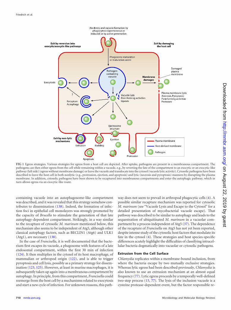
organisms makes it necessary to examine each stage individually.The identification and characterization of novel egress strategiescontinue, and some exotic mechanisms, such as the egress of mi-crosporidia from intestinal cells in Caenorhabditis elegans (149),might be more widespread than assumed. In the following sec-tions, the various exit strategies known to date and examples fromthe recent literature are presented in order of increasing damageinflicted to the host cell, from nonlytic to death-inflicting pro-cesses. A schematic overview of the various egress strategies ispresented in Fig. 1.
EGRESS STRATEGIES WITH NEITHER VACUOLE LYSIS NORCELL LYSIS
Exocytosis and Autophagy-Related Mechanisms
Arguably, the simplest mechanistic exit strategy for an intracellu-lar pathogen is to remain in a vacuole during replication and in-duce the fusion of that vacuole with the host plasma membranewhen the time has come for transmission. This strategy has theadvantage of perfectly preserving the host cell. For example,Legionella can reemerge from cells of its primary amoeba hosts,Dictyostelium discoideum and Acanthamoeba castellani, via exocy-tosis (35, 36). Two pathogen factors, LepA and LepB, were iden-tified, which, based on structural similarities, could mimic theSNARE function and steer the pathogen into the exocytic path-way. In the case of the common periodontal pathogen Porphy-romonas gingivalis, the endocytic recycling pathway leads to theexocytosis of bacteria from epithelial cells (142). However, de-pending on the host cell type, P. gingivalis not only localizes toendosomes but also may be found in the cytosol or autophago-somes. Therefore, the authors of that study suggested that otherexit mechanisms are likely to exist. Indeed, another group pro-posed the existence of a protrusion-like strategy without the ex-posure of the pathogen to the extracellular milieu (158).
The facultative intracellular pathogenic yeast Cryptococcus neo-formans proliferates inside the phagosome of macrophages andmonocytes. The exit of progeny from phagolysosomes has beendescribed to occur by either lytic egress or a nonlytic exocytosis-like expulsion or “vomocytosis” mechanism. Whereas lytic egressappears to be the result of intracellular replication and is precededby the permeabilization of the phagosomal membrane (150), ex-pulsion seems to depend on a yeast factor, indicating that it mightbe an active process (9, 92). Those studies also noted an inhibitoryeffect of the host cell actin. More recently, actin flashes were ob-served around the phagosome prior to expulsion (80), likely incoordination with a WASH-mediated V-ATPase retrieval mecha-nism (31). Indeed, the Arp2/3 activator WASH localizes to Dictyoste-lium endosomes and phagosomes during the reneutralization phasethat follows the (phago)lysosomal acidic stage and leads to a near-neutral postlysosomal compartment. WASH-driven actin polymer-ization was shown to be necessary for V-ATPase retrieval (31). How-ever, whether the actin flashes play a direct role in the Cryptococcusexit mechanism remains to be defined. In addition, a direct cell-to-cell spreading mechanism between macrophages without an expo-sure of the pathogen to the extracellular milieu was proposed, but itsdetails and significance remain unclear (8, 91).
A particular way to use an exocytosis-like mechanism was re-cently reported for Brucella and may also apply to Francisella,which might exploit an autophagy-like process to gain access tothe cell exterior and reinvade naïve bystanders (34, 138). Interest-
Friedrich et al.
708 mmbr.asm.org Microbiology and Molecular Biology Reviews
on August 22, 2019 by guest
http://mm
br.asm.org/
Dow
nloaded from
ingly, this newly revealed strategy has gained interest at the sametime as another (maybe distinct or not) autophagy-related path-way has been implicated in unconventional protein secretion (26,110). In both cases, cytoplasmic (including vacuolar and cytoso-lic) bacteria and proteins are sequestered in a membrane compart-ment that fuses with the plasma membrane and releases themoutside.
Brucella usually spends much energy and a good share of itsvirulence program to establish a replication niche that acquiresmost features of the endoplasmic reticulum (ER) (153). The stepsfrom entry to the establishment of this replication compartmentare well understood, but how Brucella finally escapes its primaryhost cell at later stages and spreads infection has long been a ne-glected field of study. Recently, the transformation of the Brucella-
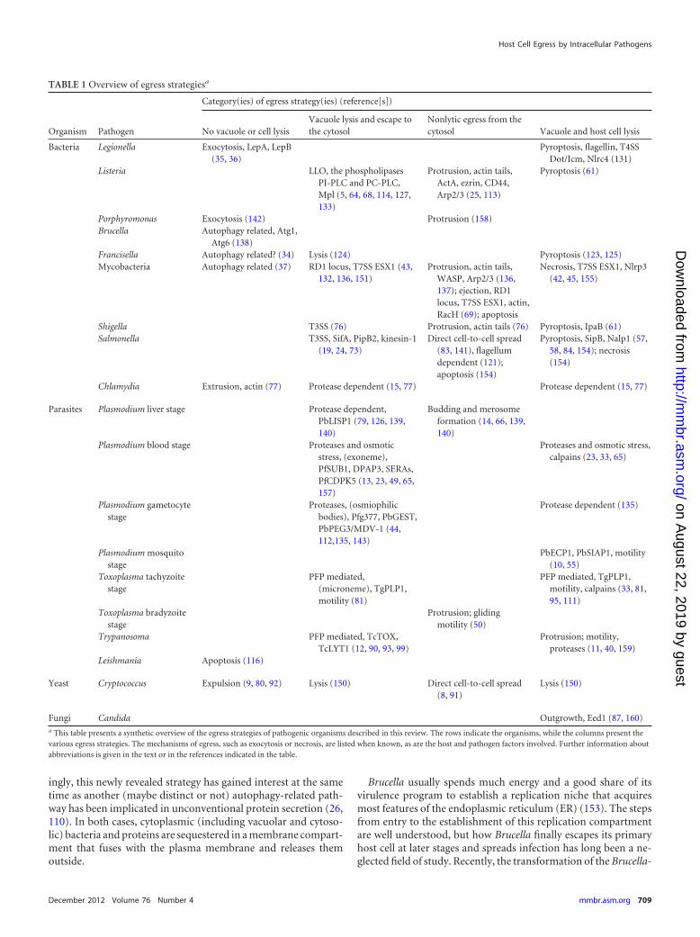
TABLE 1 Overview of egress strategiesa
Organism Pathogen
Category(ies) of egress strategy(ies) (reference[s])
No vacuole or cell lysisVacuole lysis and escape tothe cytosol
Nonlytic egress from thecytosol Vacuole and host cell lysis
Bacteria Legionella Exocytosis, LepA, LepB(35, 36)
Pyroptosis, flagellin, T4SSDot/Icm, Nlrc4 (131)
Listeria LLO, the phospholipasesPI-PLC and PC-PLC,Mpl (5, 64, 68, 114, 127,133)
Protrusion, actin tails,ActA, ezrin, CD44,Arp2/3 (25, 113)
Pyroptosis (61)
Porphyromonas Exocytosis (142) Protrusion (158)Brucella Autophagy related, Atg1,
Atg6 (138)Francisella Autophagy related? (34) Lysis (124) Pyroptosis (123, 125)Mycobacteria Autophagy related (37) RD1 locus, T7SS ESX1 (43,
132, 136, 151)Protrusion, actin tails,
WASP, Arp2/3 (136,137); ejection, RD1locus, T7SS ESX1, actin,RacH (69); apoptosis
Necrosis, T7SS ESX1, Nlrp3(42, 45, 155)
Shigella T3SS (76) Protrusion, actin tails (76) Pyroptosis, IpaB (61)Salmonella T3SS, SifA, PipB2, kinesin-1
(19, 24, 73)Direct cell-to-cell spread
(83, 141), flagellumdependent (121);apoptosis (154)
Pyroptosis, SipB, Nalp1 (57,58, 84, 154); necrosis(154)
Chlamydia Extrusion, actin (77) Protease dependent (15, 77) Protease dependent (15, 77)
Parasites Plasmodium liver stage Protease dependent,PbLISP1 (79, 126, 139,140)
Budding and merosomeformation (14, 66, 139,140)
Plasmodium blood stage Proteases and osmoticstress, (exoneme),PfSUB1, DPAP3, SERAs,PfCDPK5 (13, 23, 49, 65,157)
Proteases and osmotic stress,calpains (23, 33, 65)
Plasmodium gametocytestage
Proteases, (osmiophilicbodies), Pfg377, PbGEST,PbPEG3/MDV-1 (44,112,135, 143)
Protease dependent (135)
Plasmodium mosquitostage
PbECP1, PbSIAP1, motility(10, 55)
Toxoplasma tachyzoitestage
PFP mediated,(microneme), TgPLP1,motility (81)
PFP mediated, TgPLP1,motility, calpains (33, 81,95, 111)
Toxoplasma bradyzoitestage
Protrusion; glidingmotility (50)
Trypanosoma PFP mediated, TcTOX,TcLYT1 (12, 90, 93, 99)
Protrusion; motility,proteases (11, 40, 159)
Leishmania Apoptosis (116)
Yeast Cryptococcus Expulsion (9, 80, 92) Lysis (150) Direct cell-to-cell spread(8, 91)
Lysis (150)
Fungi Candida Outgrowth, Eed1 (87, 160)a This table presents a synthetic overview of the egress strategies of pathogenic organisms described in this review. The rows indicate the organisms, while the columns present thevarious egress strategies. The mechanisms of egress, such as exocytosis or necrosis, are listed when known, as are the host and pathogen factors involved. Further information aboutabbreviations is given in the text or in the references indicated in the table.
Host Cell Egress by Intracellular Pathogens
December 2012 Volume 76 Number 4 mmbr.asm.org 709
on August 22, 2019 by guest
http://mm
br.asm.org/
Dow
nloaded from
containing vacuole into an autophagosome-like compartmentwas described, and it was revealed that this strategy somehow con-tributes to dissemination (138). Indeed, the formation of infec-tion foci in epithelial cell monolayers was strongly promoted bythe capacity of Brucella to stimulate the generation of that lateautophagy-dependent compartment. Strikingly, in a way similarto the recapture of cytosolic M. marinum mentioned below, thismechanism also seems to be independent of Atg5, although otherclassical autophagy factors, such as BECLIN1 (Atg6) and ULK1(Atg1), are necessary (138).
In the case of Francisella, it is well documented that the bacte-rium first escapes its vacuole, a phagosome with features of a lateendosomal compartment, within the first 30 min of infection(124). It then multiplies in the cytosol of its host macrophage, ofmammalian or arthropod origin (122), and is able to triggerpyroptosis and cell lysis, possibly as a primary strategy for dissem-ination (123, 125). However, at least in murine macrophages, it issubsequently taken up again into a membranous compartment byautophagy. In principle, from this compartment, Francisella couldreemerge from the host cell by a mechanisms related to exocytosisand start a new cycle of infection. For unknown reasons, this path-
way does not seem to prevail in arthropod phagocytic cells (4). Apossible similar recapture mechanism was reported for cytosolicM. marinum (see “Vacuole Lysis and Escape to the Cytosol” for adetailed presentation of mycobacterial vacuole escape). Thatpathway was described to be similar to autophagy and leads to thesequestration of ubiquitinated M. marinum in a vacuolar com-partment by a process independent of Atg5 (37). The dependenceof the recapture of Francisella on Atg5 has not yet been reported,despite intense study of the cytosolic host factors that modulate itsfate in the cytosol (4). These strategies and host species-specificdifferences acutely highlight the difficulties of classifying intracel-lular bacteria dogmatically into vacuolar or cytosolic pathogens.
Extrusion from the Cell Surface
Chlamydia replicates within a membrane-bound inclusion, fromwhere the bacteria escape by two mutually exclusive strategies.Whereas lytic egress had been described previously, Chlamydia isalso known to use an extrusion mechanism at an almost equalfrequency (77). Lytic egress proceeds by a temporally well-definedtwo-step process (15, 77). The lysis of the inclusion vacuole is acysteine protease-dependent event, but the factor responsible re-
FIG 1 Egress strategies. Various strategies for egress from a host cell are depicted. After uptake, pathogens are present in a membranous compartment. Thepathogens can then either egress from the cell while remaining within a vacuole, e.g., by reverting the fate of the compartment to an exocytic or an exocytic-likepathway (left side) (egress without membrane damage) or leave the vacuole and translocate into the cytosol (vacuole lytic activity). Cytosolic pathogens have beendescribed to leave the host cell in both nonlytic (e.g., protrusion, ejection, and apoptosis) and lytic (necrosis and pyroptosis) manners by disrupting the plasmamembrane. In addition, cytosolic pathogens have been shown to be recaptured into membranous compartments and enter the autophagic pathway, which inturn allows egress via an exocytic-like route.
Friedrich et al.
710 mmbr.asm.org Microbiology and Molecular Biology Reviews
on August 22, 2019 by guest
http://mm
br.asm.org/
Dow
nloaded from
mains unknown. Subsequently, the host cell plasma membraneruptures, triggering the influx of extracellular calcium and therelease of cytosolic bacteria. To prevent complete lysis, the hostcell responds with the activation of the lysosome fusion-mediatedplasma membrane repair mechanism (15). In contrast, extrusionis a nonlytic process in which protruding Chlamydia inclusionsare partially pinched off from the host cell in an actin-dependentmanner. The resulting extracellular inclusion bodies are sur-rounded by the host cell cytosol and the host plasma membrane.Both the host cell and the residual inclusion remain intact (77),which might play an important role in the persistence of infection.
VACUOLE LYSIS AND ESCAPE TO THE CYTOSOL
Vacuole lysis can be part of an exit strategy that is either nonlyticor lytic for the host cell (see Strategies for Nonlytic Egress from theCytosol as well as Egress Strategies Involving Lysis of the Vacuoleand Host Cell). Controlled vacuolar escape is crucial for patho-gens replicating in the cytosol but can also be necessary for intra-vacuolar progeny to pursue a particular egress strategy (for exam-ple, the budding of Plasmodium liver-stage parasites). Shigellareplicates in the cytosol, and vacuolar escape depends on a func-tional type III secretion system (T3SS) along with its translocon(105). While the exact mechanism of vacuolar lysis remains un-known, this event is accompanied by the recruitment of cytosolicgalectins, possibly to glycosylations decorating the exposed bacte-ria but surely to membranes of the ruptured phagosome (105, 115,144). This phenomenon has been observed not only for Gram-negative Shigella and Salmonella spp. but also for the Gram-posi-tive bacterium Listeria monocytogenes as well as during steriledamage and therefore appears to be a generic cytosolic defenseresponse to phagosomal damage (105, 144). In particular, galec-tin-8 is able to restrict Salmonella proliferation by binding to spe-cific carbohydrates on the inner leaflet of the breaking vacuole,leading to the activation of antibacterial autophagy (144).
In contrast to Shigella, Salmonella usually replicates within thevacuole. Salmonella secretes factors similar to those secreted byShigella via its T3SS but in addition releases SifA and PipB2, whichinterfere with kinesin-mediated vacuolar dynamics, delay vacuo-lar lysis, and, hence, ensure intravacuolar replication (24, 73). ASalmonella sifA mutant rapidly becomes cytosolic, and its viru-lence is strongly attenuated in mice (19). While vacuolar lysis mostprobably occurs during lytic egress (see Egress Strategies InvolvingLysis of the Vacuole and Host Cell), it might not be required for amechanism of direct cell-to-cell transfer, relying on the kinesin-dependent repositioning of the Salmonella-containing vacuolefrom the juxtanuclear region to the periphery (83, 141).
Listeria secretes the well-characterized PFP listeriolysin O(LLO). LLO, like T. cruzi TcTOX, is nonfunctional in the pH-neutral cytosol but is activated at a low pH (12, 93, 127), as occursin the course of phagosome maturation. The pH dependence con-fines these molecules to a specific role in pathogen escape from thevacuole into the host cell cytosol while keeping the host plasmamembrane intact (90). The dysregulation of LLO activity impactsthe virulence of Listeria in mice (64). LLO activity might also beaffected by a perturbation of host cell ion homeostasis, whichinhibits phagosomal escape and also results in decreased virulence(114). Listeria resides in single-membrane primary vacuoles whentaken up by professional or nonprofessional phagocytic cells or indouble-membrane secondary vacuoles when resulting from cell-to-cell spread by protrusion. Although not required in several
human epithelial cell lines, LLO appears to be essential for vacu-olar escape in most other cell types by acting on primary vacuolesand the outer membrane of secondary vacuoles (5). The earlysecretion of LLO after the uptake of bacteria by macrophages leadsto the permeabilization of the primary vacuole, the breakdown ofion gradients, and the inhibition of phagolysosome fusion, pre-serving an environment from which the bacteria can escape effi-ciently (72, 128). Membrane disruption by LLO is assisted by twophospholipases (phosphatidylinositol-phospholipase C [PI-PLC]and phosphatidylcholine-phospholipase C [PC-PLC]). PC-PLC isactivated through proteolytic cleavage by the bacterial acid-de-pendent metalloprotease Mpl, which also cleaves the actin nucle-ator ActA that is necessary for protrusion (see Strategies for Non-lytic Egress from the Cytosol) and could be responsible fordelaying bacterial motility in the cytoplasm (94, 117). The twolipases have overlapping functions in the dissolution of primaryand secondary vacuoles and appear to be particularly importantfor the disruption of the inner membrane of secondary vacuoles(5, 133). Moreover, in epithelial cells, where LLO expression is notrequired, PC-PLC activity was shown to be necessary for the lysisof these single- and double-membrane compartments (68).
Mycobacteria from the tuberculosis group are classically con-sidered vacuolar intracellular pathogens, but recent evidence hasseriously challenged this view. The first blow to this dogma camefrom studies of M. marinum, the closest relative of the tuberculo-sis group, a pathogen responsible for fish tuberculosis and foropportunistic skin infections of humans. A mycobacterium wasobserved to escape from its vacuole to the cytosol of its host mac-rophage and even to move around the cytoplasm, powered by anactin tail, much like Listeria and Shigella (136). Further work dem-onstrated the involvement of the Arp2/3 complex in this motility,and this mechanism was also proposed to play a role in the spreadof infection (see “Protrusion at the Plasma Membrane”). A poten-tial but still hotly debated breakthrough was reported in studies ofM. tuberculosis and Mycobacterium leprae in myeloid cells. Carefulmorphometric quantitations indeed revealed that a sizable frac-tion of these mycobacteria can escape their compartment and in-vade the cytosol of their hosts (151). Interestingly, the attenuatedvaccine strain Mycobacterium bovis BCG was apparently incapac-itated in this vacuole escape, raising speculations about the role ofcytosolic mycobacteria in the immune response mounted duringvarious stages of a tuberculosis infection. The RD1 locus is themajor region of difference between virulent M. tuberculosis andBCG (75) and was shown to encode the type VII secretion systemESX1 as well as its secreted factors, such as early secreted antigentarget 6 (ESAT-6) (reviewed in reference 1). The ESX1 machinerylocalizes to and secretes at the new poles of growing bacteria (30).It was hypothesized that ESAT-6 plays a key role in vacuole escape,as it was shown to possess membranolytic activity (43) and in-duces membrane pores of approximately 4.5 nm in diameter in acontact-dependent manner (134). The perforation of and exitfrom vacuoles by M. marinum and M. tuberculosis were recentlyconfirmed by a very elegant experimental strategy (132). Using anapproach similar to the one already validated for Shigella (115),those authors monitored the exposure of a fluorescence resonanceenergy transfer (FRET) substrate loaded into the cytosol of thehost to M. tuberculosis cell wall-associated �-lactamase activity.That study also confirmed the requirement for a functional ESX1secretion system for vacuole escape.
The genetic dissection of host and pathogen factors involved in
Host Cell Egress by Intracellular Pathogens
December 2012 Volume 76 Number 4 mmbr.asm.org 711
on August 22, 2019 by guest
http://mm
br.asm.org/
Dow
nloaded from
vacuolar escape and egress from the host cell made rapid progressthanks to the establishment of a mycobacterium-host system in analternative model organism, Dictyostelium discoideum. This socialamoeba naturally grazes on soil bacteria by efficient phagocytosis;it interacts with a large variety of bacteria and fungi and has de-veloped defense processes to survive in such complex environ-ments, which are precursors to the cell-intrinsic resistance mech-anisms further refined in phagocytes of the innate immune system(38). As described above for macrophages, within Dictyostelium,M. marinum also modifies the composition of the phagosome.The association of M. marinum with the vacuolar H�-ATPase isdecreased compared to that of nonpathogenic mycobacteria andis practically undetectable at 6 h postinfection (70). Furthermore,during the ensuing proliferation phase, M. marinum avoids thedelivery of cathepsin D, a lysosomal protease, to the phagosomallumen. Surprisingly, Dictyostelium vacuolin (a flotillin homo-logue and a raft and late phagolysosomal marker) strongly accu-mulates around the niche, making it possible to observe and quan-titate the dynamics of escape from the vacuole. Even morestriking, upon infection of Dictyostelium, M. tuberculosis was alsoable to escape to the cytosol to a certain extent. That study not onlyconfirmed the evolutionary conservation of tubercular mycobac-terium virulence mechanisms and host processes but also elabo-rated the crucial role of ESAT-6. Indeed, M. marinum RD1 mu-tants (equivalent to the BCG strain) showed a greatly reducedvacuole escape efficiency, which was partially rescued by trans-complementation, namely, the expression of the bacterial viru-lence factor directly in the cytosol of the host Dictyostelium (69).
As mentioned above, the vacuolar escape of T. cruzi dependson the PFP TcTOX. While the molecular identity of TcTOX re-mains unknown, TcLYT1, a protein with similar characteristicsand suspected to be identical to TcTOX, appears to contribute tothe hemolytic activity. A TcLYT1�/� strain showed diminishedinfectivity in cell cultures (93) and attenuated virulence in themouse model (159). The vacuolar escape of T. cruzi may dependon the differentiation of metacyclic trypomastigotes into amasti-gotes (93, 99), as hemolytic activity has been detected for onlysome developmental stages. A trans-sialidase localized on the sur-face of T. cruzi trypomastigotes facilitates exit from the PV (120),but it remains uncertain if and how this relates to the action ofTcTOX. The sialidase catalyzes the transfer of host cell sialic acidresidues onto mucin-like proteins on the parasite surface. Para-sites exit from sialic acid-deficient CHO cells earlier, suggestingthat sialic acid inside the PV might constitute a barrier for theparasite.
STRATEGIES FOR NONLYTIC EGRESS FROM THE CYTOSOL
Protrusion at the Plasma Membrane
Protrusion mechanisms play an important role in the egress ofprokaryotic intracellular pathogens (28, 76). Protrusion inducedby Listeria and Shigella has been extensively described and relieson the polymerization of host cell actin on the bacterial surface.For example, cytosolic Listeria uses its factor ActA to stimulateF-actin nucleation by mimicking the host WASP family proteins(25). Through the formation of actin tails, the bacteria move andinduce filopodia extending from the host plasma membrane.Filopodia contain the bacterium at their tip and are engulfed byneighboring cells. This transmission process does not seem to in-volve the death of the initially infected cell (62). Actin filaments in
filopodia are relatively stable (117). The formation of the protru-sion and effective spreading rely on the ezrin-radixin-moesin(ERM) family of proteins, which link the actin cytoskeleton totransmembrane proteins, and in particular, ezrin and CD44 wereshown to be important (113). A similar transmission process wasproposed for the fish and frog pathogen M. marinum in macro-phages. The motility of cytosolic M. marinum by WASP-inducedactin polymerization has been described (136, 137); however, M.marinum has not yet been observed in double-membrane com-partments, which would result from such a transmission strategy.
Despite several eukaryotic pathogens carrying their own ma-chinery for motility, there is so far surprisingly little documenta-tion of protrusion mechanisms for eukaryotic pathogens. Oneexception might be Toxoplasma gondii bradyzoites, the parasiteform characteristic of chronic infections, contrasting with the lyticegress of acute-stage T. gondii tachyzoites (see “Concerted Egressfrom the Vacuole and the Host Cell”). Visual observations sug-gested that bradyzoites use a nonlytic strategy reminiscent of pro-trusion mechanisms involving the parasites’ gliding motility, al-lowing the invasion of neighboring cells without exposure to theexternal medium (50).
Another way of exploiting cytoskeleton dynamics to spreadfrom cell to cell is the stabilization of direct, membranous, andF-actin-containing connections between an infected and a nonin-fected target cell. Physical connections, such as nanotubes orfilopodium bridges, are used by bacteria (3, 104, 158) to cross adistance to neighboring cells. Conceptually, this transfer can oc-cur intracellularly, within the tubes in a one-dimensional diffu-sion model; however, its mechanism is not yet understood.
Ejection at the Plasma Membrane
Apart from cell death-mediated mechanisms (see “Apoptosis as aNonlytic Exit Strategy” and “Unconcerted Egress from the Vacu-ole and the Host Cell”), there is evidence for an alternative strategyof intercellular spreading for cytosolic M. marinum and M. tuber-culosis in epithelial cell layers (27, 32) as well as in amoebae (69).RD1, the major virulence locus of pathogenic mycobacteria that isabsent in attenuated strain M. bovis BCG, has been implicated incell lysis, egress, and dissemination (42, 75, 82, 152). The exactmechanism behind this plethora of effects is unclear, but effectorsassociated with the ESX1 secretion system are secreted at the bac-terial poles (30) and might be ideally positioned to initiate anegress strategy when they contact the host cell cortex. This view issupported by observations of M. marinum escape from theamoeba Dictyostelium. In this system, the nonlytic ejection ofcytosolic bacteria across the host plasma membrane was ob-served to take place via a structure of dense F-actin, termed theejectosome (69). A bacterial mutant lacking the RD1 locus wasnot able to escape from the host cell, and this inhibition couldbe partially trans-complemented by expressing ESAT-6 di-rectly in the host cell cytosol (69). Interestingly, a host mutantlacking the small GTPase RacH was also deficient in formingthe ejectosome.
It remains to be shown whether other pathogenic mycobacteriause an ejectosome-based egress strategy in addition to cell death-based mechanisms. However, because virulent M. tuberculosis andM. leprae strains, but not the avirulent strain M. bovis BCG, cantranslocate into the cytosol for replication like M. marinum (151),at least under some conditions, we speculate that transmission willhave to involve some form of protrusion or ejection. Sustaining
Friedrich et al.
712 mmbr.asm.org Microbiology and Molecular Biology Reviews
on August 22, 2019 by guest
http://mm
br.asm.org/
Dow
nloaded from
this hypothesis, M. marinum was observed to induce long filopo-dium-like structures reminiscent of Listeria-induced protrusionsin infected macrophages. These structures were observed to servecell-to-cell transmission between macrophages in vitro (137) andinside infected zebrafish at early stages of infection (41). Recently,microsporidian obligate intracellular parasites have been shownto follow an actin-dependent nonlytic egress strategy to exit intes-tinal epithelial cells of the worm Caenorhabditis elegans (149).While the details of the egress process are not fully clarified, itappears to be preceded by the remodeling of a dense layer of actinfilaments, followed by the egress itself, which is nonlytic and doesnot appear to damage the cells (55a).
Budding from the Plasma Membrane
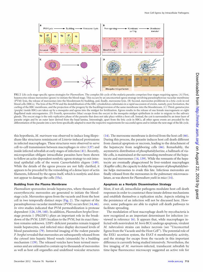
Plasmodium sporozoites invade hepatocytes, where thousands ofexoerythrocytic merozoites are generated to initiate the blood-stage cycle. Merozoites egress from the vacuole and from the hostcell in two temporally distinct steps (Fig. 2). The rupture of theparasitophorous vacuolar membrane (PVM) occurs first (14, 66).In vitro studies indicated that PVM permeabilization is proteasedependent (126, 139, 140). In addition, Plasmodium berghei liver-stage protein 1 (PbLISP1) plays an important role in the break-down of the PVM. LISP1 localizes to the PVM, but its exact func-tion remains unknown. LISP1-deficient parasites remain trappedinside hepatocytes, and infected mice display decreased levels ofblood parasitemia (79). Intravital imaging of the rodent parasiteP. berghei revealed that merozoites are subsequently released fromthe cytosol into hepatic blood vessels (sinusoids) by a buddingmechanism (139). The released vesicles have been termed mero-somes and are estimated to contain up to thousands of merozoitesas well as host cell organelles and undefined vesicular structures
(14). The merosome membrane is derived from the host cell (66).During this process, the parasite induces host cell death differentfrom classical apoptosis or necrosis, leading to the detachment ofthe hepatocyte from neighboring cells (66). Remarkably, theasymmetric distribution of phosphatidylserine, a hallmark of via-ble cells, is maintained at the surrounding membrane of the hepa-tocyte and merosomes (14, 139). While the remnants of the hepa-tocyte are eventually phagocytosed by liver-resident macrophages(Kupffer cells), the capacity to delay the exposure of phosphatidylser-ine helps merosomes to evade this fate. Infectious merozoites arefinally released from the merosomes in the pulmonary microvascu-lature, as was shown for Plasmodium yoelii in mice (14).
Apoptosis as a Nonlytic Dissemination Strategy
Most, if not all, intracellular pathogens modulate host cell deathresponses in order to counteract these innate defense mechanismsand establish themselves a replication niche. Aspects relating tothe persistence of an infection will not be discussed here. How-ever, some pathogens are able to exploit cell death pathways tofacilitate spreading.
The modulation of host macrophage death by mycobacteria isnow recognized as an important determinant for infection (re-viewed in reference 16). It appears that, while macrophages in-fected with nonvirulent M. bovis BCG undergo apoptosis, virulentM. tuberculosis strains can induce necrosis (see “UnconcertedEgress from the Vacuole and the Host Cell”). The potential role ofthe ESX1 secretion system, the ESAT-6 membranolytic activity,and the strategy for escape from the vacuole in this importantdifference is currently being studied intensively. Nevertheless, thelive imaging of M. marinum-infected, translucent zebrafish bytime-lapse fluorescence microscopy suggested an active role for
FIG 2 Life cycle stage-specific egress strategies for Plasmodium. The complex life cycle of the malaria parasite comprises four stages requiring egress. (A) First,hepatocytes release merozoites (green) to initiate the blood stage. This occurs by an unconcerted egress strategy involving parasitophorous vacuolar membrane(PVM) lysis, the release of merosomes into the bloodstream by budding, and, finally, merosome lysis. (B) Second, merozoites proliferate in a lytic cycle in redblood cells (RBCs). The lysis of the PVM and the destabilization of the RBC cytoskeleton culminates in a rapid succession of events, namely, pore formation, thecurling of the RBC membrane, and the projection of the progeny by the buckling/eversion of the same membrane into the bloodstream. (C) Third, gametocytes(purple) inside RBCs are taken up by a mosquito and egress into the midgut for fertilization. Egress results in the release of one female macrogamete or eightflagellated male microgametes. (D) Fourth, sporozoites (blue) escape from the oocyst in the mosquito midgut epithelium in order to migrate to the salivaryglands. The oocyst stage is the only replicative phase of the parasite that does not take place within a host cell. Instead, the cyst is surrounded by an inner layer ofparasite origin and by an outer layer derived from the basal lamina. Interestingly, apart from the lytic cycle in RBCs, all other egress events are preceded by thedifferentiation of the parasite into a new form specifically adapted to meet the respective requirements for successful egress and to initiate the next stage of the life cycle.
Host Cell Egress by Intracellular Pathogens
December 2012 Volume 76 Number 4 mmbr.asm.org 713
on August 22, 2019 by guest
http://mm
br.asm.org/
Dow
nloaded from
intragranuloma apoptotic events in the spread of infection by vir-ulent M. marinum (42). Cell death attracted macrophages, whichengulfed bacteria, spreading infection and inducing new granulo-matous structures. Granulomas can persist over long periods oftime and have traditionally been associated with the containmentof infection in mammals. Interestingly, M. avium, which is classi-fied as a ubiquitous nontuberculous mycobacterium that cancause pulmonary diseases in immunocompromised individuals,was reported to use apoptotic macrophages for spread (51). Whilesome M. avium strains appear to use the apoptotic bodies as avehicle to infect naïve macrophages that engulf them, others seemto be able to lyse both the vacuole and the apoptotic body toreinfect neighboring macrophages. Therefore, apoptosis can act asan innate defense mechanism against mycobacteria but mightplay different roles in different hosts.
EGRESS STRATEGIES INVOLVING LYSIS OF THE VACUOLEAND HOST CELL
Vacuolar lysis and host cell lysis often take place simultaneously orin rapid succession, but there are also examples where they arespaced in time. According to this difference in the timing of thetwo steps, we distinguish concerted from unconcerted lytic egress.Unconcerted egress can, for example, result from pathogen repli-cation in the cytoplasm or from mechanistically different strate-gies for lysing the vacuole and the host cell. In principle, a patho-gen could also trigger host cell lysis without previously exiting thevacuole. However, to our knowledge, there is no example of suchan egress strategy to date.
Perhaps the most striking example for a (superficially) “uncon-trolled” egress strategy is the one used by the dimorphic fungusCandida albicans to exit from mucosal epithelial cells. Micro-scopic observations suggested that the simple physical penetrationof membrane barriers driven by outgrowth might result in“egress” and the spread of infection. The fungus’ ability to un-dergo a morphological switch from spherical yeast to filamentoushyphae leads to high mechanical stress on the host cell and hasbeen linked to its virulence (87). A �eed1 (epithelial escape anddissemination) mutant incapable of hyphal formation in responseto a diverse range of stimuli is strongly attenuated in causing thetissue damages characteristic of advanced infections (160). In re-constituted human oral epithelial cells, spherical forms of thisstrain were endocytosed and proliferated within cells but re-mained trapped inside.
Unconcerted Egress from the Vacuole and the Host Cell
Many intracellular pathogens have been shown to subvert host celldeath pathways for exit from the host cell and dissemination.Pyroptosis is a caspase-1-dependent programmed cell death re-sulting in cell lysis and the release of proinflammatory cytokines.It is therefore considered a potent innate defense mechanism. De-spite this, some pathogens, such as Francisella (123, 125) and Le-gionella (131), exit host cells that have undergone this type of celldeath. As another example, Salmonella enterica induces the extru-sion of infected cells from the intestinal epithelium for dissemina-tion into the gut and the environment. Homeostatic extrusioninvolves apoptosis, but extrusion triggered by the bacteria is ac-companied by pyroptosis (84). That work also showed the exis-tence of a subpopulation of flagellated, motile bacteria that residein the cytosol and, in contrast to intravacuolar bacteria, expressinvasion factors such as T3SS1. Interestingly, in this context, an-
other study demonstrated the existence of a nonlytic, flagellum-dependent egress mechanism in oncotic macrophages (121).
As different host cell types undergoing different types of celldeath may be involved during the course of an infection, it issometimes difficult to establish which events are beneficial to thehost or to the pathogen during infection in vivo. This has beendiscussed for systemic infections of Salmonella, and new tools toinvestigate the population dynamics of pathogens in the hostshould help resolve these questions (reviewed in reference 154).Salmonella enterica serovar Typhimurium induces apoptosis inintestinal epithelial cells (which might be of importance to crossthis barrier and initiate infection) and in a subset of infected mac-rophages. However, pyroptosis and necrosis appear to be numer-ically dominant in systemic infections (154). S. Typhimurium in-duces pyroptosis in macrophages and is able to delay host celldeath to improve replication and systemic dissemination (58).The activation of pyroptosis by Salmonella is similar to but distinctfrom the pathway induced by the anthrax lethal toxin. The formerseems to act via Ipaf, whereas the latter is known to require theinflammasome adaptor Nalp1 (57). Interestingly, Salmonella in-duces both caspase-1 activation and the release of inflamma-tory cytokines, associated with or possibly triggering massive(phago)lysosome exocytosis (18). It is not yet clear whether themicrobicidal activities released from lysosomes participate in theantimicrobial nature of pyroptosis (18). Shigella spp. also inducepyroptosis of their host macrophages (61). Similarly to Salmo-nella, a pathogenic factor, Shigella IpaB or Salmonella SipB, is se-creted via a T3SS and can directly bind to and activate caspase-1.The pyroptosis of Shigella-infected macrophages recruits poly-morphonuclear leukocytes, which can be infected by capturingthese dying cells (61). Interestingly, while Listeria induces apop-tosis in a variety of cell types, in macrophages, the bacterium ap-pears to make use of pyroptosis to spread the infection (61).
As mentioned in “Apoptosis as a Nonlytic Exit Strategy,” themodulation of host macrophage death by mycobacteria is emerg-ing as an important determinant of infection (reviewed in refer-ence 16). Virulent M. tuberculosis cells induce necrosis in a com-plex pathway that depends on the ESX1 secretion system. Themanipulation of the host eicosanoid pathways leads to the inhibi-tion of cellular membrane repair, affecting mitochondria and theplasma membrane and possibly affecting other organelles, such asthe replication vacuole. This results in the steering of the macro-phage into necrosis (45). The release of bacteria by this mecha-nism not only avoids apoptosis as an innate defense mechanismbut also delays the establishment of adaptive immunity in mice, asnecrosis prevents the cross-presentation of antigens by dendriticcells (46). The induction of necrosis by virulent mycobacteriamight also be important for the outcome of infection in otherhosts, because susceptibility has been linked to a mutation affect-ing eicosanoid biosynthesis in zebrafish and humans (145, 146).The exact mechanisms of the induction of necrosis are understudy, but a recent report signaled a critical role for the NLRP3inflammasome in this process (155). A contact-dependent hemo-lysin activity linked to a functional ESX1 system induces Ca2�-dependent lysosome secretion that is concurrent with the releaseof proinflammatory cytokines from infected macrophages (85).ESAT-6 appears to generate vacuole damage that is sensed by thehost and recruits various signaling molecules, such as galectin-3,leading to the Syk tyrosine kinase-dependent activation of NLRP3(85, 155). This pathway is reminiscent of Syk-dependent NLRP3
Friedrich et al.
714 mmbr.asm.org Microbiology and Molecular Biology Reviews
on August 22, 2019 by guest
http://mm
br.asm.org/
Dow
nloaded from
activation during fungal and malarial infections (67, 130). Re-cently, such a link between M. tuberculosis vacuole damage and/orcomplete lysis and host cell death by necrosis was confirmed in-dependently (132).
Legionella infects Acanthamoeba but is also an opportunistichuman pathogen found within an intracellular vacuole in alveolarepithelial cells and macrophages. At later stages of infection, bac-terial flagellin induces pyroptosis in macrophages in a mechanismdependent on the bacterial type IVB secretion system Dot/Icmand on the host Nlrc4 inflammasome (131). As part of the pyrop-totic process, a host-derived pore-forming activity is induced, theaction of which precedes host cell lysis and the release of the bac-teria (131). These findings might have a general significance forvarious intracellular pathogens inducing host cell pyroptosis.Legionella mutants that do not induce this host-derived pore-forming activity are able to replicate but are severely delayed inexit from the host cell (6). Similar observations were made foramoeba hosts (60). However, it remains unknown whether pore-forming activity is critical for bacterial release, because other fac-tors of the pyroptotic response may contribute.
Although it is unclear whether it occurs as a concerted or un-concerted strategy, there is another example illustrating the im-portance of host cell death pathways in Leishmania major infec-tion. L. major is a parasite that usually replicates within thephagosome. After a bite by a sand fly, neutrophils phagocytose thetransmitted parasites (108). Neutrophils undergoing apoptosisappear to promote the release and subsequent transfer of the par-asite into macrophages and, in addition, are critical for the mod-ulation of macrophage and dendritic cell functions (discussed inreference 109). The efficient capture of infected neutrophils bydendritic cells in the skin inhibits the early antileishmania re-sponse (116).
Other unconcerted lytic egress strategies unrelated to cell death-mediated mechanisms do exist. In T. cruzi, for example, motilityappears to be an important contributor to egress. After replica-tion, nonmotile amastigotes redifferentiate in the cytosol intoflagellated trypomastigotes. Based on visual observations, the lat-ter breach the host plasma membrane shortly after becoming mo-tile (11). Trypomastigotes are capable of protrusion from cy-tochalasin D-treated host cells immediately after uptake (156).Therefore, under normal conditions, a potential protrusionmechanism would probably require the destabilization of the hostcell actin cytoskeleton by parasite factors. These may include acysteine protease activity that has been implicated in the egress ofT. cruzi trypomastigotes from host cells (40).
Concerted Egress from the Vacuole and the Host Cell
Concerted egress strategies are common among apicomplexanparasites, although there are some notable exceptions, such as thebudding of Plasmodium liver-stage parasites (see “Budding fromthe Plasma Membrane”). This illustrates that in organisms withcomplex life cycles, the mechanisms of exit from the host cell canbe stage specific (Fig. 2). The egress of blood-stage Plasmodiumparasites from red blood cells (RBCs) is governed by the release ofparasite proteases and a calcium-dependent kinase (49). Merozo-ites are very short lived in the blood circulation, and hence, egressneeds to be tightly controlled to optimize dissemination. The lysisof the PVM and the lysis of the host plasma membrane requiredifferent effectors and occur in rapid succession (reviewed in ref-erence 23). The rupture of RBCs also involves osmotic stress (65).
Other classes of effectors, such as PFPs (52) and lipases (20), mightalso be involved, but there is currently no direct support for thishypothesis. Modifications of the RBC membrane and cytoskele-ton appear central for the efficient dispersal of the nonmotileprogeny, enabling the RBC membrane to undergo a sequence ofevents described as curling-buckling-eversion-vesiculation (2).Indeed, the removal of adaptor proteins and cytoskeletal elementsfrom the RBC membrane has been reported (96). At least in vitro,the subversion of host calpain proteases facilitates the egress ofPlasmodium falciparum and T. gondii by cleaving several cytoskel-etal elements (33). However, the reticulocyte-invading rodentmalaria parasite P. yoelii develops equally in calpain�/� mice com-pared to wild-type mice (71).
The subtilisin-like serine protease SUB1 is essential for theegress of P. falciparum from RBCs (157). PfSUB1 is released intothe parasitophorous vacuole (PV) from a novel type of parasitesecretory organelle, called the exoneme, just prior to egress, butnothing is known about the stimulus that leads to its secretion.PfSUB1 is a Ca2�-dependent protease, and the low-Ca2� environ-ment of the PV space could represent a second level of control inaddition to regulated exoneme discharge. PfSUB1 and the cysteineprotease dipeptidyl aminopeptidase 3 (DPAP3) process papain-like proteins of the serine repeat antigen (SERA) family, which iscrucial for parasite egress (13, 157). The existence of a proteolyticcascade has been postulated; however, evidence for protease activ-ity has so far been obtained only for PfSERA5 (74). One P. bergheiSERA homologue, called egress cysteine protease 1 (PbECP1), wasshown to be essential for the egress of the mosquito stage of theparasite (10). Despite the activation of parasite motility, PbECP1-negative sporozoites are unable to leave the oocyst. Nevertheless,motility does contribute to efficient egress in this stage, becausesporozoites lacking productive gliding locomotion also display apartial defect in oocyst egress (55).
The exit of Plasmodium gametocytes from RBCs occurs con-comitantly with differentiation into gametes. Gametogenesis andegress depend on activation by external stimuli (temperature dropand xanthurenic acid) in the mosquito midgut and on parasiteprotease activities (135). Parasite organelles called osmiophilicbodies, found predominantly in female gametocytes, were postu-lated to be functionally equivalent to exonemes in the blood stage(44). Pfg377 is a protein located in the osmiophilic bodies thatplays a pivotal role in the biogenesis of these organelles, and par-asites deficient in Pfg377 showed a reduced emergence of femalegametes from RBCs (but no effect on male gametes) and reducedinfectivity toward mosquitoes. PbGEST and the protein of earlygametocyte 3 (PEG3) (also called male development 1 [MDV-1])are released from osmiophilic bodies into the PV and are impor-tant for the egress of both male and female P. berghei gametocytes(112, 143). Besides its role in vertebrate-mosquito transmission,PbGEST also functions in cell traversal by sporozoites, which isrequired for productive transmission back to the vertebrate host(143).
In the apicomplexan model organism T. gondii, perforin-likeprotein 1 (TgPLP1) plays a key role in the egress of tachyzoites, theform characteristic of acute infections (81). The protein is secretedinto the PV space from parasite organelles called micronemes,presumably just prior to egress. TgPLP1-deficient parasites areavirulent in mice and display severely delayed and temporally het-erogeneous egress in cell culture, which suggested that parasitegliding motility is able to force exit from the host cell eventually.
Host Cell Egress by Intracellular Pathogens
December 2012 Volume 76 Number 4 mmbr.asm.org 715
on August 22, 2019 by guest
http://mm
br.asm.org/
Dow
nloaded from
TgPLP1 destabilizes the PVM and possibly also attacks other hostcell membranes (81). The fact that TgPLP1 is released from mi-cronemes has important implications not only for T. gondii butpotentially also for other apicomplexan parasites. Microneme se-cretion in apicomplexans is thought to resemble Ca2�-mediatedexocytosis in other organisms and has traditionally been associ-ated with the activation of gliding motility and host cell invasion(21, 102). Microneme secretion and egress can be artificially in-duced by Ca2� ionophores in a variety of parasites (17, 54). Im-portantly, T. gondii ionophore-induced-egress mutants show adefect in the establishment of in vivo infections (89). Both inducedand noninduced egress requires the release of TgPLP1 (81). Inaddition, induced egress relies on parasite motility (95, 100, 111).In contrast, noninduced egress involves increased vacuolar pres-sure but does not require gliding motility (88). In addition toparasite factors, the release of Ca2� from the ER leads to the acti-vation of host cell calpains that participate in parasite egress bydestabilizing the host cell cytoskeleton (33). The action of TgPLP1likely results in the breakdown of ion gradients in the host cell andwas proposed to trigger a feedback-loop mechanism leading to theamplification of microneme secretion (118).
While the replication of T. gondii by successive rounds of binaryfission is compatible with induced/premature egress, this is notnecessarily the case for other parasites. For example, Plasmodiummultiplies by schizogony, which means that premature egresscould be fatal due to incomplete cell division.
Interestingly, in the case of Eimeria tenella infection, the hostmight rely on immune-mediated mechanisms (cytokines and an-tibodies) to force the premature exit of sporozoites beforeschizogony starts (47). For T. gondii, it was hypothesized that in-duced/premature egress reflects the parasite’s ability to react toenvironmental changes, whereas noninduced egress is the re-sponse to an intrinsic stimulus presumably coupled to replication.The significance of proliferation-dependent versus prematureegress in in vivo infection was investigated in a recent study inmice. During acute infection, T. gondii undergoes a rapid turnoverof egress and reinvasion in macrophages (147). Activated macro-phages were suspected to be involved in the external trigger forpremature egress, but it remains to be determined if the parasite orthe host ultimately benefits from this rapid turnover. Prolifera-tion-dependent egress was also observed in vivo in mesothelialcells in a way similar to what is known from cell culture conditions(147).
STIMULI AND SIGNALING LEADING TO EGRESS
To date, the stimuli and the ensuing signaling leading to egressremain largely unknown for most intracellular pathogens. An ex-ception is the apicomplexan parasite T. gondii. The phytohor-mone abscisic acid (ABA) acts as a natural agonist of egress in T.gondii tachyzoites (101). ABA induces the formation of the sec-ondary messenger cyclic ADP-ribose (cADPR), which in turn trig-gers Ca2�-dependent protein secretion from micronemes. Thesuccessful completion of replication appears to be coupled to par-asite ABA biosynthesis (101). Conversely, a metabolic block ofABA biosynthesis severely delays egress and results in the differ-entiation of parasites into the semidormant bradyzoites charac-teristic of chronic infection (101). In addition to this intrinsicegress stimulus, the T. gondii tachyzoite is able to monitor the stateof the host cell and reacts to extrinsic stimuli with egress from itshost cell. Such premature egress can be an ultimate solution in
response to a life-threatening situation. The egress of T. gondii canbe triggered by cytotoxic T lymphocytes through the induction ofdeath receptor-mediated apoptosis (107) and possibly by acti-vated macrophages (147). The means by which the pathogensenses environmental changes are only beginning to be under-stood. Although not essential for egress, a drop in the host cellcytosolic K� concentration is sufficient to trigger egress throughthe activation of a parasite PI-PLC, which in turn leads to anincrease in the intraparasitic Ca2� concentration (59, 88, 100). Inaccordance with this, PFPs secreted by cytotoxic T or NK cellswere shown to induce egress (106, 107). Alternatively, an insuffi-cient maintenance of the K� gradient across the host plasmamembrane due to an energy exhaustion of the host cell couldcause a decrease in the concentration of host cell cytosolic K� andthus trigger egress.
CONCLUSIONS AND PERSPECTIVES
Intracellular pathogens comprise a very diverse collection of or-ganisms. Egress from their host cell is a vital event in their lifecycle. The release of progeny is important for the spread and es-tablishment of infection in the host organism and for transmis-sion to a new host. Failure to egress has been associated with con-siderably reduced virulence in a variety of organisms, indicatingthat the targeting of effectors of egress may have therapeutic po-tential.
The analysis of egress has been technically challenging: it isdifficult to investigate factors that are involved in the release of thepathogen from a vacuolar compartment and/or exit from the hostcell independently, as the first is a prerequisite of the second. Toaddress this issue, a number of elegant methods have been devel-oped. Inducible expression systems have proven to be a valuabletool in this regard (5, 68, 105). Recently, fluorescence microscopyusing a FRET-based approach and/or galectin-3 as a marker forvacuolar lysis was established as a new tool to identify host cellularfactors implicated in vacuolar egress by Shigella, Listeria, Salmo-nella, mycobacteria, and potentially other intracellular pathogens(105, 115, 132). High spatiotemporal resolution also allows thelocalization of these factors during the event, as was demonstratedfor RhoA and Rac1, which were recruited to the site of vacuolerupture by Shigella (115). Another useful technique is a hypotonicshock protocol, which allows the artificial delivery of mycobacte-ria from the vacuole into the host cell cytosol (134). Furthermore,the establishment of powerful forward genetic screens in T. gondiifor the isolation of egress mutants is promising to shed light onsignaling pathways leading to egress (53). Most importantly, theincreasing use of alternative host model systems has significantlyadvanced our understanding of host-pathogen interactions and,specifically, of egress strategies. Translucent zebrafish embryos asa model host organism make it possible to monitor mycobacterialinfections over time in a living organism. Furthermore, amoebaeare gaining attention as single-cell host model systems. Environ-mental amoebae are a natural breeding ground for pathogenicbacteria, and the concept is emerging that pathogenic strategies,including egress, emerged from these very early interactions (35,36, 69).
Although host cell egress by different pathogens sometimesneeds to meet very different requirements, the various strategiesshare some common principles. From what is known to date, itappears that most eukaryotic intracellular parasites have opted fora lytic egress strategy, which might simply be due to their relatively
Friedrich et al.
716 mmbr.asm.org Microbiology and Molecular Biology Reviews
on August 22, 2019 by guest
http://mm
br.asm.org/
Dow
nloaded from
large sizes compared to the sizes of bacteria, for which nonlyticegress strategies are more common (76). As a consequence, thesepathogens need to be prepared to face the hostile extracellularenvironment. In order to minimize exposure to the host immunesystem, many of these pathogens have developed rapid and highlycontrolled exit mechanisms that ensure the fast release of progenythat are ready for the immediate invasion of neighboring cells. Infact, it has become clear that, at least in some apicomplexans,egress and subsequent invasion are linked (22, 86). In T. gondiitachyzoites, the connection between invasion and egress has beenachieved by the storage of molecular effectors for invasion andegress in the same set of secretory organelles, the micronemes (21,22, 81, 102). Furthermore, Ca2� signaling appears to connect thecontrol of microneme discharge with the activation of the glideo-some machinery functioning in invasion and egress (21, 95, 100,102, 111). Similarly, exonemes in Plasmodium play an importantrole in egress and invasion. PfSUB1 not only is essential for egressbut also contributes directly to the efficient invasion of parasitesinto RBCs (86). Plasmodium also harbors micronemes, but incontrast to T. gondii, these appear to be dispensable for egress, atleast for blood-stage parasites (56). It remains to be establishedwhether corresponding and/or different secretory organelles op-erate in related organisms, as has been proposed for rhoptries andmicrospheres in the escape of the Theileria sporozoite from itsvacuole into the lymphocyte cytosol (129).
With new assays and model systems and the improvements inpathogen gene expression analyses and regulated gene expressionsystems, we are well equipped to deepen our understanding ofegress as a pathogen-controlled process. From future work, we canexpect to see a broader picture of the implications of egress inhost-pathogen interactions and virulence mechanisms.
ACKNOWLEDGMENTS
We thank V. B. Carruthers and K. Matuschewski for critical reading ofearlier versions of the manuscript.
This work was supported by the Swiss National Foundation (grantFN3100A0-116722 to D.S.-F. and grant 31003A_132995 to T.S.) and ispart of the activities of the BioMalPar European Network of Excellence,supported by a European grant (LSHP-CT-2004-503578) from the Prior-ity 1 Life Sciences, Genomics, and Biotechnology for Health in FP6.
REFERENCES1. Abdallah AM, et al. 2007. Type VII secretion—mycobacteria show the
way. Nat. Rev. Microbiol. 5:883– 891.2. Abkarian M, Massiera G, Berry L, Roques M, Braun-Breton C. 2011.
A novel mechanism for egress of malarial parasites from red blood cells.Blood 117:4118 – 4124.
3. Abounit S, Zurzolo C. 2012. Wiring through tunneling nanotubes—from electrical signals to organelle transfer. J. Cell Sci. 125:1089 –1098.
4. Akimana C, Al-Khodor S, Abu Kwaik Y. 2010. Host factors required formodulation of phagosome biogenesis and proliferation of Francisellatularensis within the cytosol. PLoS One 5:e11025. doi:10.1371/journal.pone.0011025.
5. Alberti-Segui C, Goeden KR, Higgins DE. 2007. Differential function ofListeria monocytogenes listeriolysin O and phospholipases C in vacuolardissolution following cell-to-cell spread. Cell. Microbiol. 9:179 –195.
6. Alli OA, et al. 2000. Temporal pore formation-mediated egress frommacrophages and alveolar epithelial cells by Legionella pneumophila.Infect. Immun. 68:6431– 6440.
7. Almeida-Campos FR, Horta MF. 2000. Proteolytic activation of leish-porin: evidence that Leishmania amazonensis and Leishmania guyanen-sis have distinct inactive forms. Mol. Biochem. Parasitol. 111:363–375.
8. Alvarez M, Casadevall A. 2007. Cell-to-cell spread and massive vacuoleformation after Cryptococcus neoformans infection of murine macro-phages. BMC Immunol. 8:16. doi:10.1186/1471-2172-8-16.
9. Alvarez M, Casadevall A. 2006. Phagosome extrusion and host-cellsurvival after Cryptococcus neoformans phagocytosis by macrophages.Curr. Biol. 16:2161–2165.
10. Aly AS, Matuschewski K. 2005. A malarial cysteine protease is necessaryfor Plasmodium sporozoite egress from oocysts. J. Exp. Med. 202:225–230.
11. Andrade LO, Andrews NW. 2005. The Trypanosoma cruzi-host-cellinterplay: location, invasion, retention. Nat. Rev. Microbiol. 3:819 – 823.
12. Andrews NW, Abrams CK, Slatin SL, Griffiths G. 1990. A T. cruzi-secreted protein immunologically related to the complement compo-nent C9: evidence for membrane pore-forming activity at low pH. Cell61:1277–1287.
13. Arastu-Kapur S, et al. 2008. Identification of proteases that regulateerythrocyte rupture by the malaria parasite Plasmodium falciparum.Nat. Chem. Biol. 4:203–213.
14. Baer K, Klotz C, Kappe SH, Schnieder T, Frevert U. 2007. Release ofhepatic Plasmodium yoelii merozoites into the pulmonary microvascu-lature. PLoS Pathog. 3:e171. doi:10.1371/journal.ppat.0030171.
15. Beatty WL. 2007. Lysosome repair enables host cell survival and bacterialpersistence following Chlamydia trachomatis infection. Cell. Microbiol.9:2141–2152.
16. Behar SM, Divangahi M, Remold HG. 2010. Evasion of innate immu-nity by Mycobacterium tuberculosis: is death an exit strategy? Nat. Rev.Microbiol. 8:668 – 674.
17. Behrendt JH, Taubert A, Zahner H, Hermosilla C. 2008. Studies onsynchronous egress of coccidian parasites (Neospora caninum, Toxo-plasma gondii, Eimeria bovis) from bovine endothelial host cells medi-ated by calcium ionophore A23187. Vet. Res. Commun. 32:325–332.
18. Bergsbaken T, Fink SL, den Hartigh AB, Loomis WP, Cookson BT.2011. Coordinated host responses during pyroptosis: caspase-1-dependent lysosome exocytosis and inflammatory cytokine maturation.J. Immunol. 187:2748 –2754.
19. Beuzon CR, et al. 2000. Salmonella maintains the integrity of its intra-cellular vacuole through the action of SifA. EMBO J. 19:3235–3249.
20. Bhanot P, Schauer K, Coppens I, Nussenzweig V. 2005. A surfacephospholipase is involved in the migration of Plasmodium sporozoitesthrough cells. J. Biol. Chem. 280:6752– 6760.
21. Billker O, Lourido S, Sibley LD. 2009. Calcium-dependent signalingand kinases in apicomplexan parasites. Cell Host Microbe 5:612– 622.
22. Black MW, Arrizabalaga G, Boothroyd JC. 2000. Ionophore-resistantmutants of Toxoplasma gondii reveal host cell permeabilization as anearly event in egress. Mol. Cell. Biol. 20:9399 –9408.
23. Blackman MJ. 2008. Malarial proteases and host cell egress: an ‘emerg-ing’ cascade. Cell. Microbiol. 10:1925–1934.
24. Boucrot E, Henry T, Borg JP, Gorvel JP, Meresse S. 2005. The intra-cellular fate of Salmonella depends on the recruitment of kinesin. Science308:1174 –1178.
25. Boujemaa-Paterski R, et al. 2001. Listeria protein ActA mimics WASpfamily proteins: it activates filament barbed end branching by Arp2/3complex. Biochemistry 40:11390 –11404.
26. Bruns C, McCaffery JM, Curwin AJ, Duran JM, Malhotra V. 2011.Biogenesis of a novel compartment for autophagosome-mediated un-conventional protein secretion. J. Cell Biol. 195:979 –992.
27. Byrd TF, Green GM, Fowlston SE, Lyons CR. 1998. Differentialgrowth characteristics and streptomycin susceptibility of virulent andavirulent Mycobacterium tuberculosis strains in a novel fibroblast-mycobacterium microcolony assay. Infect. Immun. 66:5132–5139.
28. Carlsson F, Brown EJ. 2006. Actin-based motility of intracellular bac-teria, and polarized surface distribution of the bacterial effector mole-cules. J. Cell. Physiol. 209:288 –296.
29. Carlsson F, Brown EJ. 2009. Cell biology. The art of making an exit.Science 323:1678 –1679.
30. Carlsson F, Joshi SA, Rangell L, Brown EJ. 2009. Polar localization ofvirulence-related Esx-1 secretion in mycobacteria. PLoS Pathog.5:e1000285. doi:10.1371/journal.ppat.1000285.
31. Carnell M, et al. 2011. Actin polymerization driven by WASH causesV-ATPase retrieval and vesicle neutralization before exocytosis. J. CellBiol. 193:831– 839.
32. Castro-Garza J, King CH, Swords WE, Quinn FD. 2002. Demonstra-tion of spread by Mycobacterium tuberculosis bacilli in A549 epithelialcell monolayers. FEMS Microbiol. Lett. 212:145–149.
33. Chandramohanadas R, et al. 2009. Apicomplexan parasites co-opt host
Host Cell Egress by Intracellular Pathogens
December 2012 Volume 76 Number 4 mmbr.asm.org 717
on August 22, 2019 by guest
http://mm
br.asm.org/
Dow
nloaded from
calpains to facilitate their escape from infected cells. Science 324:794 –797.
34. Checroun C, Wehrly TD, Fischer ER, Hayes SF, Celli J. 2006. Au-tophagy-mediated reentry of Francisella tularensis into the endocyticcompartment after cytoplasmic replication. Proc. Natl. Acad. Sci. U. S. A.103:14578 –14583.
35. Chen J, et al. 2004. Legionella effectors that promote nonlytic releasefrom protozoa. Science 303:1358 –1361.
36. Chen J, Reyes M, Clarke M, Shuman HA. 2007. Host cell-dependentsecretion and translocation of the LepA and LepB effectors of Legionellapneumophila. Cell. Microbiol. 9:1660 –1671.
37. Collins CA, et al. 2009. Atg5-independent sequestration of ubiquiti-nated mycobacteria. PLoS Pathog. 5:e1000430. doi:10.1371/journal.ppat.1000430.
38. Cosson P, Soldati T. 2008. Eat, kill or die: when amoeba meets bacteria.Curr. Opin. Microbiol. 11:271–276.
39. Costales J, Rowland EC. 2005. Human chagasic serum contains anti-bodies capable of inhibiting Trypanosoma cruzi egress from tissue cul-ture cells. J. Parasitol. 91:950 –953.
40. Costales J, Rowland EC. 2007. A role for protease activity and host-cellpermeability during the process of Trypanosoma cruzi egress from in-fected cells. J. Parasitol. 93:1350 –1359.
41. Davis JM, et al. 2002. Real-time visualization of mycobacterium-macrophage interactions leading to initiation of granuloma formation inzebrafish embryos. Immunity 17:693–702.
42. Davis JM, Ramakrishnan L. 2009. The role of the granuloma in expan-sion and dissemination of early tuberculous infection. Cell 136:37– 49.
43. de Jonge MI, et al. 2007. ESAT-6 from Mycobacterium tuberculosisdissociates from its putative chaperone CFP-10 under acidic conditionsand exhibits membrane-lysing activity. J. Bacteriol. 189:6028 – 6034.
44. de Koning-Ward TF, et al. 2008. The role of osmiophilic bodies andPfg377 expression in female gametocyte emergence and mosquito infec-tivity in the human malaria parasite Plasmodium falciparum. Mol. Mi-crobiol. 67:278 –290.
45. Divangahi M, et al. 2009. Mycobacterium tuberculosis evades macro-phage defenses by inhibiting plasma membrane repair. Nat. Immunol.10:899 –906.
46. Divangahi M, Desjardins D, Nunes-Alves C, Remold HG, Behar SM.2010. Eicosanoid pathways regulate adaptive immunity to Mycobacte-rium tuberculosis. Nat. Immunol. 11:751–758.
47. Dong X, et al. 2011. Enhanced egress of intracellular Eimeria tenellasporozoites by splenic lymphocytes from coccidian-infected chickens.Infect. Immun. 79:3465–3470.
48. Duprez L, Wirawan E, Vanden Berghe T, Vandenabeele P. 2009. Majorcell death pathways at a glance. Microbes Infect. 11:1050 –1062.
49. Dvorin JD, et al. 2010. A plant-like kinase in Plasmodium falciparumregulates parasite egress from erythrocytes. Science 328:910 –912.
50. Dzierszinski F, Nishi M, Ouko L, Roos DS. 2004. Dynamics of Toxo-plasma gondii differentiation. Eukaryot. Cell 3:992–1003.
51. Early J, Fischer K, Bermudez LE. 2011. Mycobacterium avium usesapoptotic macrophages as tools for spreading. Microb. Pathog. 50:132–139.
52. Ecker A, Pinto SB, Baker KW, Kafatos FC, Sinden RE. 2007. Plasmo-dium berghei: plasmodium perforin-like protein 5 is required for mos-quito midgut invasion in Anopheles stephensi. Exp. Parasitol. 116:504 –508.
53. Eidell KP, Burke T, Gubbels MJ. 2010. Development of a screen todissect Toxoplasma gondii egress. Mol. Biochem. Parasitol. 171:97–103.
54. Ellison SP, Greiner E, Dame JB. 2001. In vitro culture and synchronousrelease of Sarcocystis neurona merozoites from host cells. Vet. Parasitol.95:251–261.
55. Engelmann S, Silvie O, Matuschewski K. 2009. Disruption of Plasmo-dium sporozoite transmission by depletion of sporozoite invasion-associated protein 1. Eukaryot. Cell 8:640 – 648.
55a.Estes KA, Szumowski SC, Troemel ER. 2011. Non-lytic, actin-basedexit of intracellular parasites from C. elegans intestinal cells. PLoSPathog. 7:e1002227. doi:10.1371/journal.ppat.1002227.
56. Farrell A, et al. 2012. A DOC2 protein identified by mutational profilingis essential for apicomplexan parasite exocytosis. Science 335:218 –221.
57. Fink SL, Bergsbaken T, Cookson BT. 2008. Anthrax lethal toxin andSalmonella elicit the common cell death pathway of caspase-1-dependent pyroptosis via distinct mechanisms. Proc. Natl. Acad. Sci.U. S. A. 105:4312– 4317.
58. Fink SL, Cookson BT. 2007. Pyroptosis and host cell death responsesduring Salmonella infection. Cell. Microbiol. 9:2562–2570.
59. Fruth IA, Arrizabalaga G. 2007. Toxoplasma gondii: induction of egressby the potassium ionophore nigericin. Int. J. Parasitol. 37:1559 –1567.
60. Gao LY, Kwaik YA. 2000. The mechanism of killing and exiting theprotozoan host Acanthamoeba polyphaga by Legionella pneumophila.Environ. Microbiol. 2:79 –90.
61. Gao LY, Kwaik YA. 2000. The modulation of host cell apoptosis byintracellular bacterial pathogens. Trends Microbiol. 8:306 –313.
62. Gedde MM, Higgins DE, Tilney LG, Portnoy DA. 2000. Role of list-eriolysin O in cell-to-cell spread of Listeria monocytogenes. Infect. Im-mun. 68:999 –1003.
63. Geoffroy C, Gaillard JL, Alouf JE, Berche P. 1987. Purification, char-acterization, and toxicity of the sulfhydryl-activated hemolysin listerio-lysin O from Listeria monocytogenes. Infect. Immun. 55:1641–1646.
64. Glomski IJ, Decatur AL, Portnoy DA. 2003. Listeria monocytogenesmutants that fail to compartmentalize listerolysin O activity are cyto-toxic, avirulent, and unable to evade host extracellular defenses. Infect.Immun. 71:6754 – 6765.
65. Glushakova S, Yin D, Li T, Zimmerberg J. 2005. Membrane transfor-mation during malaria parasite release from human red blood cells. Curr.Biol. 15:1645–1650.
66. Graewe S, et al. 2011. Hostile takeover by Plasmodium: reorganizationof parasite and host cell membranes during liver stage egress. PLoS Pat-hog. 7:e1002224. doi:10.1371/journal.ppat.1002224.
67. Gross O, et al. 2009. Syk kinase signalling couples to the Nlrp3 inflam-masome for anti-fungal host defence. Nature 459:433– 436.
68. Grundling A, Gonzalez MD, Higgins DE. 2003. Requirement of theListeria monocytogenes broad-range phospholipase PC-PLC during in-fection of human epithelial cells. J. Bacteriol. 185:6295– 6307.
69. Hagedorn M, Rohde KH, Russell DG, Soldati T. 2009. Infection bytubercular mycobacteria is spread by nonlytic ejection from theiramoeba hosts. Science 323:1729 –1733.
70. Hagedorn M, Soldati T. 2007. Flotillin and RacH modulate the intra-cellular immunity of Dictyostelium to Mycobacterium marinum infec-tion. Cell. Microbiol. 9:2716 –2733.
71. Hanspal M, Goel VK, Oh SS, Chishti AH. 2002. Erythrocyte calpain isdispensable for malaria parasite invasion and growth. Mol. Biochem.Parasitol. 122:227–229.
72. Henry R, et al. 2006. Cytolysin-dependent delay of vacuole maturationin macrophages infected with Listeria monocytogenes. Cell. Microbiol.8:107–119.
73. Henry T, et al. 2006. The Salmonella effector protein PipB2 is a linker forkinesin-1. Proc. Natl. Acad. Sci. U. S. A. 103:13497–13502.
74. Hodder AN, et al. 2003. Enzymic, phylogenetic, and structural charac-terization of the unusual papain-like protease domain of Plasmodiumfalciparum SERA5. J. Biol. Chem. 278:48169 – 48177.
75. Hsu T, et al. 2003. The primary mechanism of attenuation of bacillusCalmette-Guerin is a loss of secreted lytic function required for invasionof lung interstitial tissue. Proc. Natl. Acad. Sci. U. S. A. 100:12420 –12425.
76. Hybiske K, Stephens RS. 2008. Exit strategies of intracellular pathogens.Nat. Rev. Microbiol. 6:99 –110.
77. Hybiske K, Stephens RS. 2007. Mechanisms of host cell exit by theintracellular bacterium Chlamydia. Proc. Natl. Acad. Sci. U. S. A. 104:11430 –11435.
78. Idone V, Tam C, Andrews NW. 2008. Two-way traffic on the road toplasma membrane repair. Trends Cell Biol. 18:552–559.
79. Ishino T, et al. 2009. LISP1 is important for the egress of Plasmodiumberghei parasites from liver cells. Cell. Microbiol. 11:1329 –1339.
80. Johnston SA, May RC. 2010. The human fungal pathogen Cryptococcusneoformans escapes macrophages by a phagosome emptying mechanismthat is inhibited by Arp2/3 complex-mediated actin polymerisation.PLoS Pathog. 6:e1001041. doi:10.1371/journal.ppat.1001041.
81. Kafsack BF, et al. 2009. Rapid membrane disruption by a perforin-likeprotein facilitates parasite exit from host cells. Science 323:530 –533.
82. Kaku T, Kawamura I, Uchiyama R, Kurenuma T, Mitsuyama M. 2007.RD1 region in mycobacterial genome is involved in the induction ofnecrosis in infected RAW264 cells via mitochondrial membrane damageand ATP depletion. FEMS Microbiol. Lett. 274:189 –195.
83. Kaniuk NA, et al. 2011. Salmonella exploits Arl8B-directed kinesinactivity to promote endosome tubulation and cell-to-cell transfer. Cell.Microbiol. 13:1812–1823.
84. Knodler LA, et al. 2010. Dissemination of invasive Salmonella via bac-
Friedrich et al.
718 mmbr.asm.org Microbiology and Molecular Biology Reviews
on August 22, 2019 by guest
http://mm
br.asm.org/
Dow
nloaded from
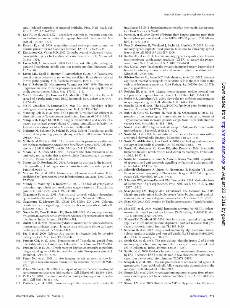
terial-induced extrusion of mucosal epithelia. Proc. Natl. Acad. Sci.U. S. A. 107:17733–17738.
85. Koo IC, et al. 2008. ESX-1-dependent cytolysis in lysosome secretionand inflammasome activation during mycobacterial infection. Cell. Mi-crobiol. 10:1866 –1878.
86. Koussis K, et al. 2009. A multifunctional serine protease primes themalaria parasite for red blood cell invasion. EMBO J. 28:725–735.
87. Kumamoto CA, Vinces MD. 2005. Contributions of hyphae and hypha-co-regulated genes to Candida albicans virulence. Cell. Microbiol.7:1546 –1554.
88. Lavine MD, Arrizabalaga G. 2008. Exit from host cells by the pathogenicparasite Toxoplasma gondii does not require motility. Eukaryot. Cell7:131–140.
89. Lavine MD, Knoll LJ, Rooney PJ, Arrizabalaga G. 2007. A Toxoplasmagondii mutant defective in responding to calcium fluxes shows reducedin vivo pathogenicity. Mol. Biochem. Parasitol. 155:113–122.
90. Ley V, Robbins ES, Nussenzweig V, Andrews NW. 1990. The exit ofTrypanosoma cruzi from the phagosome is inhibited by raising the pH ofacidic compartments. J. Exp. Med. 171:401– 413.
91. Ma H, Croudace JE, Lammas DA, May RC. 2007. Direct cell-to-cellspread of a pathogenic yeast. BMC Immunol. 8:15. doi:10.1186/1471-2172-8-15.
92. Ma H, Croudace JE, Lammas DA, May RC. 2006. Expulsion of livepathogenic yeast by macrophages. Curr. Biol. 16:2156 –2160.
93. Manning-Cela R, et al. 2001. LYT1 protein is required for efficient invitro infection by Trypanosoma cruzi. Infect. Immun. 69:3916 –3923.
94. Marquis H, Hager EJ. 2000. pH-regulated activation and release of abacteria-associated phospholipase C during intracellular infection byListeria monocytogenes. Mol. Microbiol. 35:289 –298.
95. Meissner M, Schluter D, Soldati D. 2002. Role of Toxoplasma gondiimyosin A in powering parasite gliding and host cell invasion. Science298:837– 840.
96. Millholland MG, et al. 2011. The malaria parasite progressively disman-tles the host erythrocyte cytoskeleton for efficient egress. Mol. Cell. Pro-teomics 10:M111.010678. doi:10.1074/mcp.M111.010678.
97. Moore-Lai D, Rowland E. 2004. Discovery and characterization of anantibody, anti-egressin, that is able to inhibit Trypanosoma cruzi egressin vitro. J. Parasitol. 90:524 –530.
98. Moore-Lai D, Rowland EC. 2004. Antiegressin acts late in the intracel-lular growth cycle of Trypanosoma cruzi to inhibit parasite egress. J.Parasitol. 90:85–91.
99. Mortara RA, et al. 2005. Mammalian cell invasion and intracellulartrafficking by Trypanosoma cruzi infective forms. An. Acad. Bras. Cienc.77:77–94.
100. Moudy R, Manning TJ, Beckers CJ. 2001. The loss of cytoplasmicpotassium upon host cell breakdown triggers egress of Toxoplasmagondii. J. Biol. Chem. 276:41492– 41501.
101. Nagamune K, et al. 2008. Abscisic acid controls calcium-dependentegress and development in Toxoplasma gondii. Nature 451:207–210.
102. Nagamune K, Moreno SN, Chini EN, Sibley LD. 2008. Calciumregulation and signaling in apicomplexan parasites. Subcell.Biochem. 47:70 – 81.
103. Noronha FS, Cruz JS, Beirao PS, Horta MF. 2000. Macrophage damageby Leishmania amazonensis cytolysin: evidence of pore formation on cellmembrane. Infect. Immun. 68:4578 – 4584.
104. Onfelt B, et al. 2006. Structurally distinct membrane nanotubes betweenhuman macrophages support long-distance vesicular traffic or surfing ofbacteria. J. Immunol. 177:8476 – 8483.
105. Paz I, et al. 2009. Galectin-3, a marker for vacuole lysis by invasivepathogens. Cell. Microbiol. 12:530 –544.
106. Persson CM, et al. 2009. Transmission of Toxoplasma gondii frominfected dendritic cells to natural killer cells. Infect. Immun. 77:970 –976.
107. Persson EK, et al. 2007. Death receptor ligation or exposure to perforintrigger rapid egress of the intracellular parasite Toxoplasma gondii. J.Immunol. 179:8357– 8365.
108. Peters NC, et al. 2008. In vivo imaging reveals an essential role forneutrophils in leishmaniasis transmitted by sand flies. Science 321:970 –974.
109. Peters NC, Sacks DL. 2009. The impact of vector-mediated neutrophilrecruitment on cutaneous leishmaniasis. Cell. Microbiol. 11:1290 –1296.
110. Pfeffer SR. 2010. Unconventional secretion by autophagosome exocyto-sis. J. Cell Biol. 188:451– 452.
111. Plattner F, et al. 2008. Toxoplasma profilin is essential for host cell
invasion and TLR11-dependent induction of an interleukin-12 response.Cell Host Microbe 3:77– 87.
112. Ponzi M, et al. 2009. Egress of Plasmodium berghei gametes from theirhost erythrocyte is mediated by the MDV-1/PEG3 protein. Cell. Micro-biol. 11:1272–1288.
113. Pust S, Morrison H, Wehland J, Sechi AS, Herrlich P. 2005. Listeriamonocytogenes exploits ERM protein functions to efficiently spreadfrom cell to cell. EMBO J. 24:1287–1300.
114. Radtke AL, et al. 2011. Listeria monocytogenes exploits cystic fibrosistransmembrane conductance regulator (CFTR) to escape the phago-some. Proc. Natl. Acad. Sci. U. S. A. 108:1633–1638.
115. Ray K, et al. 2010. Tracking the dynamic interplay between bacterial andhost factors during pathogen-induced vacuole rupture in real time. Cell.Microbiol. 12:545–556.
116. Ribeiro-Gomes FL, Peters NC, Debrabant A, Sacks DL. 2012. Efficientcapture of infected neutrophils by dendritic cells in the skin inhibits theearly anti-leishmania response. PLoS Pathog. 8:e1002536. doi:10.1371/journal.ppat.1002536.
117. Robbins JR, et al. 1999. Listeria monocytogenes exploits normal hostcell processes to spread from cell to cell. J. Cell Biol. 146:1333–1350.
118. Roiko MS, Carruthers VB. 2009. New roles for perforins and proteasesin apicomplexan egress. Cell. Microbiol. 11:1444 –1452.
119. Rosado CJ, et al. 2008. The MACPF/CDC family of pore-forming tox-ins. Cell. Microbiol. 10:1765–1774.
120. Rubin-de-Celis SS, Uemura H, Yoshida N, Schenkman S. 2006. Ex-pression of trypomastigote trans-sialidase in metacyclic forms ofTrypanosoma cruzi increases parasite escape from its parasitophorousvacuole. Cell. Microbiol. 8:1888 –1898.
121. Sano G, et al. 2007. Flagella facilitate escape of Salmonella from oncoticmacrophages. J. Bacteriol. 189:8224 – 8232.
122. Santic M, et al. 2009. Intracellular fate of Francisella tularensis withinarthropod-derived cells. Environ. Microbiol. 11:1473–1481.
123. Santic M, Al-Khodor S, Abu Kwaik Y. 2010. Cell biology and molecularecology of Francisella tularensis. Cell. Microbiol. 12:129 –139.
124. Santic M, Molmeret M, Klose KE, Abu Kwaik Y. 2006. Francisellatularensis travels a novel, twisted road within macrophages. Trends Mi-crobiol. 14:37– 44.
125. Santic M, Pavokovic G, Jones S, Asare R, Kwaik YA. 2010. Regulationof apoptosis and anti-apoptosis signalling by Francisella tularensis. Mi-crobes Infect. 12:126 –134.
126. Schmidt-Christensen A, Sturm A, Horstmann S, Heussler VT. 2008.Expression and processing of Plasmodium berghei SERA3 during liverstages. Cell. Microbiol. 10:1723–1734.
127. Schuerch DW, Wilson-Kubalek EM, Tweten RK. 2005. Molecular basisof listeriolysin O pH dependence. Proc. Natl. Acad. Sci. U. S. A. 102:12537–12542.
128. Shaughnessy LM, Hoppe AD, Christensen KA, Swanson JA. 2006.Membrane perforations inhibit lysosome fusion by altering pH and cal-cium in Listeria monocytogenes vacuoles. Cell. Microbiol. 8:781–792.
129. Shaw MK. 2003. Cell invasion by Theileria sporozoites. Trends Parasitol.19:2– 6.
130. Shio MT, et al. 2009. Malarial hemozoin activates the NLRP3 inflam-masome through Lyn and Syk kinases. PLoS Pathog. 5:e1000559. doi:10.1371/journal.ppat.1000559.
131. Silveira TN, Zamboni DS. 2010. Pore formation triggered by Legionellaspp. is an Nlrc4 inflammasome-dependent host cell response that pre-cedes pyroptosis. Infect. Immun. 78:1403–1413.
132. Simeone R, et al. 2012. Phagosomal rupture by Mycobacterium tuber-culosis results in toxicity and host cell death. PLoS Pathog. 8:e1002507.doi:10.1371/journal.ppat.1002507.
133. Smith GA, et al. 1995. The two distinct phospholipases C of Listeriamonocytogenes have overlapping roles in escape from a vacuole andcell-to-cell spread. Infect. Immun. 63:4231– 4237.
134. Smith J, et al. 2008. Evidence for pore formation in host cell membranesby ESX-1-secreted ESAT-6 and its role in Mycobacterium marinum es-cape from the vacuole. Infect. Immun. 76:5478 –5487.
135. Sologub L, et al. 2011. Malaria proteases mediate inside-out egress ofgametocytes from red blood cells following parasite transmission to themosquito. Cell. Microbiol. 13:897–912.
136. Stamm LM, et al. 2003. Mycobacterium marinum escapes from phago-somes and is propelled by actin-based motility. J. Exp. Med. 198:1361–1368.
137. Stamm LM, et al. 2005. Role of the WASP family proteins for Mycobac-
Host Cell Egress by Intracellular Pathogens
December 2012 Volume 76 Number 4 mmbr.asm.org 719
on August 22, 2019 by guest
http://mm
br.asm.org/
Dow
nloaded from
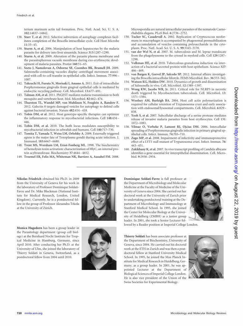
terium marinum actin tail formation. Proc. Natl. Acad. Sci. U. S. A.102:14837–14842.
138. Starr T, et al. 2012. Selective subversion of autophagy complexes facil-itates completion of the Brucella intracellular cycle. Cell Host Microbe11:33– 45.
139. Sturm A, et al. 2006. Manipulation of host hepatocytes by the malariaparasite for delivery into liver sinusoids. Science 313:1287–1290.
140. Sturm A, et al. 2009. Alteration of the parasite plasma membrane andthe parasitophorous vacuole membrane during exo-erythrocytic devel-opment of malaria parasites. Protist 160:51– 63.
141. Szeto J, Namolovan A, Osborne SE, Coombes BK, Brumell JH. 2009.Salmonella-containing vacuoles display centrifugal movement associ-ated with cell-to-cell transfer in epithelial cells. Infect. Immun. 77:996 –1007.
142. Takeuchi H, Furuta N, Morisaki I, Amano A. 2011. Exit of intracellularPorphyromonas gingivalis from gingival epithelial cells is mediated byendocytic recycling pathway. Cell. Microbiol. 13:677– 691.
143. Talman AM, et al. 2011. PbGEST mediates malaria transmission to bothmosquito and vertebrate host. Mol. Microbiol. 82:462– 474.
144. Thurston TL, Wandel MP, von Muhlinen N, Foeglein A, Randow F.2012. Galectin 8 targets damaged vesicles for autophagy to defend cellsagainst bacterial invasion. Nature 482:414 – 418.
145. Tobin DM, et al. 2012. Host genotype-specific therapies can optimizethe inflammatory response to mycobacterial infections. Cell 148:434 –446.
146. Tobin DM, et al. 2010. The lta4h locus modulates susceptibility tomycobacterial infection in zebrafish and humans. Cell 140:717–730.
147. Tomita T, Yamada T, Weiss LM, Orlofsky A. 2009. Externally triggeredegress is the major fate of Toxoplasma gondii during acute infection. J.Immunol. 183:6667– 6680.
148. Trent MS, Worsham LM, Ernst-Fonberg ML. 1998. The biochemistryof hemolysin toxin activation: characterization of HlyC, an internal pro-tein acyltransferase. Biochemistry 37:4644 – 4652.
149. Troemel ER, Felix MA, Whiteman NK, Barriere A, Ausubel FM. 2008.
Microsporidia are natural intracellular parasites of the nematode Caeno-rhabditis elegans. PLoS Biol. 6:2736 –2752.
150. Tucker SC, Casadevall A. 2002. Replication of Cryptococcus neofor-mans in macrophages is accompanied by phagosomal permeabilizationand accumulation of vesicles containing polysaccharide in the cyto-plasm. Proc. Natl. Acad. Sci. U. S. A. 99:3165–3170.
151. van der Wel N, et al. 2007. M. tuberculosis and M. leprae translocatefrom the phagolysosome to the cytosol in myeloid cells. Cell 129:1287–1298.
152. Volkman HE, et al. 2010. Tuberculous granuloma induction via inter-action of a bacterial secreted protein with host epithelium. Science 327:466 – 469.
153. von Bargen K, Gorvel JP, Salcedo SP. 2012. Internal affairs: investigat-ing the Brucella intracellular lifestyle. FEMS Microbiol. Rev. 36:533–562.
154. Watson KG, Holden DW. 2010. Dynamics of growth and disseminationof Salmonella in vivo. Cell. Microbiol. 12:1389 –1397.
155. Wong KW, Jacobs WR, Jr. 2011. Critical role for NLRP3 in necroticdeath triggered by Mycobacterium tuberculosis. Cell. Microbiol. 13:1371–1384.
156. Woolsey AM, Burleigh BA. 2004. Host cell actin polymerization isrequired for cellular retention of Trypanosoma cruzi and early associa-tion with endosomal/lysosomal compartments. Cell. Microbiol. 6:829 –838.
157. Yeoh S, et al. 2007. Subcellular discharge of a serine protease mediatesrelease of invasive malaria parasites from host erythrocytes. Cell 131:1072–1083.
158. Yilmaz O, Verbeke P, Lamont RJ, Ojcius DM. 2006. Intercellularspreading of Porphyromonas gingivalis infection in primary gingival ep-ithelial cells. Infect. Immun. 74:703–710.
159. Zago MP, et al. 2008. Impairment of infectivity and immunoprotectiveeffect of a LYT1 null mutant of Trypanosoma cruzi. Infect. Immun. 76:443– 451.
160. Zakikhany K, et al. 2007. In vivo transcript profiling of Candida albicansidentifies a gene essential for interepithelial dissemination. Cell. Micro-biol. 9:2938 –2954.
Nikolas Friedrich obtained his Ph.D. in 2009from the University of Geneva for his work inthe laboratory of Professor Dominique Soldati-Favre and Dr. Mike Blackman (National Insti-tute for Medical Research, London, UnitedKingdom). Currently, he is a postdoctoral fel-low in the group of Professor Alexandra Trkolaat the University of Zürich.
Monica Hagedorn has been a group leader inthe Parasitology department (group cell biol-ogy) at the Bernhard Nocht Institute for Trop-ical Medicine in Hamburg, Germany, sinceApril 2010. After conducting her Ph.D. at theUniversity of Ulm, she joined the laboratory ofThierry Soldati in Geneva, Switzerland, as apostdoctoral fellow from 2004 until 2010.
Dominique Soldati-Favre is full professor atthe Department of Microbiology and MolecularMedicine at the Faculty of Medicine of the Uni-versity of Geneva since 2004. She carried out herdoctoral work at the University of Zurich priorto undertaking postdoctoral training at the De-partment of Microbiology and Immunology atStanford Medical School. In 1995, she joinedthe Center for Molecular Biology at the Univer-sity of Heidelberg (ZMBH) as a junior groupleader. In 2001, she took a Senior Lecturer fol-lowed by a Reader position at Imperial College London.
Thierry Soldati has been associate professor atthe Department of Biochemistry, University ofGeneva, since 2004. He carried out his doctoralwork at the ETH in Zurich and was then a post-doctoral fellow at Stanford University MedicalSchool. In 1995, he joined the Max Planck In-stitute for Medical Research in Heidelberg, Ger-many, as a group leader. In 2001, he was ap-pointed Lecturer at the Department ofBiological Sciences of Imperial College London.He is also vice president of the Union of theSwiss Societies for Experimental Biology.
Friedrich et al.
720 mmbr.asm.org Microbiology and Molecular Biology Reviews
on August 22, 2019 by guest
http://mm
br.asm.org/
Dow
nloaded from