Embed Size (px)
Citation preview
Submitted 27 February 2018Accepted 15 May 2018Published 4 June 2018
Corresponding authorKenneth B. Storey,[email protected]
Academic editorVladimir Uversky
Additional Information andDeclarations can be found onpage 17
DOI 10.7717/peerj.4911
Copyright2018 Logan and Storey
Distributed underCreative Commons CC-BY 4.0
OPEN ACCESS
Pro-inflammatory AGE-RAGE signalingis activated during arousal fromhibernation in ground squirrel adiposeSamantha M. Logan and Kenneth B. StoreyInstitute of Biochemistry, Departments of Biology and Chemistry, Carleton University, Ottawa, Ontario,Canada
ABSTRACTBackground. Inflammation is generally suppressed during hibernation, but selecttissues (e.g. lung) have been shown to activate both antioxidant and pro-inflammatorypathways, particularly during arousal from torpor when breathing rates increase andoxidative metabolism fueling the rewarming process produces more reactive oxygenspecies. Brown and white adipose tissues are now understood to be major hubs for theregulation of immune and inflammatory responses, yet how these potentially damagingprocesses are regulated by fat tissues during hibernation has hardly been studied.The advanced glycation end-product receptor (RAGE) can induce pro-inflammatoryresponses when bound by AGEs (which are glycated and oxidized proteins, lipids, ornucleic acids) or damage associated molecular pattern molecules (DAMPs, which arereleased from dying cells).Methods. Since gene expression and protein synthesis are largely suppressed duringtorpor, increases in AGE-RAGE pathway proteins relative to a euthermic control couldsuggest some role for these pro-inflammatorymediators during hibernation. This studydetermined how the pro-inflammatory AGE-RAGE signaling pathway is regulated atsix major time points of the torpor-arousal cycle in brown and white adipose froma model hibernator, Ictidomys tridecemlineatus. Immunoblotting, RT-qPCR, and acompetitive ELISA were used to assess the relative gene expression and protein levelsof key regulators of the AGE-RAGE pathway during a hibernation bout.Results. The results of this study revealed that RAGE is upregulated as animals arousefrom torpor in both types of fat, but AGE and DAMP levels either remain unchangedor decrease. Downstream of the AGE-RAGE cascade, nfat5 was more highly expressedduring arousal in brown adipose.Discussion. An increase in RAGE protein levels and elevated mRNA levels of thedownstream transcription factor nfat5 during arousal suggest the pro-inflammatoryresponse is upregulated in adipose tissue of the hibernating ground squirrel. It isunlikely that this cascade is activated by AGEs or DAMPs. This research sheds lighton how a fat-but-fit organism with highly regulated metabolism may control the pro-inflammatory AGE-RAGE pathway, a signaling cascade that is often dysregulated inother obese organisms.
Subjects Animal Behavior, Molecular BiologyKeywords AGE-RAGE, Inflammation, 13-lined ground squirrel, Damage-associated molecularpattern molecule, Hibernation, Adipose, Torpor-arousal cycle
How to cite this article Logan and Storey (2018), Pro-inflammatory AGE-RAGE signaling is activated during arousal from hibernation inground squirrel adipose. PeerJ 6:e4911; DOI 10.7717/peerj.4911
INTRODUCTIONThe advanced-glycation end product (AGE) and AGE receptor (AGE-RAGE) pathway isemerging as an important signal transduction pathway that influences an immune andoxidative stress response via the activation of mitogen-activated protein kinase (MAPK)and nuclear factor kappa-light-chain-enhancer of activated B cells (NF-κB) pathways(Sparvero et al., 2009). RAGE is normally expressed in a range of tissues including lung,heart, vasculature, brain, liver, embryonic tissue, etc., but becomes overexpressed in areas oflocalized inflammation (for instance, post-trauma) or in tissues affected by inflammatorydisease (like diabetes, cancer, neurodegeneration, etc.) (Brett et al., 1993; Sparvero et al.,2009). This pro-inflammatory pathway can contribute to disease pathogenesis since itsactivation (via ligand binding to RAGE) often leads to cellular dysfunction and ultimatelytissue damage (Ramasamy, Yan & Schmidt, 2011).
During the winter months, hibernating ground squirrels use a range of molecularadaptations to maintain tissue homeostasis until arousal in the spring. These animalsregulate tissue viability through antioxidant (Morin et al., 2008; Ni & Storey, 2010; Rouble,Tessier & Storey, 2014) and anti-apoptotic (Rouble et al., 2013; Logan et al., 2016; Logan,Luu & Storey, 2016) pathways to prevent the conditions (e.g., hypoxia, cell death) thatcould give rise to localized inflammation and subsequent tissue damage (Bouma et al.,2013; Bogren et al., 2014). However, there is evidence that pro-inflammatory pathwayscould be upregulated upon arousal from hibernation from reports showing increasedoxidized proteins, lipid peroxides, and pro-inflammatory cytokines (TNF-alpha, IFN-γ)during arousal from hibernation in ground squirrels and bats (Lee, Choi & Park, 2002;Kurtz & Carey, 2007; Orr et al., 2009). Hibernating ground squirrels are well known fortheir extraordinary ability tomaintain whole-body homeostasis during periods of cell stress,so it is essential to understand how they control inflammation, a process that could resultin cerebral ischemia, muscle wasting, and overconsumption of ATP if left unchecked,but does not in ground squirrels (Frerichs et al., 1994; Bogren et al., 2014; Ruf & Geiser,2015; Tessier & Storey, 2016). Most studies on inflammation in hibernators focus on howanti-inflammatory/protective signaling pathways could be involved in this whole-bodyhomeostasis but few studies have aimed to identify potential pro-inflammatory pathwaysthat could be activated to warrant the increase in anti-inflammatory signaling, in the firstplace. The regulation of the AGE-RAGE pathway has never been studied in an animal thatundergoes metabolic suppression (to the best of our knowledge). Thus, it is essential toassess how the pro-inflammatory AGE-RAGE pathway may be regulated throughout thetorpor-arousal cycle in the 13-lined ground squirrel (Ictidomys tridecemlineatus, a modelhibernator).
Specifically, the brown and white adipose tissues (BAT and WAT, respectively) of13-lined ground squirrels are of interest because these tissues undergo major physiologicalchanges before and during hibernation that involve managing oxidative stress andinflammation. Hibernator BAT undergoes a 2- to 3-fold increase in weight during the pre-hibernation phase in Arctic ground squirrels (Spermophilus parryii) (Boyer & Barnes, 1999)
Logan and Storey (2018), PeerJ, DOI 10.7717/peerj.4911 2/22
since BAT is essential for the generation of heat during arousals from torpor via non-shivering thermogenesis (Storey & Storey, 2005). The arousal process involves an increasedproduction of reactive oxygen species (ROS) (accompanying increased oxygen intakeand elevated oxidative metabolism) which can lead to tissue damage and inflammation.Evidence of increased ROS production during torpor and/or arousal in mammals includesincreased lipid peroxidation, HSP70 expression, and NF-κB activity in ground squirrelintestine (Carey, Frank & Seifert, 2000; Carey, Rhoads & Aw, 2003). Adipose tissues fromhibernating European ground squirrel (Spermophilus citellus) and 13-lined ground squirrelsshow increases in antioxidant enzymes such as superoxide dismutases 1 and 2, and catalase,possibly to counteract increases in the generation of ROS (Vucetic et al., 2013; Rouble,Tessier & Storey, 2014). WAT also rapidly expands in the summer months before torporfollowing hyperphagia (a period of intense eating needed to lay down large depots ofstored fatty acids) (Sheriff et al., 2013; Schwartz, Hampton & Andrews, 2015). WAT is alsoan important mediator of the inflammatory response since it can produce both pro-and anti-inflammatory cytokines termed adipokines (Trayhurn & Wood, 2004). Oxidativestress and chronic inflammation in humans promotes the dysregulation of adipokines(e.g., leptin, TNF-α, IL-6, adiponectin, etc.) synthesis and secretion which can ultimatelylead to secondary metabolic disorders including insulin resistance, cardiovascular disease,and cancer (Kayser & Verges, 2013; He et al., 2015). By contrast, the 13-lined groundsquirrel is ‘‘fat but fit’’, providing an excellent model of controlled energy metabolism andregulated inflammatory/immune responses. Thus, it is essential to study the AGE-RAGEpathway and the pro-inflammatory pathways it regulates (i.e., MAPK, JNK, NFAT) in afat-storing hibernator this is capable of reversible insulin resistance and that is resistant totissue damage brought on by oxidative stress.
RAGE is a receptor for protein ligands including glycated proteins (AGEs), damageassociated molecular patterns (DAMPs), and pathogen associated molecular patterns(PAMPs) (not part of the current study). AGEs are formed by the non-enzymaticattachment of sugars to proteins, lipids, or nucleic acids by the Maillard reaction, acomplicated process that can yield hundreds of AGE species and has been discussedelsewhere (Ott et al., 2014; Boyer et al., 2015). AGE accumulation is a symptom ofinflammatory states including normal aging and metabolic disorders like diabetes whereglucose is openly available (Cooper, 2004; Vlassara, Brownlee & Cerami, 1985). Damage-associated molecular pattern (DAMP) molecules are intracellularly derived proteinsreleased into the extracellular space by dead or dying cells, and are RAGE ligands that caninvoke immune, pro-inflammatory, and oxidative stress signaling cascades (Rubartelli &Lotze, 2007). DAMPs that bind RAGE include S100 calcium-binding protein B (S100B)and high mobility group box 1 protein (HMGB1), both of which are highly conservedbetween mammalian species and are expressed in adipose tissue (Buckman et al., 2014;Ghosh et al., 2016). AGEs or DAMPs binding to RAGE, activates ERK1/2, JNK, and p38signaling pathways induce pro-inflammatory gene expression (Sparvero et al., 2009; Ottet al., 2014). The focus of this study is the MAPK signaling pathway involving ERK1/2because this pathway was activated during the stress response in BAT andWAT from torpidprimates (Biggar et al., 2015). Raf is a serine/threonine MAPK kinase kinase (MAPKKK)
Logan and Storey (2018), PeerJ, DOI 10.7717/peerj.4911 3/22
which is involved in the phosphorylation of MAPK kinases (MAPKK) like MEK1/2,which phosphorylates MAPKs (ERK1/2) to activate them. Once activated, ERK1/2 canphosphorylate transcription factors from the Ets, NFAT, and Jun families to promotethe expression of inflammatory/immune genes (Cowan & Storey, 2003; MacDonald &Storey, 2005a; Lu & Xu, 2006). Experiments on obese human WAT (a good system forstudying oxidative stress and chronic inflammation in adipose) showed that Ets1 candirectly regulate inflammation in adipocytes by interacting with the TNF-α promoter andpromoting TNF-α expression (Zhang et al., 2016). ERK1/2 also regulates the activationof the Rel family of transcription factors, including NF-κB and NFAT1-5 (Ueno et al.,2013). NFATs regulate immune responses and angiogenesis, making them interesting tostudy in the context of expanded adipose tissue. NFAT5 is expressed in both WAT andBAT, is known to associate with NF-κB to increase its pro-inflammatory activity, and canregulate the expression of important immune and osmoregulatory genes like monocytechemoattractant protein 1 (MCP1) and TNF-α (Ueno et al., 2013). Finally, Jun, one of thesubunits of the AP-1 transcription factor, is an important mediator of inflammation inadipocytes. Elevated leptin levels activate Jun and AP-1 DNA binding to increase reactiveoxygen species production and to propagate pro-inflammatory signaling (Gullicksen et al.,2003;Makki, Froguel & Wolowczuk, 2013).
In the current study, total protein levels of full length RAGE, HMGB1 and S100Bwere assessed via Western blotting. RAGE activation via AGEs and DAMPs can induceERK1/2 signaling (Lander et al., 1997; Reddy et al., 2006) so RT-qPCR was used to evaluatethe expression of rage, hmgb1, one MAPK gene upstream of ERK1/2 (araf ) and selectdownstream transcription factors known to be activated by ERK1/2 (ets1, nfat5, jun)(Fig. 1). The results of this study suggest that RAGE signaling is activated in BAT andWAT upon arousal from hibernation, but CML-AGE, HMGB1 and S100B are probablynot responsible for the activation of this pathway.
METHODSAnimal experimentsWild-captured 13-lined ground squirrels weighing approximately 150–300 g, were caughtand transported by a United States Department of Agriculture-licensed trapper (TLSResearch, Bloomingdale, IL, USA) to Dr. J.M. Hallenback’s laboratory at the AnimalHibernation Facility, National Institute of Neurological Disorders and Stroke (NIH,Bethesda, MD, USA), where the hibernation experiments were performed. NINDS animalcare and use committee (ACUC) animal housing and experimental procedures werefollowed (protocol number ASP 1223-05). Each ground squirrel was anesthetized with 5%isofluorane and fitted with a sensor chip (IPTT-300; Bio Medic Data Systems, Seaford,DE, USA) injected under the skin. Each squirrel was housed individually in a shoeboxcage at 21 ◦C. Animals were fed a standard rodent diet and water ad libitum until theygained sufficient lipid stores to enter hibernation. To enable a natural transition intotorpor, animals were transferred to an environmental chamber at ∼5 ◦C in constantdarkness. Sampling points throughout the torpor-arousal cycle were chosen based on body
Logan and Storey (2018), PeerJ, DOI 10.7717/peerj.4911 4/22
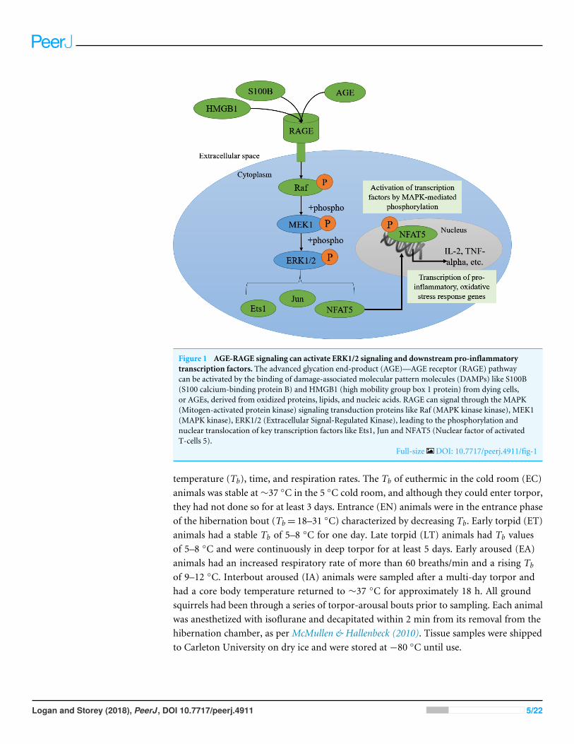
Figure 1 AGE-RAGE signaling can activate ERK1/2 signaling and downstream pro-inflammatorytranscription factors. The advanced glycation end-product (AGE)—AGE receptor (RAGE) pathwaycan be activated by the binding of damage-associated molecular pattern molecules (DAMPs) like S100B(S100 calcium-binding protein B) and HMGB1 (high mobility group box 1 protein) from dying cells,or AGEs, derived from oxidized proteins, lipids, and nucleic acids. RAGE can signal through the MAPK(Mitogen-activated protein kinase) signaling transduction proteins like Raf (MAPK kinase kinase), MEK1(MAPK kinase), ERK1/2 (Extracellular Signal-Regulated Kinase), leading to the phosphorylation andnuclear translocation of key transcription factors like Ets1, Jun and NFAT5 (Nuclear factor of activatedT-cells 5).
Full-size DOI: 10.7717/peerj.4911/fig-1
temperature (Tb), time, and respiration rates. The Tb of euthermic in the cold room (EC)animals was stable at∼37 ◦C in the 5 ◦C cold room, and although they could enter torpor,they had not done so for at least 3 days. Entrance (EN) animals were in the entrance phaseof the hibernation bout (Tb= 18–31 ◦C) characterized by decreasing Tb. Early torpid (ET)animals had a stable Tb of 5–8 ◦C for one day. Late torpid (LT) animals had Tb valuesof 5–8 ◦C and were continuously in deep torpor for at least 5 days. Early aroused (EA)animals had an increased respiratory rate of more than 60 breaths/min and a rising Tb
of 9–12 ◦C. Interbout aroused (IA) animals were sampled after a multi-day torpor andhad a core body temperature returned to ∼37 ◦C for approximately 18 h. All groundsquirrels had been through a series of torpor-arousal bouts prior to sampling. Each animalwas anesthetized with isoflurane and decapitated within 2 min from its removal from thehibernation chamber, as per McMullen & Hallenbeck (2010). Tissue samples were shippedto Carleton University on dry ice and were stored at −80 ◦C until use.
Logan and Storey (2018), PeerJ, DOI 10.7717/peerj.4911 5/22
Total protein extractionFrozen brown adipose tissue (BAT) and white adipose tissue (WAT) samples were weighedfor immunoblotting (∼70 mg of 4 biological replicates from EC, EN, ET, LT, EA andIA) and carboxymethyl-lysine-ELISAs (∼50 mg of 4 biological replicates from EC andLT ground squirrels). Frozen samples were weighed and crushed into small pieces underliquid nitrogen. Five to seven piston strokes of a glass Dounce pestle homogenized thetissues in ice-cold Cell Signaling Lysis Buffer (#43-040; EMD Millipore, Burlington, MA,USA), added in a 1:3 w:v ratio and supplemented with 1 mM sodium orthovanadate,10mM sodium fluoride, 10 mM β-glycerophosphate, and 10 µL/mL Protease Inhibitorcocktail (#PIC001.1; BioShop, Burlington, Canada). Each sample was placed on ice andgently vortexed every 10 min for 30 min before centrifugation at 13,500×g for 20 min at4 ◦C. The supernatant containing soluble proteins was collected for each sample and totalprotein concentration was determined for immunoblotting and ELISA samples using theBio-Rad method (Cat#500-0005; Bio-Rad, Hercules, CA, USA). Appropriate amounts ofhomogenization buffer were added to the samples intended for immunoblotting such thatthey were all standardized to 10 µg/µL. Then, 2X sodium dodecyl sulfate (SDS) loadingbuffer (100 mM Tris-base adjusted to pH 6.8, 4% w:v SDS, 20% v:v glycerol, 0.2% w:vbromophenol blue, 10% v:v 2-mercaptoethanol) was added to each sample in a 1:1 v:v ratioand the samples were mixed and boiled. The final 5 µg/µL protein samples were stored at−40◦C until use. The samples intended for ELISAs were standardized to 1.4 µg/µL (WAT)and 20 µg/µL (BAT) by adding appropriate amounts of phosphate buffered saline (PBS)containing 0.1% v:v bovine serum albumin (BSA; Sigma, #A4503), as per the commercialELISA manufacturer’s instructions.
Western blottingEqual amounts (25 µg) of prepared protein homogenate and 4–5uL of 10.5–175 kDaPiNK Plus pre-stained protein ladder (#PM005-0500; FroggaBio, Toronto, Canada) wereloaded onto 10–15% SDS-PAGE gels, electrophoresed for 80–100 min at 180 V usingthe BioRad Mini Protean III system in Tris-glycine running buffer (0.25 M Tris-base pH8.0, 2.45 M glycine, 0.035 M SDS). Proteins were wet-transferred onto polyvinylidenefluoride (PVDF), either at 160 mA for 90 min (RAGE) or at 30 V for 45 min (S100Band HMGB1) in Tris-glycine transfer buffer (25 mM Tris pH 8.5, 192 mM glycineand 10% v:v methanol). Blots were blocked with 2.5–10.0% w:v milk in Tris-bufferedsaline with Tween-20 (TBST; 50 mM Tris–HCl, 150 mM NaCl, 0.05% v:v Tween-20,pH 6.8) for 15–30 min. The blots were incubated overnight (RAGE) or twice overnight(S100B and HMGB1) at 4 ◦C on a rocker with primary antibodies (diluted 1:1,000 v:vin TBST). RAGE (#GTX23611; GeneTex, Irvine, CA, USA) and S100B (#GTX129573;GeneTex, Irvine, CA, USA) were rabbit polyclonal primary antibodies and the HMGB1(University of Iowa, #PCRP-HMGB1-3A7) was a mouse monoclonal primary antibody.Following primary antibody removal, the membranes were incubated for 15 min with anHRP-linked anti-rabbit IgG secondary antibody (RAGE and S100B) or an HRP-linkedanti-mouse IgG secondary antibody (HMGB1), diluted 1:8,000 v:v with TBST. Enhancedchemiluminescence (ECL) reagents were used to visualize the protein bands. The amount
Logan and Storey (2018), PeerJ, DOI 10.7717/peerj.4911 6/22
of protein in each lane was reassessed using Coomassie Blue staining (0.25%w:v Coomassiebrilliant blue, 7.5% v:v acetic acid, 50% methanol) of the PVDF membranes.
Carboxymethyl lysine-advanced glycation end product (CML-AGE)ELISACML-AGE was assessed using a protein ELISA because it is the most abundant subtypeof AGE, it is formed from the oxidation of protein and lipid precursors, it is a bona fideRAGE ligand, and it induces strong inflammatory signaling through RAGE. The OxiSelectN ε-(carboxymethyl) lysine (CML) competitive ELISA (#STA-816; Cell Biolabs Inc., SanDiego, CA, USA) was performed as directed by the manufacturer. All materials listed wereincluded with the kit. Briefly, 100 µL 1X CML Conjugate (prepared by diluting the 1000XCML Conjugate with 1X CML Diluent, prepared in 1X PBS buffer) incubated in CMLconjugate-coated wells overnight at 4 ◦C. Wells of the were washed twice with 1X PBS andthen wells were blocked with 200 µL Assay Diluent for 1 h at room temperature whilerocking on an orbital shaker. The CML-BSA standard curve was prepared from 1 mg/mLCML-BSA Standard, diluted with Assay Diluent to concentrations between 0–12.5 µg/mL.Then, 50 µL of the standards or WAT (1.4 µg/µL) or BAT (20 µg/µL) samples (EC vs. LT)were added to the CML-Conjugate coated wells in duplicate and incubated for 10 min atroom temperature on an orbital shaker. Following a 1-hour (room temperature, shaking)incubation with 50 µL of anti-CML primary antibody (diluted 1:1000 v:v in Assay Diluent),the wells were washed with 1X Wash Buffer. Then, 100 µL of the secondary antibody-HRPConjugate (diluted 1:1,000 v:v in Assay Diluent) was incubated for 1 h (room temperature,shaking). The wells were washed with 1X Wash Buffer and then 100 µL room temperatureSubstrate Solution was added to each well and incubated for 3 min. 100 µL of Stop Solutionwas added to stop the enzyme reaction and the plate was read at 450 nm using a BioTekPowerWave HT spectrophotometer.
RNA extraction, cDNA synthesis and quantitative RT-PCRRNA extractions were performed using the TRIzol (Invitrogen, #15596026) method asdirected by the manufacturer and as previously described (Logan et al., 2016). Briefly,approximately 100 mg of BAT and WAT was weighed and homogenized in 1 mL ofTRIzol using a Polytron PT1200 homogenizer. Four biological replicates were made foreach tissue from independent animals for each of the 6 experimental time points, exceptthree WAT biological replicates were prepared for EN and EA due to tissue availabilityof these time points. RNA purity was assessed using the ratio of absorbances 260/280nm and RNA integrity was assessed by visualizing 18S and 26S ribosomal bands on a 1%agarose gel with SybrGreen staining. RNA samples were normalized in a total volume of10 µL DEPC-autoclaved ddH2O such that each biological replicate contained 4 µg (brownadipose tissue) or 1.14 µg (white adipose tissue) of RNA. The normalized RNA sampleswere incubated with 1µL ofOligo-dT (200 ng/µL 5′-TTTTTTTTTTTTTTTTTTTTTTV-3′;where V = A, G, or C; Sigma Genosys) and placed in a thermocycler at 65 ◦C for 5 min,then chilled on ice for 5 min. Reverse transcription was performed with 4 µL of 5Xfirst-strand buffer (Invitrogen, #18057018), 2 µL of 0.1 M DTT (Invitrogen, #D1532), 1 µL
Logan and Storey (2018), PeerJ, DOI 10.7717/peerj.4911 7/22
of 10 mM dNTPs (#NUC001; BioShop, Burlington, Canada), and 1 µL of MMLV Reversetranscriptase (Invitrogen, # 28025013). Following a 45 min incubation in the thermocyclerat 42 ◦C, RT-qPCR was performed as described previously (Pellissier et al., 2006), using aBioRad CFX Connect apparatus. Forward and reverse primers used for RT-qPCR were asfollows: rage, 5′-AAGCGGGAGAAGCAGAAAGT-3′, 5′-GTGCCAGCTAAGAGTTCCCT-3′; hmgb1, 5′-AGAGCGGAGAGAGTGAGGAG-3′, 5′-TGACATTTTGCCTCTCGGCT-3′; araf, 5′-TGTACCTGCCCAACAAGCAA-3′, 5′-GTAGACCACGCAGCAATCCT-3′;jun, 5′-GAGAGATTGTCGGGGCTGAG-3′, 5′-CCCTTGGCTTTAGTCCTCGG-3′; ets1,5′-AAGCTCTAAGGTGGTCTCAGT-3′, 5′-GCTTCACTTTTCCAATGGGGTC-3′; nfat5,5′-AGCATCCATCAACCCCGAAG-3′, 5′-CCAATCCACACCCCTCATCC-3′; acta1, 5′-AGAACAGCAGGTGTAGTCACG-3′, 5′- AGCCATTGTCACACACGAGG-3′; and tbp,5′- AGAGTGTGCTGGGAATGCTC-3′, 5′-CAGGCTGCTGTTCTGATCCA-3′. Finally,isolated PCR products were sequenced (BioBasic, Markham, ON) and BLASTn was usedto confirm the identity of the amplified genes.
Statistical analysesWestern blot bands were imaged using the Chemi-Genius Bioimaging system (Syngene,Frederick, MD) and were quantified using GeneTools software. Background was accountedfor. PVDF membranes were stained using Coomassie Blue staining (0.25% w:v Coomassiebrilliant blue, 7.5% v:v acetic acid, 50% methanol) to visualize the total amount of proteinin each lane. Chemiluminescent band density in each lane was standardized against thesummed intensity of a group of Coomassie stained protein bands in the same lane (Eaton etal., 2013). Data are expressed as means ± SEM, n= 4 independent samples from differentanimals for all experiments except an n= 3 was used for WAT EN and EA for RT-qPCRexperiments, and an n= 3 was used for BAT LT in the CML-AGE ELISA experiment, dueto limited sample availability. CML-AGE protein concentrations were determined usingthe CML-BSA standard curve (a graph of mean OD 450 nm± standard deviation collectedfor known concentration values). A Student’s t -test was used to evaluate the results ofthe CML-ELISA. Statistical analysis of RT-qPCR experiments involved converting raw Ctvalues obtained from each PCR run to a linear form using 2-̂Ct calculations (1Ct). Thenthe1Ct values for each gene of interest were normalized against the reference genes (acta1for WAT and tbp for BAT) (11Ct). For time-course experiments (Western blotting andRT-qPCR), any differences between control and other torpor-arousal time points wereanalyzed using a one-way ANOVA with Tukey’s post-hoc test. Statistical analyses wereperformed on SigmaPlot software and considered statistically significant the tests yieldeda result of p< 0.05.
RESULTSRelative changes in protein levels of RAGE and its ligands, S100Band HMGB1, over the torpor arousal cycle in brown and whiteadipose tissueImmunoblotting was used to determine relative total protein levels of RAGE, S100B andHMGB1 in brown adipose tissue (BAT) over six time points of the torpor-arousal cycle,
Logan and Storey (2018), PeerJ, DOI 10.7717/peerj.4911 8/22
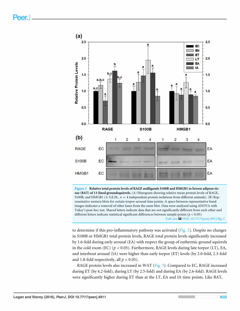
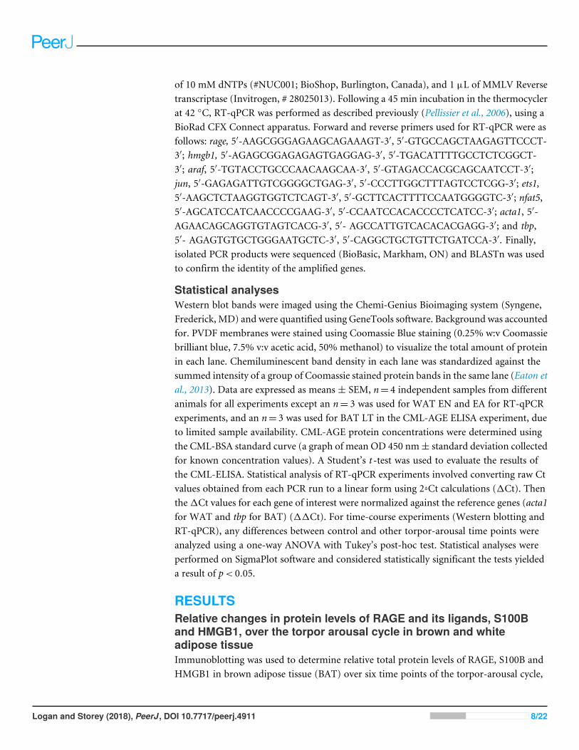
Figure 2 Relative total protein levels of RAGE andligands S100B and HMGB1 in brown adipose tis-sue (BAT) of 13-lined groundsquirrels. (A) Histogram showing relative mean protein levels of RAGE,S100B, and HMGB1 (± S.E.M., n = 4 independent protein isolations from different animals). (B) Rep-resentative western blots for certain torpor-arousal time points. A space between representative bandimages indicates a removal of other lanes from the same blot. Data were analyzed using ANOVA withTukey’s post-hoc test. Shared letters indicate data that are not significantly different from each other anddifferent letters indicate statistical significant differences between sample points (p< 0.05).
Full-size DOI: 10.7717/peerj.4911/fig-2
to determine if this pro-inflammatory pathway was activated (Fig. 2). Despite no changesin S100B or HMGB1 total protein levels, RAGE total protein levels significantly increasedby 1.6-fold during early arousal (EA) with respect the group of euthermic ground squirrelsin the cold room (EC) (p< 0.05). Furthermore, RAGE levels during late torpor (LT), EA,and interbout arousal (IA) were higher than early torpor (ET) levels (by 2.0-fold, 2.3-foldand 1.8-fold respectively, all p< 0.05).
RAGE protein levels also increased in WAT (Fig. 3). Compared to EC, RAGE increasedduring ET (by 4.2-fold), during LT (by 2.5-fold) and during EA (by 2.6-fold). RAGE levelswere significantly higher during ET than at the LT, EA and IA time points. Like BAT,
Logan and Storey (2018), PeerJ, DOI 10.7717/peerj.4911 9/22
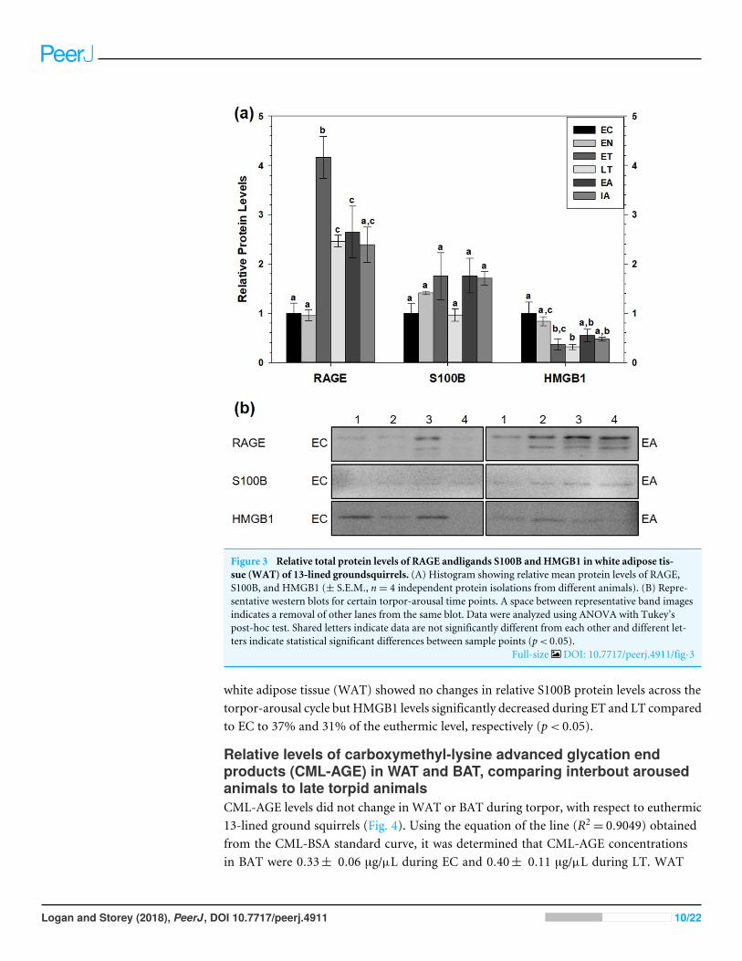
Figure 3 Relative total protein levels of RAGE andligands S100B and HMGB1 in white adipose tis-sue (WAT) of 13-lined groundsquirrels. (A) Histogram showing relative mean protein levels of RAGE,S100B, and HMGB1 (± S.E.M., n= 4 independent protein isolations from different animals). (B) Repre-sentative western blots for certain torpor-arousal time points. A space between representative band imagesindicates a removal of other lanes from the same blot. Data were analyzed using ANOVA with Tukey’spost-hoc test. Shared letters indicate data are not significantly different from each other and different let-ters indicate statistical significant differences between sample points (p< 0.05).
Full-size DOI: 10.7717/peerj.4911/fig-3
white adipose tissue (WAT) showed no changes in relative S100B protein levels across thetorpor-arousal cycle but HMGB1 levels significantly decreased during ET and LT comparedto EC to 37% and 31% of the euthermic level, respectively (p< 0.05).
Relative levels of carboxymethyl-lysine advanced glycation endproducts (CML-AGE) in WAT and BAT, comparing interbout arousedanimals to late torpid animalsCML-AGE levels did not change in WAT or BAT during torpor, with respect to euthermic13-lined ground squirrels (Fig. 4). Using the equation of the line (R2
= 0.9049) obtainedfrom the CML-BSA standard curve, it was determined that CML-AGE concentrationsin BAT were 0.33± 0.06 µg/µL during EC and 0.40± 0.11 µg/µL during LT. WAT
Logan and Storey (2018), PeerJ, DOI 10.7717/peerj.4911 10/22
Figure 4 Calculated concentrations of carboxymethyl-lysine (CML) AGE in 13-lined ground squirrelBAT andWAT. Calculated concentrations of carboxymethyl-lysine (CML) AGE in 13-lined ground squir-rel BAT and WAT, using a BSA-AGE standard curve (File S1). Values are expressed as mean protein con-centrations (± S.E.M., n = 4 for all time points except for n = 3 for BAT LT). Data were analyzed with aStudent’s t -test and amount of CML-AGE was deemed not significantly different between LT and EC.
Full-size DOI: 10.7717/peerj.4911/fig-4
CML-AGE concentrations were much lower with EC levels at 0.13 ± 0.05 and LT valuesaround 0.08± 0.02 µg/µL.
Assessment of RAGE, HMGB1 transcript levels and downstreamMAPK signaling and transcription factor mRNA levels in brown andwhite adipose tissueTranscript levels of rage and hmgb1were assessed over the course of the torpor-arousal cyclein BAT (Fig. 5) and WAT (Fig. 6) of 13-lined ground squirrel. BAT rage levels remainedconstant until EA when they increased 4.7-fold (relative to EC) before returning to EClevels during IA. BAT hmgb1 levels also remained relatively constant but increased duringEA relative to EN (to 2.7-fold of EC) but this change was not significant with respect to thecontrol. There was no difference in WAT rage and hmgb1 levels over the torpor-arousalcycle.
RT-qPCR was also used to assess the relative change in gene expression of select MAPKsignaling transducers and downstream transcription factors known to be involved in theimmune response and oxidative stress response. BAT jun levels decreased between EN andEA to 43–55% of the EC level before increasing to euthermic levels during IA (Fig. 5). BATnfat5 levels increased significantly during IA to 2.8-fold the EC value, however, araf andets1 mRNA levels did not change. Similarly, there were virtually no changes in araf, jun,ets1, or nfat5mRNA levels in WAT, except for a significant decrease in araf levels betweenduring EA relative to ET (Fig. 6).
Logan and Storey (2018), PeerJ, DOI 10.7717/peerj.4911 11/22
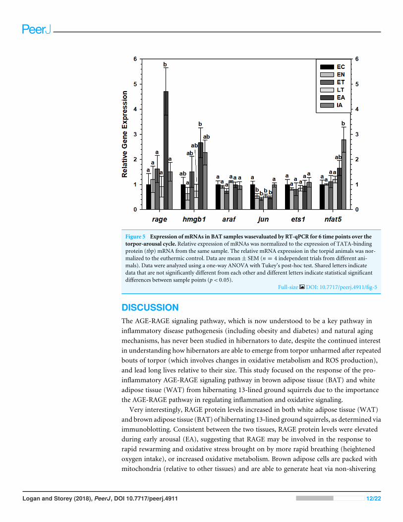
Figure 5 Expression of mRNAs in BAT samples wasevaluated by RT-qPCR for 6 time points over thetorpor-arousal cycle. Relative expression of mRNAs was normalized to the expression of TATA-bindingprotein (tbp) mRNA from the same sample. The relative mRNA expression in the torpid animals was nor-malized to the euthermic control. Data are mean± SEM (n = 4 independent trials from different ani-mals). Data were analyzed using a one-way ANOVA with Tukey’s post-hoc test. Shared letters indicatedata that are not significantly different from each other and different letters indicate statistical significantdifferences between sample points (p< 0.05).
Full-size DOI: 10.7717/peerj.4911/fig-5
DISCUSSIONThe AGE-RAGE signaling pathway, which is now understood to be a key pathway ininflammatory disease pathogenesis (including obesity and diabetes) and natural agingmechanisms, has never been studied in hibernators to date, despite the continued interestin understanding how hibernators are able to emerge from torpor unharmed after repeatedbouts of torpor (which involves changes in oxidative metabolism and ROS production),and lead long lives relative to their size. This study focused on the response of the pro-inflammatory AGE-RAGE signaling pathway in brown adipose tissue (BAT) and whiteadipose tissue (WAT) from hibernating 13-lined ground squirrels due to the importancethe AGE-RAGE pathway in regulating inflammation and oxidative signaling.
Very interestingly, RAGE protein levels increased in both white adipose tissue (WAT)and brown adipose tissue (BAT) of hibernating 13-lined ground squirrels, as determined viaimmunoblotting. Consistent between the two tissues, RAGE protein levels were elevatedduring early arousal (EA), suggesting that RAGE may be involved in the response torapid rewarming and oxidative stress brought on by more rapid breathing (heightenedoxygen intake), or increased oxidative metabolism. Brown adipose cells are packed withmitochondria (relative to other tissues) and are able to generate heat via non-shivering
Logan and Storey (2018), PeerJ, DOI 10.7717/peerj.4911 12/22
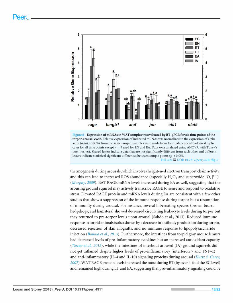
Figure 6 Expression of mRNAs inWAT samples wasevaluated by RT-qPCR for six time points of thetorpor-arousal cycle. Relative expression of indicated mRNAs was normalized to the expression of alphaactin (acta1) mRNA from the same sample. Samples were made from four independent biological repli-cates for all time points except n= 3 used for EN and EA. Data were analyzed using ANOVA with Tukey’spost-hoc test. Shared letters indicate data that are not significantly different from each other and differentletters indicate statistical significant differences between sample points (p< 0.05).
Full-size DOI: 10.7717/peerj.4911/fig-6
thermogenesis during arousals, which involves heightened electron transport chain activity,and this can lead to increased ROS abundance (especially H2O2 and superoxide [O2]•−)(Murphy, 2009). BAT RAGE mRNA levels increased during EA as well, suggesting that thearousing ground squirrel may actively transcribe RAGE to sense and respond to oxidativestress. Elevated RAGE protein and mRNA levels during EA are consistent with a few otherstudies that show a suppression of the immune response during torpor but a resumptionof immunity during arousal. For instance, several hibernating species (brown bears,hedgehogs, and hamsters) showed decreased circulating leukocyte levels during torpor butthey returned to pre-torpor levels upon arousal (Sahdo et al., 2013). Reduced immuneresponse in torpid animals is also shownby a decrease in antibody productionduring torpor,decreased rejection of skin allografts, and no immune response to lipopolysaccharideinjection (Bouma et al., 2013). Furthermore, the intestines from torpid gray mouse lemurshad decreased levels of pro-inflammatory cytokines but an increased antioxidant capacity(Tessier et al., 2015), while the intestines of interbout aroused (IA) ground squirrels didnot get inflamed despite higher levels of pro-inflammatory (interferon γ and TNF-α)and anti-inflammatory (IL-4 and IL-10) signaling proteins during arousal (Kurtz & Carey,2007).WATRAGE protein levels increased themost during ET (by over 4-fold the EC level)and remained high during LT and EA, suggesting that pro-inflammatory signaling could be
Logan and Storey (2018), PeerJ, DOI 10.7717/peerj.4911 13/22
important in WAT during torpor, despite evidence of low inflammatory responses duringtorpor in other hibernator tissues. Indeed, acute inflammatory responses are generallyprotective, as they attract anti-inflammatory, immune, and antioxidative agents to the siteof inflammation. Based on unchanging WAT rage transcript levels, WAT RAGE may betranslated from stored mRNA pools in stress granules rather than actively transcribed overthe torpor-arousal cycle. Together, these data suggest RAGE is regulated in a tissue-specificmanner, where dormant ground squirrels increase RAGE activity throughout torpor andarousal in preparation for threats to the immune system or elevated oxidative stress uponrewarming.
DAMP proteins HMGB1 and S100Bwere identified as potential targets for assessing pro-inflammatory signaling in hibernating ground squirrels because both are highly expressed inadipose tissues, and are upregulated in diet-induced obesitymicemodels and following highfat diets in mice (Buckman et al., 2014; Song et al., 2014; Ghosh et al., 2016). By contrast,most other S100 proteins including S100A8, A9, or A12 are expressed by immune cells(granulocytes, keratinocytes) or epithelial cells (Foell et al., 2007; Buckman et al., 2014).S100B and HMGB1 protein levels remained relatively constant over the torpor-arousalcycle in 13-lined ground squirrel WAT and BAT, except that WAT HMGB1 protein levelsdecreased during ET and LT. Relatively little change in the levels of these DAMPs couldsuggest that there are few damaged cells over the course of a torpor-bout and they likely donot play a role in RAGE-mediated signaling. However, low (nano-molar range) levels ofS100B are often protective since they bind RAGE to induce adaptive functions like neuriteoutgrowth in the brain or cell migration. High concentrations of S100B (micro-molarrange) produce ROS and induce apoptosis (Sparvero et al., 2009). Thus, constant S100Blevels could indicate that ground squirrel WAT and BAT are protected from injury duringmetabolic suppression and upon arousal. Hmgb1 transcript levels did not change in WATbut BAT hmgb1 increased during EA with respect to EN. Hmgb1 mRNA levels were shownto increase following thermal injury in rats (Fang et al., 2002), so this slight increase inBAT hmgb1 levels during arousal could suggest hmgb1 is more expressed during heat stressas ground squirrel body temperature rises from ∼5 ◦C to euthermic levels. Furthermore,elevated hmgb1 levels from adipose correlate with pro-inflammatory marker levels andmetabolic syndrome or tissue damage (Jialal et al., 2015; Qiu et al., 2016), suggesting thatground squirrels may upregulate hmgb1 during EA in response to oxidative stress. Again,these changes are not observed at the protein level, indicating ground squirrels mayinhibit HMGB1 protein expression as a means to reduce the pro-inflammatory response.CML-AGE levels did not change in BAT or WAT either, suggesting that ground squirrelslikely prevent AGE accumulation before torpor. A previous study done by our lab revealedthat 13-lined ground squirrel WAT and BAT adapt to changes in oxidative stress byincreasing antioxidant enzyme levels during torpor (but not during arousal) with respectto a euthermic control. Specifically, thioredoxin 1 (TRX1) increased during LT in BATand WAT, while superoxide dismutases 1 and 2 (SOD1, SOD2) increased in WAT duringLT (Rouble, Tessier & Storey, 2014). Increased antioxidant enzyme levels and activity coulddeplete the pools of ROS that are required to catalyze the non-enzymatic formation ofAGEs. Overall, no changes or decreases in DAMPs (S100B and HMGB1) and CML-AGE
Logan and Storey (2018), PeerJ, DOI 10.7717/peerj.4911 14/22
reverberate the notion that pro-inflammatory and immune responses are repressed duringtorpor while the antioxidant response is intensified since these three ligands are typicallyhighly abundant and upregulated in pro-inflammatory environments.
To assess if AGE-RAGE could induce pro-inflammatory and immune responses atany time point of the torpor-arousal cycle in ground squirrels, RT-qPCR was used toanalyze the relative transcript abundance of key MAPK signaling proteins. There is abreadth of literature that indicates that RAGE ligands (AGEs and DAMPs) signal throughRAGE to activate MAPKs (specifically, the ERK1/2, JNK, and p38 pathways) (Lander et al.,1997; Qiu et al., 2016) and this leads to increases in gene expression by phosphorylatedpro-inflammatory transcription factors (e.g., NFATs, AP1 subunit c-jun, Ets, etc.) (Pulvereret al., 1991;MacDonald & Storey, 2005a; Lu & Xu, 2006; Tsai et al., 2007;Wang et al., 2014)and the activation of transcription activators like p90 RSK S6 kinase (Cowan & Storey,2003). Furthermore, studies on various ground squirrel tissues indicate that MAPKsare differentially regulated over the torpor-arousal cycle (MacDonald & Storey, 2005b;Rouble, Tessier & Storey, 2014), making them important to discuss in the context of RAGEactivation. In ground squirrel WAT, there were no changes upstream of ERK1/2 kinase(i.e., araf transcript levels) or in the expression of any transcription factors downstreamof ERK1/2 (i.e., jun, ets1, and nfat5). These results are consistent with decreased ERK1/2phosphorylation during EN, ET and LT, previously described in 13-lined ground squirrelWAT (Rouble, Tessier & Storey, 2014). Overall, the results suggest that RAGE signalingmay be active in ground squirrel WAT but it is not inducing downstream MAPK signaling(Fig. 7). Instead, RAGE may induce pro-inflammatory signaling through the NF-κBsignaling pathway, since RAGE signaling can increase the de novo synthesis of NF-κB(Sparvero et al., 2009), but this has yet to be confirmed in hibernator WAT. NF-κB hasbeen shown to increase in hibernator intestine during torpor, relative to a summer activecontrol, but was undetectable in brown adipose (Carey, Frank & Seifert, 2000). There isalso evidence that NF-κB subunits (p50/p65) and the kinase (IKK) that activates NF-κBby inhibiting NF-κB inhibitor (IκB) increase during arousal in 13-lined ground squirrelmuscle, while IκB is more phosphorylated during this time, leading to IκB degradation(Allan & Storey, 2012). Thus, RAGE may regulate inflammation through NF-κB in WATas opposed to MAPK signaling.
Consistent with increased BAT RAGE during arousal, this study showed an increase inBAT nfat5 transcript expression during arousal. These results alsomake sense with previousreports of increased BATERK1/2 phosphorylation during EA, assessed in I. tridecemlineatus(Rouble, Tessier & Storey, 2014). By contrast, BAT jun transcript levels decreased betweenEN and EA, suggesting that this transcription factor is not involved in pro-inflammatorysignaling during torpor or arousal. This result is consistent with decreased phosphorylatedJNK levels (a kinase that phosphorylates the protein c-jun), observed in BAT andWAT from13-lined ground squirrels (Rouble, Tessier & Storey, 2014), further emphasizing that thistranscription factor is an unlikely mediator of pro-inflammatory signaling in hibernatoradipose. Future studies should be directed towards quantifying the relative changes in totaland phosphorylated NFAT5 levels to determine if the change in transcript levels matchesthose at the protein level. Together with the data from the Rouble, Tessier & Storey (2014)
Logan and Storey (2018), PeerJ, DOI 10.7717/peerj.4911 15/22
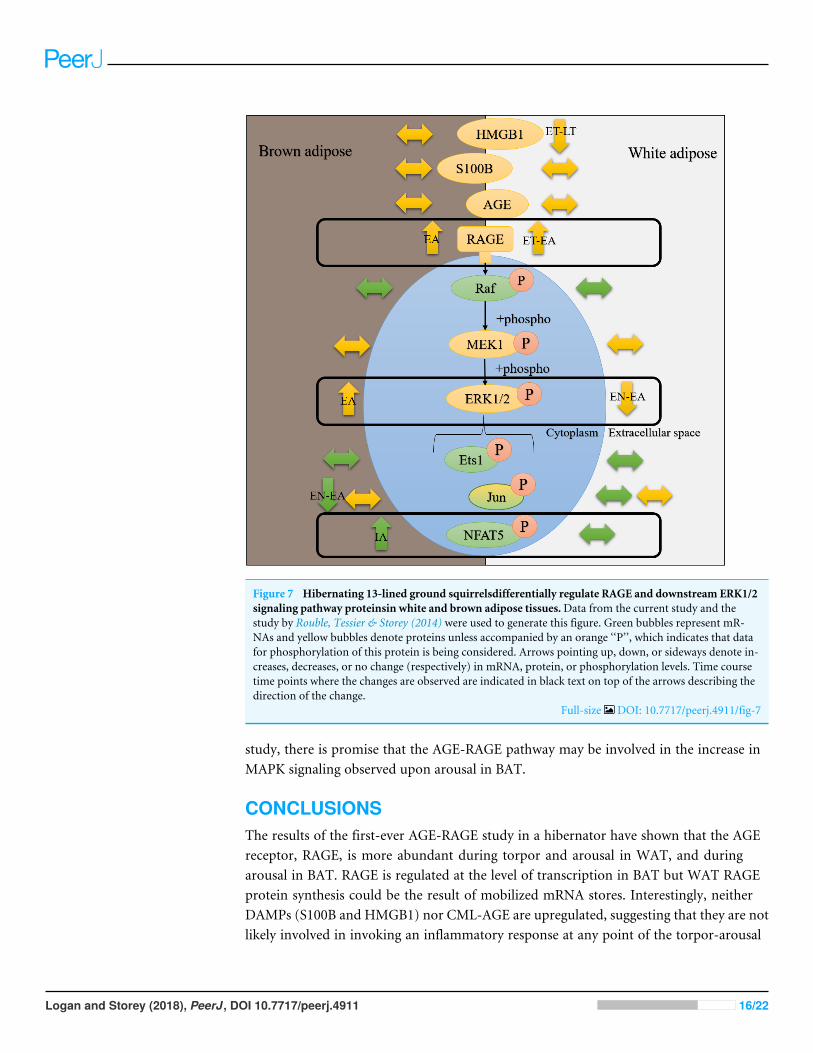
Figure 7 Hibernating 13-lined ground squirrelsdifferentially regulate RAGE and downstream ERK1/2signaling pathway proteinsin white and brown adipose tissues.Data from the current study and thestudy by Rouble, Tessier & Storey (2014) were used to generate this figure. Green bubbles represent mR-NAs and yellow bubbles denote proteins unless accompanied by an orange ‘‘P’’, which indicates that datafor phosphorylation of this protein is being considered. Arrows pointing up, down, or sideways denote in-creases, decreases, or no change (respectively) in mRNA, protein, or phosphorylation levels. Time coursetime points where the changes are observed are indicated in black text on top of the arrows describing thedirection of the change.
Full-size DOI: 10.7717/peerj.4911/fig-7
study, there is promise that the AGE-RAGE pathway may be involved in the increase inMAPK signaling observed upon arousal in BAT.
CONCLUSIONSThe results of the first-ever AGE-RAGE study in a hibernator have shown that the AGEreceptor, RAGE, is more abundant during torpor and arousal in WAT, and duringarousal in BAT. RAGE is regulated at the level of transcription in BAT but WAT RAGEprotein synthesis could be the result of mobilized mRNA stores. Interestingly, neitherDAMPs (S100B and HMGB1) nor CML-AGE are upregulated, suggesting that they are notlikely involved in invoking an inflammatory response at any point of the torpor-arousal
Logan and Storey (2018), PeerJ, DOI 10.7717/peerj.4911 16/22
cycle. Although CML-AGE is usually the most abundant and most potent inducer ofRAGE signaling, there are countless other AGE species that could bind RAGE duringtorpor to increase its upregulation. RT-qPCR analysis implicate that MAPK signalingand downstream transcription factors Jun, NFAT5 or Ets1 were not upregulated over thetorpor-arousal cycle in WAT, suggesting that these transcription factors are likely notinvolved in the pro-inflammatory, immune or oxidative stress responses in hibernatorWAT during hibernation. However, NFAT5 transcript levels increased during arousal inbrown adipose, which correlated with BAT RAGE levels and previous studies showingincreases in phosphorylated ERK1/2 levels during arousal in BAT. Future studies shouldinclude the analysis of the key RAGE-induced downstream transcription factor, NF-κB, inground squirrel adipose over the course of the torpor-arousal cycle.
ADDITIONAL INFORMATION AND DECLARATIONS
FundingThis work was supported by a discovery grant from the Natural Sciences and EngineeringResearch Council (NSERC) of Canada (6793) to Kenneth Storey and the Canada ResearchChairs program. Samantha Logan holds an NSERC Alexander Graham Bell CanadaGraduate Scholarship—Doctoral award. The funders had no role in study design, datacollection and analysis, decision to publish, or preparation of the manuscript.
Grant DisclosuresThe following grant information was disclosed by the authors:Natural Sciences and Engineering Research Council (NSERC) of Canada: 6793.Canada Research Chairs program.NSERC Alexander Graham Bell Canada Graduate Scholarship—Doctoral award.
Competing InterestsKenneth B. Storey is an Academic Editor for PeerJ.
Author Contributions• SamanthaMLogan conceived and designed the experiments, performed the experiments,analyzed the data, prepared figures and/or tables, authored or reviewed drafts of thepaper, approved the final draft.• Kenneth B. Storey conceived and designed the experiments, contributed reagents/-materials/analysis tools, authored or reviewed drafts of the paper, approved the finaldraft.
Animal EthicsThe following information was supplied relating to ethical approvals (i.e., approving bodyand any reference numbers):
All animal procedures were approved by the Animal Care and Use Committee of theNational Institute of Neurological Disorders and Stroke (protocol number ASP 1223-05).
Logan and Storey (2018), PeerJ, DOI 10.7717/peerj.4911 17/22
Data AvailabilityThe following information was supplied regarding data availability:
Logan, Samantha (2018): AGE competitive elisa. figshare. Dataset. https://doi.org/10.6084/m9.figshare.5885032.v1
Logan, Samantha (2018): BioRadCFX files for RT-PCR data. figshare. Fileset.https://doi.org/10.6084/m9.figshare.5885026.v1
Logan, Samantha (2018): AGE-RAGE western blots. figshare. Fileset. https://doi.org/10.6084/m9.figshare.5885029.
Supplemental InformationSupplemental information for this article can be found online at http://dx.doi.org/10.7717/peerj.4911#supplemental-information.
REFERENCESAllanME, Storey KB. 2012. Expression of NF-kB and downstream antioxidant genes in
skeletal muscle of hibernating ground squirrels, Spermophilus tridecemlineatus. CellBiochemistry and Function 30:166–174 DOI 10.1002/cbf.1832.
Biggar KK,Wu C-W, Tessier SN, Zhang J, Pifferi F, Perret M, Storey KB. 2015. Primatetorpor: regulation of stress-activated protein kinases during daily torpor in the graymouse lemur,Microcebus murinus. Genomics, Proteomics & Bioinformatics 13:81–90DOI 10.1016/j.gpb.2015.03.002.
Bogren L, Olson J, Carpluk J, Moore J, Drew K. 2014. Resistance to systemic inflamma-tion and multi-organ damage after global ischemia/reperfusion in the arctic groundsquirrel. PLOS ONE 9:e94225 DOI 10.1371/journal.pone.0094225.
BoumaHR, Henning RH, Kroese FGM, Carey HV. 2013.Hibernation is associatedwith depression of T-cell independent humoral immune responses in the 13-lined ground squirrel. Developmental and Comparative Immunology 39:154–160DOI 10.1016/j.dci.2012.11.004.
Boyer BB, Barnes BM. 1999.Molecular and metabolic aspects of mammalian hiberna-tion. BioScience 49:713–724 DOI 10.2307/1313595.
Boyer F, Vidot JB, Dubourg AG, Rondeau P, EssopMF, Bourdon E. 2015. Oxidativestress and adipocyte biology: focus on the role of AGEs. Oxidative Medicine andCellular Longevity 2015:1–9 DOI 10.1155/2015/534873.
Brett J, Schmidt AM, Yan SD, Zou YS,Weidman E, Pinsky D, Nowygrod R, NeeperM, Przysiecki C, Shaw A. 1993. Survey of the distribution of a newly characterizedreceptor for advanced glycation end products in tissues. The American Journal ofPathology 143:1699–1712.
Buckman LB, Anderson-Baucum EK, Hasty AH, Ellacott KL. 2014. Regulationof S100B in white adipose tissue by obesity in mice. Adipocyte 3:215–220DOI 10.4161/adip.28730.
Carey HV, Frank CL, Seifert JP. 2000.Hibernation induces oxidative stress andactivation of NK-kappaB in ground squirrel intestine. Journal of Comparative
Logan and Storey (2018), PeerJ, DOI 10.7717/peerj.4911 18/22
Physiology. B, Biochemical, Systemic, and Environmental Physiology 170:551–559DOI 10.1007/s003600000135.
Carey HV, Rhoads CA, Aw TY. 2003.Hibernation induces glutathione redox imbalancein ground squirrel intestine. Journal of Comparative Physiology B: Biochemical, Sys-temic, and Environmental Physiology 173:269–276 DOI 10.1007/s00360-003-0330-3.
Cooper M. 2004. Importance of advanced glycation end products in diabetes-associatedcardiovascular and renal disease. American Journal of Hypertension 17:31S–38SDOI 10.1016/j.amjhyper.2004.08.021.
Cowan KJ, Storey KB. 2003.Mitogen-activated protein kinases: new signaling pathwaysfunctioning in cellular responses to environmental stress. Journal of ExperimentalBiology 206:1107–1115 DOI 10.1242/jeb.00220.
Eaton SL, Roche SL, Llavero HurtadoM, Oldknow KJ, Farquharson C, GillingwaterTH,Wishart TM. 2013. Total protein analysis as a reliable loading control forquantitative fluorescent western blotting. PLOS ONE 8:1–9DOI 10.1371/journal.pone.0072457.
FangW-H, Yao Y-M, Shi Z-G, Yu Y,Wu Y, Lu L-R, Sheng Z-Y. 2002. The significanceof changes in high mobility group-1 protein mRNA expression in rats after thermalinjury. Shock 17:329–333 DOI 10.1097/00024382-200204000-00016.
Foell D,Wittkowski H, Vogl T, Roth J. 2007. S100 proteins expressed in phagocytes: anovel group of damage-associated molecular pattern molecules. Journal of LeukocyteBiology 81:28–37 DOI 10.1189/jlb.0306170.
Frerichs KU, Kennedy C, Sokoloff L, Hallenbeck JM. 1994. Local cerebral blood flowduring hibernation, a model of natural tolerance to ‘‘cerebral ischemia’’. Journal ofCerebral Blood Flow and Metabolism 14:193–205 DOI 10.1038/jcbfm.1994.26.
Ghosh AR, Bhattacharya R, Bhattacharya S, Nargis T, Rahaman O, Duttagupta P,Raychaudhuri D, Liu CSC, Roy S, Ghosh P, Khanna S, Chaudhuri T, TantiaO, Haak S, Bandyopadhyay S, Mukhopadhyay S, Chakrabarti P, Ganguly D.2016. Adipose recruitment and activation of plasmacytoid dendritic cells fuelmetaflammation. Diabetes 65:3440–3452 DOI 10.2337/db16-0331.
Gullicksen PS, Hausman DB, Dean RG, Hartzell DL, Baile CA. 2003. Adipose tissuecellularity and apoptosis after intracerebroventricular injections of leptin and 21 daysof recovery in rats. International Journal of Obesity and Related Metabolic Disorders27:302–312 DOI 10.1038/sj.ijo.0802205.
He Z, Li M, Zheng D, Chen Q, LiuW, Feng L. 2015. Adipose tissue hypoxia and low-grade inflammation: a possible mechanism for ethanol-related glucose intolerance?British Journal of Nutrition 113:1355–1364 DOI 10.1017/S000711451500077X.
Jialal I, Devaraj S, Bettaieb A, Haj F, Adams-Huet B. 2015. Increased adipose tissuesecretion of fetuin-A, lipopolysaccharide-binding protein and high-mobilitygroup box protein 1 in metabolic syndrome. Atherosclerosis 241:130–137DOI 10.1016/j.atherosclerosis.2015.04.814.
Kayser B, Verges S. 2013.Hypoxia, energy balance and obesity: from pathophysi-ological mechanisms to new treatment strategies. Obesity Reviews 14:579–592DOI 10.1111/obr.12034.
Logan and Storey (2018), PeerJ, DOI 10.7717/peerj.4911 19/22
Kurtz CC, Carey HV. 2007. Seasonal changes in the intestinal immune system of hiber-nating ground squirrels. Developmental and Comparative Immunology 31:415–428DOI 10.1016/j.dci.2006.07.003.
Lander HM, Tauras JM, Ogiste JS, Hori O, Moss RA, Schmidt AM. 1997. Activationof the receptor for advanced glycation end products triggers a p21ras-dependentmitogen-activated protein kinase pathway regulated by oxidant stress. Journal ofBiological Chemistry 272:17810–17814 DOI 10.1074/jbc.272.28.17810.
LeeM, Choi I, Park K. 2002. Activation of stress signaling molecules in bat brainduring arousal from hibernation. Journal of Neurochemistry 82:867–873DOI 10.1046/j.1471-4159.2002.01022.x.
Logan SM, Luu BE, Storey KB. 2016. Turn down genes for WAT? Activation of anti-apoptosis pathways protects white adipose tissue in metabolically depressedthirteen-lined ground squirrels.Molecular And Cellular Biochemistry 416:47–62DOI 10.1007/s11010-016-2695-0.
Logan SM, Tessier SN, Tye J, Storey KB. 2016. Response of the JAK-STAT pathway tomammalian hibernation in 13-lined ground squirrel striated muscle.Molecular andCellular Biochemistry 414:115–127 DOI 10.1007/s11010-016-2665-6.
Lu Z, Xu S. 2006. ERK1/2 MAP kinases in cell survival and apoptosis. IUBMB Life(International Union of Biochemistry and Molecular Biology: Life) 58:621–631DOI 10.1080/15216540600957438.
MacDonald JA, Storey KB. 2005a. Tyrosine phosphorylation and the control of cellularinformation. In: Storey KB, ed. Functional metabolism: regulation and adaptation.Hoboken: John Wiley & Sons, Inc., 125–151 DOI 10.1002/047167558X.ch5.
MacDonald JA, Storey KB. 2005b.Mitogen-activated protein kinases and selecteddownstream targets display organ-specific responses in the hibernating groundsquirrel. International Journal of Biochemistry and Cell Biology 37:679–691DOI 10.1016/j.biocel.2004.05.023.
Makki K, Froguel P, Wolowczuk I. 2013. Adipose tissue in obesity-related inflammationand insulin resistance: cells, cytokines, and chemokines. ISRN Inflammation2013:139239 DOI 10.1155/2013/139239.
McMullen DC, Hallenbeck JM. 2010. Regulation of Akt during torpor in the hi-bernating ground squirrel, Ictidomys tridecemlineatus. Journal of ComparativePhysiology B: Biochemical, Systemic, and Environmental Physiology 180:927–934DOI 10.1007/s00360-010-0468-8.
Morin P, Ni Z, McMullen DC, Storey KB. 2008. Expression of Nrf2 and its downstreamgene targets in hibernating 13-lined ground squirrels, Spermophilus tridecemlinea-tus.Molecular and Cellular Biochemistry 312:121–129DOI 10.1007/s11010-008-9727-3.
MurphyMP. 2009.How mitochondria produce reactive oxygen species. BiochemicalJournal 417:1–13 DOI 10.1042/BJ20081386.
Ni Z, Storey KB. 2010.Heme oxygenase expression and Nrf2 signaling during hi-bernation in ground squirrels. Canadian Journal of Physiology and Pharmacology88:379–387 DOI 10.1139/Y10-017.
Logan and Storey (2018), PeerJ, DOI 10.7717/peerj.4911 20/22
Orr AL, Lohse LA, Drew KL, Hermes-LimaM. 2009. Physiological oxidative stress afterarousal from hibernation in Arctic ground squirrel. Comparative Biochemistry &Physiology 153:213–221 DOI 10.1016/j.cbpa.2009.02.016.Physiological.
Ott C, Jacobs K, Haucke E, Navarrete Santos A, Grune T, SimmA. 2014. Role ofadvanced glycation end products in cellular signaling. Redox Biology 2:411–429DOI 10.1016/j.redox.2013.12.016.
Pellissier F, Glogowskib CM, Heinemannb SF, Balliveta M, Ossipowa V. 2006.Lab assembly of a low-cost, robust SYBR green buffer system for quantitativereal-time polymerase chain reaction. Analytical Biochemistry 350:310–312DOI 10.1016/j.ab.2005.12.002.
Pulverer BJ, Kyriakis JM, Avruch J, Nikolakaki E, Woodgett JR. 1991. Phosphorylationof c-jun mediated by MAP kinases. Nature 353:670–674 DOI 10.1038/353670a0.
Qiu Y, Chen Y, Zeng T, GuoW, ZhouW, Yang X. 2016.High-mobility group box-B1(HMGB1) mediates the hypoxia-induced mesenchymal transition of osteoblastcells via activating ERK/JNK signaling. Cell Biology International 40:1152–1161DOI 10.1002/cbin.10616.
Ramasamy R, Yan SF, Schmidt AM. 2011. Receptor for AGE (RAGE): signaling mecha-nisms in the pathogenesis of diabetes and its complications. Annals of the New YorkAcademy of Sciences 1243:88–102 DOI 10.1111/j.1749-6632.2011.06320.x.
ReddyMA, Li S-L, Sahar S, Kim Y-S, Xu Z-G, Lanting L, Natarajan R. 2006. Key roleof src kinase in S100B-induced activation of the receptor for advanced glycationend products in vascular smooth muscle cells. Journal of Biological Chemistry281:13685–13693 DOI 10.1074/jbc.M511425200.
Rouble AN, Hefler J, Mamady H, Storey KB, Tessier SN. 2013. Anti-apoptotic sig-naling as a cytoprotective mechanism in mammalian hibernation. PeerJ 1:e29DOI 10.7717/peerj.29.
Rouble AN, Tessier SN, Storey KB. 2014. Characterization of adipocyte stress responsepathways during hibernation in thirteen-lined ground squirrels.Molecular andCellular Biochemistry 393:271–282 DOI 10.1007/s11010-014-2070-y.
Rubartelli A, Lotze MT. 2007. Inside, outside, upside down: damage-associatedmolecular-pattern molecules (DAMPs) and redox. Trends in Immunology28:429–436 DOI 10.1016/j.it.2007.08.004.
Ruf T, Geiser F. 2015. Daily torpor and hibernation in birds and mammals. BiologicalReviews of the Cambridge Philosophical Society 90:891–926 DOI 10.1111/brv.12137.
Schwartz C, HamptonM, AndrewsMT. 2015.Hypothalamic gene expression underlyingpre-hibernation satiety. Genes Brain Behavior 14:310–318 DOI 10.1111/gbb.12199.
Sheriff MJ, Fridinger RW, Tøien Ø, Barnes BM, Buck CL. 2013.Metabolic rate andprehibernation fattening in free-living arctic ground squirrels. Physiological andBiochemical Zoology 86:515–527 DOI 10.1086/673092.
Song F, Del Pozo CH, Rosario R, Zou YS, Ananthakrishnan R, Xu X, Patel PR, BenoitVM, Yan SF, Li H, Friedman RA, Kim JK, Ramasamy R, Ferrante AW, SchmidtAM. 2014. RAGE regulates the metabolic and inflammatory response to high-fatfeeding in mice. Diabetes 63:1948–1965 DOI 10.2337/db13-1636.
Logan and Storey (2018), PeerJ, DOI 10.7717/peerj.4911 21/22
Sparvero LJ, Asafu-Adjei D, Kang R, Tang D, Amin N, Im J, Rutledge R, Lin B,Amoscato AA, Zeh HJ, Lotze MT. 2009. RAGE (receptor for advanced glycationendproducts), RAGE ligands, and their role in cancer and inflammation. Journal ofTranslational Medicine 7:Article 17 DOI 10.1186/1479-5876-7-17.
Storey KB, Storey JM. 2005. Mammalian hibernation: biochemical adaptation and geneexpression. In: Storey KB, ed. Functional metabolism: regulation and adaptation.Hoboken: John Wiley & Sons, Inc., 443–472.
Tessier SN, Katzenback BA, Pifferi F, Perret M, Storey KB. 2015. Cytokine andantioxidant regulation in the intestine of the gray mouse lemur (Microcebusmurinus) during torpor. Genomics, Proteomics and Bioinformatics 13:127–135DOI 10.1016/j.gpb.2015.03.005.
Tessier SN, Storey KB. 2016. Lessons from mammalian hibernators: molecular insightsinto striated muscle plasticity and remodeling. Biomolecular Concepts 7:69–92DOI 10.1515/bmc-2015-0031.
Trayhurn P,Wood IS. 2004. Adipokines: inflammation and the pleiotropic role of whiteadipose tissue. British Journal of Nutrition 92:347–355 DOI 10.1079/BJN20041213.
Tsai TT, Guttapalli A, Agrawal A, Albert TJ, Shapiro IM, RisbudMV. 2007.MEK/ERKsignaling controls osmoregulation of nucleus pulposus cells of the intervertebraldisc by transactivation of TonEBP/OREBP. Journal of Bone and Mineral Research22:965–974 DOI 10.1359/jbmr.070322.
UenoM, ShenW-J, Patel S, Greenberg AS, Azhar S, Kraemer FB. 2013. Fat-specificprotein 27 modulates nuclear factor of activated T-cells 5 and the cellular responseto stress. Journal of Lipid Research 54:734–743 DOI 10.1194/jlr.M033365.
Vlassara H, Brownlee M, Cerami A. 1985.High-affinity-receptor-mediated uptake anddegradation of glucose-modified proteins: a potential mechanism for the removalof senescent macromolecules. Proceedings of the National Academy of Sciences of theUnited States of America 82:5588–5592 DOI 10.1073/pnas.82.17.5588.
Vucetic M, Stancic A, Otasevic V, Jankovic A, Korac A, Markelic M, VelickovicK, Golic I, Buzadzic B, Storey KB, Korac B. 2013. The impact of cold accli-mation and hibernation on antioxidant defenses in the ground squirrel (Sper-mophilus citellus): an update. Free Radical Biology and Medicine 65:916–924DOI 10.1016/j.freeradbiomed.2013.08.188.
WangWK, Lu QH, Zhang JN,Wang B, Liu XJ, An FS, QinWD, Chen XY, DongWQ,Zhang C, Zhang Y, ZhangMX. 2014.HMGB1 mediates hyperglycaemia-inducedcardiomyocyte apoptosis via ERK/Ets-1 signalling pathway. Journal of Cellular andMolecular Medicine 18:2311–2320 DOI 10.1111/jcmm.12399.
Zhang XM, Guo L, Huang X, Li QM, Chi MH. 2016. 4-Hydroxynonenal regulatesTNF-alpha gene transcription indirectly via ETS1 and microRNA-29b in Humanadipocytes induced from adipose tissue-derived stromal cells. Anatomical Record299:1145–1152 DOI 10.1002/ar.23371.
Logan and Storey (2018), PeerJ, DOI 10.7717/peerj.4911 22/22