Embed Size (px)
Citation preview
Proceedings of the 62nd Annual National
Breeders Roundtable
May 2-3, 2013
Airport Marriott Hotel
St. Louis, Missouri
Sponsored by:
Poultry Breeders of America
and
U.S. Poultry & Egg Association
1
Table of Contents
Section I
Table of Contents…………………………………………………………………………………1
2012 National Breeders Roundtable Organizing Committee……………………………………..2
2012 National Breeders Roundtable Speaker Contact Information………...…………………….3
Section II
Veterinary Medical Genetics: Identification, Control and Treatment of Genetic Disease in
Animals
Dr. N. Matthew Ellinwood……………………….………………….……… No Paper Submitted
Ancestral Development Potential: A New Tool for Animal Breeding
Dr. Ehab Abouheif………..….………………………………………………………..………….5
Epigenetic Instability and Virus-Host Interactions in Chickens
Dr. Jiuzhou Song………...….…………………………………………………………………..19
Estimation of Genetic Parameters for Behavioral Assessment Scores in Labrador Retrievers,
German Shepherd Dogs and Golden Retrievers
Kelly Schultz.............…………………………………………………………………………….26
Application of Genomics in Pigs
Dr. Joseph Deeb…………….…...……………………………………………No Paper Submitted
Genetic Modification of Pigs: Expanding Their Utility as Biomedical Models
Dr. Jason Ross…………………………………………………………………………………..32
White Striping in Broiler Breast Meat
Dr. Casey Owens…...…………………………………………………………………………....39
Growth Hormone Transgenic Atlantic Salmon: Opportunities, Risks and Risk Management
Dr. Eric Hallerman………...…………….…………………………………………………..….47
What do RFI, Host Responses to PRRS Virus Infection and SCID Pigs as a Biomedical Model
Have in Common?
Dr. Jack Dekkers…...……………………………..………………………………..………..…..56
2
2013 National Breeders Roundtable
Organizing Committee
Dr. Neil O’Sullivan Chairman
Hy-Line International
Dallas Center, IA
Dr. Mark Cooper Session Chairman
Cobb-Vantress, Inc.
Siloam Springs, AR
Dr. Frank Siewerdt Session Chairman
Cobb-Vantress, Inc.
Siloam Springs, AR
3
2013 National Breeders Roundtable
Speaker Contact Information
Dr. Ehab Abouheif
Department of Biology 1205 Avenue Docteur
McGill University Penfield, Montreal Quebec H3A 1B1
Phone: 514-398-7190 [email protected]
Dr. Joseph Deeb
Genus PLC 100 Bluegrass Commons Blvd.
Suite 2200 Hendersonville, TN 37075
Phone: 645-265-2774 [email protected]
Dr Jack Dekkers
Professor of Animal Science 339 Kildee Hall
Iowa State University Ames, IA 50011-3150 Phone: 515-294-2293 [email protected]
Dr. N. Matthew Ellinwood
Associate Professor 2356D Kildee Hall
Iowa State University Ames, IA 50011
Phone: 515-294-5136 [email protected]
Dr. Eric Hallerman
Professor of Fisheries and Wildlife Conservation 100 Cheatham Hall
Virginia Tech Blacksburg, VA 20461 Phone: 540-231-5573
4
Dr. Casey Owens Center of Excellence for Poultry Science
University of Arkansas Fayetteville, AR 72701 Phone 479-575-4281 [email protected]
Dr. Jason Ross
2356 Kildee Hall Iowa State University
Ames, IA 50011 Phone: 515-294-8647 [email protected]
Kelly M. Schulz 109 Kildee Hall
Iowa State University Ames, IA 50011
Phone: 515-294-2293 [email protected]
Dr. Jiuzhou Song
Department of Animal and Avian Sciences Building 142
University of Maryland, College Park College Park, MD 20742
Phone: 301-405-5943 [email protected]
5
Ancestral Developmental Potential: a new tool for animal breeding?
Rajendhran Rajakumar1 and Ehab Abouheif1*
1McGill University Department of Biology
1205 avenue Docteur Penfield Montreal, Quebec
H3A 1B1 Canada
*To whom correspondence should be sent: [email protected] This proceeding article should be cited as:
Rajakumar, R. and Abouheif, E (2013) Ancestral developmental potential: a new tool for animal breeding? In N. O’Sullivan, M. Cooper, & F. Siewerdt (Eds.), Proceeding of the 62nd Annual National Breeders Roundtable. Paper presented at The 2013 National Breeders Roundtable, St. Louis, Missouri, 2-‐3 May (pp. 5-‐18). Tucker, GA: US Poultry & Egg. “… I wish to guard the reader against supposing that reversion is due to some rare or accidental combination of circumstances. When a character, lost during hundreds of generations, suddenly reappears, no doubt some combination must occur; but reversions may constantly be observed, at least to the immediately preceding generations, in the offspring of most unions … Reversion is most likely the rule, as Mr Sedgwick has shown, with certain diseases...” This insightful quote from Darwin’s (1868) classic The Variation of Animals and Plants under
Domestication illustrates that although a trait is lost during the evolution of a lineage, the
potential to produce that trait is retained, such that it may reappear in individuals in modern
populations. Whales and dolphins, for example, are closely related to hoofed animals but lost
their hind limbs ~34-‐41 million years ago when they re-‐entered water
6
(Thewissen et al., 2006). Today, several anomalous individuals of this group have been
discovered with partial hind limbs indicating that hind limbs have “reappeared” in these
individuals (Hall, 2003; Tomić & Meyer-Rochow, 2011). The sudden reappearance of such
ancestral traits in these anomalous individuals is most often called “atavism,” which is
derived from the word “atavus” or ancestor (Darwin 1868). Atavistic traits can suddenly
reappear in individuals even though they had been lost for hundreds, thousands, and even
millions of years (Collin & Miglietta, 2008; Wiens, 2011).
Unfortunately, modern evolutionary biologists appear to have undervalued the true
significance of atavisms for evolutionary theory. The consensus view is that atavisms are
rare mistakes in the developmental system that only provides evidence of ancestry
(Levinton, 1986). Apparently, as stated above in his quote, Darwin failed in “guarding the
reader against supposing that reversion is due to some rare or accidental combination of
circumstances” and as a consequence, atavisms are currently viewed as rare “freaks of
nature” that contribute little to the raw material that natural selection can act upon
(Blumberg, 2010). We argue in the following paragraphs that Darwin was right about his
observation that reversions/atavisms are a common occurrence in nature and that this type
of variation is likely to be “the rule” and not the exception. This is especially true for poultry,
where ancestral traits frequently reappear in individuals in modern populations and can also
be induced experimentally (Darwin 1868; Harris et al., 2006; Hampe, 1959; Müller, 1989).
Furthermore, we summarize evidence from our recent study (Rajakumar et al. 2012)
showing that reversions/atavisms reflect “ancestral developmental potentials” that, when
induced, provide raw material that natural selection can act upon to facilitate adaptive
evolution. Giving reversions/atavisms their proper place in evolutionary theory (West-
Eberhard, 2003; Stiassny, 2003), brings forth entirely new perspectives on how “ancestral
developmental potentials” can be used to improve animal breeding and understand
complex disease, especially with respect to poultry. We will finish by briefly outlining these
perspectives.
Reversions/Atavisms occur frequently in poultry. The field of experimental embryology
provided some of the first evidence that atavisms in poultry can be experimentally induced.
First, Hampe (1959) and Müller (1989) inserted a physical barrier within a specific region of
the chicken hindlimb during its development. The outcome of this experiment was startling
7
– the adult bone and musculature now resembled that of reptiles. Because birds descended
from reptiles (Lee, 2001; Hugall et al., 2007), this classic experiment demonstrates that it is
possible to revert the hindlimb in chickens to its reptilian state. Another celebrated example
of atavism in poultry is the induction of teeth in talpid2 mutant chickens. Although birds lost
teeth approximately 70-80 million years ago, Harris et al. (2006) discovered that teeth had
reappeared (or at least had initiated development) in talpid2 mutant chickens. In both these
examples, the atavistic traits had been lost for millions of years, showing that ancestral
developmental potentials can be retained for vast periods of time. Darwin (1868), however,
shows how these potentials can be frequently induced by natural means in populations
after the trait has been lost for generations. Darwin (1868) states: “The best yet simplest
characters lying dormant are, perhaps, those previously given, in which chickens and young
pigeons, raised from a cross between differentially colored birds, are at first of one color, but
in a year or two acquire feathers of the color of the other parent; for in this case the
tendency to a change in plumage is clearly latent in the young bird.” These examples of
atavism illustrate that ancestral developmental potentials are retained for variable lengths
of time and in many traits. Indeed, we have just scratched the surface in terms of surveying
the ancestral potentials that exist in poultry.
“Supersoldier ants” show that reversions/atavisms reflect ancestral developmental
potential that can facilitate adaptive evolution. Most new perspectives in science come
from unexpected sources and in roundabout ways. In our case, we provide a new
perspective on the biological significance of atavistic traits by studying the evolutionary
developmental biology of ants. One of the most exciting discoveries to emerge from the
field of evolutionary developmental biology (also known as EvoDevo) is the deep
conservation, over hundreds of millions of years of evolution, of the genes that regulate
development of an organism (Carroll, 2005; Carroll et al., 2009). For example, in all animals,
including, ants, chickens, and humans, the developmental regulatory gene hedgehog is
essential in the formation of limbs (Riddle et al., 1993; Ingham & McMahon, 2001), and the
gene Pax6 functions to specify where an eye will develop (Gehring & Ikeo, 1999). A team
led by Walter Gehring conducted an experiment that beautifully illustrates the functional
conservation of these developmental regulatory genes: they genetically inserted and
expressed the mouse Pax 6 gene in the developing wing or leg of a fruit fly, and
8
demonstrated that adult compound eyes (resembling those of a fruit fly) appear on wings or
legs in the adult fly (Halder et al., 1995). How is it that these developmental regulatory
genes, which are so conserved in their function and expression, can account for the amazing
morphological diversity in the animal kingdom? The Abouheif Lab’s approach to answering
this fundamental question has been to study how these genes interact with their
environment in the context of the complex societies of ants.
In the most advanced ant societies, the non-reproductive worker caste is very large,
and can be composed of thousands and even millions of individuals (Hölldobler & Wilson,
1990; Wilson, 2003; Hölldobler & Wilson, 2009). The individual workers in these highly
advanced societies are morphologically differentiated into several subcastes such that they
can efficiently divide labor in the colony (Hölldobler & Wilson, 1990). In the genus Pheidole,
which is one of the most evolutionarily diverse genera of ants with over 1000 species, the
worker caste is divided into “minor workers” and “soldiers” (Wilson, 2003; Moreau, 2008).
Minor workers forage and nurse the young, whereas soldiers defend the nest and help
process food. In 8 of the ~1000 species in this genus a third “supersoldier” subcaste has
evolved (Moreau, 2008). This supersoldier subcaste has a massive head which functions to
block the nest entrance when attacked by army ants (Huang, 2010).
In our recent study (Rajakumar et al. 2012), we found that the ancestral species of all
Pheidole had a supersoldier subcaste that was subsequently lost in most of the ~1000
species for ~30 to 65 million years. Supersoldiers then re-evolved multiple times
independently in the genus. By applying a key growth hormone, called Juvenile Hormone, to
larvae at a very specific time in development, we discovered that supersoldiers can be
experimentally induced in species that had lost the supersoldier subcaste for over 30 to 65
million years (see fig. 3 & fig. S9 in Rajakumar et al. 2012). This means that although the
supersoldier phenotype had been lost for millions of years, all Pheidole species retain an
ancestral developmental potential to produce supersoldiers. We demonstrated that
experimentally induced supersoldiers are produced through the same developmental
pathway as naturally evolved supersoldiers, and as adults, both experimentally induced and
naturally evolved supersoldiers are significantly larger than regular soldiers. The only
distinct morphological difference we observed between experimentally induced and
naturally evolved supersoldiers was the appearance of tiny wing vestiges on the thorax of
those that were experimentally induced (See Fig. 3 in Rajakumar et al 2012). These wing
9
vestiges are thought to be detrimental; they do not allow individuals to efficiently maneuver
underground, and therefore, are a negative side consequence of experimentally inducing
supersoldiers. These tiny wing vestiges are important for understanding how ancestral
developmental potential relate to complex disease, as we will soon describe below.
We also discovered in wild colonies of one Pheidole species, which does not have a
supersoldier subcaste, several anomalous individuals that looked very similar to
supersoldiers but also had tiny wing vestiges on the thorax (see fig. 2 Rajakumar et al. 2012).
Furthermore, anomalous supersoldier-like individuals were also found in wild colonies by
other researchers in different species (Wheeler, 1902; Goetsche, 1937), meaning that this
ancestral developmental potential is being induced in nature all the time and is therefore a
source of raw material for natural selection to act upon. In addition, these researchers
found that it was changes in nutrition that likely caused the induction of supersoldier-like
anomalies in wild colonies (Wheeler, 1902; Goetsche, 1937; Gregg, 1942). As mentioned
above, both experimentally induced and anomalous supersoldier-like individuals exhibit a
potentially adaptive trait (large size) but also a maladaptive trait (tiny wing vestiges). One
possible explanation is that not enough time has passed for the re-evolution of mechanisms
that can suppress these wing vestiges during the development of induced or anomalous
supersoldiers. Therefore, in order for such individuals to evolve as a functional subcaste of
the colony, there must be selection on large size and on elimination of wing vestiges.
We showed that an evolutionary process known as genetic accommodation was
responsible for the re-evolution of supersoldiers in Pheidole. This process occurs when an
environmentally induced phenotype becomes fixed in populations through natural selection
(West-Eberhard, 2003). Specifically, genetic accommodation of phenotypes occurs by
selecting on genes responsible for increasing the frequency and adjusting the form of a trait.
In the case of the re-evolution of Pheidole supersoldiers: first, to increase frequency the
supersoldier-like anomalies in the colony, selection occurred on genes that increase the
environmental sensitivity for producing supersoldiers, such that supersoldiers become
regularly induced by recurrent variation in nutrition or other environmental cues and their
frequency increases to approximately 4% of the colony; and second, to adjust the form of
supersoldier-like anomalies, selection occurred on genes that eliminate the production of
wing vestiges. To summarize, Rajakumar et al. 2012 shows that the induction of ancestral
developmental potentials, like that which produces supersoldiers, occurs frequently in
10
natural populations and that they are neither hopeless monsters nor freaks of nature. On
the contrary, they are raw materials for natural selection to act upon. This conclusion has
important consequences for animal breeding and understanding complex disease as we
outline in the following sections.
The association between ancestral developmental potential and complex disease in
poultry. Inducing the ancestral developmental potential for supersoldiers not only
illuminates the fact that ancestral potentials offer a rich source of raw material for natural
selection to act upon, but has also illuminated another important fact – that inducing
ancestral potentials in natural populations is often accompanied with the induction of
detrimental or negative side consequences (West-Eberhard, 2003; Rajakumar et al. 2012).
As we discussed above, we were never able to induce supersoldiers in species that normally
lack them without also inducing the appearance of the tiny, but detrimental, wing vestiges
(Rajakumar et al. 2012). This observation appears to be generally applicable to other
species, including poultry. In the quote at the very beginning of this article, Darwin’s (1868)
remark that “Reversion is most likely the rule, as Mr Sedgwick has shown, with certain
diseases...” indicates that he was well aware of the association between the induction of
reversions/atavisms and the appearance of disease. Darwin follows this remark with an
example in poultry describing the association between the induction of ancestral potentials
and ovarian cancer: “I will here add a somewhat different case, as it connects in a striking
manner latent characters of two classes. Mr. Hewitt possessed an excellent Seabright gold-
laced hen bantam, which, as she became old, grew diseased in her ovaria, and assumed
male characters. In this breed the males resemble the females in all respects, except in their
combs, wattles, spurs, and instincts; hence it would have been expected that the diseased
hen would have assumed only those masculine characters which are proper to the breed, but
she acquired, in addition well-arched tail sickle-feathers quite a foot in length, saddle-
feathers on the loins, and hackles on the neck,─ ornaments, which, as Mr. Hewitt remarks
“would be held abominable in this breed.” The Seabright bantam is known to have
originated about the year 1800 from a cross between a common bantam and a Polish fowl,
recrossed en-tailed bantam, and carefully selected; hence there can be hardly a doubt that
the sickle-feathers and hackles which appeared in the old hen were derived from the Polish
fowl or common bantam; and we thus see that not only certain masculine characters proper
11
to the Seabright bantam, but other masculine characters derived from the first progenitors
of the breed, removed by a period of above sixty years, were lying latent in this hen-bird
ready to be evolved as soon as her ovaria became diseased.” This intricate association
between the induction of ancestral developmental potential and negative side
consequences leading to disease is likely to be the rule and not the exception in poultry.
How to use ancestral developmental potentials to select for desirable features in poultry.
In the previous section, we briefly described the negative role that ancestral developmental
potentials can play in animal breeding through its association with negative side
consequences leading to complex disease. In this section, we show how ancestral potentials
can also play a positive role in animal breeding. After the publication of our article on the
role of ancestral developmental potential in the origin and evolution of supersoldiers
(Rajakumar et al. 2012), we received a storm of media attention (all you have to do is type
“supersoldier ants” into Google for this to become immediately obvious). One of the media
posts by Iain Thompson in The Register entitled “Boffins hack evolution, create
SUPERSOLDIER ANTS: Genetic prestidigitation could engineer new species” makes the
following statements about the implications of our findings: “For example, the aurochs – the
massive ancestor to modern cattle that was hunted to extinction by the 1600s – may be
recreatable by examining a cow’s genome and finding a way to activate the processes that
would cause the much larger and more aggressive aurochs to develop. In the plant world
too, crops could be subjected to environmental and chemical stressing to see if the dormant
genotypes could be activated. This could usher in new crops that can better deal with current
conditions – not to mention changing conditions as climate change wreaks its havoc.” Could
this really be possible? We argue that it is, so long as the focus is on resurrecting specific
desirable traits and not whole species. This means that researchers and animal breeders
would need to be very familiar with the ancestral traits in the group of interest. The
challenge, as we discussed above, will be to find a way to eliminate any negative side
consequences that may also be induced. In the following sections we briefly outline two
ways that ancestral developmental potentials could be induced in poultry for further
artificial selection:
12
Crossing as a tool to release ancestral developmental potential for artificial selection.
Once again we return to Darwin’s ingenious observations in 1868 in The Variation of
Animals and Plants under Domestication. He recognized that crossing different lineages or
species generally induced the appearance of atavistic traits in animals and plants: “...When
two races or species are crossed there is strongest tendency to the reappearance in the
offspring of long lost characters, possessed by neither parent nor immediate progenitor.” He
then goes on to give a remarkable example of his observation in poultry: “I raised several
chickens from a Polish hen by a Spanish cock, –breeds which do not incubate, –and none of
the young hens at first recovered the instinct, and this appeared to afford a well-marked
exception to the foregoing rule; but one of these hens, the only one which was preserved, in
the third year sat well on her eggs and reared a brood of chickens. So that here we have the
appearance with advancing age of a primitive instinct, in the same manner as we have seen
that the red plumage of the Gallus bankiva is sometimes reacquired by crossed and purely-
bred fowls of various kinds as they grow old.” This example beautifully shows that crossing is
not only a useful tool for inducing the reappearance of lost physical traits, like plumage, but
can also be used as a tool for inducing the reappearance of lost behavioral traits. Indeed,
ancestral developmental potential is already being considered key in the generation of
behavioral variation and the re-evolution of complex behavioral traits (Foster, 2013).
Environmental stress as a tool of releasing ancestral developmental potential for artificial
selection. The induction of ancestral developmental potential in supersoldier ants shows
that environmental factors, such as nutrition, hormones, temperature, and even particular
chemicals, can potentially be used as tools to induce ancestral developmental potentials in
animals and plants for artificial selection. William Morton Wheeler (1902) and Goetsch
(1937) showed that increased nutrition in colonies could give rise to supersoldier-like
anomalies in species that normally lack supersoldiers. Because nutrition is so closely linked
to levels of particular hormones, it becomes clear why high levels of juvenile hormone could
induce the development of supersoldiers in species that lack them. Therefore, it is entirely
possible that in poultry, ancestral developmental potential could be induced by alternating
sudden increases in nutrition (or hormones) with regular amounts of nutrition in the feed.
Temperature and chemical shocks could also be used (Waddington, 1953, Waddington,
1956). For instance, Waddington (1956) performed a classic experiment in fruit flies, where
13
he applied chemical (ether) to developing fruit fly embryos. Flies by definition only have one
pair of wings, whereas all other insects have two pairs. This means that flies lost the second
pair of wings during their evolution. The environmental shocks that Waddington (1956)
applied induced the reappearance of hindwings, producing adult flies with four wings.
Therefore, a range of environmental shocks can be used during poultry development to
induce ancestral developmental potentials for artificial selection.
Fixation of induced ancestral developmental potentials and suppression of negative side
consequences through artificial selection. Although supersoldiers with no wing vestiges
took millions of years to evolve, ancestral developmental potentials in poultry can be
induced and desirable traits can be fixed through artificial selection in just a few
generations. Classic experiments by Suzuki and Nijhout (2006) and Waddington (1956) in
insects have demonstrated that ancestral traits induced by temperature or chemical shock,
such as pigmentation in caterpillars or the presence of hindwings in fruit flies, can be fixed
through artificial selection in as little as 7 generations. Darwin also acknowledged the speed
with which induced ancestral potentials can be fixed by artifical selection: “By the aid of a
little selection, carried on during a few generations, most of our cultivated plants could
probably be brought back, without any great change in their conditions of life, to a wild or
nearly wild condition” (Darwin, 1868). Therefore, to select for desirable ancestral traits in
poultry, breeders should induce ancestral potentials by crossing or by administering
environmental shocks each generation, followed by artificial selection. Furthermore, several
studies suggest that there are two ways to repress maladaptive traits of induced ancestral
potentials: first, studies in chickens suggest that diet regulation, such as calorie-restriction
and content, can be manipulated to repress ovarian cancer in chickens (Ansenberger et al.,
2010; Carver et al., 2011), and second, studies in insects suggest that artificial selection can
be used to simultaneously select positively for desirable ancestral traits while selecting
against the appearance of any maladaptive traits (Suzuki & Nijhout 2008). In summary, to
use ancestral developmental potentials to enhance genetic lines, poultry breeders should:
(1) induce ancestral developmental potentials by crossing different breeds or species of
poultry and/or by exposing individuals to environmental shock each generation; (2)
artificially select on the induced trait to increase its frequency; and finally (3) repress the
14
development of maladaptive traits by manipulating diet or selecting against negative
maladaptive traits while selecting on positive ancestral traits.
Conclusion. We are grateful for the opportunity to have exposed our basic research on the
evolution and development of complex societies in ants. We hope to have convinced you or
at least provoked discussion on the possibility that ancestral developmental potential is a
powerful tool for improving breeding and understanding complex disease in poultry. We
maintain that basic research is the fuel of innovation, and that insights gained from the
highly organized societies of ants may actually serve as good models for understanding the
mechanisms underlying the positive and negative roles of inducing ancestral potentials for
animal breeding.
References
Ansenberger, K., Richards, C., Zhuge, Y., Barua, A., Bahr, J. M., Luborsky, J. L., & Hales, D. B.
(2010). Decreased severity of ovarian cancer and increased survival in hens fed a flaxseed-enriched diet for 1 year. Gynecologic oncology, 117(2), 341-347.
Blumberg, Mark S. Freaks of nature: What anomalies tell us about development and evolution. Oxford
University Press, 2010. Carroll, S. B. (2005). Endless forms most beautiful: the new science of evo devo and the making of the animal
kingdom (No. 54). WW Norton & Company. Carroll, S. B., Grenier, J., & Weatherbee, S. (2009). From DNA to diversity: molecular genetics and the evolution
of animal design. Wiley-Blackwell.
Carver, D. K., Barnes, H. J., Anderson, K. E., Petitte, J. N., Whitaker, R., Berchuck, A., &
Rodriguez, G. C. (2011). Reduction of ovarian and oviductal cancers in calorie-restricted laying chickens. Cancer Prevention Research, 4(4), 562-567.
Collin, R., & Miglietta, M. P. (2008). Reversing opinions on Dollo’s Law. Trends in ecology &
evolution, 23(11), 602-609. Darwin, C. (1868). The variation of animals and plants under domestication (Vol. 2). John
Murray. Foster, S. A. (2013). Evolution of behavioural phenotypes: influences of ancestry and
expression. Animal Behaviour. http://dx.doi.org/10.1016/j.anbehav.2013.02.008 Gehring, W. J., & Ikeo, K. (1999). Pax 6: mastering eye morphogenesis and eye evolution. Trends in Genetics,
15(9), 371-377.
15
Goetsch, W. (1937). Die Entstehung der “Soldaten” im Ameisenstaat. Naturwissenschaften, 25(50), 803-808.
Halder, G., Callaerts, P., & Gehring, W. J. (1995). Induction of ectopic eyes by targeted expression of the
eyeless gene in Drosophila. Science, 267(5205), 1788-1792.
Hall, B. K. (2003). Descent with modification: the unity underlying homology and homoplasy
as seen through an analysis of development and evolution. Biological Reviews, 78(3), 409-433.
Hampe, A., (1959). Contribution a l'etude du developement et de la regulation des
deficiencies et des excédents dans la patte de l'embryon de poulet, Arch. Anat.
Microscop. Morphol. Exp., 48:347–478.
Harris, M. P., Hasso, S. M., Ferguson, M. W., & Fallon, J. F. (2006). The development of
archosaurian first-generation teeth in a chicken mutant. Current Biology, 16(4), 371-
377.
Huang, M. H. (2010). Multi-phase defense by the big-headed ant, Pheidole obtusospinosa,
against raiding army ants. Journal of Insect Science, 10. Hugall, A. F., Foster, R., & Lee, M. S. (2007). Calibration choice, rate smoothing, and the pattern of tetrapod
diversification according to the long nuclear gene RAG-1. Systematic Biology, 56(4), 543-563.
Hölldobler, B., & Wilson, E. O. (1990). The ants. Belknap Press.
Hölldobler, B., & Wilson, E. O. (2009). The superorganism: the beauty, elegance, and
strangeness of insect societies. WW Norton & Company. Ingham, P. W., & McMahon, A. P. (2001). Hedgehog signaling in animal development: paradigms and
principles. Genes & development, 15(23), 3059-3087. Lee, M. S. (2001). Molecules, morphology, and the monophyly of diapsid reptiels. Contributions to Zoology,
70(1), 1-22.
Levinton, J.S. (1986). Developmental constraints and evolutionary saltations: A discussion
and critique. In Genetics, Development and Evolution, ed. J.P. Gustafson, G.L.
Stebbins, and F.J. Ayala, 253-288. New York: Plenum.
Moreau, C. S. (2008). Unraveling the evolutionary history of the hyperdiverse ant genus
Pheidole (Hymenoptera: Formicidae). Molecular Phylogenetics and Evolution, 48(1),
224-239.
Müller, G. B. (1989). Ancestral patterns in bird limb development: A new look at Hampe's
experiment. Journal of Evolutionary Biology, 2(1), 31-47.
16
Patel, N. H., Martin-Blanco, E., Coleman, K. G., Poole, S. J., Ellis, M. C., Kornberg, T. B., & Goodman, C. S. (1989).
Expression of engrailed proteins in arthropods, annelids, and chordates. Cell, 58(5), 955-968. Riddle, R. D., Johnson, R. L., Laufer, E., & Tabin, C. (1993). Sonic hedgehog mediates the polarizing activity of
the ZPA. Cell, 75(7), 1401-1416.
Stiassny, M.L.J. in Keywords and Concepts in Evolutionary Developmental Biology (Harvard
Univ. Press, Cambridge, MA, 2003), pp. 10–14. Suzuki, Y., & Nijhout, H. F. (2006). Evolution of a polyphenism by genetic accommodation. Science, 311(5761),
650-652. Suzuki, Y., & Nijhout, H. F. (2008). Constraint and developmental dissociation of phenotypic integration in a
genetically accommodated trait. Evolution & Development, 10(6), 690-699.
Thewissen, J. G. M., Cohn, M. J., Stevens, L. S., Bajpai, S., Heyning, J., & Horton, W. E. (2006).
Developmental basis for hind-limb loss in dolphins and origin of the cetacean bodyplan. Proceedings of the National Academy of Sciences, 103(22), 8414-8418.
Tomić, N., & Meyer-Rochow, V. B. (2011). Atavisms: medical, genetic, and evolutionary
implications. Perspectives in Biology and Medicine, 54(3), 332-353. Waddington, C. H. (1953). Genetic assimilation of an acquired character. Evolution, 118-126. Waddington, C. H. (1956). Genetic assimilation of the bithorax phenotype. Evolution, 1-13. West-Eberhard, M. J. (2003). Developmental plasticity and evolution. Oxford University
Press, USA. Wheeler, W. M. (1902). A neglected factor in evolution. Science, 15(385), 766-774. Wiens, J. J. (2011). Re-evolution of lost mandibular teth in frogs after more than 200 million
years, and re-evaluating Dollo’s law. Evolution, 65(5), 1283-1296. Wilson, E. O. (2003). Pheidole in the New World: a dominant, hyperdiverse ant genus.
Harvard University Press. Questions for Speaker: Dr. Ehab Abouheif Question: Dr. Jiuzhou Song After induction by the hormone did you check other genes’ behavior? Answer: We focused on the expresson of the spalt gene as a “read out” of the gene network responsible for wing development because it’s position is relatively far downstream in the network and has a unique pattern of expression in the vestigial wing imaginal discs (precursor cells that will develop into the adult wing) of soldier larvae relative to those
17
queens and minor workers. More importantly, spalt expression is associated with apoptosis in the vestigial wing imaginal discs of soldiers, which means that spalt may play an important role in eliminating wing vestiges before metamorphosis is complete. We therefore considered spalt to be a key gene, which not only serves as a “read out” for the whole network, but also serves to characterize the similarities and differences between castes, including the naturally evolved and induced supersoldiers. Question: Dr. Jiuzhou Song Is it a single gene or one of gene family? Answer: spalt itself is a single gene, but it is part of the gene network responsible for wing development. Once again, spalt expression can be used as a proxy to indicate the overall expression of this network. Future work will have to formally characterize the expression of other genes in this network before and after induction by the hormone. Question: Dr. Sue Lamont Following on your thought of the positive aspects of “release of genetic variation” by uncovering ancestral potential, what would you speculate as good nutritional manipulations? Methyl donors, to enhance epigenetic changes? Answer: Methyl donors would be a very interesting group of molecules to manipulate. There is a possibility that supplementing methyl donors in food given to the animals may lead to effects on epigenetic pathways. Since epigenetic pathways (ex: DNA methylation) use folate as a substrate and, along with hormonal pathways, work hand in hand to translate environmental status (ex: nutrition) into effects on development. Therefore, perturbing epigenetic pathways may be as efficient or even more so than perturbing nutrition. On the other hand, perturbing environmental factors like nutrition can induce ancestral developmental potentials, like in the case of ants, where protein has been proposed to be a potential candidate that might specifically be involved in the induction of supersoldiers. That being said, many different nutrients might be involved, and to determine which nutrients are worth testing, we recommend close examination of the diet and natural history of the ancestors of the poultry line of interest. Question: Dr. Frank Siewerdt Where should we look for clues to explore specific ancestral developmental potential? For example, how could we find out if chickens may lay 3 eggs a day, or sows developing 40 functional teats will one day become a possibility? Answer: The first question one should ask is “what are their ancestors like?” If their (remote or recent) ancestors exhibited a particular trait, then there is a good chance that the potential to produce that trait is laying dormant in the genome of the contemporary domesticated animal that you are working with. It is also important to note that when ancestral potentials are induced and variation is released, new and different combinations of ancestral traits can arise and be artifically selected for. This process, which is called “developmental recombination” (West-Eberhard, 2003), makes it possible to create new combinations from
18
ancient ancestral traits. So in theory, chickens that lay 3 eggs a day, or sows developing 40 functional teats may one day become a possibility using ancestral developmental potentials, although it may not be easy. Starting to explore the deep and recent evolutionary history, ecology, development, physiology, and life history of domesticated animals and plants is of primary importance for using ancestral developmental potential to improve animal breeding. Question: Dr. Gerald Herbert Your talk may infer that “long-term” epigenetic effects and “long-term” multi generational imprinting may play a major role in evolution. This contrasts with the concept of mutations in DNA increasing and decreasing in frequency, (changes in gene frequency) as the mechanism underlying evolution. Evolution may change partially due to activation and inactivation of genes by environmental influences. Answer: Your last sentence elegantly summarizes the general implications of our work. However, it is currently thought that although it is indeed the perturbation of hormonal pathways or epigenetic mechanisms may facilitate the initial emergence of an ancestral phenotype following an environmental induction, it is the fixation of standing genetic variation or de novo genetic mutation that preserves the induced phenotype across generations. However, it is entirely possible that trans-generational epigenetic imprinting may be a mechanism which permits the environment to have a persistent effect across generations, which is only subsequently followed by the “genetic fixation” of the initial imprint. Clearly, much research remains to be done in this new and exciting area of research.
19
The Current and Future of Epigenetics of Marek’s Disease in Chickens Yanghua He, Jiuzhou Song*
Department of Animal and Avian Sciences, University of Maryland, College Park, MD, USA. Email: [email protected]
Abstract
Epigenetic regulation in host-pathogen interaction is important in the etiology of Marek’s
disease (MD). These epigenetic regulatory mechanisms, including DNA methylation and
histone modifications, are influenced by Marek’s disease virus infection. To facilitate
epigenetic strategy and methods in animal health, it is of interest to investigate how and
what the new epigenetics and genetics influence the infectious disease. With this aim, we
summarize the current state of knowledge in order to provide insight in this emerging
epigenetic field. We also present the evidence for epigenetic effects for MD control.
Introduction
Marek’s disease (MD) is a lymphoproliferative disease in chickens caused by Marek’s disease
virus (MDV) and characterized by T cell lymphoma. The MDV is a naturally oncogenic, highly
contagious, and cell associated alpha-herpesvirus (NIIKURA et al. 2007). The disease is
characterized by a mononuclear infiltration of the peripheral nerves, gonads, iris, various
viscera, muscles, and the skin. Partial or complete paralysis is a common symptom of MD
due to accumulation and proliferation of tumor cells in peripheral nerves. During the 1960s,
as the industry converted to high-intensity rearing, MD generated tremendous economic
losses. Since the 1970s, MD has been controlled by vaccination. However, although
vaccination prevents the formation of lymphoma and other MD symptoms, it does not
prevent MDV infection, replication, or horizontal spread (PURCHASE and OKAZAKI 1971). Thus,
to advance MDV evolution escalating to higher virulence, knowledge supporting
development of etiology of MD and new strategies for control of MD need to be developed.
For a long time, resistance to MD and disease risk have long been thought to be influenced
by genetic, environmental factors, and the combination of which contributes to the
observed outcome in an individual. To augment vaccination measures, host genetic resistant
to MD becomes obviously more and more important. To control MD efficiently, we need to
further explore the mechanisms of host-virus interactions. However, most of researches
focused on the genetic differences between resistant and susceptible chickens to elucidate
20
the mechanisms of MD resistance (KAISER et al. 2003; SARSON et al. 2008). The more details of
host-MDV interaction are not understood. In our research, we believe that the interaction is
an intrinsic epigenetic mechanism and needs to be characterized thoroughly. The
advancement of the host-virus interactions will help us understand the etiology of MD and
facilitate epigenetic methods in resistance to MD, which, in turn, should result in a deep
insight on disease resistance breeding in chickens.
DNA methylation analysis in Marek’s disease
Epigenetics involving phenomena such as DNA methylation, chromatin modifications and
non-coding RNAs constitutes a dynamic regulatory framework linking genotypes with
environmental factors in differential disease responses among individuals having high
genetic similarity (MITRA et al. 2012). Although several efforts have attempted to pinpoint
factors responsible for conferring MD resistance (LIU et al. 2001; WAIN et al. 1998), many
confounding factors, such as, tissue types, virus strains and ages of birds have made it
difficult to find a consensus for the complex disease. In our studies, therefore, we took
epigenomics approach in highly inbred lines to identify the mechanisms that contribute to
the neoplastic diseases by utilizing the tractable and powerful combination of DNA
methylation, histone methylation, microRNAs, statistical genomics and computational
methods. The line 63 at the Avian Disease and Oncology Laboratory (ADOL) is relatively
resistant to MD tumors but is susceptible to Marek’s disease virus (MDV). However, another
line 72 is susceptible to both MDV and MD tumors (BACON et al. 2000). Therefore, these
inbred lines with high degree of genetic similarity constitute unique models for epigenetic
research because they make it possible to explore mechanisms of resistance and
susceptibility to neoplastic diseases.
Methylation of cytosines at CpG dinucleotides is an epigenetic modification of DNA that has
been shown to play important roles during tumorigenesis, embryonic development, and X
chromosome inactivation in mammals (MORGAN et al. 2005). The function of DNA
methylation is further heightened by the association of epigenetic dysfunction with various
diseases, in particular neoplastic diseases. Generally, DNA methylation is known as a post-
replication modification generated and maintained by three methyltransferases-DNMT1,
DNMT3a, and DNMT3b (KOROCHKIN 2006). Notably, we found two DNA mutations in
DNMT3b (YU et al. 2008b) and a higher promoter methylation level of ALVE and TVB in the
21
spleen of MD-susceptible chickens (L72) compared to that of MD-resistant chickens (L63) (YU
et al. 2008a), and the methylation level in CD4 promoter region was down regulated in the
former but not in the later at 21dpi (LUO et al. 2011). To advance the understanding of
functional patterns of DNA methylation in disease resistance or susceptibility, we extended
the scope of examination to interested genes (LUO et al. 2012b), which include genes related
to immune that the expression levels of these genes are alterable upon MDV challenge
(HEIDARI et al. 2010; THANTHRIGE-DON et al. 2010). We found DNA methylation heterogeneity
between the MD-resistant L63 and –susceptible L72 chickens. Since MDV induces a dynamic
expression change in DNMTs, differential methylation changes have been observed
between resistant and susceptible chickens after MDV infection. To thoroughly ascertain the
methylation variation in induced by MDV infection in both chicken lines, we mapped the
genome-wide DNA methylation profiles in each line using Methyl-MAPS (Methylation
Mapping Analysis by Paired-end Sequencing). We found that the methylation levels were
reduced in chickens from the resistant line 63 after MDV infection and 11,512 infection
induced differential methylation regions (iDMRs) were identified. The number of iDMRs was
larger in line 72 than in line 63, and most of iDMRs found in line 63 were overlapped with the
iDMRs found in line 72. Importantly, we further demonstrated that in vitro methylation
levels were associated with MDV replication, and found that MDV propagation in the
infected cells was restricted by pharmacological inhibition of DNA methylation. The results
suggested that DNA methylation changes in the host may be associated with disease
resistance or susceptibility. The methylation variations induced by the viral infection may
consequentially change the host transcriptome and result in diverse disease outcomes. All
in all, the differential DNA methylation levels and its change induced by MDV challenge
between the lines of chickens suggested that DNA methylation may play a role in host
resistance and/or susceptibility to MD (LUO et al. 2012b; TIAN et al. 2013).
Histone modifications in Marek’s disease
Although our study found that MD-resistant and susceptible birds with different DNA
methylation levels on several candidate genes, indicating the potential functions of
epigenetic factors in inducing different tumor incidence rates. However, little was known
about the histone modification patterns in these two chicken lines before. Therefore, to
22
gain more insight into the function of histone modifications in MD, we performed a histone
landscape analysis using ChIP-Seq in the unique MD-resistant (L63) and –susceptible (L72)
chicken lines both before and after MDV infection. Large number of line-specific H3K4me3
modifications and their underlying genes in immune response and cell adhesion in L63
chicken were found. Interestingly, we also found that the virus-induced specific H3K27me3
patterns in L72 chicken overlapped with some miRNAs which target genes involved in novel
pathways that may be related to MD-susceptibility (LUO et al. 2012a). Besides, WaveSeq, a
novel data-driven method of detecting regions of significant enrichment in ChIP-Seq data
was developed. The distribution-free method utilizes the wavelet transform, and is robust to
diverse data characteristics such as low signal-to-noise ratios and broad enrichment
patterns. The WaveSeq can detect both narrow and broad peaks with a high degree of
accuracy even in low signal-to-noise ratio data sets, and it is also suited for application in
complex experimental scenarios, helping make biologically relevant functional discoveries
(MITRA and SONG 2012).
Gene Expression Analysis to Marek’s Disease
Natural resistance to MDV can be divided into two categories: major histocompatibility
complex (MHC)-associated resistance, wherein different MHC haplotypes at the B blood
group locus confer varying levels of resistance and non-MHC associated resistance in which
birds having the same MHC haplotype exhibit vastly different responses to MDV infection.
Inbred lines 63 and 72 we used in this study fall into the latter category. These lines share a
high degree of genetic similarity but have divergent responses to MDV infection completely
independent of the MHC. Earlier reports have shown that the MHC haplotype as well as
non-MHC genes are responsible for genetic resistance to MD (BACON and WITTER 1993; BACON
and WITTER 1994; BACON and WITTER 1995). To further explore transcriptome differences
after MDV infection, besides significant expressed genes, we also identified other non-
coding RNAs during the development of MD and screened miRNAs that were sensitive to
Marek's disease virus (MDV) infection. Our results indicated that differential expression of
miRNA in resistant and susceptible chickens was caused by MDV infection, which effectively
influenced protein expression of ATF2 that might be related to Marek's disease
resistance/susceptibility (TIAN et al. 2012). The results above will facilitate the discoveries of
etiology mechanisms and chromosome loci which convey MD resistance.
23
Genetic Variations to Marek’s Disease
Aside from epigenetics, genetics and environmental factors are also causative agents to MD.
Some studies indicated that genetic variations are associated with the susceptibility of MD
(EMARA et al. 2001). To identify new genetic markers, we used high density SNP chips to
screen potential genetic markers and detect CNVs in the chicken lines. Besides, the DNA
mutations of DNA methyltransferases (DNMT3a, DNMT3b and DNMT1) were found and
their relationships with genes expressions of themselves were analyzed (YU et al. 2008b).
Meanwhile, the complexities and structural characteristics of these genes in entropy view
were analyzed to help us elucidate mechanisms and effects of the genes in epigenetic
processes (XIE X 2010). To have a full understanding of vaccination in MD control, we also
examined the effect of host genetic variation on vaccine efficacy (CHANG et al. 2010) and
comparative evaluation of MD virus infection in a series of recombinant congenic strains
(CHANG et al. 2011). In these studies, the lipoprotein metabolism in Marek’s disease
susceptible and resistant chickens was characterized (YUAN P 2012). All of these are around
the Marek's disease to expose its real biological mechanism.
Future of epigenetics of Marek’s Disease
Epigenetics is an active and exciting area of research. It is being driven by the massive
amounts of new information being generated by next generation sequencing methods.
Current epigenetics offers perhaps the greatest potential for animal health. Although we
don’t understand the mechanisms, we will identify unique epigenetic factors that could be
potentially used as epigenetic biomarkers. The knowledge of host-pathogen interaction will
provide a better understanding of epigenetic modifications at a 'systems level' and will serve
as mechanistic studies aimed at defining epigenetic roles that underlie disease resistance.
Most importantly, we believe an improved strategy for epigenetically preventive measures
against disease will subsequently pave the way for more focused and efficient application of
marker-assisted selection (MAS) or genomic selection in poultry breeding program in the
near future.
24
References BACON, L. D., H. D. HUNT and H. H. CHENG, 2000 A review of the development of chicken lines
to resolve genes determining resistance to diseases. Poult Sci 79: 1082-1093. BACON, L. D., and R. L. WITTER, 1993 Influence of B-haplotype on the relative efficacy of
Marek's disease vaccines of different serotypes. Avian Dis 37: 53-59. BACON, L. D., and R. L. WITTER, 1994 Serotype specificity of B-haplotype influence on the
relative efficacy of Marek's disease vaccines. Avian Dis 38: 65-71. BACON, L. D., and R. L. WITTER, 1995 Efficacy of Marek's disease vaccines in Mhc heterozygous
chickens: Mhc congenic x inbred line F1 matings. J Hered 86: 269-273. CHANG, S., Z. DING, J. R. DUNN, L. F. LEE, M. HEIDARI et al., 2011 A comparative evaluation of the
protective efficacy of rMd5deltaMeq and CVI988/ Rispens against a vv+ strain of Marek's disease virus infection in a series of recombinant congenic strains of White Leghorn chickens. Avian Dis 55: 384-390.
CHANG, S., J. R. DUNN, M. HEIDARI, L. F. LEE, J. SONG et al., 2010 Genetics and vaccine efficacy: host genetic variation affecting Marek's disease vaccine efficacy in White Leghorn chickens. Poult Sci 89: 2083-2091.
EMARA, M. G., M. A. ABDELLATIF, D. L. POLLOCK, M. SADJADI, S. S. CLOUD et al., 2001 Genetic variation in susceptibility to Marek's disease in a commercial broiler population. Avian Dis 45: 400-409.
HEIDARI, M., A. J. SARSON, M. HUEBNER, S. SHARIF, D. KIREEV et al., 2010 Marek's disease virus-induced immunosuppression: array analysis of chicken immune response gene expression profiling. Viral Immunol 23: 309-319.
KAISER, P., G. UNDERWOOD and F. DAVISON, 2003 Differential cytokine responses following Marek's disease virus infection of chickens differing in resistance to Marek's disease. J Virol 77: 762-768.
KOROCHKIN, L. I., 2006 [What is epigenetics]. Genetika 42: 1156-1164. LIU, H. C., H. H. CHENG, V. TIRUNAGARU, L. SOFER and J. BURNSIDE, 2001 A strategy to identify
positional candidate genes conferring Marek's disease resistance by integrating DNA microarrays and genetic mapping. Anim Genet 32: 351-359.
LUO, J., A. MITRA, F. TIAN, S. CHANG, H. ZHANG et al., 2012a Histone methylation analysis and pathway predictions in chickens after MDV infection. PLoS One 7: e41849.
LUO, J., Y. YU, S. CHANG, F. TIAN, H. ZHANG et al., 2012b DNA Methylation Fluctuation Induced by Virus Infection Differs between MD-resistant and -susceptible Chickens. Front Genet 3: 20.
LUO, J., Y. YU, H. ZHANG, F. TIAN, S. CHANG et al., 2011 Down-regulation of promoter methylation level of CD4 gene after MDV infection in MD-susceptible chicken line. BMC Proc 5 Suppl 4: S7.
MITRA, A., J. LUO, H. ZHANG, K. CUI, K. ZHAO et al., 2012 Marek's disease virus infection induces widespread differential chromatin marks in inbred chicken lines. BMC Genomics 13: 557.
MITRA, A., and J. SONG, 2012 WaveSeq: a novel data-driven method of detecting histone modification enrichments using wavelets. PLoS One 7: e45486.
MORGAN, H. D., F. SANTOS, K. GREEN, W. DEAN and W. REIK, 2005 Epigenetic reprogramming in mammals. Hum Mol Genet 14 Spec No 1: R47-58.
NIIKURA, M., T. KIM, H. D. HUNT, J. BURNSIDE, R. W. MORGAN et al., 2007 Marek's disease virus up-regulates major histocompatibility complex class II cell surface expression in infected cells. Virology 359: 212-219.
25
PURCHASE, H. G., and W. OKAZAKI, 1971 Effect of vaccination with herpesvirus of turkeys (HVT) on horizontal spread of Marek's disease herpesvirus. Avian Dis 15: 391-397.
SARSON, A. J., P. PARVIZI, D. LEPP, M. QUINTON and S. SHARIF, 2008 Transcriptional analysis of host responses to Marek's disease virus infection in genetically resistant and susceptible chickens. Anim Genet 39: 232-240.
THANTHRIGE-DON, N., L. R. READ, M. F. ABDUL-CAREEM, H. MOHAMMADI, A. I. MALLICK et al., 2010 Marek's disease virus influences the expression of genes associated with IFN-gamma-inducible MHC class II expression. Viral Immunol 23: 227-232.
TIAN, F., J. LUO, H. ZHANG, S. CHANG and J. SONG, 2012 MiRNA expression signatures induced by Marek's disease virus infection in chickens. Genomics 99: 152-159.
TIAN, F., F. ZHAN, N. D. VANDERKRAATS, J. F. HIKEN, J. R. EDWARDS et al., 2013 DNMT gene expression and methylome in Marek's disease resistant and susceptible chickens prior to and following infection by MDV. Epigenetics 8.
WAIN, H. M., A. A. TOYE, S. HUGHES and N. BUMSTEAD, 1998 Targeting of marker loci to chicken chromosome 16 by representational difference analysis. Anim Genet 29: 446-452.
XIE X, Y. Y., LIU G, YUAN Z, SONG J., 2010 Complexity and Entropy Analysis of DNA Methyltransferase. Journal of Data Mining in Genomics & Proteomics 1: 1-7.
YU, Y., H. ZHANG, F. TIAN, L. BACON, Y. ZHANG et al., 2008a Quantitative evaluation of DNA methylation patterns for ALVE and TVB genes in a neoplastic disease susceptible and resistant chicken model. PLoS One 3: e1731.
YU, Y., H. ZHANG, F. TIAN, W. ZHANG, H. FANG et al., 2008b An integrated epigenetic and genetic analysis of DNA methyltransferase genes (DNMTs) in tumor resistant and susceptible chicken lines. PLoS One 3: e2672.
YUAN P, Y. Y., LUO J, TIAN F, ZHANG H, CHANG S, RAMACHANDRAN R, SONG J., 2012 Comparative study of lipoprotein metabolism differs between Marek's disease susceptible and resistant chickens. Poultry Science 91: 2598-2605.
26
Estimation of genetic parameters for behavioral assessment scores in Labrador Retrievers, German Shepherd Dogs and Golden Retrievers.
K. M. Schulz1, D. W. Moser2, J. M. Bormann2, E. A. Leighton3
Kelly M. Schulz Department of Animal Science
Iowa State University Ames, IA 50011
Phone: 515-294-2933 E-mail: [email protected]
1Iowa State University, Ames, IA; 2Kansas State University, Manhattan, KS, USA, 3The Seeing Eye, Inc., Morristown, NJ, USA
Invited Paper prepared for presentation at the National Breeder’s Roundtable 2013, St. Louis, Missouri, May 2-3, 2013.
Copyright 2013 by Kelly M. Schulz, Daniel W. Moser, Jennifer M. Bormann, and Eldin A. Leighton. All rights reserved. Readers may make verbatim copies of this document for non-commercial purposes by any means, provided that the copyright notice appears on all such copies. Interested readers are directed to http://krex.k-state.edu/dspace/bitstream/handle/2097/13660/KellySchiefelbein2012.pdf?sequence=1.
The Guide Dog Service Industry
The use of dogs guiding blind people is speculated to date as far back as the
Pleistocene Age (Coon, 1959) and today, guide dogs are ranked as one of the most noble
and useful occupations of all working dogs (Willis, 1995). To keep pace with increasingly
more complex working environments where guide dogs are asked to assist their blind
masters, guide dog breeding programs must constantly improve the working ability and
general health of the puppies they produce. Two guide dog schools in the U.S., Guiding Eyes
for the Blind and The Seeing Eye, Inc., have undertaken this challenge by setting the
ultimate goals of their breeding programs to increase the number of healthy, high-quality
dogs available for training while minimizing the number of dogs released.
Guiding Eyes for the Blind Headquarters and Training Center is located in Yorktown
Heights, New York, while their Canine Development Center (Breeding Center) is located in
27
Patterson, New York. Guiding Eyes for the Blind is an internationally accredited guide dog
school with a mission to provide greater independence, dignity, and new horizons of
opportunity to the visually impaired. Their selective breeding program began in the mid-
1960s and the organization continually strives to genetically improve the health and working
ability of its guide dogs.
The Seeing Eye, Inc., located in Morristown, New Jersey, was founded in 1929 and is
the oldest existing guide dog school in the world. It is an internationally accredited provider
of Seeing Eye® dogs and owns the trademark for and is technically the only source for a dog
with this designation. The Seeing Eye, Inc. is an international philanthropic organization,
providing Seeing Eye® dogs to blind people in both the U.S. and Canada. It is renowned as a
research leader in canine genetics, breeding, disease control, and behavior. Their overall
mission is to enhance the independence, dignity, and self-confidence of blind people
through the use of Seeing Eye® dogs.
In addition to Guiding Eyes for the Blind and The Seeing Eye, there are at least 8
other schools in the U.S. that provide dogs as trained guides for blind people. It is estimated
that about 10,000 blind people in the U.S. use a guide dog as part of their daily walk through
life.
Breeding and Selection of Guide Dogs
Obtaining an adequate number of high-quality dogs suitable for breeding or training
to work as guides for blind people is a constant challenge for many guide dog schools. To
meet their needs, many schools now maintain their own breeding colonies to ensure an
adequate supply of high-quality dogs with predictable traits. The most effective way to use
information about the traits in a population, to produce predictable stock and to improve it
over time, is to apply the time-proven principles of population genetics and quantitative
genetics to manage the population over successive generations (Bourdon, 2000).
Over the past three decades, advances in computing power have enabled the study
of genetic complexity in more animals and in greater detail than ever before. Researchers
can now predict changes in quantitative genetic traits in future generations by using
complex statistical models that take into account all the animals in the pedigree. Several
traits can be studied simultaneously and relationships among traits can be established.
28
Most parameters of behavior are complex combinations of genetic and environmental
influences. With sufficient data on each of many animals in an extended pedigree, it is
possible to assess the extent to which these traits are heritable. Furthermore, researchers
can also forecast the likely results of selection in the future, which enables comparisons of
the possible results of different breeder selection strategies (Bourdon, 2000).
One of many issues facing guide dog organizations is that there is no single perfect
“type” of guide dog. Because substantial variation exists among the people who use guide
dogs, there must also be variation among the dogs available for matching with their blind
masters. This means that guide dog schools are not breeding for one specific kind of "super
dog", as show breeders predominantly desire. Rather, it is essential to have dogs with
varying degrees of characteristics such as breed, temperament, size, and energy to enable
guide dog schools to properly match dogs with blind people who are equally varying in their
needs and desires. After all, the dog with which they are ultimately matched will literally live
with them 24 hours per day.
This task of finding the “right” dog for each client is one of the hardest jobs faced by
the training staff of a guide dog school. The trainer has to assign the dog to the
client/handler early on in the training course as they have a short period of time with the
clients and dogs together to determine if they have made a successful match. It is very
difficult to switch a pair late in the training period as bonding between the client and dog
will already have begun to occur. Each trainer works with a small group of clients and dogs
at the same time and therefore has potential to create confusion for the dog when
reassigning them with a different client, but having to remain around their original handler.
It is also important that the dog’s temperament matches the home environment of the
client. For example, it would be unwise to pair a high energy large dog with a short elderly
client who doesn't often leave their home.
Behavior issues top the list of most common reasons for rejecting dogs from working
as guides (Goddard and Beilharz, 1982, 1982/83). The criteria used in the breeder selection
process at guide dog organizations are crucial to the success of working guides. A potential
tool for this selection process is known as the Canine Behavioral Assessment and Research
Questionnaire (C-BARQ, 2012). The C-BARQ was developed by researchers at the Center for
the Interaction of Animals and Society at the University of Pennsylvania and was designed to
provide standardized evaluations of canine behavior and temperament. Information the C-
29
BARQ provides may be useful to guide dog schools as a way to learn more about the nature
of optimal behavioral traits leading to the production of dogs that are more successful in
their ability to work as guides.
The C-BARQ was validated in two separate, but similar studies (Serpell and Hsu,
2001; Hsu and Serpell, 2003) confirming that the questionnaire factors and the behavior and
temperament traits they represent were stable and consistent among different populations
of dogs. The authors (Serpell and Hsu, 2001) also evaluated the overall construct validity of
the C-BARQ, which was deemed valid as well as capable of discriminating among dogs that
succeed in training versus dogs that fail training for behavioral reasons. Complete details
describing the process used to validate the C-BARQ and assess its reliability have been
reported (Serpell and Hsu, 2001, 2005; Hsu and Serpell, 2003).
While this canine behavioral assessment instrument has been extensively tested for
reliability and validity, prior to this ongoing research a comprehensive genetic analysis of
the C-BARQ had not been completed. Thus, the objectives of this research were to estimate
the magnitude of heritability for each of the 101 questions and each of the 12 subscale
factors, as well as exploration of breeding strategy implications based on the results
obtained.
Current Research Results
At 6- and 12-months of age respectively, questionnaire responses were obtained on
3,149 and 3,348 Labrador Retrievers from Guiding Eyes for the Blind and 989 and 1,187
Labrador Retrievers, 608 and 692 Golden Retrievers, and 966 and 1,348 German Shepherd
Dogs from The Seeing Eye, Inc. The C-BARQ questionnaires were completed by the puppy
raiser families, who as volunteer families, raise these puppies in home environments from
about 7-8 weeks of age until they return to the school to begin training at 14-18 months of
age. He/she is asked to rate his/her dog’s typical response to a range of commonly
encountered events, situations, and environmental stimuli (Serpell Hsu, 2001, 2005; Hsu
and Serpell, 2003).
The estimates of heritability and standard errors from The Seeing Eye, Inc. dogs
indicate that there is much genetic variation that could be exploited in selection against
“Familiar dog-directed aggression/fear” of Golden Retrievers at 6-months, “Chasing” of
30
Golden Retrievers at 6-months, and “Nonsocial fear” of Golden Retrievers at 12-months or
in selection for improved “Trainability” of Labrador Retrievers, German Shepard Dogs, and
Golden Retrievers at 12-months. Heritability values obtained from the current study can be
used as a guideline to approximate the outcome of various selection decisions (Willis, 1995).
Furthermore, some of the subscale factors identified as being moderate or highly heritable
might also prove useful for predicting which puppies may be at risk of failing from the guide
dog program. In general, the remaining factors and most of the 101 questions were found
to be lowly heritable (< 0.10). These estimates are useful to understand more about the
nature of behavioral traits leading to the production of successful working guides and
provide fodder for future research.
Future Research
There is a great need to develop a common language in describing temperament
traits of the dog to enable comparisons across populations. Estimating the direction and
magnitude of genetic correlations between the subscale factors would be beneficial to
determine if any two traits are influenced by common genes. The C-BARQ may also be a
potential tool to predict which puppies may be most at risk of failing from the guide dog
program as well as determining more specifically how the scores relate to success.
References
Bourdon, R. M. 2000. Understanding Animal Breeding. 2nd ed. Prentice Hall, Inc. Upper Saddle River, New Jersey.
Coon, N. 1959. A brief history of dog guides for the blind. The Seeing Eye, Inc. Morristown, New Jersey.
Goddard, M. E. and R. G. Beilharz. 1982. Genetic and environmental factors affecting the suitability of dogs as guide dogs for the blind. Theor. Appl. Genet. 62:97-102.
Goddard, M. E. and R. G. Beilharz. 1982/83. Genetics of traits which determine the suitability of dogs as guide-dogs for the blind. Appl. Anim. Ethol. 9:299-315.
Hsu, Y., and J. A. Serpell. 2003. Development and validation of a questionnaire for measuring behavior and temperament traits in pet dogs. J. Am. Vet. Med. Assoc. 223:1293-1300.
31
Serpell, J. A. and Y. Hsu. 2001. Development and validation of a novel method for evaluating behavior and temperament in guide dogs. Appl. Anim. Behav. Sci. 72:347-364.
Serpell, J. A. and Y. Hsu. 2005. Effects of breed, sex, and neuter status on trainability in dogs. Anthrozoös. 18:196-207.
Willis, M. B. 1995. Genetics of dog behavior with particular reference to working dogs. Pages 51-64 in The Domestic Dog, it's evolution, behavior and interactions with people. J. A. Serpell, ed. Cambridge University Press. Cambridge, England.
Questions for Speaker: Kelly Schulz
Speaker: Kelly Schulz
Question: Dr. Sammy Aggrey
Would you be willing to reanalyze your data using categorical trait model?
Yes, that is my current plan.
Question From: Dr. Jesus Arango
Q1). Sex was the fixed effect in the MME: Did you find differences between sexes for any of
the analyzed traits?
Yes.
Q2). You analyzed traits at two ages; did you carry bi-variate analyses to explore genetic
correlations between traits?
It is in our current plans to do so for the subscale factors across both time points.
Q3). You had long pedigrees for both populations; did you estimate inbreeding and change of
inbreeding in these populations?
Answer: No, as both organizations calculate their respective inbreeding coefficients and
monitor it continually.
Question From: Dr. N Matthew Ellinwood
Question: Did you incorporate a dam effect in your model?
Answer: No, it was not found to be statistically significant in the preliminary analysis.
For more details, please visit: http://krex.k-
state.edu/dspace/bitstream/handle/2097/13660/KellySchiefelbein2012.pdf?sequence=1
32
Genetic Modification of Pigs: Expanding their Utility as Biomedical Models
Jason W. Ross1, Joshua T. Selsby1, Dan J. Nonneman2 1 Department of Animal Science, Iowa State University, Ames, IA 50011 2USDA-ARS, U.S. Meat Animal Research Center, Clay Center, NE 68933
Introduction
Development of transgenic animals has become technically more feasible over the
past several decades creating opportunities for the genetic modification of numerous
species. This dramatic expansion in the number of genetic modifications in pigs, in
particular, is due to the advancement of molecular techniques that facilitate genetic
modification as well as their agricultural importance and physiological characteristics, which
make them an ideal model to conduct translational biomedical research. Genetic
modification of pigs can address both basic and applied research questions with implications
for agriculture and medicine.
Pig Biomedical Utility
Pigs are increasingly being utilized as biomedical models. Even without genetic
modification, pigs have been used by many investigators to model cardiovascular
physiology, reproduction, skin physiology, respiratory function and infectious disease
(Lunney 2007; Prather et al. 2008; Yang and Ross 2012). However, precise manipulations of
pig genomes allows the generation of pig models for specific genetic diseases. Strategies for
introducing genetic modifications into the swine genome have accelerated their utility as
biomedical models by facilitating rapid development of numerous transgenic pig models.
Multiple approaches have been explored to create transgenic animals that have been
previously characterized with respect to the strengths and weaknesses of each approach
(Ross and Prather 2010; Yang and Ross 2012).
Strategies for Developing Transgenic Pigs
Milestones achieved in the development of transgenic pigs often follows shortly
after similar achievements in mice. For example, the first transgenic mice were developed
over 30 years ago (Brinster et al. 1981; Costantini and Lacy 1981; Gordon et al. 1980;
Wagner et al. 1981) and the technique was further refined to include utilization of
33
embryonic stem (ES) cells coupled with homologous recombination to created mice with
targeted genetic modifications (Doetschman et al. 1987; Thomas and Capecchi 1987).
Pronuclear injection of linearized DNA into the single cell zygote pronuclei, however,
is not an efficient process. Nevertheless, not long after publication of the first knockout
mice, the technique was used to produce transgenic pigs (Bleck et al. 1998; Hammer et al.
1985; Petters et al. 1997; Vize et al. 1988) and other large animal models (Bondioli et al.
1991; Wang et al. 2002). Sperm mediated transgenesis (SMTG) relies on the ability of
mammalian sperm to bind DNA consequently serving as the vehicle through which
exogenous DNA constructs can be delivered into the newly created zygote. SMTG can occur
via insemination or intracytoplasmic sperm injection (ICSI ) (Kurome et al. 2007; Kurome et
al. 2006; Lavitrano et al. 1997; Pereyra-Bonnet et al. 2008; Perry et al. 1999; Webster et al.
2005). In the case of oocyte transduction, AAV vectors integrate into metaphase
chromosomes while the oocyte is arrested in metaphase II of meiosis. The strategy was
first accomplished in cattle (Chan et al. 1998) and later, a similar approach in pigs was used
to create the first eGFP transgenic pigs (Cabot et al. 2001).
While those strategies have been useful in creating transgenic pigs, the most
commonly utilized approach is the genetic modification of a somatic cell followed by nuclear
transfer. Since the birth of Dolly in 1996 (Wilmut et al. 1997) somatic cell cloning of other
large animal species (Baguisi et al. 1999; Cibelli et al. 1998; Galli et al. 2003; Jang et al. 2007;
Woods et al. 2003; Yin et al. 2008) (Oh et al. 2008), including pigs (Polejaeva et al. 2000) has
been widely used .
Genetic Modification followed by Somatic Cell Nuclear Transfer
Generation of cloned transgenic pigs through nuclear transfer requires the utilization
of a donor cell whose genome can be effectively remodeled and reprogrammed to complete
term gestation. Importantly, that cell type must be capable of being genetically modified
prior to SCNT. While numerous cell types in pigs have been successfully used (Beebe et al.
2007; Brunetti et al. 2008; Hornen et al. 2007; Hyun et al. 2003; Kurome et al. 2008; Lai et al.
2002a; Lai et al. 2002b; Tomii et al. 2005), we have had success primarily with fetal
fibroblasts (Ross et al. 2012; Ross et al. 2010; Zhao et al. 2009). Fetal fibroblasts are used as
they generally do not senesce until after ~30 days making genetic engineering and selection
within their lifespan feasible. Efficient generation of genetic modifications in donor cell
34
genome relies not only on the targeting/integration strategy but also on the efficient
introduction of the exogenous DNA into the potential donor cell and subsequent selection
of stable integrated cells. In pigs, a variety of methods have been quite effective at
introducing exogenous DNA into the somatic cell, including lipid based delivery (Hyun et al.
2003; Lee et al. 2005), viral delivery (Rogers et al. 2008), and electroporation (Arat et al.
2001; Ramsoondar et al. 2003; Ross et al. 2010). These strategies have been used by us and
others to create genetically modified pigs that are contributing to biomedical research
(Prather et al. 2013).
Specific Models
Retinitis Pigmentosa
Using some of the above mentioned approaches we have developed a miniature
swine model of retinitis pigmentosa (RP) (Ross et al. 2012). In this case, we utilized the
human rhodopsin gene containing the P23H mutation, the most common mutation in the
rhodopsin gene resulting in RP in humans. The construct was linearized, and electroporated
into the cells using the strategies we developed for transfection of pig fetal fibroblasts (Ross
et al. 2010). The result was the production of six founder P23H miniature pigs. Each of the
founders demonstrated a unique disease phenotype with respect to disease onset and
progression. These differences in phenotype are likely the result of copy number variation
of the integrated transgene and the different integration sites in the genome between the
founders, as demonstrated by in in situ hybridization and southern blot analysis. This model
is now being utilized as a translational research model for investigators working towards
developing strategies to mitigate the devastating effects of RP in humans.
Muscular Dystrophy
While genetic modifications to pigs can be made to create novel and useful
biomedical models of disease, some can be serendipitously discovered, as was the case with
the discovery of a pig model of Becker muscular dystrophy (BMD) by a research group at the
USDA and its subsequent characterization. BMD, caused by a dystrophin insufficiency, is a
progressive muscle disease where over time whole muscles become more fibrotic and less
functional. It is related to the more severe Duchenne muscular dystrophy (dystrophin
deficiency), which is a 100% fatal disease due to respiratory or cardiac failure in the mid-20s.
BMD patients may have disease phenotype that closely matches that of DMD or a milder
35
phenotype, depending on the causative mutation. A line of pigs that died at a high rate
during transport was found to have a mutation in the dystrophin gene and a 60-90%
reduction in dystrophin protein abundance (Nonneman et al. 2012). In conjunction with this
group, we found recently that this mutation led to muscle injury in the diaphragm and
longissimus but not the psoas at only eight weeks of age. This is important because the
diaphragm and longissimus are used far more frequently than the psoas in penned animals
pointing toward a use-dependent disease severity. Also, dystrophin insufficiency led to a
failure of the dystrophin-glycoprotein complex to correctly assemble, which is also
consistent with the disease observed in humans. Our next step will be to determine the
extent to which dystrophin insufficiency leads to muscle injury and loss of muscle function
as the disease progresses.
Conclusion
Pigs are and will continue to be utilized as biomedical research models.
Development of strategies to improve the efficiency of genetic modifications, such as the
utilization of zinc finger nucleases and Tal effector nucleases will further enable precise
genetic manipulation of pigs to expand their utility as biomedical models (Carlson et al.
2012; Whyte and Prather 2012).
References Cited
Arat S, Rzucidlo SJ, Gibbons J, Miyoshi K, Stice SL. 2001. Production of transgenic bovine embryos by
transfer of transfected granulosa cells into enucleated oocytes. Mol Reprod Dev 60(1):20-26. Baguisi A, Behboodi E, Melican DT, Pollock JS, Destrempes MM, Cammuso C, Williams JL, Nims SD,
Porter CA, Midura P, Palacios MJ, Ayres SL, Denniston RS, Hayes ML, Ziomek CA, Meade HM, Godke RA, Gavin WG, Overstrom EW, Echelard Y. 1999. Production of goats by somatic cell nuclear transfer. Nat Biotechnol 17(5):456-461.
Beebe L, McIlfatrick S, Grupen C, Boquest A, Harrison S, Faast R, Ashman R, Wengle J, Hamilton H, Nottle M. 2007. A comparison of two in vitro maturation media for use with adult porcine oocytes for adult somatic cell nuclear transfer. Cloning Stem Cells 9(4):564-570.
Bleck GT, White BR, Miller DJ, M.B. W. 1998. Production of bovine alpha-lactalbumin in the milk of transgenic pigs. Journal of Animal Science 76(12):3072-3078.
Bondioli KR, Biery KA, Hill KG, Jones KB, De Mayo FJ. 1991. Production of transgenic cattle by pronuclear injection. Biotechnology 16:265-273.
Brinster RL, Chen HY, Trumbauer M, Senear AW, Warren R, Palmiter RD. 1981. Somatic expression of herpes thymidine kinase in mice following injection of a fusion gene into eggs. Cell 27(1 Pt 2):223-231.
36
Brunetti D, Perota A, Lagutina I, Colleoni S, Duchi R, Calabrese F, Seveso M, Cozzi E, Lazzari G, Lucchini F, Galli C. 2008. Transgene expression of green fluorescent protein and germ line transmission in cloned pigs derived from in vitro transfected adult fibroblasts. Cloning Stem Cells 10(4):409-419.
Cabot RA, Kuhholzer B, Chan AWS, Lai L, Park KW, Chong KY, Schatten G, Murphy CN, Abeydeera LR, Day BN, Prather RS. 2001. Transgenic pigs produced using in vitro matured oocytes infected with a retroviral vector. Animal Biotechnology 12(2):205-214.
Carlson DF, Tan W, Lillico SG, Stverakova D, Proudfoot C, Christian M, Voytas DF, Long CR, Whitelaw CB, Fahrenkrug SC. 2012. Efficient TALEN-mediated gene knockout in livestock. Proceedings of the National Academy of Sciences of the United States of America 109(43):17382-17387.
Chan AWS, Homan EJ, Ballou LU, Burns JC, Bremel RD. 1998. Transgenic Cattle Produced by Reverse-Transcribed Gene Transfer in Oocytes. Proceedings of the National Academy of Sciences of the United States of America 95(24):14028-14033.
Cibelli JB, Stice SL, Golueke PJ, Kane JJ, Jerry J, Blackwell C, Ponce de Leon FA, Robl JM. 1998. Cloned transgenic calves produced from nonquiescent fetal fibroblasts. Science 280(5367):1256-1258.
Costantini F, Lacy E. 1981. Introduction of a rabbit beta-globin gene into the mouse germ line. Nature 294(5836):92-94.
Doetschman T, Gregg RG, Maeda N, Hooper ML, Melton DW, Thompson S, Smithies O. 1987. Targetted correction of a mutant HPRT gene in mouse embryonic stem cells. Nature 330(6148):576-578.
Galli C, Lagutina I, Crotti G, Colleoni S, Turini P, Ponderato N, Duchi R, Lazzari G. 2003. Pregnancy: a cloned horse born to its dam twin. Nature 424(6949):635.
Gordon JW, Scangos GA, Plotkin DJ, Barbosa JA, Ruddle FH. 1980. Genetic transformation of mouse embryos by microinjection of purified DNA. Proc Natl Acad Sci U S A 77(12):7380-7384.
Hammer RE, Pursel VG, Rexroad CE, Jr., Wall RJ, Bolt DJ, Ebert KM, Palmiter RD, Brinster RL. 1985. Production of transgenic rabbits, sheep and pigs by microinjection. Nature 315(6021):680-683.
Hornen N, Kues WA, Carnwath JW, Lucas-Hahn A, Petersen B, Hassel P, Niemann H. 2007. Production of viable pigs from fetal somatic stem cells. Cloning Stem Cells 9(3):364-373.
Hyun S, Lee G, Kim D, Kim H, Lee S, Nam D, Jeong Y, Kim S, Yeom S, Kang S, Han J, Lee B, Hwang W. 2003. Production of nuclear transfer-derived piglets using porcine fetal fibroblasts transfected with the enhanced green fluorescent protein. Biol Reprod 69(3):1060-1068.
Jang G, Kim MK, Oh HJ, Hossein MS, Fibrianto YH, Hong SG, Park JE, Kim JJ, Kim HJ, Kang SK, Kim DY, Lee BC. 2007. Birth of viable female dogs produced by somatic cell nuclear transfer. Theriogenology 67(5):941-947.
Kurome M, Saito H, Tomii R, Ueno S, Hiruma K, Nagashima H. 2007. Effects of sperm pretreatment on efficiency of ICSI-mediated gene transfer in pigs. J Reprod Dev 53(6):1217-1226.
Kurome M, Tomii R, Ueno S, Hiruma K, Matsumoto S, Okumura K, Nakamura K, Matsumoto M, Kaji Y, Endo F, Nagashima H. 2008. Production of cloned pigs from salivary gland-derived progenitor cells. Cloning Stem Cells 10(2):277-286.
Kurome M, Ueda H, Tomii R, Naruse K, Nagashima H. 2006. Production of transgenic-clone pigs by the combination of ICSI-mediated gene transfer with somatic cell nuclear transfer. Transgenic Res 15(2):229-240.
Lai L, Park KW, Cheong HT, Kuhholzer B, Samuel M, Bonk A, Im GS, Rieke A, Day BN, Murphy CN, Carter DB, Prather RS. 2002a. Transgenic pig expressing the enhanced green fluorescent protein produced by nuclear transfer using colchicine-treated fibroblasts as donor cells. Mol Reprod Dev 62(3):300-306.
Lai LX, Kolber-Simonds D, Park KW, Cheong HT, Greenstein JL, Im GS, Samuel M, Bonk A, Rieke A, Day BN, Murphy CN, Carter DB, Hawley RJ, Prather RS. 2002b. Production of alpha-1,3-
37
galactosyltransferase knockout pigs by nuclear transfer cloning. Science 295(5557):1089-1092.
Lavitrano M, Forni M, Varzi V, Pucci L, Bacci ML, Di Stefano C, Fioretti D, Zoraqi G, Moioli B, Rossi M, Lazzereschi D, Stoppacciaro A, Seren E, Alfani D, Cortesini R, Frati L. 1997. Sperm-mediated gene transfer: production of pigs transgenic for a human regulator of complement activation. Transplant Proc 29(8):3508-3509.
Lee SL, Ock SA, Yoo JG, Kumar BM, Choe SY, Rho GJ. 2005. Efficiency of gene transfection into donor cells for nuclear transfer of bovine embryos. Mol Reprod Dev 72(2):191-200.
Lunney JK. 2007. Advances in swine biomedical model genomics. Int J Biol Sci 3(3):179-184. Nonneman DJ, Brown-Brandl T, Jones SA, Wiedmann RT, Rohrer GA. 2012. A defect in dystrophin
causes a novel porcine stress syndrome. BMC Genomics 13:233. Oh HJ, Kim MK, Jang G, Kim HJ, Hong SG, Park JE, Park K, Park C, Sohn SH, Kim DY, Shin NS, Lee BC.
2008. Cloning endangered gray wolves (Canis lupus) from somatic cells collected postmortem. Theriogenology 70(4):638-647.
Pereyra-Bonnet F, Fernandez-Martin R, Olivera R, Jarazo J, Vichera G, Gibbons A, Salamone D. 2008. A unique method to produce transgenic embryos in ovine, porcine, feline, bovine and equine species. Reprod Fertil Dev 20(7):741-749.
Perry AC, Wakayama T, Kishikawa H, Kasai T, Okabe M, Toyoda Y, Yanagimachi R. 1999. Mammalian transgenesis by intracytoplasmic sperm injection. Science 284(5417):1180-1183.
Petters RM, Alexander CA, Wells KD, Collins EB, Sommer JR, Blanton MR, Rojas G, Hao Y, Flowers WL, Banin E, Cideciyan AV, Jacobson SG, Wong F. 1997. Genetically Engineered Large Animal Model for Studying Cone Photoreceptor Survival and Degeneration in Retinitis Pigmentosa. Nature Biotechnology 15(10):965-970.
Polejaeva IA, Chen SH, Vaught TD, Page RL, Mullins J, Ball S, Dai Y, Boone J, Walker S, Ayares DL, Colman A, Campbell KH. 2000. Cloned pigs produced by nuclear transfer from adult somatic cells. Nature 407(6800):86-90.
Prather RS, Lorson M, Ross JW, J.J. W, Walters E. 2013. Genetically Engineered Pig Models for Human Diseases. Annual Reviews in Animal Biosciences 1:203-220.
Prather RS, Shen M, Dai Y. 2008. Genetically modified pigs for medicine and agriculture. Biotechnology and Genetic Engineering Reviews 25:245-246.
Ramsoondar JJ, Machaty Z, Costa C, Williams BL, Fodor WL, Bondioli KR. 2003. Production of alpha 1,3-galactosyltransferase-knockout cloned pigs expressing human alpha 1,2-fucosylosyltransferase. Biol Reprod 69(2):437-445.
Rogers CS, Hao Y, Roklina T, Samuel M, Stoltz DA, Li Y, Petroff E, Vermeer DW, Kabel AC, Yan Z, Spate L, Wax D, Murphy CN, Rieke A, Whitworth K, Linville ML, Korte SW, Engelhardt JF, Welsh MJ, Prather RS. 2008. Production of CFTR-null and CFTR-deltaF508 heterozygous pigs by adeno-associated virus-mediated gene targeting and somatic cell nuclear transfer. Journal of Clinical Investigation 118(4):1571-1577.
Ross JW, Fernandez-Castro J, Zhao J, Samuel M, Walters EM, Rios C, Bray-Ward P, Wang W, Zhou L, Noel JM, McCall MA, DeMarco P, Prather RS, Kaplan HJ. 2012. Generation of an inbred miniature pig model of retinitis pigmentosa. Investigative Ophthalmology and Vision Sciences 53(1):501-507.
Ross JW, Prather RS, editors. 2010. Transgenics and Modern Reproductive Technologies: CAB International
Ross JW, Whyte JJ, Zhao J, Samuel M, Wells KD, Prather RS. 2010. Optimization of square-wave electroporation for transfection of porcine fetal fibroblasts. Transgenic Res 19(4):611-620.
Thomas KR, Capecchi MR. 1987. Site-directed mutagenesis by gene targeting in mouse embryo-derived stem cells. Cell 51(3):503-512.
Tomii R, Kurome M, Ochiai T, Wako N, Ueda H, Hirakawa K, Kano K, Nagashima H. 2005. Production of cloned pigs by nuclear transfer of preadipocytes established from adult mature adipocytes. Cloning Stem Cells 7(4):279-288.
38
Vize PD, Michalska AE, Ashman R, Lloyd B, Stone BA, Quinn P, Wells JR, Seamark RF. 1988. Introduction of a porcine growth hormone fusion gene into transgenic pigs promotes growth. J Cell Sci 90 ( Pt 2):295-300.
Wagner EF, Stewart TA, Mintz B. 1981. The human beta-globin gene and a functional viral thymidine kinase gene in developing mice. Proc Natl Acad Sci U S A 78(8):5016-5020.
Wang B, Baldassarre H, Tao T, Gauthier M, Neveu N, Zhou JF, Leduc M, Duguay F, Bilodeau AS, Lazaris A, Keefer C, Karatzas CN. 2002. Transgenic goats produced by DNA pronuclear microinjection of in vitro derived zygotes. Mol Reprod Dev 63(4):437-443.
Webster NL, Forni M, Bacci ML, Giovannoni R, Razzini R, Fantinati P, Zannoni A, Fusetti L, Dalpra L, Bianco MR, Papa M, Seren E, Sandrin MS, Mc Kenzie IF, Lavitrano M. 2005. Multi-transgenic pigs expressing three fluorescent proteins produced with high efficiency by sperm mediated gene transfer. Mol Reprod Dev 72(1):68-76.
Whyte JJ, Prather RS. 2012. Cell Biology Symposium: Zinc finger nucleases to create custom-designed modifications in the swine (Sus scrofa) genome. Journal of animal science 90(4):1111-1117.
Wilmut I, Schnieke AE, McWhir J, Kind AJ, Campbell KH. 1997. Viable offspring derived from fetal and adult mammalian cells. Nature 385(6619):810-813.
Woods GL, White KL, Vanderwall DK, Li GP, Aston KI, Bunch TD, Meerdo LN, Pate BJ. 2003. A mule cloned from fetal cells by nuclear transfer. Science 301(5636):1063.
Yang CX, Ross JW, editors. 2012. Genetic Modification of Domestic Animals for Agriculture and Biomedical Applications: InTech Publishing.
Yin XJ, Lee HS, Yu XF, Choi E, Koo BC, Kwon MS, Lee YS, Cho SJ, Jin GZ, Kim LH, Shin HD, Kim T, Kim NH, Kong IK. 2008. Generation of cloned transgenic cats expressing red fluorescence protein. Biol Reprod 78(3):425-431.
Zhao J, Ross JW, Hao Y, Spate LD, Walters EM, Samuel MS, Rieke A, Murphy CN, Prather RS. 2009. Significant Improvement in Cloning Efficiency of an Inbred Miniature Pig by Histone Deacetylase Inhibitor Treatment After Somatic Cell Nuclear Transfer. Biol Reprod.
39
White Striping in Broiler Breast Meat
Casey M. Owens University of Arkansas Fayetteville, AR 72701
E-mail: [email protected]
Summary
White striping is an emerging issue in the broiler meat industry which causes
concerns among the producers. The condition is characterized by the occurrence of white
striations, parallel to the direction of muscle fibers, in broiler breast fillets. Varying levels of
severity can occur. The present work reviews studies that determined the consumer
acceptance, meat quality and pathological changes associated with different degrees of
white striping. The occurrence of moderate and severe degrees was associated with heavier
birds or increased growth rate in broilers. Serologic profile and the histopathological
findings indicated chronic myopathic tissue changes associated with higher degrees of white
striping along with an increase in fat. White striping in fillets reduced the consumer
acceptance of the visual appearance of breast fillets, but did not have major effects on meat
quality. White striping is a growth associated myopathic condition seen in fast growing
broilers resulting in a product defect.
Introduction
Boneless broiler breast meat is a popular product in the global poultry industry. It is
basic form of adding value, resulting high value cuts. Broiler breast meat is used in a variety
of markets including retail, further processing, and foodservice. Though variation around
the world can exist in the amount of broiler breast meat on the market, it is or will be a
leading product, depending on the country. Due to the popularity of boneless breast meat,
processors have been using large, high breast yielding broilers as their source in recent
years. The high yielding birds in the big bird market allows for increased yields and pounds
per man hour in processing. In the U.S., the average live weight of birds in this segment is
now approximately 3.4 kg, but can range from 2.7 to 4 kg or more. The big bird market
(>2.7 kg) segment makes up over 40% of the overall market in the U.S. with approximately
18% being birds 3.7 kg. This trend for increased bird size and the increased percentage of
40
the market will likely continue. Like the U.S., in other countries, broilers are also growing at
a fast rate with high breast yields.
One issue that has become more apparent in recent years in the U.S., Brazil, and the
overall global poultry market is the appearance of white striations in the breast muscle,
typically in larger broilers (Kuttappan et al., 2009; Kuttappan et al., 2012; Ferreira, 2012).
The appearance of white striations in muscle has also been commonly referred to the
condition called white striping. White striping is a condition characterized grossly by the
appearance of white striations, parallel to the direction of the muscle fibers, mainly on the
ventral surface of broiler breast fillets. It can also be observed in thighs and tenders. The
severity of the condition can vary from none (normal) to severe. Kuttappan et al., (2012)
developed a classification system based on visual appearance. Fillets have been classified
into multiple categories including normal (NORM), moderate (MOD), or severe (SEV) based
on the severity of the condition. However, more extensive categories can also be used if
needed.
This condition is an emerging problem in the poultry industry, worldwide, and
therefore, the research associated with it is relatively recent as well. Recent research in our
lab, has been conducted to evaluate various aspects associated with the condition. The
objectives have been to characterize the condition, evaluate meat quality and sensory
attributes, and to determine the causes and/or influencing factors.
Description of the Condition
The condition is characterized by white striations, or “stripes,” that run parallel to
the muscle fibers often beginning at the cranial portion of the fillet near the wing
attachment where it is most concentrated. The striping is readily visible even in moderate
cases across the fillet, but can be distracting in severe cases where the lines are very
prominent. As the condition worsens as in severe cases, the stripes, or striations, are visible
throughout the fillet (length of muscle fibers and from the cranial to caudal portion of fillet)
and can become wider in appearance. Other reports from industry show that in extreme
cases, the ‘white’ portion can cover a much greater area (i.e., not only a striation). While
this may be less common at the time, the industry could potentially see these extreme cases
more frequently in the future as the industry continues to make advancements in broiler
performance.
41
Upon histological examination, the striped areas of the fillet are characterized by an
increase in fat cells (lipidosis) and connective tissue (fibrosis) along with areas of muscle
fiber degeneration. It is likely that fat and connective tissue infiltrate the areas where fiber
degeneration occurs (Kuttappan et al., 2009; 2013b). It has been suggested that the
condition is a form of muscular dystrophy with unknown causes. Analysis of serum enzymes
showed that SEV fillets has elevated levels of enzymes related to muscle damage (Kuttappan
et al., 2013a) supporting the histological findings. Furthermore, evaluation of proximate
analysis show increased fat percentage which also supports the histological analysis
(Kuttappan et al., 2012a, 2013b). The histological and proximate changes are more
associated with the ventral surface location of the fillet (Kuttappan et al., 2013b). Kuttappan
et al (2013a) evaluated hematologic profiles of NORM and SEV birds and results suggested
that white striping was not a result of any infections, inflammatory or stress condition.
Body Size and Growth Rate
Previous studies and communication with the industry indicate that the condition is
associated with fast growth rates and heavy birds (high breast yielding broilers). In a recent
study, high incidences (>50%) of white striping were observed when processing broilers at
approximately 60 d of age (Kuttappan et al., 2009). Other studies have shown that the
incidence of white striping in breast increases when birds are processed at older ages
and/or heavier weights (e.g., 8 wk vs. 6 wk) (Bauermeister et al., 2009). Bauermeister et al.
(2011) also reported that birds with higher breast meat yield exhibited a higher incidence of
striping compared to birds with lower breast meat yield. Furthermore, males tend to have a
greater incidence of striping (or severity of) than females, but this is likely due to body and
fillet size differences (Kuttappan et al., 2009). Though the condition is associated with
heavier broilers, it does not seem to be strain specific. Differences that may exist among
strains are likely to occur due to different growth rates/breast yields associated with the
various strains. Growth rate has also been evaluated and a faster growth rate has been
associated with a higher incidence of birds exhibiting white striping (Kuttappan et al.,
2012a). Trends in the industry over the past few decades have been to produce a broiler
with a typical market weight in a shorter period of time (i.e., faster growth rate). Since
1925, the average market weight has more than doubled (1.1 to 2.6 kg) in less than half the
growout time (112 d to 47 d) (NCC, 2011). Breast meat yield has also increased significantly
during that period (Havenstein et al., 2003). More recent trends in the poultry meat
42
industry are to produce birds to heavier weights (thus, older birds) in order to gain more
breast meat per bird for portioning and further processing. Therefore, it is likely that a high
percentage of today’s birds will be affected by moderate or severe white striping now and in
the future. Also, a percentage of birds may exhibit severe or extreme cases of this condition
more frequently as well.
Meat Quality
To date, general meat quality attributes have not been affected by the white striping
condition. Kuttappan et al. (2009) evaluated water holding capacity, color and tenderness
and reported no differences in those attributes that would affect eating quality. However,
when evaluating fillet dimensions, breast fillets exhibiting the white striping condition were
also larger in width and height compared to normal fillets (Kuttappan et al., 2009) which is
expected since this condition is associated with larger broilers, or those with a rapid growth
rate. Due to the limited research available, further research is still warranted in this area
especially to evaluate the most extreme cases of severity. The condition could potentially
lead to issues with product quality in the future.
Appearance
Because white striping is visually apparent on raw breast fillets, the consumers
evaluated the visual appearance of raw breast fillets and determined acceptability.
Kuttappan et al. (2012b) showed that consumer acceptance of raw fillets significantly
decreased as severity increased. In that study, a high negative correlation between image
analysis of ‘white area’ and consumer hedonic score was noted. This suggests that image
analysis could be used for future quality control measures. It was also reported that
consumer purchase intent significantly decreased when any degree (moderate or severe) of
white striping was apparent. The major reason for the consumer dislike, indicated by
consumer comments, was the “fatty” or “marbled” look of the product. These findings are
important because consumer acceptance of products can have a major impact on their
purchasing choices. In the U.S., fresh, raw breast fillets are common in the retail market
(i.e., grocery store) and size of fillets can vary between specific products and brands, coming
from birds from a wide variety of sizes (e.g., 22 to 3.6+ kg). Broiler breast meat is also
commonly exported from Brazil where this is also a quality issue (Ferriera, 2012).
Future Outlook
43
There are still no known causes for the condition other than its relationship to
growth rate. It has been suggested that the causes may be related to nutritional
deficiencies, but this has not been fully determined yet. Kuttappan et al. (2012c) evaluated
the effect of various levels of vitamin E, ranging from 15 to 400 IU of vitamin E/kg of feed,
and determined that dietary vitamin E had no effect on the incidence of moderate or severe
striping in broilers. More research is needed to determine other dietary factors that may be
involved.
Regardless of the cause, processors have to deal with the problem now. For product
going into the raw retail market, it would be advantageous for processors to sort the
product based on striping severity. Generally, fillets categorized as severe have stripes that
are very prominent and distracting to the eye. Additionally, using image analysis could
potentially be a tool for sorting fillets though the methods would have to be optimized.
Regardless, fillets exhibiting the most severe striping should be diverted away from products
that are sold as raw where consumers are able to choose product based on appearance.
The white striping condition does not appear to be an issue in cooked fillets based on
appearance or meat quality. Therefore, fillets with severe striping may be used for other
products such as further processed, pre-cooked fillets or breaded products (whole muscle or
comminuted). However, research is needed even in the area of further processing and the
incorporation of striped fillets into products. It is possible that in the severe cases, product
quality may be affected.
There is still a great deal of research that is needed in this area. This condition has been
observed over the past several years and with the way birds are marketed today (e.g., large,
high breast yielding), it is likely that white striping in broiler meat will continue to be an
issue.
References Bauermeister, L.J., A.U. Morey, E.T. Moran, M.Singh, C.M. Owens, and S.R. McKee. 2009. Occurrence of white striping in chicken breast fillets in relation to broiler size. Poult. Sci. 88 (Suppl.1): 104 (Abstr.) Bauermeister, L.J., A. Morey, C. M. Owens, E. T. Moran, and S. R. McKee. 2011. Occurrence of white striping in two different fast growing strains of broilers. Poult. Sci. SPSS Meeting Abstr. 51 Ferreira, T. Z. 2012. Características histopatológicas da miopatia white striping de frangos de corte. http://hdl.handle.net/10183/65931 Accessed February 2013.
44
Havenstein, G. B., P. R. Ferket, and M. A. Qureshi. 2003. Carcass Composition and Yield of 1957 Versus 2001 Broilers When Fed Representative 1957 and 2001 Broiler Diets. Poult. Sci. 82:1509–1518 Kuttappan, V. A., V. B. Brewer, F. D. Clark, S. R. McKee, J.F. Meullenet, J.L. Emmert and C. M. Owens. 2009. Effect of white striping on the histological and meat quality characteristics of broiler fillets. Poult. Sci. 88 (Suppl.1): 447P (Abstr.) Kuttappan, V.A., V. B. Brewer, P. W. Waldroup, and C. M. Owens. 2012a. Influence of growth rate on the occurrence of white striping in broiler breast fillets. Poult. Sci. 91:2677-2685 Kuttappan, V.A., Y. S. Lee, G. F. Erf, J-F. C. Meullenet, S. R. McKee and C. M. Owens. 2012b. Consumer acceptance of visual appearance of broiler breast meat with varying degrees of white striping. Poult. Sci. 91:1240-1247 Kuttappan, V.A., S. D. Goodgame, C. D. Bradley, A. Mauromoustakos, B. M. Hargis, P. W. Waldroup and C. M. Owens. 2012c. Effect of different levels of dietary vitamin E (DL-α-tocopherol acetate) on the occurrence of different degrees of white striping on broiler breast fillets. Poult. Sci. 91:3230-3235 Kuttappan, V.A., G. R. Huff, W. E. Huff, B. M. Hargis, J. K. Apple, C. Coon and C.M. Owens. 2013a. Comparison of hematologic and serologic profiles of broiler birds with normal (NORM) and severe (SEV) degrees of white striping in breast fillets. Poult. Sci. 92:339-345 Kuttappan, V.A., H. L. Shivaprasad, D. P. Shaw, B. A. Valentine, B. M. Hargis, F. D. Clark, S. R. McKee, and C. M. Owens. 2013b. Pathological changes associated with white striping in broiler breast muscles. Poult. Sci. 92:331-338 National Chicken Council. 2011. U.S. Broiler Performance. http://www.nationalchickencouncil.org/about-the-industry/statistics/u-s-broiler-performance/ Accessed February 2013.
Questions for Speaker: Dr. Casey Owens Hanning Question From: Dr. Gerald Herbert Question: Do you feel controlling growth by means of a suitable lighting program (light restriction) would help control the expression of the condition? Answer: Increased growth rate (especially in high breast yielding birds) has been shown to result in increased incidence. While no research has been conducted to evaluate lighting programs to determine its effect on white striping, it is plausible that slowing growth by controlling light programs could reduce the incidence, or severity of. However, desired body weights would not be achieved in same period of time thereby affecting efficiency. It would be a trade-off to consider. Question From: Dr. Marco Quiroz
45
Question: What is the average incidence of white striping in a commercial 7 lbs flock? Answer: Some degree of white striping is common in flocks of this size, based upon personal observations. Moderately striped fillets would be more prevalent than severely striped fillets though it is likely there would still be some prevalence of severely striped fillets. However, no study has been conducted in the US to determine incidence in the marketplace (as opposed to research settings). In Italy, Petracci et al. (2013) recently surveyed 56 flocks (28,000) broilers (approx. 6 lb liveweight) and reported up to 26% in some flocks (moderate and severe degrees) with the average over all flocks at 12% (8.9% moderate and 3.1% severe). It is safe to assume that if broilers were processed at a larger weight, the % incidence would increase. Petracci also reported that the high breast yielding strains had a higher incidence of striping compared to standard yielding broilers. Petracci , M., S. Mudalal , A. Bonfiglio , and C. Cavani. 2013. Occurrence of white striping under commercial conditions and its impact on breast meat quality in broiler chickens. Poult. Sci. 92:1670–1675 Question From: Dr. Derek Emmerson Question: Is this condition on the radar of USDA as a potential cause for condemnation of product? Answer: For the most part, no. However, I have been asked that question in the past on if the issue should result in condemnation and my answer has always been no. This is a quality issue. Question From: Dr. Derek Emmerson Question: Were amino acid concentrations varied in your experiment where you evaluated the influence of high energy diets on the incidence of white striping? Answer: The diets for low energy and high energy treatments are below:
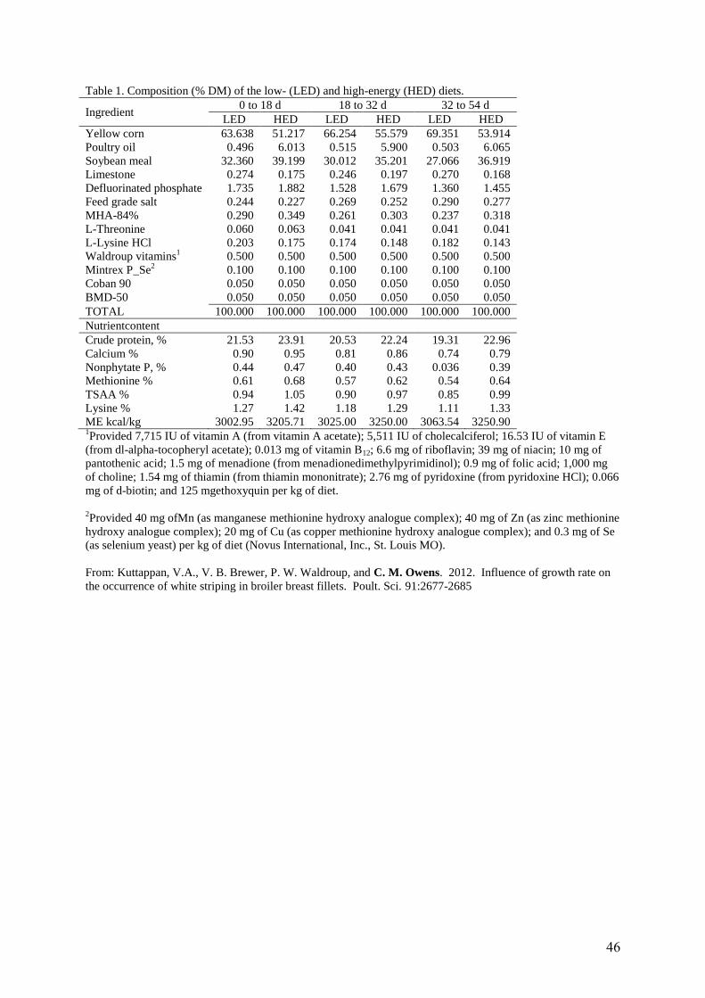
46
Table 1. Composition (% DM) of the low- (LED) and high-energy (HED) diets.
Ingredient 0 to 18 d 18 to 32 d 32 to 54 d
LED HED LED HED LED HED
Yellow corn 63.638 51.217 66.254 55.579 69.351 53.914
Poultry oil 0.496 6.013 0.515 5.900 0.503 6.065
Soybean meal 32.360 39.199 30.012 35.201 27.066 36.919
Limestone 0.274 0.175 0.246 0.197 0.270 0.168
Defluorinated phosphate 1.735 1.882 1.528 1.679 1.360 1.455
Feed grade salt 0.244 0.227 0.269 0.252 0.290 0.277
MHA-84% 0.290 0.349 0.261 0.303 0.237 0.318
L-Threonine 0.060 0.063 0.041 0.041 0.041 0.041
L-Lysine HCl 0.203 0.175 0.174 0.148 0.182 0.143
Waldroup vitamins1
0.500 0.500 0.500 0.500 0.500 0.500
Mintrex P_Se2
0.100 0.100 0.100 0.100 0.100 0.100
Coban 90 0.050 0.050 0.050 0.050 0.050 0.050
BMD-50 0.050 0.050 0.050 0.050 0.050 0.050
TOTAL 100.000 100.000 100.000 100.000 100.000 100.000
Nutrientcontent
Crude protein, % 21.53 23.91 20.53 22.24 19.31 22.96
Calcium % 0.90 0.95 0.81 0.86 0.74 0.79
Nonphytate P, % 0.44 0.47 0.40 0.43 0.036 0.39
Methionine % 0.61 0.68 0.57 0.62 0.54 0.64
TSAA % 0.94 1.05 0.90 0.97 0.85 0.99
Lysine % 1.27 1.42 1.18 1.29 1.11 1.33
ME kcal/kg 3002.95 3205.71 3025.00 3250.00 3063.54 3250.90 1Provided 7,715 IU of vitamin A (from vitamin A acetate); 5,511 IU of cholecalciferol; 16.53 IU of vitamin E
(from dl-alpha-tocopheryl acetate); 0.013 mg of vitamin B12; 6.6 mg of riboflavin; 39 mg of niacin; 10 mg of
pantothenic acid; 1.5 mg of menadione (from menadionedimethylpyrimidinol); 0.9 mg of folic acid; 1,000 mg
of choline; 1.54 mg of thiamin (from thiamin mononitrate); 2.76 mg of pyridoxine (from pyridoxine HCl); 0.066
mg of d-biotin; and 125 mgethoxyquin per kg of diet.
2Provided 40 mg ofMn (as manganese methionine hydroxy analogue complex); 40 mg of Zn (as zinc methionine
hydroxy analogue complex); 20 mg of Cu (as copper methionine hydroxy analogue complex); and 0.3 mg of Se
(as selenium yeast) per kg of diet (Novus International, Inc., St. Louis MO).
From: Kuttappan, V.A., V. B. Brewer, P. W. Waldroup, and C. M. Owens. 2012. Influence of growth rate on
the occurrence of white striping in broiler breast fillets. Poult. Sci. 91:2677-2685
47
Growth hormone transgenic Atlantic salmon: Opportunities, risks, and risk management
Eric M. Hallerman
Department of Fish and Wildlife Conservation Virginia Polytechnic Institute and State University
Blacksburg, Virginia 2406-0321
A large international effort has developed growth hormone-transgenic fishes of 18
species for potential use in aquaculture (Hallerman et al. 2007). The leading candidate for
commercial production is the AquAdvantage salmon, an Atlantic salmon expressing an
introduced copy of the Chinook salmon growth hormone (GH) gene (Du et al. 1992). It
exhibits 4-6-fold growth rate enhancement early in life (Fletcher et al. 2004) and a 10–20%
improvement in feed conversion efficiency (Entiss 1997, Cook et al. 2000a), offering the
prospect of shorter production time, reduced costs, and improved efficiency and
profitability. AquaBounty Technologies seeks U.S. Food and Drug Administration approval
for limited production of the fish under strict confinement in Panama (FDA 2010a); full-scale
commercial production is not sought at this time. Key issues pertain to food safety and
environmental risks associated with its production (NRC 2002).
Consumers concerns regarding genetically modified foods are prominent, including
concerns regarding food products from transgenic animals. The level of food safety concern
associated with a GM animal product varies with the application (NRC 2002). Focusing on
the AquaBounty salmon, bioactivity of the transgene does not pose food safety concern;
that is, neither salmon growth hormone, not its fragments, nor hormones secreted in
response to growth hormone pose risk to consumers. Regarding allergenicity, the growth
hormone molecule is a small peptide, present at low titer, and has no known allergenic
epitopes; further, consumers allergic to fish would not buy this fish product. Against this
background, the Food and Drug Administration Veterinary Medicine Advisory Committee
concluded that products from the AquAdvantage Atlantic salmon posed no food safety
issues (FDA 2010a). Despite the lack of science-based food safety issues, many consumers
want to know whether foods are GM and make informed product choices. In the European
Union and Japan, labeling of GM-derived food products is required. In the United States,
however, such labeling is not required and is the subject of ongoing debate (FDA 2010b).
48
The issue of ecological risk posed by production of transgenic fishes has a recognized
scientific basis (NRC 2002, Kapuscinski et al. 2007). The classical framework for assessing
ecological risk (NRC 2002) has five steps. First, risk assessors identify potential harms, i.e.,
ecological or genetic outcomes that we would not want to become realized. Second, they
identify the hazard that might lead to those harms, in this case the transgenic fish. Third,
they assess the probability of exposure, in this context the likelihood of escape and
persistence of transgenics in a particular receiving ecosystem. Fourth, in the most critical
step, they assess the probability of harm becoming realized given exposure to the hazard.
Finally, ecological risk is calculated as the probability of exposure to the hazard times the
probability of harm being realized given exposure.
Empirical studies of the growth hormone-transgenic Atlantic salmon have shown that
transgene expression affects not only growth rate, but also a range of morphological,
physiological, and behavioral traits. These studies have shown mostly maladaptive
alterations of oxygen and energy metabolism (Stevens et al. 1998, Stevens and Sutterlin
1999, Cook et al. 2000b, 2000c), feeding behavior (Abrahams and Sutterlin 1999),
smoltification (Saunders et al. 1998), and cardiorespiratory function (Deitsch et al. 2006).
These negative impacts have led some observers to suggest that the transgenics would pose
no significant environmental risk. However, empirical observations of GH-transgenic fishes
also have shown heightened growth rate, heightened food conversion efficiency, in some
cases larger ultimate size (which may confer mating advantage), and increased
osmoregulatory ability. Noting that trait-by-trait assessments of fitness do not address the
integrated phenotype of an individual, especially if there are tradeoffs among fitness-related
traits, how should we predict the fate of the transgene in receiving populations and hence,
likelihood of harm? The answer is that we should consider the net effect of transgene
expression on the fitness of individuals (Muir and Howard 1999, 2001, 2002). Against this
background, I was the principal investigator for a recent collaborative project involving
Ewen McLean (then at Virginia Tech) and Ian Fleming and Garth Fletcher (Memorial
University of Newfoundland). Our goal was to develop empirical data useful for quantifying
ecological and genetic risks posed by GH transgenic Atlantic salmon in the wild.
49
The first broad question that we asked was whether heightened competition, predation,
or other processes pose ecological harms to receiving ecosystems. Focusing upon the
survival component of fitness, we found no difference between transgenics and controls
regarding oxygen consumption rate, developmental rate, survival until emergence from
gravel, fry behavior, or growth and survival in an artificial stream (Moreau et al. 2011a). In a
second set of experiments, we asked whether transgenics are compromised in the face of
environmental stress. We applied ecologically and aquaculturally relevant stressors –
starvation, low Dissolved oxygen, and handling – and followed stress response variables
including hematocrit, pH, pCO2, Ca++, K+, Na+, Cl-, glucose, and cortisol. Cnaani et al. (in
review) found that transgenic fish were more stressed than wild-type fish, showing faster
and more pronounced stress responses. Overall, these data sets indicate that the GH-
transgenic Atlantic salmon are equally or less fit than wild type salmon; yet, under a range
of ecological conditions, fitness could be sufficiently high that there could be considerable
risk of ecological harm becoming realized.
The second broad question that we asked was whether interbreeding of transgenic fish
with wild populations poses genetic and evolutionary harms to receiving populations; that is
we sought to quantify the reproductive component of fitness. We should note that
introgression of the transgene into a wild population is a risk pathway, but not a risk
endpoint, i.e., it is not a harm in and of itself. Possible harms would include loss of
adaptation, reduced genetically effective population size, and in the extreme case,
extinction of receiving population. Noting that rapid growth rate is related to early
maturation in salmon, we predicted that precocious maturation of the salmon as parr may
pose a potent route for introgression of GH transgene. However, transgenics matured as
parr less frequently than non-transgenics (Moreau and Fleming 2012), and so the
opportunity to introgress is likely reduced for transgenic parr. In our most critical
experiment, we compared the behavior and reproductive success of transgenic and control
males in artificial spawning channels. We had trials where transgenic and non-transgenic
anadromous males competed for breeding opportunities and other trials where a transgenic
or non-transgenic anadromous male had sole access to female. We found that transgenic
parr were inferior competitors relative to wild-type parr in terms of nest fidelity, spawn
participation, and fertilization success (Moreau et al. 2011b). We found that transgenic
50
anadromous males were outcompeted in terms of nest fidelity, quivering frequency, and
spawn participation. Tying together the results of both spawning experiments, transgenic
males exhibiting either reproductive strategy exhibited low but non-zero reproductive
fitness. Hence, the risk associated with reproduction of GH-transgenic Atlantic salmon may
generally be low, but it is non-zero.
Pulling together all our results to predict the net fitness of GH-transgenic Atlantic salmon
and transgene fate in near-natural ecosystems, we found that the survival component of
fitness was less than or equal to that of the wild type, and reproductive fitness decreased
relative to the wild type. The upshot is that net fitness is reduced, and the transgene would
be expected to be purged from a receiving population following a single episode of
introduction. We did not, however, address the outcome should introductions recur via
continuing escapes from aquaculture operations.
The adoption of risk management measures affects the assessment of associated risk;
that is, ecological risk may be minimized by culturing transgenic fish under strict
confinement. Hence, the AquaBounty proposal before the U.S. Food and Drug
Administration is for pilot-scale production of GH-transgenic Atlantic salmon in Panama, in
indoor recirculating aquaculture systems with redundant physical confinements and
reproductive confinement (FDA 2010a). This production experiment is aimed at evaluating
production economics and demonstrating effective confinement. Should the outcomes
prove positive, AquaBounty would have to seek further regulatory approval for any
expanded production.
It becomes clear that regulatory approval is key to the future of the AquAdvantage
salmon. To date, no genetically modified animal intended for use as food by humans has
received regulatory approval. However, on December 26, 2012, the U.S. Food and Drug
Administration released it draft environmental assessment (FDA 2012a) and its preliminary
finding of no significant impact (FDA 2012b) for the proposed pilot-scale experiment for
production of the GH-transgenic Atlantic salmon. The public comment period was open
through April 26, 2013. Whatever the regulatory outcome, the FDA decision on the
AquaBounty salmon will be critical to the commercialization of transgenic fishes and
51
transgenic animals more generally. There are other transgenic fishes in the R&D and
regulatory pipeline, including other GH-transgenic fishes, disease resistant lines, biopharm
lines, and reversibly sterile lines. Other transgenic animals in development include goats
expressing lysozyme, which reduces milk bacterial loads. The “Enviropig” expressed phytase,
conferring better utilization and lower excretion of dietary phosphorous, but recently it was
euthanized because of ongoing cost and regulatory uncertainty. Porcine models of disease
are hung up in regulatory review because they could potentially be eaten by humans
(Maxmen 2012). A recent review concluded that regulatory uncertainty is stifling
development of genetically modified animals (Van Eenennaam et al. 2011)
Acknowledgments I gratefully acknowledge AquaBounty Farms, Inc. for access to transgenic Atlantic salmon and the U.S. Department of Agriculture Biotechnology Risk Assessment Grant Program for support. I thank the Poultry Breeders Roundtable for the invitation to present my views and for travel support. Literature cited Abrahams, M.V., and A. Sutterlin. 1999. The foraging and antipredator behavior of growth-
enhanced transgenic Atlantic salmon. Animal Behavior 58:933–942. Cnaani, A., E. McLean, and E.M. Hallerman. Effects of growth hormone transgene expression
and triploidization on hematological response to acute stress in Atlantic salmon (Salmo salar L.). Manuscript in review.
Cook, J.T., M.A. McNiven, G.F. Richardson, and A.M. Sutterlin. 2000a. Growth rate, body
composition, and feed digestibility/conversion of growth-enhanced transgenic Atlantic salmon. Aquaculture 188:15-32.
Cook, J.T., M.A. McNiven, and A.M. Sutterlin. 2000b. Metabolic rate of pre-smolt growth-
enhanced transgenic Atlantic salmon (Salmo salar). Aquaculture 188:33-45. Cook, J.T., A.M. Sutterlin, and M.A. McNiven. 2000c. Effect of food deprivation on oxygen
consumption and body composition of growth-enhanced transgenic Atlantic salmon (Salmo salar). Aquaculture 188:47-63.
Deitch, E., G.L. Fletcher, L.H. Peterson, I.A.S.F. Costa, M.A. Shears, W.R. Driedzic, and A.K.
Gamperl. 2006. Cardiorespiratory modifications, and limitations, in post-smolt growth
52
hormone transgenic Atlantic salmon (Salmo salar). Journal of Experimental Biology 209: 1310-1325.
Du, S.J., Z.Y. Gong, G.L. Fletcher, M.A. Shears, M.J. King, D.R. Idler, and C.L. Hew. 1992.
Growth enhancement in transgenic Atlantic salmon by the use of an all-fish chimeric growth hormone gene construct. Biotechnology 10:176-181.
Entiss, E., 1997. Aquabiotech: A blue revolution? World Aquaculture 28:12-15. FDA (U.S. Food and Drug Administration). 2010a. Briefing packet: AquAdvantage salmon for
public meeting of September 20, 2010. Center for Veterinary Medicine, Veterinary Medicine Advisory Committee, http://www.fda.gov/downloads/AdvisoryCommittees/CommitteesMeetingMaterials/VeterinaryMedicineAdvisoryCommittee/UCM224762.pdf.
FDA (U.S. Food and Drug Administration). 2010b. Background document: Public hearing on
the labeling of food made from the AquAdvantage salmon. http://www.fda.gov/downloads/Food/IngredientsPackagingLabeling/LabelingNutrition/FoodLabelingGuidanceRegulatoryInformation/Topic-SpecificLabelingInformation/UCM223913.pdf.
FDA (U.S. Food and Drug Administration). 2012a. AquAdvantage salmon: Draft
environmental assessment. http://www.fda.gov/downloads/AnimalVeterinary/DevelopmentApprovalProcess/GeneticEngineering/GeneticallyEngineeredAnimals/UCM333102.pdf.
FDA (U.S. Food and Drug Administration). 2012b. Preliminary finding of no significant
impact: AquAdvantage salmon. http://www.fda.gov/downloads/AnimalVeterinary/DevelopmentApprovalProcess/GeneticEngineering/GeneticallyEngineeredAnimals/UCM333105.pdf.
Fletcher, G.L., M.A. Shears, E.S. Yaskowiak, M.J. King, and S.V. Goddard. 2004. Gene transfer:
potential to enhance the genome of Atlantic salmon for aquaculture. Australian Journal of Experimental Agriculture 44:1095-1100.
Hallerman, E.M., E. McLean, and I.A. Fleming. 2007. Effects of growth hormone transgenes
on the behavior and welfare of aquacultured fishes: a review identifying research needs. Applied Animal Behavior Science 104:265-294.
Kapuscinski, A.R., K.R. Hayes, S. Li, G. Dana, E.M. Hallerman, and P.J. Schei. 2007.
Environmental risk assessment of genetically modified organisms, volume 3: methodologies for transgenic fish. CABI Press, Cambridge, MA.
Maxmen, A. 2012. Model pigs face messy path. Nature 486:453. Moreau, D.T.R., and I.A. Fleming. 2012. Enhanced growth reduces precocial male
maturation in Atlantic salmon. Functional Ecology 26:399-405.
53
Moreau, D.T.R., I.A. Fleming, G.L. Fletcher, and J.A. Brown. 2011a. Growth hormone
transgenesis does not influence territorial dominance or growth and survival of first-feeding Atlantic salmon Salmo salar in food-limited stream microcosms. Journal of Fish Biology 78:726-740.
Moreau, D.T.R., C. Conway, and I.A. Fleming. 2011b. Reproductive performance of
alternative male phenotypes of growth hormone transgenic Atlantic salmon (Salmo salar). Evolutionary Applications 4:736-748.
Muir, W.M., and R.D. Howard. 1999. Possible ecological risks of transgenic organism release
when transgenes affect mating success: sexual selection and the Trojan gene hypothesis. Proceedings of the National Academy of Sciences U.S.A. 96:13853-13856.
Muir, W.M., and R.D. Howard. 2001. Fitness components and ecological risk of transgenic
release: a model using Japanese medaka (Oryzias latipes). American Naturalist 158:1–16. Muir, W.M., and R.D. Howard. 2002. Assessment of possible ecological risks and hazards of
transgenic fish with implications for other sexually reproducing organisms. Transgenic Research 11:101–114.
NRC (National Research Council). 2002. Animal Biotechnology: Science-Based Concerns.
National Academy Press, Washington, DC, www.nap.edu. Saunders, R.L., G.L. Fletcher, and C.L. Hew. 1998. Smolt development in growth hormone
transgenic Atlantic salmon. Aquaculture 168:177-193. Stevens, E.D., A. Sutterlin, and T. Cook. 1998. Respiratory metabolism and swimming
performance in growth hormone transgenic Atlantic salmon. Canadian Journal of Fisheries and Aquatic Sciences 55:2028-2035.
Van Eenennaam, A.L., E.M. Hallerman, and W.M. Muir. 2011. The science and regulation of
food from genetically engineered animals. CAST Commentary QTA2011-2. Council for Agricultural Science and Technology, Ames, Iowa, http://www.cast-
science.org/publications/?the_science_and_regulation_of_food_from_genetically_engineered_animals&show=product&productID=21628.
Questions for Speaker: Dr. Eric Hallerman Question From: Dr. Karim Akbar Question: What is the mechanics of measuring feed efficiency in salmon or other experimental fish? You mentioned that feed efficiency in fish is good. If the test Is done in tank with fish having no access to other sources of feed, that is fine, but some reports are based on experiments in open net enclosures, and only feed that is added is considered.
54
What about sources of feed that are already in the water and are not considered in the calculation? Answer: Measurement of feed efficiency in fish generally is quantified as mass of feed provided per unit of mass gained, e.g., 1.6 grams of feed per gram gained. Dr. Akbar is on target, some past studies have reported excellent feed conversion ratios that actually were unreliable because fish held in outdoor systems had access to natural food items. Yet, fish really do have excellent feed conversion efficiency. Lovell (1989) compared feed conversion ratios of 1.2 for channel catfish to 2.1 for broiler chickens and 7.7 for beef cattle. Why is it, then, that fish have such good feed conversion ratios? First, they are cold-blooded and allocate no energy to maintaining body temperature. Lovell notes that fish are able to assimilate diets that are high in protein because of their lower dietary energy requirement. Second, living in water, in which they are neutrally buoyant, fish allocate little energy and material resource to development of their skeleton. Third, fish convert food that is 90% dry matter to fish flesh that is ~15% dry matter. Hence, while feed conversion efficiency in fish is excellent, it is not miraculous. Question From: Dr. Casey Owens Hanning Question: Are there any meat quality differences due to transgenic Atlantic salmon? Answer: Many aspects of meat quality of the AquaBounty Atlantic salmon were quantified in the context of the company’s application to the Food and Drug Administration for approval of their proposed pilot-scale production trial. Results were presented at a public meeting held in 2010 (FDA 2010). Of the proximate, vitamin, mineral and amino acid analytes quantified, only three – vitamin B6, folic acid, and niacin – were present at levels in transgenic salmon that were statistically significantly different from those in control salmon. Based on all criteria considered, the FDA Veterinary Medicine Advisory Committee concluded that the levels of all proximate, vitamin, mineral and amino acid analytes in transgenic salmon except vitamin B6 were similar to levels in one or more appropriate groups of control salmon. They did note, however, that sample sizes in some cases were not particularly large. Question From: Dr. Jesus Arango Question: Public perception is an important driver in product demand. Fish and salmon meat in particular, have seen an explosion in demand mainly due to the perception of being healthy. However, there are several sources that warn about the presence of heavy metals in fish meat. What is your opinion? Would it vary depending on sources? Should one be really concerned about this issue? Answer: Heavy metals tend to bioaccumulate up marine food chains, such that high-level predators such as tunas and billfishes can carry high levels of mercury relative to their environment. Consumption advisories have been issued for certain such species, typically recommending no more than one serving per month. The most prominent issue pertaining to cultured fishes, however, pertains to organic compounds. Hites et al. (2004) analyzed farmed and wild salmon products for
55
organochlorine contaminants and showed that these contaminants (e.g., PCBs, DDT, and dioxins) were significantly higher in farmed salmon than in the wild. Interestingly, European-raised salmon had significantly higher contaminant loads that those raised in North and South America. Risk analysis indicated that consumption of farmed Atlantic salmon may pose health risks that detract from the beneficial effects of fish consumption. The Hite et al. report led to an active exchange of letters and responses; see Science for 23 July 2004. My recommendation is to enjoy salmon in moderation as part of a balanced diet. Question From: Damarius Fleming Question: In the face of resistance to GM foods by commercial markets (groceries, etc.) do you feel that researchers are doing enough to dispel some of the rumors/trepidation of consumers? Answer: Many consumers perceive risks from consumption of genetically modified foods. To the extent that these perceptions stem from lack of information about GM foods, they can be addressed by access to science-based information. It is incumbent upon researchers in the academic and agribusiness communities to develop appropriately targeted information packages and present them for consideration by the general public in appropriate fora. For example, Van Eenennaam et al. (2011) explained the science and regulation of food from genetically engineered animals, engaging the public through the Council for Agricultural Science and Technology. The Cooperative Extension Service in every state engages a wide cross-section of Americans, and is a useful outlet for effectively communicated scientific information. Public acceptance of GM foods ultimately determines the success or failure of such products. Literature cited FDA. 2010. Briefing packet: Food and Drug Administration Center for Veterinary Medicine
Veterinary Medicine Advisory Committee, September 20, 2010. http://www.fda.gov/downloads/AdvisoryCommittees/CommitteesMeetingMaterials/VeterinaryMedicineAdvisoryCommittee/UCM224762.pdf. Accessed May 17, 2013.
Hites, R.A., J.A. Foran, D.O. Carpenter, M.C. Hamilton, B.A Knuth, and S.J. Schwager. 2004. Global assessment of organic contaminants in farmed salmon. Science 303: 226-229.
Lovell, T. 1989. Nutrition and feeding of fish. Van Nostrand Reinhold, New York. Van Eenennaam, A.L., E.M. Hallerman, and W.M Muir. 2011. The science and regulation of
food from genetically engineered animals. CAST Commentary QTA2011-2, Council for Agricultural Science and Technology, Ames, Iowa. http://www.cast-
science.org/publications/?the_science_and_regulation_of_food_from_genetically_engineered_animals&show=product&productID=21628. Accessed May 17, 2013.
56
What do RFI, PRRS, and SCID have in Common?
Jack C. M. Dekkers Department of Animal Science
Iowa State University
Introduction
RFI refers to Residual Feed Intake, a measure of feed efficiency defined as the difference
between an animal’s observed feed intake and it’s expected feed intake based on expected
energy requirements for maintenance and production, based on observed performance
(e.g. body weight, growth rate, fatness). It has been used as a measure of feed efficiency in
most livestock species, including chickens and pigs.
PRRS stands for Porcine Reproductive and Respiratory Syndrome. Caused by an RNA virus, it
is the most costly disease in the pig industry, both in the US and globally. Annual costs to the
US industry alone are estimated at 664 million (Holtkamp et al. 2013). PRRS affects both the
breeding (piglet production) and the growing pig sectors of the pork industry through a
decrease in reproductive health, an increase in deaths, and reductions in the rate and
efficiency of growth. PRRS also functions as a cofactor in other disease syndromes. Efforts to
control the disease through improvements in veterinary health management, biosecurity,
disease elimination, and vaccination have been met with some success but not enough to
stem the spread of the disease. Host genetics is another route that is currently being
pursued.
SCID stands for Severe Combined Immune Deficiency. It is a category of devastating genetic
diseases that results in lack of or severe impairment of the adaptive immune system. It can
be caused by a mutation in genes that are crucial to the development of antibodies. It has
been identified in dogs, horses and humans (David Vetter, the Bubble boy) and has been
artificially created in mice, rats, and recently pigs (Suzuki et al. 2012). SCID mice and rats are
extensively used as models for biomedical research into cancer, vaccine development, etc.
So what do RFI, PRRS, and SCID have in common, apart from the fact that all three are
acronyms? The purpose of this paper is to tell the story of how the paths of these three
rather distinct phenomena crossed in research involving our selection lines for RFI in pigs at
Iowa State University, thereby providing insight into the sometimes serendipitous route that
57
research can take and into findings that may also be of relevance to the poultry breeding
industry.
The ISU RFI lines in Yorkshire Pigs
In order to enable study of the genetic and physiological basis of feed efficiency in pigs, a
selection experiment in Yorkshire pigs was started at Iowa State University in 2001, with the
aim to develop lines that differ in RFI during the grow-finish phase (Cai et al., 2008). Using
purebred Yorkshire pigs sourced from the Midwest, a selection line for low RFI (= increased
efficiency) and a randomly selected control line were initiated by splitting litters. In
generation 5, selection for increased RFI (= reduced efficiency) was initiated in the randomly
selected control line. Now in its 8th generation of selection, the low RFI line consumes 376
g/d less than the high RFI line and has 241 g/d lower RFI (Figure 1). Selection for RFI has
resulted in an improvement in feed conversion ratio, with the low RFI line requiring 0.22 g
feed less per g weight gain than the high RFI line in generation 8. Other performance traits
that have changed as a result of selection for RFI are average daily gain and ultrasonic
backfat and loin eye area, with low RFI pigs growing 79 g/d slower, having 2.5 mm less
backfat, and 1.5 cm2 larger loins than pigs from the high RFI in generation 8.
In recent years, the focus of research using the ISU RFI lines has shifted towards
understanding the biological and physiological basis of the differences in feed efficiency that
have been established through selection between the lines and towards identifying bio- or
genetic markers that are associated with feed efficiency (Young and Dekkers, 2012). In
addition, research has been initiated to determine how the two lines respond to different
stressors and challenges, as part of a large USDA-NIFA grant (grant # 2011-68004-30336).
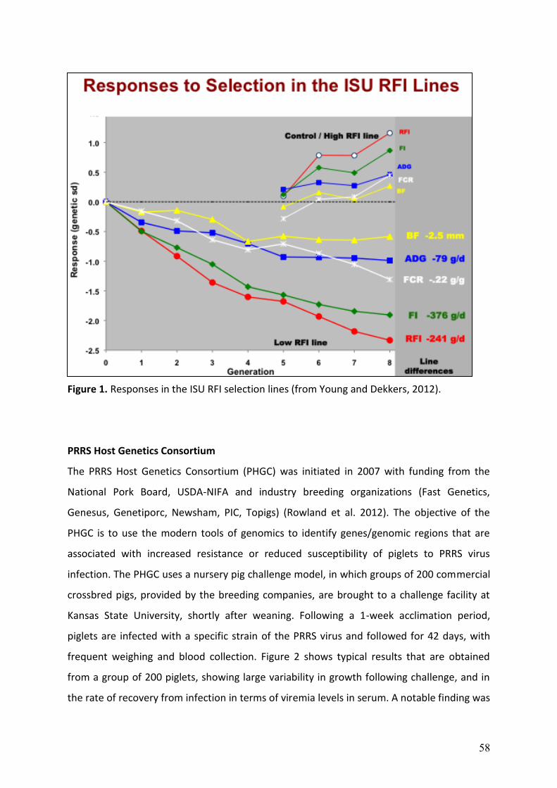
58
Figure 1. Responses in the ISU RFI selection lines (from Young and Dekkers, 2012).
PRRS Host Genetics Consortium
The PRRS Host Genetics Consortium (PHGC) was initiated in 2007 with funding from the
National Pork Board, USDA-NIFA and industry breeding organizations (Fast Genetics,
Genesus, Genetiporc, Newsham, PIC, Topigs) (Rowland et al. 2012). The objective of the
PHGC is to use the modern tools of genomics to identify genes/genomic regions that are
associated with increased resistance or reduced susceptibility of piglets to PRRS virus
infection. The PHGC uses a nursery pig challenge model, in which groups of 200 commercial
crossbred pigs, provided by the breeding companies, are brought to a challenge facility at
Kansas State University, shortly after weaning. Following a 1-week acclimation period,
piglets are infected with a specific strain of the PRRS virus and followed for 42 days, with
frequent weighing and blood collection. Figure 2 shows typical results that are obtained
from a group of 200 piglets, showing large variability in growth following challenge, and in
the rate of recovery from infection in terms of viremia levels in serum. A notable finding was
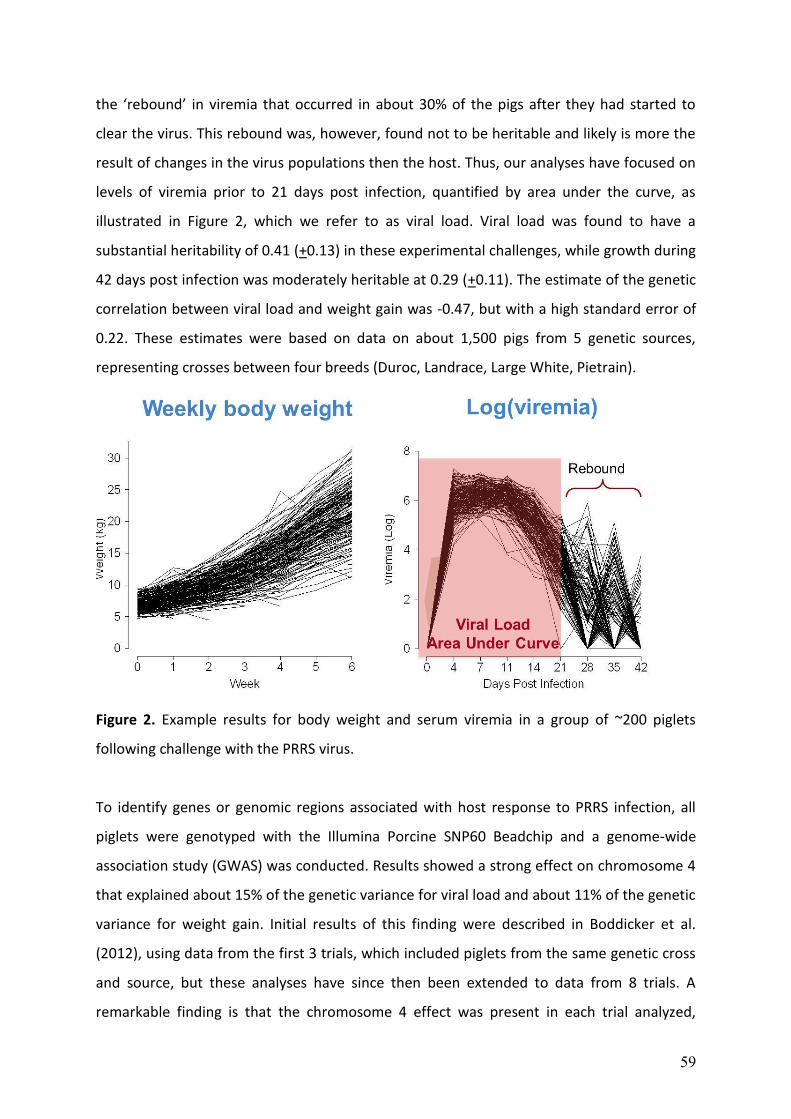
59
the ‘rebound’ in viremia that occurred in about 30% of the pigs after they had started to
clear the virus. This rebound was, however, found not to be heritable and likely is more the
result of changes in the virus populations then the host. Thus, our analyses have focused on
levels of viremia prior to 21 days post infection, quantified by area under the curve, as
illustrated in Figure 2, which we refer to as viral load. Viral load was found to have a
substantial heritability of 0.41 (+0.13) in these experimental challenges, while growth during
42 days post infection was moderately heritable at 0.29 (+0.11). The estimate of the genetic
correlation between viral load and weight gain was -0.47, but with a high standard error of
0.22. These estimates were based on data on about 1,500 pigs from 5 genetic sources,
representing crosses between four breeds (Duroc, Landrace, Large White, Pietrain).
Figure 2. Example results for body weight and serum viremia in a group of ~200 piglets
following challenge with the PRRS virus.
To identify genes or genomic regions associated with host response to PRRS infection, all
piglets were genotyped with the Illumina Porcine SNP60 Beadchip and a genome-wide
association study (GWAS) was conducted. Results showed a strong effect on chromosome 4
that explained about 15% of the genetic variance for viral load and about 11% of the genetic
variance for weight gain. Initial results of this finding were described in Boddicker et al.
(2012), using data from the first 3 trials, which included piglets from the same genetic cross
and source, but these analyses have since then been extended to data from 8 trials. A
remarkable finding is that the chromosome 4 effect was present in each trial analyzed,
60
representing different breed crosses and genetic sources. Although the exact gene and
mutation have not yet been identified, the favorable alleles for the region are present in all
breeds and lines analyzed but at a low frequency, which offers great perspectives for
genetic improvement through marker-assisted selection on this region.
PRRS and the ISU RFI lines
In order to evaluate whether the ISU RFI lines responded differently to a PRRS challenge,
100 piglets from each line were sent to Kansas State University, where they were challenged
with the PRRS virus, following the PHGC protocol. Our hypothesis was that, having been
selected under relatively high-health conditions, the low RFI line (high efficiency) would not
have the resources to mount as effective an immune response to the PRRS virus as the high
RFI line. However, our hypothesis was proven wrong since, although differences were not
significant at p<0.05, the low RFI line tended to have a lower viral load (p=0.09) and a higher
growth rate following challenge (p=0.17). Mortality was less than 10%, similar to that
observed in other PHGC trials, and was not different between the two lines. This finding is
consistent with other results from the two lines, that suggest that the efficient line is better
able to mobilize resources and direct it where they are needed to deal with a challenge or
demand. For example, the low RFI line has slightly greater litter size, both at birth and at
weaning, and low RFI piglets are slightly heavier, both at birth and at weaning. The low RFI
sow is able to provide the required extra nutrients to its piglets not by eating more but by
mobilizing more body reserves (Young et al. 2010).
SCID, PRRS, and the ISU RFI lines
One of the routine procedures in the Kansas State University PRRS challenge studies is to
conduct a necropsy on pigs that died or that had to be euthanized prior to the end of the
experiment, in order to determine the cause of death. Routine necropsy of 4 of the piglets
from the low RFI line showed some interesting findings: a very small thymus and lymph
nodes and other abnormal histopathology, and the piglets had no detectable antibodies to
the PRRS virus. These findings were recognized as being consistent with SCID, resulting in
the first reported cases of SCID in pigs (Cino Ozuna et al. 2012). Remating the six parents of
these four piglets resulted in litters that each produced SCID piglets in an approximately 1:3
ratio, indicating that it segregates in an autosomal recessive manner.
61
At birth, SCID piglets appear healthy without a weight difference between affected and
normal littermates. Pre-suckling umbilical cord blood of SCID piglets were shown to have no
detectable immunoglobulins. However, suckling provides affected piglets with maternal
antibodies, which allow SCID piglets to remain healthy for several weeks after farrowing. But
maternal antibody levels diminish after weaning, leaving SCID piglets susceptible to many
pathogens and they will succumb to these, unless they are maintained in a pathogen-free
environment, similar to the Bubble Boy. SCID piglets have been shown to have no or very
low levels of B and T cells, which are needed to mount an adaptive immune response. The
SCID piglets do have natural killer cells but it is not clear whether they are functional in the
SCID pig.
To identify the gene or genomic region responsible for SCID, the Illumina Porcine SNP60
Beadchip was used to genotype 20 affected piglets, 6 known carrier parents, 50 unaffected
littermates, and 97 ancestors. Using GWAS, a 5.6 Mb region that likely contains the
causative mutation was identified. This region contains a strong candidate gene based on
interspecies comparative information. Phasing the SNPs in a 1 Mb region surrounding the
candidate gene showed two haplotypes that segregate with affected status, either in a
homozygous or compound heterozygous state. The identified haplotypes were traced
through the pedigree of the RFI line back to the founder generation, which were sourced
from the purebred Yorkshire population. Allele-specific primers were developed to allow
identification of carriers and affected pigs in the ISU RFI population. Work is ongoing to
identify the causative mutation.
SCID Pigs as a Biomedical Model.
Transgenic SCID mice are extensively used as a biomedical model for immune system
research, cancer research, studies of the effects of disease, cell and tissue transplantation,
and testing new vaccines and therapeutic agents for immuno-compromised individuals.
However, it has been well established that pigs are much better biomedical models for
humans than mice. Until our finding, and the recent production of a transgenic X-linked SCID
pig in Japan (Suzuki et al., 2012), no such pig models were available. The transgenic SCID pig
developed by Suzuki et al. (2012) is X-linked and produces a different immunophenotype
than our SCID pig.
62
To demonstrate the utility of our SCID pig as a biomedical model, 3 SCID piglets and 3
normal littermates were sent to Kansas State University, where human melanoma and
human pancreatic carcinoma cells were injected in the left and right ears of the piglets
(Basel et al., 2012). While the normal piglets immediately rejected and cleared the human
cells, in the SCID piglets, these human cells formed visible tumors. This confirms that the
SCID piglets do not have the ability to reject foreign cells and demonstrates that they
provide a useful model for cancer and other research.
Because SCID carriers are phenotypically normal, SCID piglets can be produced by carrier by
carrier matings. However, this only produces 25% affected piglets on average, which does
not make a very reliable and efficient system to produce SCID piglets for research. In
humans, as well as other species, the standard procedure for curing SCID is by giving the
patient an adaptive immune system through bone marrow transfer. Thus, in order to rescue
affected SCID piglets such that they can be raised to breeding age and be available for
breeding as SCID homozygotes, nine affected piglets from four litters were given bone
marrow transfers from normal donors that were matched based on major class I and II
haplotypes of the MHC. Five recipients showed signs of graft versus host disease after
transfer and were euthanized. The four surviving transplanted piglets were documented to
have a reconstituted adaptive immune system and are providing a pool of frozen semen for
use to breed carrier females to produce litters with 50/50 SCID/normal piglets.
Acknowledgements
The RFI work is a large collaborative effort that has included contributions from ISU faculty
(Tom Baas, Rohan Fernando, Dorian Garrick, Max Rothschild, Chris Tuggle, Elisabeth
Lonergan, Steven Lonergan, Nick Gabler, John Patience, Mike Spurlock, Lloyd Anderson,
Anna Johnson, Peng Liu, Dan Nettleton, and Vasant Honavar), graduate students (David
Casey, Weiguo Cai, Jennifer Young, Nick Boddicker, Dinesh Thekkoot, Emily Waide, Andrew
Hess, Danielle Gorbach, Oliver Couture, Rachel Smith, Kyle Grubbs, Shannon Cruzen, Emily
Arkfeld, Venkatesh Mani, Amanda Harris, Jessica Jenkins, Sender Lkhagvadorj, Larry Saddler,
and Long Qu), post-docs and research associates (Jennifer Young, Suneel Onteru, Anoosh
Rakhshandeh, and Ed Steadham), and staff at the Lauren Christian Swine Breeding Research
Center, along with contributions from external collaborators, including Brian Kerr from
USDA-ARS, Kim Bunter and Frank Dunshea from Australia, and Rob Bergsma and Egbert Knol
63
from IPG & Wageningen University. The RFI project was made possible by the donation of
FIRE feeders by PIC/Genus and Newsham Choice Genetics and funding from USDA-CSREES
NRI Grants #2010-65206-20670 and #2011-68004-30336, the National Pork Board, the Iowa
Pork Producers Association, the ISU Center for Integrated Animal Genomics, Iowa State and
Hatch Funds, Pfizer Animal Health, and the USDA Swine Genome Coordinator.
The PHGC is a collaborative effort involving the research groups of Bob Rowland at Kansas
State University, Joan Lunney at USDA-ARS-Beltsville and James Reecy, Chris Tuggle, and
Jack Dekkers at ISU. GWAS were conducted by graduate student Nick Boddicker. The PHGC
was supported by the USDA NIFA PRRS CAP Award 2008-55620-19132, the National Pork
Board, and the NRSP-8 Swine Genome and Bioinformatics Coordination projects, and the
PRRS Host Genetics Consortium, consisting of USDA ARS, Kansas State University, Iowa State
University, Michigan State University, Washington State University, Purdue University, the
University of Nebraska-Lincoln, PIC/Genus, Newsham Choice Genetics, Fast Genetics,
Genetiporc, Inc., Genesus, Inc., PigGen Canada, Inc., IDEXX Laboratories, and Tetracore, Inc.
Technical assistance is acknowledged from Juan Pedro Steibel for the algorithm to compute
area under the curve, Max Rothschild for assistance with genotyping, Nader Deeb for
assistance with data and analysis, Eric Fritz for database management, Ania Wolc and
Dinesh Thekkoot for statistical assistance, and the lab of Bob Rowland, specifically Becky
Eaves, Maureen Kerrigan, Ben Trible, Jessica Otradovec, Brooke Bloomberg, Aubree Gottlob,
Laura O’Brien, and Ranjini Chand for animal care and sample collection, and the lab of Joan
Lunney, specifically Samuel Abrams and Amber Tietgens for preparation of all genomic DNA
samples for SNP genotyping.
The SCID project is a collaborative effort involving Emily Waide (graduate student supported
by USDA NIFA National Needs grant 2010-38420-20328), Chris Tuggle, Jason Ross, Matthew
Ellinwood, Nick Boddicker, Dinesh Thekkoot, and Jennifer Young from ISU, Bob Rowland,
Carol Wyatt, Deryl Troyer, Giselle Cino, Maureen Kerrigan, Matthew Basel, and Ben Tribble
from Kansas State University, and Sam Ho from the Michigan Gift of Life. The SCID work was
supported by Iowa State University Research Foundation and the Iowa State University
VPRED office.
64
References Basel, M.T., S. Balivada, A.P. Beck, M.A. Kerrigan, M.M. Pyle, J.C.M. Dekkers, C.R. Wyatt, R.R.R. Rowland, D.E. Anderson, S.H. Bossmann, and D.L. Troyer. 2012. Human Xenografts are not rejected in a naturally occurring immunodeficient porcine line: a human tumor model in pigs. BioResearch Open Access. 1: 63-68. Boddicker N.J., Waide E.H., Rowland R.R.R., Lunney J.K., Garrick D.J., Reecy J.M., & Dekkers J.C.M. (2012) Evidence for a major QTL associated with host response to Porcine Reproductive and Respiratory Syndrome Virus challenge. Journal of Animal Science 90, 1733-1746. Cai, W., D. S. Casey, and J. C. M. Dekkers. 2008. Selection response and genetic parameters for residual feed intake in Yorkshire swine. J. Anim. Sci. 86:287-298. Cino Ozuna, A.G., R.R.R. Rowland, J.C. Nietfeld, J.C.M. Dekkers, and C.R. Wyatt. 2012. Preliminary findings of a previously unrecognized porcine primary immunodeficiency disorder. Veterinary Pathology doi:10.1177/0300985812457790 Holtkamp D.J., Kliebenstein J.B., Neumann E.J., Zimmerman J.J., Rotto H., Yoder T.K., Wang C., Yeske P., Mowrer C. & Haley C. 2013. Assessment of the economic impact of porcine reproductive and respiratory syndrome virus on United States pork producers. J. Swine Health Prod. 21(2):72-84. Rowland R.R.R., Lunney J.K., and Dekkers J.C.M. 2012. Control of porcine reproductive and respiratory syndrome (PRRS) through genetic improvements in disease resistance and tolerance. Frontiers in Genetics 3, 260. Suzuki S., Iwamoto M., Saito Y., Fuchimoto D., Sembon S., Suzuki M., Mikawa S., Hashimoto M., Aoki Y., Najima Y., Takagi S., Suzuki N., Suzuki E., Kubo M., Mimuro J., Kashiwakura Y., Madoiwa S., Sakata Y., Perry A.C., Ishikawa F., and Onishi A. 2012. Il2rg gene-targeted severe combined immunodeficiency pigs. Cell Stem Cell 10: 753-758. Young, J. M., R. Bergsma, E. F. Knol, J. F. Patience, and J. C. M. Dekkers. 2010. Effect of selection for residual feed intake on sow reproductive performance and lactation efficiency. Proc. 9th WCGALP. Paper #223. Young, J.M., and J.C.M. Dekkers. 2012. The genetic and biological basis of residual feed intake as a measure of feed efficiency. Chapter 7, Pp. 153-166 In: J.F. Patience (Ed): Feed Efficiency in Swine. Wageningen Academic Publishers, Wageningen, The Netherlands.
Questions for Speaker: Dr. Jack Dekkers
Question From: Dr. Neil O’Sullivan Question: Signal found on SSC4 for growth rate. Does this signal show up in tests done on a normal clean farm? Answer: We have not been able to find reports of QTL for this region in the literature. Most/all of the reported QTL studies would have been conducted without presence of PRRS and in relatively ‘clean’ environment. So this suggests that this region may not affect performance in a ‘clean’ environment.
65
Question From: Dr. Sue Lamont Question: Following up on Rowland’s hypothesis about macrophage permissiveness and PRRRs, what are the percentages (or absolute numbers) of macrophages present in SCID vs. normal pigs? Answer: We don’t know. To date, all our flow cytometry data to investigate cell numbers are based on blood and we have not specifically looked at macrophages. Macrophages only comprise a very small portion of the cells in blood. Question From: Dr. Sue Lamont Question: In the grafted pigs, from where did the host-genotype WBC come, which were detected in the early days post-graft? Answer: We have not determined their origin but they likely are of donor origin. We have determined that our SCID pigs are deficient in B and T cells but not in natural killer cells. In addition to representing cells other than B and T cells, they could represent lymphocytes derived from colostrum as there is some evidence that lymphocytes can cross the intestinal barrier in newborn piglets. Finally, we have not determined whether our SCID pigs are complete devoid of B and T cells; there could be some ‘leakiness’, which is often observed in SCID models, and/or the B and T cells that are there may not be functional. Question From: Dr. Gerald Herbert Question: Were the high heritability figures presented a function of the animals (the initial populations) being crossbred? Answer: That could play a role but within a trial, all piglets were of the same cross and trial was included as a fixed effect in the model of analysis, so it is unlikely that breed differences contributed to the estimate of heritability.