Embed Size (px)
Citation preview

PRODUCT DATA
™


Echelon Oval is designed around the shape of the body to accommodate the broadest patient spectrum, featuring an innovative 74cm Oval bore providing outstanding patient and technologist benefits. Oval is equipped with a suite of Workflow Integrated Technology (WIT) features, a powerful hardware platform, and a fast and easy to use computer and operating system enabling advanced image acquisition and processing capabilities.
Key Components and Specifications◾ 1.5T Superconducting Magnet with 74cm Oval Bore◾ Patient-Centric Management System
– WIT Mobile Table– WIT Gantry Monitor
◾ Gradient System — 34mT/m and 150T/m/s◾ WIT RF System with Integrated RF Receiver Coils
– 16 or 32 channel receiver– Digital Drive DX
◾ Vertex™ II Computer System and Origin™ MR Operating Software◾ Imaging Suites — Pulse Sequence and Acquisition Techniques◾ Siting Considerations and Component Dimensions
The Widest Wide Bore in the Industry

Magnet System The Echelon Oval magnet features a 74cm oval bore for maximum patient accessibility and comfort. Echelon Oval’s 1.5T magnet features high homogeneity, ultimate stability, and a full 50cm FOV in all directions. Echelon Oval also includes HOAST™ (Higher Order Active Shim Technology) applied per patient assuring exceptional magnetic field uniformity. Echelon Oval features a compact footprint and virtually zero Helium boil off.
◾ Superconducting magnet ◾ 1.5 Tesla ◾ Horizontal field ◾ Homogeneity: <0.5ppm @ 40cm DSV (VRMS) ◾ Shimming:
– Installation: Computer mapped passive shim – Patient: Linear and Higher order per patient active shim
◾ Active magnetic shielding ◾ 5G Fringe field
– Axial: 4.0m (13.1ft) – Radial: 2.5m (8.2ft)
◾ Helium cryogen ◾ Refill frequency: Once every six years with Hitachi's Customer Support Program
Patient Centric Management SystemHitachi’s mastery of patient-focused MR imaging is demonstrated in Echelon Oval’s attention to patient comfort. It begins with Echelon Oval’s bore, the widest bore (74cm) of any 1.5T. The extra width accommodates the extra-wide WIT Mobile Table (63cm), with a 550lb weight capacity, and mobility to reduce patient transfers for easy accessibility. The table’s 9+ feet of travel allows patients to enter the bore in the less stressful feet-first orientation for any exam. It lowers to 50cm allowing easy access even for wheelchair patients. Arm boards on either side accommodate patient IVs and extend vertically to ensure patient safety during transport. A positive patient experience is enhanced with adjustable airflow, lighting, and two-way communication. A gantry-mounted Workflow Integrated Technology (WIT) Monitor provides the operator with patient, coil, and gating information to further speed patient preparation.
Inspired By Your Patients
◾ Patient aperture: 74cm × 65cm ◾ Table weight capacity: 550lb (250kg) ◾ Table width: 63cm ◾ Longitudinal motion: 30cm/s ◾ Total longitudinal travel: >9ft (279cm) ◾ Vertical range: 19.7-33.2in (50–84cm) ◾ Class II laser positioning
– +/-0.5mm accuracy – Automatic movement to isocenter
◾ Table control – Up/Down – In/Out (Slow/Fast) – Table position in mm – Move to isocenter – Return to zero position – Stop – Release – Laser – Clear
◾ Scan control – Start/Abort/Pause
◾ Patient amenities – Two-way intercom – Technologist alert system – Adjustable bore illumination – Adjustable bore ventilation – Patient pads and immobilization straps

Gradient SystemHigh gradient performance is key to high performance imaging. The Echelon Oval includes a 34/150 capable gradient system. This high slew rate enables selection of low TR, TE, and IET in combination with small FOV and thin slices. This level of gradient capability positions Echelon Oval to adapt to changing MR technology and widening applications far into the future.
◾ Peak amplitude: 34 mT/m ◾ Peak slew rate: 150 T/m/s ◾ Active shielding ◾ Water cooling ◾ Gradient noise reduction: Mechanical gradient sound dampening
Radiofrequency System with WIT Receiver CoilsEchelon Oval’s RF receiver system manages multiple coil connection points on the table. The WIT RF coil system provides integrated coil arrays that can be used individually or in combination to give the operator maximum flexibility for positioning patients of all sizes. The WIT Spine coils are plugged directly into the table, so no cables get in the way. The WIT Posterior Head/Neck array can be used at either end of the table permitting head-first or feet-first imaging for all exams. The WIT receive coil system includes anterior Head, Neck, Neurovascular, Torso, and Cardiac arrays that can be easily placed on the patient for imaging.
Virtually all of Echelon Oval’s array, surface, and volumetric coils are multiple element designs for high signal uniformity, high SNR, and compatibility with RAPID™ parallel imaging for maximum clinical flexibility and image quality. Analog to Digital conversion in the scan room with optical digital transmission of MR signal data prevents electrical noise pickup and ensures highest possible SNR.
RF Transmit: ◾ Two 20kW RF power amplifiers
Available Coil Set Includes: ◾ WIT Posterior Head/Neck can be used at either end of the table for maximum patient comfort and technologist convenience
◾ WIT Posterior Spine A and B coils plug directly (no cable) into the patient table to maximize contact with the patient
◾ WIT Anterior Head ◾ WIT Anterior Neck ◾ WIT Anterior Neurovascular ◾ WIT Torso ◾ WIT Cardiac
Digital Drive Dx Receiver: ◾ 16 or 32 channels ◾ 7 coil connection points ◾ Ultra low noise coil mounted preamplifiers ◾ A/D conversion on gantry with optical digital transmission to equipment room
◾ Shoulder ◾ Extremity ◾ Hand/Wrist ◾ Foot/Ankle ◾ Flexible Extremity ◾ Bilateral Breast ◾ Peripheral Vascular ◾ Extremity Micro Coil ◾ QD T/R Body
CPU: ◾ Core i5 processor ◾ 8 GB RAM ◾ Display
– 24in LCD color monitor – Display matrix 1920x1200
◾ Magnetic disk: – 320GB storage capacity – Stores up to 400,000 images (256x256)
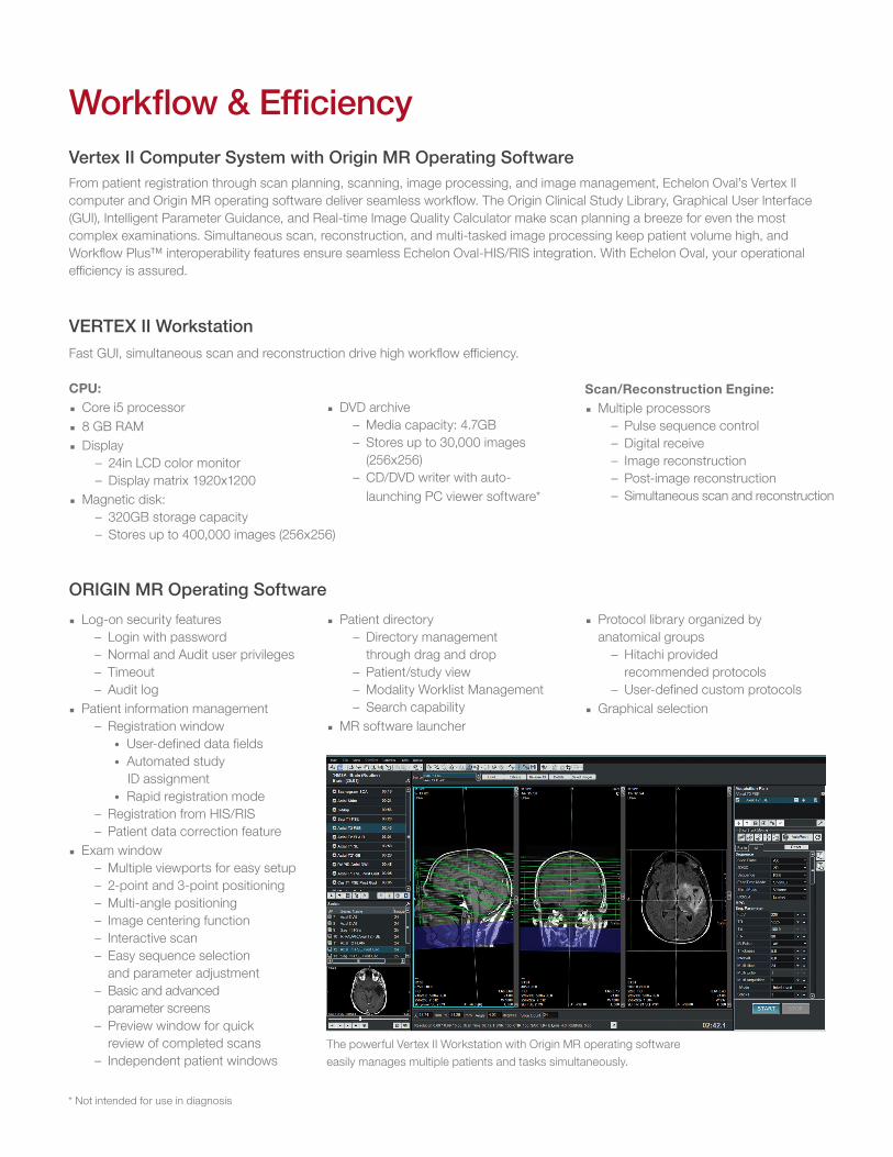
Vertex II Computer System with Origin MR Operating SoftwareFrom patient registration through scan planning, scanning, image processing, and image management, Echelon Oval’s Vertex II computer and Origin MR operating software deliver seamless workflow. The Origin Clinical Study Library, Graphical User Interface (GUI), Intelligent Parameter Guidance, and Real-time Image Quality Calculator make scan planning a breeze for even the most complex examinations. Simultaneous scan, reconstruction, and multi-tasked image processing keep patient volume high, and Workflow Plus™ interoperability features ensure seamless Echelon Oval-HIS/RIS integration. With Echelon Oval, your operational efficiency is assured.
VERTEX II WorkstationFast GUI, simultaneous scan and reconstruction drive high workflow efficiency.
Scan/Reconstruction Engine: ◾ Multiple processors
– Pulse sequence control – Digital receive – Image reconstruction – Post-image reconstruction – Simultaneous scan and reconstruction
ORIGIN MR Operating Software ◾ Log-on security features
– Login with password – Normal and Audit user privileges – Timeout – Audit log
◾ Patient information management – Registration window
• User-defined data fields • Automated study
ID assignment • Rapid registration mode
– Registration from HIS/RIS – Patient data correction feature
◾ Exam window – Multiple viewports for easy setup – 2-point and 3-point positioning – Multi-angle positioning – Image centering function – Interactive scan – Easy sequence selection and parameter adjustment
– Basic and advanced parameter screens
– Preview window for quick review of completed scans
– Independent patient windowsThe powerful Vertex II Workstation with Origin MR operating software easily manages multiple patients and tasks simultaneously.
* Not intended for use in diagnosis
Workflow & Efficiency
◾ DVD archive – Media capacity: 4.7GB – Stores up to 30,000 images (256x256)
– CD/DVD writer with auto- launching PC viewer software*
◾ Patient directory – Directory management through drag and drop
– Patient/study view – Modality Worklist Management – Search capability
◾ MR software launcher
◾ Protocol library organized by anatomical groups
– Hitachi provided recommended protocols
– User-defined custom protocols ◾ Graphical selection

◾ Processing tasks – Max/Min Intensity Projection (MIP/minIP) – Multi-Planar Reconstruction (MPR) – Vascular Volume Rendering – Signal Intensity Ratio Map (SIR Map) – Addition/subtraction – T1 and T2 calculated Images – T2 RelaxMap – T2* RelaxMap – Dynamic analysis – Perfusion analysis – Diffusion analysis
• Single direction analysis • Multi direction analysis
– ADC trace – DWI trace
– Tensor/Kurtosis analysis • Mean Diffusivity (MD) • Fractional Anisotropy (FA) • DWI trace
– Post-Reconstruction functions • Filtering
– Spectroscopy analysis • Single voxel • Dual voxel • Multi-voxel (CSI) • Breast Spectroscopy
◾ Film, Archive, and Network Functions – Flexible filming options – Drag-and-Drop Archiving/Restoring – DICOM 3.0 Compliant
• Print • Query/Retrieve • Storage • Storage Commitment • Modality Worklist Management • Modality Performed Procedure Step
– IHE Profiles • SWF/PIR • CPI • KIN • Basic Security
◾ Image review tools – WW/WL – Magnify – Pan – ROI – Image Rotation
– Measurement – Cine – Comment/Label – Statistics
◾ Sentinel™ Remote Customer SupportRemote system and cryogen monitoringRemote desktopRemote diagnosticsRemote image review

Pulse SequencesGeneral to advanced, the acquisition sequences you need to meet your clinical challenge.
◾ Spin Echo (SE) – Up to 4 echoes
◾ Inversion Recovery (IR) – FLAIR – STIR – Magnitude and Real (Real-IR) reconstruction
◾ 2D/3D Fast Spin Echo (FSE) – Echo Factors (ETL): 2–256 – User defined inter-echo time – User defined echo allocation
• Centric • Anti-centric • ADA • Sequential
– Single Shot FSE—ultra fast high echo factor acquisition for MRCP, Urography, and Myelography
– Driven Equilibrium—Increases SNR and contrast over conventional FSE without increasing TR
◾ opFSE—optimized image clarity, contrast and SNR ◾ primeFSE—user selectable receiver bandwidth ◾ isoFSE—3D isotropic acquisition (T1, T2, PD, IR) ◾ Fast Inversion Recovery (FIR)
– Echo Factors: 2–256 – Inversion Time: 20–8,000 – Driven Equilibrium – primeFIR – Double and Triple IR Black Blood acquisitions
◾ 2D/3D Gradient Echo (GE) and Multi-Echo Gradient Echo ◾ Micro TE—< 1ms TE acquisition ◾ ADAGE—combined echo imaging for high T2* contrast ◾ 3D GEIR—combined with an IR pulse for an isotropic acquisition
◾ FatSep Fat Separation (Dixon) – 2-point RSSG – 2 or 3-point FSE
Imaging SuitesThe powerful Echelon Oval imaging architecture delivers outstanding clinical imaging benefits through the Imaging Suites. The standard Imaging Suites include a broad range of acquisition sequences, sequence enhancements, and processing tools. Scanning and processing features are available to meet the clinical challenge in Neuro, Orthopedic, Body, Breast, Vascular, and Cardiac imaging.
Clinical Capabilities
◾ RADAR Motion Compensation – Spin Echo – FSE – FIR/FLAIR
– BASG – GE – TOF
◾ RAPID Parallel Imaging Acceleration – Image Based – K-space Based
◾ TIGRE™—3D volume gradient echo with RF fat saturation ◾ 2D/3D Steady-State Acquisition Rewound Gradient Echo (SARGE SG)
– RF-Spoiled SG (RSSG)—provides T1 weighted imaging – Rephased SG—flow compensation for reduced artifacts – Balanced SG (BASG)—provides high SNR and bright fluids – Phase Balanced SG (PBSG) – Phase-cycled fat suppression cardiac imaging – Time Reversed SG (TRSG)—T2 weighted fluoro imaging
◾ Diffusion Weighted Imaging (DWI) – Single Shot SE EPI
• Multi B-Factor: 0-2,000 • RF fat saturation • IR pulse
◾ Diffusion Tensor/Kurtosis—up to 30 axes ◾ Perfusion
– Dynamic Susceptibility Contrast (DSC) – ASL Perfusion (non-contrast)
◾ BSI (3D multi-shot gradient echo EPI) – Contrast from tissue susceptibility differences
◾ 2D/3D TOF ◾ BeamSat TOF and VASC-ASL—selective cylindrical beam saturation
◾ FLUTE™—fluoro triggered MRA ◾ TRAQ™—time resolved MRA ◾ Phase Contrast MRA (PC-MRA)
– Velocity encode: 5–400cm/sec, increment 1cm/sec ◾ Non-Contrast MRA
– VASC™—BASG with walking pre-sat – VASC-ASL—arterial spin labeling method – VASC-FSE—gated acquisition with image subtraction

Imaging Parameters ◾ Slice thickness
– 2D: 0.5–100mm – 3D: 0.05–10mm
◾ FOV: 3–50cm ◾ TR: 0.9–20,000ms ◾ TE: 0.25–7,680ms ◾ TI: 20–8,000ms ◾ Inter-echo time (IET)
– FSE: 4.4–30ms – EPI: 0.4–7ms
◾ Flip angle (FA) – SE: 3–120 – GE: 3–90
◾ Signals averaged: 1–99 ◾ 3D multi-slab: 32 ◾ Maximum number of 2D slices
– 256 (512 × 512) ◾ Maximum number of 3D slices
– 512 (512 × 512) ◾ Acquisition matrices
– Up to 1024 × 1024 ◾ Reconstruction matrices
– Up to 2048 × 2048 – Flexible Recon Matrix
Acquisition Features and Protocol EnhancementsScan fast and deliver excellent results using these pulse sequence enhancements and features designed to minimize artifacts and increase ease-of-use.
– RADAR radial acquisition (FSE, FIR, FLAIR, DWI, SE, primeFSE, BASG, GE, TOF)
– Gradient rephasing – Presaturation pulses-up to eight – Walking presaturation – Cardiac gating with arrhythmia rejection – Cardiac retrospective gating – Peripheral Pulse Gating with arrhythmia rejection – Respiratory gating – Diaphragm Navigation Echo – Intermittent presaturation – Beam Navi
◾ Fat suppression techniques – SINC RF fat saturation (conventional SINC pulse) – H-SINC RF fat saturation (Light mode for lipid only, Heavy mode for lipid and olefinic suppression)
– FatSep – Water Excitation (Binomial technique) – STIR, Fast STIR (FIR) – In/out of phase GE
◾ User defined variable bandwidth ◾ Dual Slice acquisition ◾ Rectangular Field of View ◾ Anti-aliasing ◾ User defined inter-echo spacing ◾ Half Scan and 3/4 scan ◾ Half Echo ◾ Asymmetric Measurement Imaging (AMI) ◾ Quantitative Mapping
– T2 RelaxMap —cartilage map – T2* RelaxMap—liver iron map – SIR Map—carotid plaque
◾ Image plane selection – Transverse, Sagittal, and Coronal – Single and Double Oblique – Multi-slice, Multi-angle – Radial for simplified MRCP, Knee acquisition planning – Multi-plane for combined Sagittal, Coronal, Axial acquisition (SC, SCA, CA, or SA)
– Interactive Scan Control (I-Scan) enables efficient plane selection and real-time image collection with slice position, scan parameter change and update for MR Fluoro
– AutoPose: Automatic slice planning for brain imaging ◾ Prescan
– RF power adjustment – Center frequency – Volume shim adjust
◾ User defined regional shim ◾ Image Processing Algorithm
– Adjustable image quality parameter – Increases image clarity and sharpness
◾ NATURAL™ image quality enhancement algorithm ◾ Coil mode search optimizes SNR for multiple coil usage ◾ Real-time image quality indicator (relative SNR, CNR) ◾ Real-time spatial resolution update shows impact of parameter changes prior to scanning
◾ Image centering—Places center of prescribed slab at magnet isocenter automatically for best image quality
◾ Auto voice ◾ Dynamic scan time table provides graphical review of dynamic scan procedure (steps and timing) for simplified study planning
◾ Motion compensation
BSI VASC ASL

Echelon Oval Site Plan (578sq.ft.)
Echelon Oval continues the Hitachi tradition of advancing MR systems beyond the technology you expect with cost-effective siting and operation. Echelon Oval’s remarkable design attributes make it accommodating to existing facilities and easily planned into new construction. As an acknowledged leader in imaging placements, Hitachi offers a wealth of site planning experience and a proven system for efficient siting, installation, and start-up.
Siting Considerations
◾ Typical room size – Scan room
• 23.8’ × 15.0’ (7.2m × 4.6m) • Min. ceiling height: 8.7’ (2.65m)
– Equipment room • 13.2’ × 9.0’ (4.0m × 2.7m) • Min. ceiling height: 8.2’ (2.5m)
– Control room • 10.1’ × 9.0’ (3.1m × 2.7m) • Min. ceiling height: 8.2’ (2.5m)
– 5 gauss line magnetic leakage flux • Axial: 13.1’ (4.0m) • Radial: 8.2’ (2.5m)
◾ RF-shielded scan room – RF noise <0dB μV/m from 10-80 MHz
◾ AC power – Voltage: 3 phase AC 460V, 480V (60Hz) – Frequency 50/60 Hz +/-1% or less – Capacity 100 kVA
◾ Air conditioning – Scan room
• Ambient operating temp: 68–75°F (20–24°C) – Equipment room
• Ambient operating temp: 64–75°F (18–24°C) – Control room
• Ambient operating temp: 64–79°F (18–26°C)
Oval Low Cost of Ownership
RFIP
Helium Comp
Heat Exchanger
Coil Storage Cabinet
Patient Table
Oval Gantry
Operator Console
Computer & Storage
Sense Unit
GCPA
Scan Room
Typical Site Layout(not to scale)
Control Room
Equipment Room

Component Dimensions
◾ Gantry – Length: 70.5in (179cm) – Width: 86.6in (220cm) – Height: 86.6in (220cm) – Weight: 9,680lbs (4,400kg)
◾ Bore – Oval design: 74cm × 65cm – Length: <63in (160cm)
◾ Patient table – Length: 89in (226cm) – Width: 29.5in (75cm) – Tabletop Width: 25in (63cm) – Height
• Max: 33.2in (84cm)• Min: 20in (50cm)
◾ Computer – QWERTY keyboard – 2-button mouse with scroll
◾ LCD monitor – 24in LCD monitor
◾ Switch/Microphone – Scan control – Patient intercom
◾ RFIP cabinet – Width: 32.3in (82cm) – Depth: 39.4in (100cm) – Height: 74in (188cm)
◾ GCPA cabinet – Width: 30.7in (78cm) – Depth: 44.3in (113cm) – Height: 74in (188cm)
◾ Helium compressor – Width: 20.8in (53cm) – Depth: 17.4in (44cm) – Height: 24.6in (62cm)
◾ Heat exchanger – Width: 30in (76cm) – Depth: 28in (71cm) – Height: 34in (86cm)
◾ Sense unit – Width: 31.4in (80cm) – Depth: 15.7in (40cm) – Height: 41.7in (106cm)

Hitachi Medical Systems America, Inc.1959 Summit Commerce ParkTwinsburg, Ohio 44087 USATel: 330.425.1313 | 800.800.3106Fax: 330.425.1410www.hitachimed.com
Hitachi, LTD. Healthcare Business UnitUeno East Tower 11F2-16-1, Higashi-UenoTaito-ku, Tokyo 110-0015, JapanTel: 81-3-3526-8407Fax: 81-3-3526-8418www.hitachi-medical.co.jp
© 2016 Hitachi Medical Systems America, Inc. All rights reserved.
0716/2000/DM#82367 v4Printed in U.S.A.
Hitachi reserves the right to change specifications described herein without prior notice. This document provides general technical descriptions of both optional and standard features.