Embed Size (px)
Citation preview

1. Introduction
X-ray single crystal structure analysis is known as the easiest and most direct approach to investigatingthe structure of the solid state. Rigaku has marketedfour-circle X-ray diffractometer systems (AFC) as astandard analysis tool in this field for many years.Rigaku has also been offering imaging-plate-mounted two-dimensional X-ray diffractometers (R-AXISseries), designed for protein structure analysis, as ahigh-speed data collection system for smallmolecules. Now, aiming primarily at small moleculesingle crystal structure analysis, Rigaku hasdeveloped a new instrument, the IP-mountedWeissenberg camera R-AXIS RAPID.
Other area detector systems typically use MoKaradiation for small molecule analysis in order tocompress the diffraction data because of the limitedapertures of these instruments. The cylindrical geo-metry of the IP in the R-AXIS RAPID extends the
scanning range (2θ) to 140°, allowing measurementof complete data sets using CuKα radiation.
A newly developed software package unifies theequipment control and data processing functions, sothat even a novice can collect data for structureanalysis without a detailed knowledge ofcrystallography. The imaging plate employed is of atype characterized by high sensitivity and low noise,so even exceedingly small crystals can be measured in a short time, thus meeting broad analytical needs from general users and professionals alike.
2. Features
(1) The X-ray tube assembly and thegoniometer are combined into a single unit tomake intense incident X-rays obtainable.
(2) The user may choose either a sealed tubeor a rotating anode X-ray source, depending onthe intended use and budget.
43 The Rigaku Journal
The Rigaku Journal
Vol. 15/ number 2/ 1998
Product Information
X-ray Single Crystal Structure Analysis System
R-AXIS RAPID

(3) As shown in Fig. 1, a newly developed 3χ goniometer permits automatic axial alignment.High-speed measurement by the screenlessWeissenberg method is practicable in addition tooscillation photography.
(4) A broad scanning range of -60 to ±140° inthe circumferential direction and ±45° in the axial direction is obtained by the use of a cylindricalimaging plate. Accordingly, a complete data setsuitable for publication in Acta Crystallographica C can be measured with not only MoKα but alsoCuKα radiation.
(5) The vertical arrangement of the IP makes a compact system with good access to the sample.
(6) A high-speed readout system significantly reduces the dead time.
(7) The double photomultiplier system hasattained a sensitivity of 1 x-ray photon/pixel and a dynamic range of 106 or more and less than 1 x-ray photon of read noise.
(8) Automatic measurement can be madeusing newly developed software.
Vol. 15 No. 2 1998 44
Fig. 1.
X-ray generator X-ray generator Sealed tube Rotating anode
Max. rated output 3 kW 18 kW
X-ray shutter Rotary shutter
Dimensions 570W x 1560 H x 780 D mm 600 W x 1700 H x 1000 D mm
X-ray tube Mo, Cu (optional)
Camera section System Vertical Weissenberg camera
Monochromator Flat graphite crystal
Camera length 127.38 mm
Angle measuring range -60 ~ +144° (circumferential direction) ±45° (axial direction)
Collimator 0.3 mm, 0.5 mm, 0.8 mm double pinhole
Beam stopper To be installed right behind crystal
CCD camera for sample observation ¼” color, to be displayed on host CPU
Goniometer System 3-axis Eulerian goniometer
ω -85 ~ +265° (in 0.002° steps)
χ -15 ~ +55° (in 0.001° steps)
φ -180 ~ +360° (in 0.002° steps)
Axial intersection precision Within φ20µm
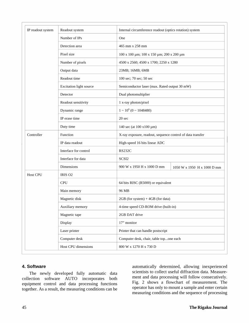
3. Hardware Specifications
4. Software
The newly developed fully automatic datacollection software AUTO incorporates bothequipment control and data processing functionstogether. As a result, the measuring conditions can be
automatically determined, allowing inexperiencedscientists to collect useful diffraction data. Measure-ment and data processing will follow consecutively.Fig. 2 shows a flowchart of measurement. Theoperator has only to mount a sample and enter certainmeasuring conditions and the sequence of processing
45 The Rigaku Journal
IP readout system Readout system Internal circumference readout (optics rotation) system
Number of IPs One
Detection area 465 mm x 258 mm
Pixel size 100 x 100 µm; 100 x 150 µm; 200 x 200 µm
Number of pixels 4500 x 2560; 4500 x 1700; 2250 x 1280
Output data 23MB; 16MB; 6MB
Readout time 100 sec; 70 sec; 50 sec
Excitation light source Semiconductor laser (max. Rated output 30 mW)
Detector Dual photomultiplier
Readout sensitivity 1 x-ray photon/pixel
Dynamic range 1 ~ 106 (0 ~ 1048480)
IP erase time 20 sec
Duty time 140 sec (at 100 x100 µm)
Controller Function X-ray exposure, readout, sequence control of data transfer
IP data readout High-speed 16 bits linear ADC
Interface for control RS232C
Interface for data SCSI2
Dimensions 900 W x 1950 H x 1000 D mm 1050 W x 1950 H x 1000 D mm
Host CPU IRIS O2
CPU 64 bits RISC (R5000) or equivalent
Main memory 96 MB
Magnetic disk 2GB (for system) + 4GB (for data)
Auxiliary memory 4-time speed CD-ROM drive (built-in)
Magnetic tape 2GB DAT drive
Display 17” monitor
Laser printer Printer that can handle postscript
Computer desk Computer desk, chair, table top...one each
Host CPU dimensions 800 W x 1270 H x 730 D

steps from Index to Scale will then be executedautomatically.
5. Measurement Example
Fig. 3 shows the measurement of cytidine(C9H13O5N3) with the R-AXIS RAPID. This exampleshows the results of the oscillation photographicmethod and the screenless Weissenberg method, using both CuKα and MoKα radiations as the X-ray source.
In the Weissenberg method, exposure is made byslowly moving the IP which is synchronized with thecrystal rotation (ω-axis oscillation) so as to preventoverlap of diffraction spots. As may be seen from Fig. 3(A) and (C), this method is a very efficient onewhich permits recording of large quantities ofdiffracted X-rays on a single IP exposure. Because the axis used for axial alignment is automatically
Vol. 15 No. 2 1998 46
AUTO FUNCTION
Crystal centering, sample name entry, etc while watching CCD camera
Determination of setting matrix from two photos ofω=0~5° and ω=90~95°, respectively
Osc/weiss: For user’s prior selection; Determination of optimalmeasurement conditions from setting matrix and latticeconstants; Calculation of completeness and redundancy.
Oscillation photo mode: Measurement of ~44 photos for an ordinary sampleWeissenberg mode: “ ~10 “
Start of measurement based on conditions automaticallydetermined by Strategy; Automatic determination of box size forintegrated reflection intensity calculation; Refinement of settingmatrix, lattice constant, etc; Integrated reflection intensity
merge:post refine:laue:scale:abscor:average:
Merge of intensity data file per frameRefinement of crystal parameters, e.g. Lattice constantsDetermination of Laue classDetermination of scale factor per frameAbsorption correctionAveraging and output of equivalent reflections
Structure analysis (Optional)
Fig. 2. Flowchart of automatic measurement

determined in the software from the result of Index the operator can be freed from complicated axialalignment work, unlike in the conventionalWeissenberg method.
CuKα radiation interacts more strongly withsamples than does MoKα radiation, and thereforedisplays higher performance in the measurement oftiny crystals. When Fig. 3(A) and (B) are compared, it
may be seen that CuKα radiation may be used tomeasure crystals having low diffracting power orlarge lattice constants (50 C or so) because it has alonger wavelength than MoKα radiation. CuKαradiation excels, furthermore, in determination ofabsolute structure by utilizing the anomalousdispersion effect. Table 1 shows the characteristics ofeach measurement mode.
47 The Rigaku Journal
MoKα CuKα
Oscillation photographicmethod
Most standard method (3~8 hrs)
Strong diffraction intensity
Crystal with large lattice parameters (~50 C)
Absolute structure determination by Anomalousdispersion effect (6~12 hrs)
Weissenberg method High-speed measurement (1~4 hrs) High-speed measurement in above conditions (4~8 hrs)
Table 1. Characteristics of each measurement mode (rough measurement time required for ordinary crystals isindicated in parentheses)
(A) Oscillation photo, MoKα, 60kV-50mAoscillation angle: 5°, exposure time: 5 min
(D) Weissenberg photo, CuKα, 60kV-50mAoscillation angle: 60°, exposure time: 5 min
(C) Weissenberg photo, MoKα, 60kV-50mAoscillation angle: 40°, exposure time: 20 min
(B) Oscillation photo, CuKα, 50kV-40mAoscillation angle: 30°, exposure time: 5 min
Fig. 3. X-ray diffraction pattern in each measurement mode (sample: cytidine C9H13O5N3)

Fig. 4 shows the structure analysis results ofcytidine (C9H13O5N3) measured by the Weissenbergmethod using MoKα radiation. The measurementtime was 60 minutes. In this case, the measurement
conditions were determined assuming that the crystalsystem is triclinic. If the Laue class is known, themeasurement time can be reduced further.
Vol. 15 No. 2 1998 48
Sample Cytidine (C9H13O5N3)
Crystal size 0.3 x 0.3 x 0.4
X-ray source MoKα 50 kV 48 mA
Measurement Weissenberg method
Exposure time 10 sec/deg (Total 60 min)
Lattice Constant13.962 14.770 5.12690.0 90.0 90.0
Crystal System orthorhombic
Laue Class mmm
Rmerge 3.2%
R 2.9%
Rw 3.0%
Fig. 4. Structure analysis result