Embed Size (px)
Citation preview
Profiling pH Gradients Across Nanocapillary ArrayMembranes Connecting Microfluidic Channels
Keqing Fa, Joseph J. Tulock, Jonathan V. Sweedler, and Paul W. Bohn*
Contribution from the Department of Chemistry and Beckman Institute for AdVanced Scienceand Technology, UniVersity of Illinois at Urbana-Champaign, 600 South Mathews AVenue,
Urbana, Illinois 61801
Received April 26, 2005; E-mail: [email protected]
Abstract: Nanocapillary array membranes (NCAMs), comprised of thin (d ∼ 5-10 µm) nuclear track-etchedpolycarbonate sheets containing ∼108 cm-2 nearly parallel nanometer-diameter capillaries, may act to gatefluid transport between microfluidic channels to effect, for example, sample collection. There is interest inH+-transport across these NCAMs because there is significant practical interest in being able to processanalyte-containing samples under different pH conditions in adjacent layers of an integrated microfluidiccircuit and because protons, with their inherently high mobility, present a challenge in separating microfluidicenvironments with different properties. To evaluate the capability of NCAMs to support pH gradients, theproton transport properties of NCAMs were studied using laser scanning confocal fluorescence microscopy(LSCFM). Spatiotemporal maps of [H+] in microfluidic channels adjacent to the NCAMs yield informationregarding diffusive and electrokinetic transport of protons. The NCAMs studied here are characterized bya positive zeta potential, ú > 0, so at small nanocapillary diameters, the overlap of electrical double layersassociated with opposite walls of the nanocapillary establish an energy barrier for either diffusion orelectrokinetic transport of cations through the nanometer-diameter capillaries due to the positive chargeon the nanocapillary surface. Proton transfer through an NCAM into microchannels is reduced for porediameters, d e 50 nm and ionic strengths I e 50 mM, while for large pore diameters or solution ionicstrengths, the incomplete overlap of electric double layer allows more facile ionic transfer across themembranes. These results establish the operating conditions for the development of multilevel integratednanofluidic/microfluidic architectures which can support multidimensional chemical analysis of mass-limitedsamples requiring sequential operations to be implemented at different pH values.
Introduction
Research exploring micro total analysis systems (µ-TAS) hasgrown exponentially over the past few years. Microfluidicallyactuatedµ-TAS techniques have been demonstrated to beapplicable to many different kinds of assays and analyticaloperations,1-5 particularly those involving the detection andcharacterization of biomolecule-containing samples. By com-bining sequentially linked operations, such as separation/detection/measurement into a single device via miniaturization,significant improvements in speed, selectivity, and detectionlimit can be realized compared to conventional bench-scaleprocedures.
Hybrid nanofluidic/microfluidic architectures for rapid samplepreparation, separation, transport, and detection are beingpursued in a number of laboratories.6-14 Capillary arraymembranes containing 10-200 nm diameter cylindrical pores
are employed as interconnects to establish controllable fluidiccommunication between micrometer-scale channels operat-ing in vertically separated planes. The microfluidic channelsare constructed either in poly(dimethylsiloxane) (PDMS) orpoly(methyl methacrylate) (PMMA), and the nanocapillaryarray membranes (NCAMs) are made of track-etched poly-carbonate (PCTE) with a poly(vinylpyrrolidone) coating. Thekey feature of nanofluidic transport is that fluid flow occursin structures with diameters,d, of the order of the Debye length,
(1) Jiang, X.; Jessamine, M. K. Ng; Stroock, A. D.; Dertinger, S. K. W.;Whitesides, G. M.J. Am. Chem. Soc.2003, 125, 5294.
(2) Peterson, D. S.Lab on a Chip2005, 5, 132.(3) Liu, J.; Pan, T.; Woolley, A. T.; Lee, M. L.Anal. Chem.2004, 76, 6948.(4) Gao, J.; Xu, J.; Locascio, L. E.; Lee, C. S.Anal. Chem.2001, 73, 2648.(5) Dai, J.; Ito, Takashi; Sun, L.; Crooks, R. M.J. Am. Chem. Soc.2003, 125,
13026.
(6) Kuo, T. C.; Cannon, D. M., Jr.; Shannon, M. A.; Bohn, P. W.; Sweedler,J. V. Sens. Actuators, A2003, 102, 223.
(7) Kuo, T. C.; Cannon, D. M., Jr.; Chen, Y. N.; Tulock, J. J.; Shannon, M.A.; Sweedler, J. V.; Bohn, P. W.Anal. Chem.2003, 75, 1861.
(8) Cannon, D. M., Jr.; Kuo, T. C.; Bohn, P. W.; Sweedler, J. V.Anal. Chem.2003, 75, 2224.
(9) Kuo, T. C.; Kim, H. K.; Cannon, D. M., Jr.; Shannon, M. A.; Sweedler, J.V.; Bohn, P. W.Angew. Chem., Int. Ed.2004, 43, 1862.
(10) Kuo, T. C.; Sloan, L. A.; Sweedler, J. V.; Bohn, P. W.Langmuir2001,17, 6298.
(11) Daiguji, H.; Yang, P. D.; Majumdar, A.Nano Lett.2004, 4, 137-142.(12) Foote, R. S.; Khandurina, J.; Jacobson, S. C.; Ramsey, J. M.Anal. Chem.
2005, 77, 57-63.(13) O’Brien, M. J.; Bisong, P.; Ista, L. K.; Rabinovich, E. M.; Garcia, A. L.;
Sibbett, S. S.; Lopez, G. P.; Brueck, S. R. J.J. Vac. Sci. Technol. B2003,21, 2941-2945.
(14) Pu, Q. S.; Yun, J. S.; Temkin, H.; Liu, S. R.Nano Lett.2004, 4, 1099-1103.
Published on Web 09/16/2005
13928 9 J. AM. CHEM. SOC. 2005 , 127, 13928-13933 10.1021/ja052708p CCC: $30.25 © 2005 American Chemical Society
κ-1. Thus, fluid motion can occur in one of two limiting regimescharacterized by the dimensionless product,κd. Whenκd . 1,the large diameter limit applies, and flow is dominated byion migration. In contrast, whenκd e 1, co-ions are repelledfrom the interior of the nanopore, and flow is dominated bycounterion motion. Sinceκ depends directly on the ionic strengthand the physical extent of the electrical double layer, themovement of fluids across the NCAM in this architecture isdriven and precisely controlled by the external electrical bias.The NCAM can, thus, act as a controllable fluidic switch,leading to applications as a microreactor and in fast chemicalmixing.9
A fully controllable gate is characterized by fluidic isolationin the “off” state and tunable fluidic transport in the “on” state.Furthermore, just understanding the volume flow rate is in-sufficient for a full description of nanofluidic flow becausedifferent ions exhibit differential transport rates in nanometer-diameter capillaries,15 leading to selective transport in electro-kinetic flow across NCAMs. For example, transient variationof the electrical current, as measured in the receiving or thesource microchannel, upon application of a forward biaspotential to an NCAM yields behavior which is, in general,much more complicated than one might be led to expect basedon a simple equivalent circuit model. It is critical to understandthe nature of differential transport of ions and molecules innanoscale confined structures because of their importance forthe development of bio-NEMS (Nano-Electro-Mechanical-Systems) andµ-TAS applications.16-26 In this regard, the surfacecharge density,σ, nanopore diameter, ionic strength of thetransporting solution,I, and the mass/charge ratio and theinherent mobility of solution constituents are key factors indetermining the observed transport behavior.
One of the goals of the NCAM architecture is controlledisolation of solutions of different composition and the abilityto process analyte-containing samples under different conditionsin adjacent layers of an integrated microfluidic circuit. The pHplays a central role in these design considerations. For example,frequently, a separation of a complex mixture is best carriedout under acidic conditions, but the fluorescent detection of thelabeled analytes is optimized under basic conditions. As anotherexample, the common electrolytes used in electrophoreticseparations often contain additives that decrease the performanceof electrospray mass spectrometry by orders of magnitude. Theability to optimize an electrophoretic separation in a microfluidicdevice and then transfer mass-limited separated analyte bandsfrom one fluidic environment into another before introducingthem to the detection system, whether fluorescence or massspectrometry, greatly increases overall system performance and
allows each “module” of the multidimensional determinationto be optimized separately.
A critical question involves how well NCAMs effect ionicisolation. We probe the H+-transport across NCAMs becauseprotons, with their inherently high mobility in aqueous media,present the most extensive challenge to preparing microfluidicenvironments with different properties. A further advantage isthat pH gradients across NCAMs can be precisely monitoredusing fluorescent probes, such as carboxyseminaphthorhoda-fluor, 1, in combination with laser scanning confocal microscopy(LSCM). The dye1 absorbs light at the absorption peak of 488nm and exhibits a shift in fluorescence emission from 588 nmin the acidic form to 640 nm in the basic form.27 Thecharacterization of pH gradients and ionic transport, by spa-tiotemporal profiling of [H+] with and without external bias,provides information critical to the performance of NCAMs inhybrid nanofluidic/microfluidic structures for multidimensional,multilevel chemical characterization of complex samples.
Experimental Section
Materials. Nanoporous polycarbonate track-etched membranes werepurchased from Osmonics (Minnetonka, MN) and stored under dry N2
prior to use. These membranes have highly monodisperse distributionsof pore diameters, with pore densities ranging from 3× 10⁸ to 6 ×108 cm-2. Table 1 shows selected properties of these membranes.
Dye 1 (Molecular Probes, Eugene, OR) was used as the fluorescentpH indicator. All solutions of1 were prepared to a concentration of16.96µM in 18 MΩ cm deionized (DI) water. A series of stock 50mM phosphate (Sigma-Aldrich, Milwaukee, WI) buffer solutions (PBS)from pH 4.5 to 9.5 were prepared in DI water.
Fabrication of the µ-TAS Devices.Standard rapid prototypingprotocols and soft lithography processes were used to fabricate PDMSlayers with embedded microchannel geometries and reservoirs.28,29First,a negative design mask was printed on a transparency (5080 dpi,Printing Services, University of Illinois at Urbana-Champaign). Thistransparency served as the photomask in photolithography to producea positive relief feature on silicon (Silicon Quest International, SantaClara, CA) using SU 8-2050 photoresist (Microlithography ChemicalCorp., Newton, MA). Prepolymer Sylgard 184 and curing agent (DowCorning Corp., Midland, MI) were thoroughly mixed in a 10:1 w:w
(15) Chaterjee, A.; Cannon, D. M., Jr.; Gatimu, E. N.; Aluru, N.; Sweedler, J.V.; Bohn, P. W.J. Nanopart. Res., in press.
(16) Loughnane, B. J.; Farrer, R. A.; Scodinu, A.; Reilly, T.; Fourkas, J. T.J.Phys. Chem. B2000, 104, 5421.
(17) Cheng, J. T.; Giordano, N.Phys. ReV. E 2002, 65, 031206.(18) Qiao, R.; Aluru, N. R.J. Chem. Phys.2003, 118, 4692.(19) Pu, Q. S.; Yun, J. S.; Temkin, H.; Liu, S. R.Nano Lett.2004, 4, 1099.(20) Daijuji, H.; Yang, P. D.; Majumdar, A.Nano Lett.2004, 4, 137.(21) Travis, K. P.; Gubbins, K. E.J. Chem. Phys.2000, 112, 1984.(22) Thompson, A. P.J. Chem. Phys.2003, 119, 7503.(23) Karlsson, R.; Karlsson, M.; Karlsson, A.; Cans, A. S.; Bergenholtz, J.;
Akerman, B.; Ewing, M.; Voinova, A. G.; Orwar, O.Langmuir2002, 18,4186.
(24) Stein, D.; Kruithof, M.; Dekker, C.Phys. ReV. Lett. 2004, 96, 035901.(25) Han, J.; Turner, S. W.; Craighead, H. G.Phys. ReV. Lett.1999, 83, 1688.(26) Kemery, P. J.; Steehler, J. K.; Bohn, P. W.Langmuir1998, 14, 2884.
(27) Molecular Probes Handbook: Eugene, OR.(28) McDonald, J. C.; Duffy, D. C.; Anderson, J. R.; Chiu, D. T.; Wu, H. K.;
Schueller, O. J. A.; Whitesides, G. M.Electrophoresis2000, 21, 27.(29) Duffy, D. C.; McDonald, J. C.; Schueller, O. J. A.; Whitesides, G. M.
Anal. Chem.1998, 70, 4974.
Table 1. Membrane Properties
pore size(nm)
pore density(cm-2)
average spacingbetween pores
(µm)number of pores fora 50 × 50 µm2 area
10 6× 10⁸ 0.41 1.5× 10⁴
50 6× 10⁸ 0.41 1.5× 10⁴
200 3× 10⁸ 0.58 7.5× 10³
pH Gradients Across Microfluidic Channels A R T I C L E S
J. AM. CHEM. SOC. 9 VOL. 127, NO. 40, 2005 13929
ratio. The mixture was poured onto the fully developed silicon masterand cured for∼18 h at room temperature. A partial curing techniquewas developed to achieve optimum sealing of the PDMS around thePCTE membrane. When the polymer mixture had reached the propercuring time, but prior to being completely cured, the sticky PDMS layerswere peeled from the silicon master, while maintaining the shape ofthe microchannels, and the PDMS microchannel/PCTE nanochannel/PDMS microchannel structures were assembled. Care was taken whiletrimming the PCTE membranes and assembling the sandwich structuresto prevent any damage to the membranes. The hybrid nanofluidic/microfluidic device was designed with a junction area of 50× 50 µm2
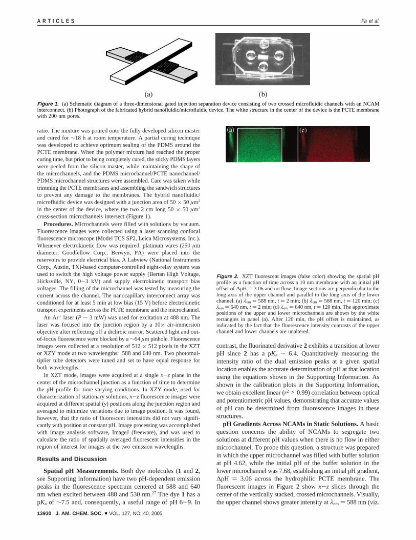
in the center of the device, where the two 2 cm long 50× 50 µm2
cross-section microchannels intersect (Figure 1).Procedures.Microchannels were filled with solutions by vacuum.
Fluorescence images were collected using a laser scanning confocalfluorescence microscope (Model TCS SP2, Leica Microsystems, Inc.).Whenever electrokinetic flow was required, platinum wires (250µmdiameter, Goodfellow Corp., Berwyn, PA) were placed into thereservoirs to provide electrical bias. A Labview (National InstrumentsCorp., Austin, TX)-based computer-controlled eight-relay system wasused to switch the high voltage power supply (Bertan High Voltage,Hicksville, NY, 0-3 kV) and supply electrokinetic transport biasvoltages. The filling of the microchannel was tested by measuring thecurrent across the channel. The nanocapillary interconnect array wasconditioned for at least 5 min at low bias (15 V) before electrokinetictransport experiments across the PCTE membrane and the microchannel.
An Ar+ laser (P ∼ 3 mW) was used for excitation at 488 nm. Thelaser was focused into the junction region by a 10× air-immersionobjective after reflecting off a dichroic mirror. Scattered light and out-of-focus fluorescence were blocked by a∼64µm pinhole. Fluorescenceimages were collected at a resolution of 512× 512 pixels in the XZTor XZY mode at two wavelengths: 588 and 640 nm. Two photomul-tiplier tube detectors were tuned and set to have equal response forboth wavelengths.
In XZT mode, images were acquired at a singlex-z plane in thecenter of the microchannel junction as a function of time to determinethe pH profile for time-varying conditions. In XZY mode, used forcharacterization of stationary solutions,x-z fluorescence images wereacquired at different spatial (y) positions along the junction region andaveraged to minimize variations due to image position. It was found,however, that the ratio of fluorescent intensities did not vary signifi-cantly with position at constant pH. Image processing was accomplishedwith image analysis software, ImageJ (freeware), and was used tocalculate the ratio of spatially averaged fluorescent intensities in theregion of interest for images at the two emission wavelengths.
Results and Discussion
Spatial pH Measurements.Both dye molecules (1 and 2,see Supporting Information) have two pH-dependent emissionpeaks in the fluorescence spectrum centered at 588 and 640nm when excited between 488 and 530 nm.27 The dye1 has apKa of ∼7.5 and, consequently, a useful range of pH 6-9. In
contrast, the fluorinated derivative2 exhibits a transition at lowerpH since 2 has a pKa ∼ 6.4. Quantitatively measuring theintensity ratio of the dual emission peaks at a given spatiallocation enables the accurate determination of pH at that locationusing the equations shown in the Supporting Information. Asshown in the calibration plots in the Supporting Information,we obtain excellent linear (r2 > 0.99) correlation between opticaland potentiometric pH values, demonstrating that accurate valuesof pH can be determined from fluorescence images in thesestructures.
pH Gradients Across NCAMs in Static Solutions.A basicquestion concerns the ability of NCAMs to segregate twosolutions at different pH values when there is no flow in eithermicrochannel. To probe this question, a structure was preparedin which the upper microchannel was filled with buffer solutionat pH 4.62, while the initial pH of the buffer solution in thelower microchannel was 7.68, establishing an initial pH gradient,∆pH ) 3.06 across the hydrophilic PCTE membrane. Thefluorescent images in Figure 2 showx-z slices through thecenter of the vertically stacked, crossed microchannels. Visually,the upper channel shows greater intensity atλem ) 588 nm (viz.
Figure 1. (a) Schematic diagram of a three-dimensional gated injection separation device consisting of two crossed microfluidic channels with an NCAMinterconnect. (b) Photograph of the fabricated hybrid nanofluidic/microfluidic device. The white structure in the center of the device is the PCTE membranewith 200 nm pores.
Figure 2. XZT fluorescent images (false color) showing the spatial pHprofile as a function of time across a 10 nm membrane with an initial pHoffset of∆pH ) 3.06 and no flow. Image sections are perpendicular to thelong axis of the upper channel and parallel to the long axis of the lowerchannel. (a)λem ) 588 nm,t ) 2 min; (b)λem ) 588 nm,t ) 120 min; (c)λem ) 640 nm,t ) 2 min; (d)λem ) 640 nm,t ) 120 min. The approximatepositions of the upper and lower microchannels are shown by the whiterectangles in panel (a). After 120 min, the pH offset is maintained, asindicated by the fact that the fluorescence intensity contrasts of the upperchannel and lower channels are unaltered.
A R T I C L E S Fa et al.
13930 J. AM. CHEM. SOC. 9 VOL. 127, NO. 40, 2005
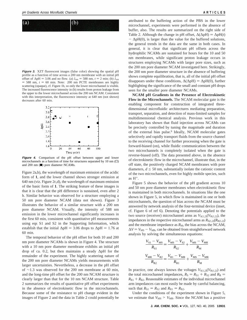
Figure 2a,b), the wavelength of maximum emission of the acidicform of 1, and the lower channel shows stronger emission at640 nm (viz. Figure 2c,d), the wavelength of maximum emissionof the basic form of1. The striking feature of these images isthat it is clear that the pH difference is sustained, even after 2h. Similar behavior was observed for a structure employing a50 nm pore diameter NCAM (data not shown). Figure 3illustrates the behavior of a similar structure with a 200 nmpore diameter NCAM. Visually, the intensity of 588 nmemission in the lower microchannel significantly increases inthe first 60 min, consistent with quantitative pH measurementsusing eqs S1 and S2 in the Supporting Information, whichestablish that the initial∆pH ) 3.06 drops to∆pH ) 1.76 at60 min.
The temporal behavior of the pH offset for both 10 and 200nm pore diameter NCAMs is shown in Figure 4. The structurewith a 10 nm pore diameter membrane exhibits an initial pHdrop of ca. 0.2, but then maintains a steady∆pH for theremainder of the experiment. The highly scattering nature ofthe 200 nm pore diameter NCAMs yields measurements withlarger uncertainties. Nevertheless, a decrease in the pH offsetof ∼1.3 was observed for the 200 nm membrane at 60 min,and the long-time pH offset for the 200 nm NCAM structure isclearly larger than that for the 10 nm NCAM structure. Table2 summarizes the results of quantitative pH offset experimentsin the absence of electrokinetic flow in the microchannels.Because some of the resistance to pH change evident in theimages of Figure 2 and the data in Table 2 could potentially be
attributed to the buffering action of the PBS in the lowermicrochannel, experiments were performed in the absence ofbuffer, also. The results are summarized on the right side ofTable 2. Although the change in pH offset,∆(∆pH) ) ∆pH(t)- ∆pH(0), is larger than the value for the buffered solutions,the general trends in the data are the same in both cases. Ingeneral, it is clear that significant pH offsets across thehydrophilic NCAMs are sustained for hours for the 10 and 50nm membranes, while significant proton leakage occurs instructures employing NCAMs with larger pore sizes, such asthe 200 nm pore diameter NCAM investigated here. Strikingly,the 200 nm pore diameter structure in the absence of bufferingshows complete equilibration, that is, all of the initial pH offsetdisappears under these conditions,∆(∆pH) ) ∆pH(0), furtherhighlighting the significance of the small and constant pH dropsseen for the smaller pore diameter NCAMs.
NCAM pH Gradients in the Presence of ElectrokineticFlow in the Microchannels.The NCAM molecular gate is theenabling component for construction of integrated three-dimensional microfluidic architectures mediating preparation,transport, separation, and detection of mass-limited samples formultidimensional chemical analysis. Previous work in thislaboratory has shown that fluid injection across NCAMs canbe precisely controlled by tuning the magnitude and durationof the external bias pulse.6 Ideally, NCAM molecular gatesselectively and rapidly transport fluids from the source channelto the receiving channel for further processing when the gate isforward-biased (on), while fluidic communication between thetwo microchannels is completely isolated when the gate isreverse-biased (off). The data presented above, in the absenceof electrokinetic flow in the microchannel, illustrate that, in theoff state, the positively charged NCAM membranes with porediameters,d e 50 nm, substantially isolate the cationic contentof the two microchannels, even for highly mobile species, suchas H+.
Figure 5 shows the behavior of the pH gradient across 10and 50 nm pore diameter membranes when electrokinetic flowis maintained in both microchannels. In situations like the oneshown in Figure 5, in which flow is maintained in one or bothmicrochannels, the question of bias across the NCAM must beanswered by network analysis of the four-terminal device (inset,cf. Figure 6 of ref 6). Denoting the potentials applied to thetwo source (receiver) microchannel arms asVS(1,2)(VR(1,2)), theimpedances in the respective microchannel arms asRS(1,2)(RR(1,2))and the membrane impedance asRM, the bias across the NCAM,∆V ) VMS - VMR, can be obtained from straightforward networkanalysis by solving the simultaneous equations:
In practice, one always knows the voltagesVS(1,2)(VR(1,2)) andthe total microchannel impedances,RS ) RS1 + RS2 andRR )RR1 + RR2. Reasonable estimates of the individual microchannelarm impedances can most easily be made by careful balancing,such thatRS1 ) RS2 andRR1 ) RR2.
Under the conditions of the experiment shown in Figure 5,we estimate thatVMS > VMR. Since the NCAM has a positive
Figure 3. XZT fluorescent images (false color) showing the spatial pHprofile as a function of time across a 200 nm membrane with an initial pHoffset of ∆pH ) 3.06 and no flow. (a)λem ) 588 nm,t ) 2 min; (b) λem
) 588 nm, t ) 60 min. Note: 200 nm PCTE membranes are highlyscattering (opaque), cf. Figure 1b, so only the lower microchannel is visible.The increased fluorescence intensity in (b) results from proton leakage fromthe upper to the lower microchannel across the 200 nm NCAM. Consistentwith this interpretation, the fluorescence intensity at 640 nm (not shown)decreases after 60 min.
Figure 4. Comparison of the pH offset between upper and lowermicrochannels as a function of time for structures separated by 10 nm (0)and 200 nm (9) pore diameter NCAMs.
VS1 - VMS
RS1)
VMS - VS2
RS2+
VMS - VMR
RM
VMR - VR2
RR2)
VR1 - VMR
RR1+
VMS - VMR
RM
pH Gradients Across Microfluidic Channels A R T I C L E S
J. AM. CHEM. SOC. 9 VOL. 127, NO. 40, 2005 13931
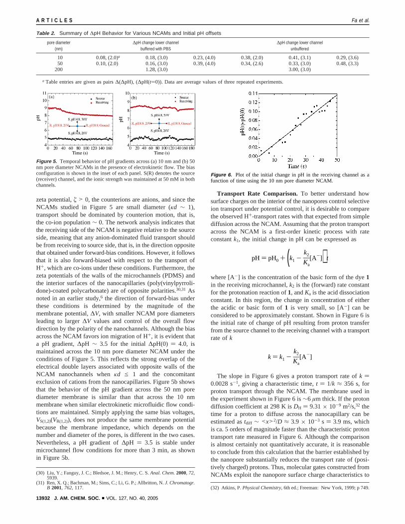
zeta potential,ú > 0, the counterions are anions, and since theNCAMs studied in Figure 5 are small diameter (κd ∼ 1),transport should be dominated by counterion motion, that is,the co-ion population∼ 0. The network analysis indicates thatthe receiving side of the NCAM is negative relative to the sourceside, meaning that any anion-dominated fluid transport shouldbe from receiving to source side, that is, in the direction oppositethat obtained under forward-bias conditions. However, it followsthat it is also forward-biased with respect to the transport ofH+, which are co-ions under these conditions. Furthermore, thezeta potentials of the walls of the microchannels (PDMS) andthe interior surfaces of the nanocapillaries (poly(vinylpyrroli-done)-coated polycarbonate) are of opposite polarities.30,31 Asnoted in an earlier study,6 the direction of forward-bias underthese conditions is determined by the magnitude of themembrane potential,∆V, with smaller NCAM pore diametersleading to larger∆V values and control of the overall flowdirection by the polarity of the nanochannels. Although the biasacross the NCAM favors ion migration of H+, it is evident thata pH gradient,∆pH ∼ 3.5 for the initial ∆pH(0) ) 4.0, ismaintained across the 10 nm pore diameter NCAM under theconditions of Figure 5. This reflects the strong overlap of theelectrical double layers associated with opposite walls of theNCAM nanochannels whenκd e 1 and the concomitantexclusion of cations from the nanocapillaries. Figure 5b showsthat the behavior of the pH gradient across the 50 nm porediameter membrane is similar than that across the 10 nmmembrane when similar electrokinetic microfluidic flow condi-tions are maintained. Simply applying the same bias voltages,VS(1,2)(VR(1,2)), does not produce the same membrane potentialbecause the membrane impedance, which depends on thenumber and diameter of the pores, is different in the two cases.Nevertheless, a pH gradient of∆pH ) 3.5 is stable undermicrochannel flow conditions for more than 3 min, as shownin Figure 5b.
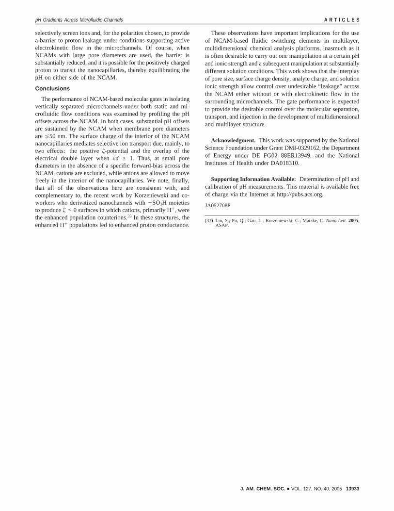
Transport Rate Comparison. To better understand howsurface charges on the interior of the nanopores control selectiveion transport under potential control, it is desirable to comparethe observed H+-transport rates with that expected from simplediffusion across the NCAM. Assuming that the proton transportacross the NCAM is a first-order kinetic process with rateconstantk1, the initial change in pH can be expressed as
where [A-] is the concentration of the basic form of the dye1in the receiving microchannel,k2 is the (forward) rate constantfor the protonation reaction of1, andKa is the acid dissociationconstant. In this region, the change in concentration of eitherthe acidic or basic form of1 is very small, so [A-] can beconsidered to be approximately constant. Shown in Figure 6 isthe initial rate of change of pH resulting from proton transferfrom the source channel to the receiving channel with a transportrate ofk
The slope in Figure 6 gives a proton transport rate ofk )0.0028 s-1, giving a characteristic time,t ) 1/k ≈ 356 s, forproton transport through the NCAM. The membrane used inthe experiment shown in Figure 6 is∼6 µm thick. If the protondiffusion coefficient at 298 K isDH ) 9.31× 10⁻⁹ m2/s,32 thetime for a proton to diffuse across the nanocapillary can beestimated astdiff ∼ <x>2/D ≈ 3.9 × 10⁻³ s ) 3.9 ms, whichis ca. 5 orders of magnitude faster than the characteristic protontransport rate measured in Figure 6. Although the comparisonis almost certainly not quantitatively accurate, it is reasonableto conclude from this calculation that the barrier established bythe nanopore substantially reduces the transport rate of (posi-tively charged) protons. Thus, molecular gates constructed fromNCAMs exploit the nanopore surface charge characteristics to(30) Liu, Y.; Fanguy, J. C.; Bledsoe, J. M.; Henry, C. S.Anal. Chem.2000, 72,
5939.(31) Ren, X. Q.; Bachman, M.; Sims, C.; Li, G. P.; Allbritton, N.J. Chromatogr.
B 2001, 762, 117. (32) Atkins, P.Physical Chemistry, 6th ed.; Freeman: New York, 1999; p 749.
Table 2. Summary of ∆pH Behavior for Various NCAMs and Initial pH offsets
pore diameter(nm)
∆pH change lower channelbuffered with PBS
∆pH change lower channelunbuffered
10 0.08, (2.0)a 0.18, (3.0) 0.23, (4.0) 0.38, (2.0) 0.41, (3.1) 0.29, (3.6)50 0.10, (2.0) 0.16, (3.0) 0.39, (4.0) 0.34, (2.6) 0.33, (3.0) 0.48, (3.3)
200 1.28, (3.0) 3.00, (3.0)
a Table entries are given as pairs∆(∆pH), (∆pH(t)0)). Data are average values of three repeated experiments.
Figure 5. Temporal behavior of pH gradients across (a) 10 nm and (b) 50nm pore diameter NCAMs in the presence of electrokinetic flow. The biasconfiguration is shown in the inset of each panel. S(R) denotes the source(receiver) channel, and the ionic strength was maintained at 50 mM in bothchannels.
Figure 6. Plot of the initial change in pH in the receiving channel as afunction of time using the 10 nm pore diameter NCAM.
pH ) pH0 + (k1 -k2
Ka[A-]) t
k ) k1 -k2
Ka[A-]
A R T I C L E S Fa et al.
13932 J. AM. CHEM. SOC. 9 VOL. 127, NO. 40, 2005
selectively screen ions and, for the polarities chosen, to providea barrier to proton leakage under conditions supporting activeelectrokinetic flow in the microchannels. Of course, whenNCAMs with large pore diameters are used, the barrier issubstantially reduced, and it is possible for the positively chargedproton to transit the nanocapillaries, thereby equilibrating thepH on either side of the NCAM.
Conclusions
The performance of NCAM-based molecular gates in isolatingvertically separated microchannels under both static and mi-crofluidic flow conditions was examined by profiling the pHoffsets across the NCAM. In both cases, substantial pH offsetsare sustained by the NCAM when membrane pore diametersaree50 nm. The surface charge of the interior of the NCAMnanocapillaries mediates selective ion transport due, mainly, totwo effects: the positiveú-potential and the overlap of theelectrical double layer whenκd e 1. Thus, at small porediameters in the absence of a specific forward-bias across theNCAM, cations are excluded, while anions are allowed to movefreely in the interior of the nanocapillaries. We note, finally,that all of the observations here are consistent with, andcomplementary to, the recent work by Korzeniewski and co-workers who derivatized nanochannels with-SO3H moietiesto produceú < 0 surfaces in which cations, primarily H+, werethe enhanced population counterions.33 In these structures, theenhanced H+ populations led to enhanced proton conductance.
These observations have important implications for the useof NCAM-based fluidic switching elements in multilayer,multidimensional chemical analysis platforms, inasmuch as itis often desirable to carry out one manipulation at a certain pHand ionic strength and a subsequent manipulation at substantiallydifferent solution conditions. This work shows that the interplayof pore size, surface charge density, analyte charge, and solutionionic strength allow control over undesirable “leakage” acrossthe NCAM either without or with electrokinetic flow in thesurrounding microchannels. The gate performance is expectedto provide the desirable control over the molecular separation,transport, and injection in the development of multidimensionaland multilayer structure.
Acknowledgment. This work was supported by the NationalScience Foundation under Grant DMI-0329162, the Departmentof Energy under DE FG02 88ER13949, and the NationalInstitutes of Health under DA018310.
Supporting Information Available: Determination of pH andcalibration of pH measurements. This material is available freeof charge via the Internet at http://pubs.acs.org.
JA052708P
(33) Liu, S.; Pu, Q.; Gao, L.; Korzeniewski, C.; Matzke, C.Nano Lett.2005,ASAP.
pH Gradients Across Microfluidic Channels A R T I C L E S
J. AM. CHEM. SOC. 9 VOL. 127, NO. 40, 2005 13933