Embed Size (px)
Citation preview

Formaldehyde Induces Micronuclei in Mouse ErythropoieticCells and Suppresses the Expansion of Human ErythroidProgenitor Cells
Zhiying Ji*, Xiyi Li*,1, Michele Fromowitz, Elizabeth Mutter-Rottmayer, Judy Tung, Martyn T.Smith, and Luoping Zhang**
Division of Environmental Health Sciences, School of Public Health, University of California,Berkeley, CA 94720
AbstractAlthough formaldehyde (FA) has been classified as a human leukemogen, the mechanisms ofleukemogenesis remain elusive. Previously, using colony-forming assays in semi-solid media, weshowed that FA exposure in vivo and in vitro was toxic to human hematopoietic stem/progenitorcells. In the present study, we have applied new liquid in vitro erythroid expansion systems tofurther investigate the toxic effects of FA (0–150 µM) on cultured mouse and humanhematopoietic stem/progenitor cells. We determined micronucleus (MN) levels in polychromaticerythrocytes (PCEs) differentiated from mouse bone marrow. We measured cell growth, cell cycledistribution, and chromosomal instability, in erythroid progenitor cells (EPCs) expanded fromhuman peripheral blood mononuclear cells. FA significantly induced MN in mouse PCEs andsuppressed human EPC expansion in a dose-dependent manner, compared with untreated controls.In the expanded human EPCs, FA slightly increased the proportion of cells in G2/M at 100 µMand aneuploidy frequency in chromosomes 7 and 8 at 50 µM. Our findings provide furtherevidence of the toxicity of FA to hematopoietic stem/progenitor cells and support the biologicalplausibility of FA-induced leukemogenesis.
Keywordsformaldehyde; erythroid progenitor; micronuclei; aneuploidy
1. IntroductionFormaldehyde (FA), the simplest aldehyde, is a high production volume industrial chemicaland a ubiquitous environmental pollutant (NTP, 2010a). FA is highly reactive and causesmultiple toxic effects in humans. The International Agency for Research on Cancer (IARC)classified FA as a human carcinogen that causes nasopharyngeal cancer in 2006 (IARC,2006) and human leukemia, particularly myeloid leukemia, in 2009 (Baan et al., 2009).
© 2013 Elsevier Ireland Ltd. All rights reserved.**Corresponding author: Luoping Zhang, PhD, Division of Environmental Health Sciences, School of Public Health, University ofCalifornia at Berkeley, 388 Li Ka-Shing Center, Berkeley, CA 94720, USA, Telephone: +1 (510) 643-5189, Fax: +1 (510) 642-0427,[email protected].*These authors contributed equally to this research.1Current Address: School of Public Health, Guangxi Medical University, Nanning, Guangxi 530021, China
Publisher's Disclaimer: This is a PDF file of an unedited manuscript that has been accepted for publication. As a service to ourcustomers we are providing this early version of the manuscript. The manuscript will undergo copyediting, typesetting, and review ofthe resulting proof before it is published in its final citable form. Please note that during the production process errors may bediscovered which could affect the content, and all legal disclaimers that apply to the journal pertain.
NIH Public AccessAuthor ManuscriptToxicol Lett. Author manuscript; available in PMC 2015 January 13.
Published in final edited form as:Toxicol Lett. 2014 January 13; 224(2): 233–239. doi:10.1016/j.toxlet.2013.10.028.
NIH
-PA Author Manuscript
NIH
-PA Author Manuscript
NIH
-PA Author Manuscript

Recently, the U.S. National Toxicology Program also classified FA as human leukemogen(NTP, 2011).
Though the mechanisms underlying FA-induced leukemogenesis remain elusive, FAexposure has been associated with adverse effects on hematopoiesis. Our review of theChinese literature reported that decreased white blood cell counts were observed in moststudies of FA-exposed workers; in the largest study, exposed workers had higherpercentages of blood abnormalities (decreased white blood cell and platelet counts andabnormal hemoglobin levels) (Tang et al., 2009). We also reported that Chinese factoryworkers exposed to high levels of FA had significantly lower counts of granulocytes,platelets, red blood cells, and lymphocytes, compared with unexposed controls (Zhang et al.,2010). The suppression of multiple lineages indicates a toxic effect of FA exposure onhematopoietic stem / progenitor cells (HSC/HPC), the target cells of leukemogenesis.
Colony-forming assays in semi-solid substrate enable the direct examination of the adverseeffects of FA on HSC/HPC circulating in peripheral blood. Using these assays, wepreviously reported that FA exposure in vivo was associated with decreased formation ofcolonies from colony forming units – granulocyte and monocyte (CFU-GM) cells and theinduction of leukemia-related aneuploidies monosomy 7 and trisomy 8 in CFU-GM in asubset of the subjects (Zhang et al., 2010). Further, we showed that FA exposure attoxicologically relevant concentrations in vitro decreased formation of CFU-GM, burstforming units – erythrocyte (BFU-E) and the more primitive colony forming units –granulocyte, erythrocyte, monocyte, and megakaryocyte (CFU-GEMM) colonies, the latterin a linearly dose-dependent manner (Zhang et al., 2010). These data supported theinhibitory effect of FA on myeloid progenitor cells indicated by the blood count data.However, limited mechanistic studies could be conducted as the colonies were formed insemi-solid medium.
Recently, in vitro methodologies were developed that utilize cytokines to drivedifferentiation or expansion and yield large numbers of mouse and human erythroidprogenitor cells, facilitating the analysis of multiple endpoints. An in vitro liquid culturemethod that recapitulates erythropoietic differentiation from mouse bone marrowprogenitors, producing polychromatic erythrocytes (PCEs) after 2–3 days in culture, wasestablished in 2007 (Shuga et al., 2007). This method forms the basis of an in vitromicronucleus (MN) genotoxicity assay that was found to generate similar results as thewidely used in vivo MN genotoxicity assay, thus generating physiologically relevant data(Shuga et al., 2007). Recently, we validated this assay in a study in which we foundincreased MN frequency in PCEs cultured from mouse bone marrow exposed to 2,5-dimethylfuran (Fromowitz et al., 2012).
A liquid culture approach to expand human erythroid progenitor cells (EPCs) fromunfractionated peripheral blood was recently described (Filippone et al., 2010). The authorsconfirmed the functional competence of the expanded EPCs by showing their permissivityto B19 parvovirus infection. We recognized in this model a unique opportunity to testhuman stem/progenitor cell toxicity of known and suspected leukemogens. To ourknowledge, we are the first researchers to use this new erythroid expansion model for thispurpose.
In the present study, we employed both of these in vitro liquid culture systems to test theeffects of FA on mouse PCEs and human EPCs. We measured MN frequency in FA-treatedand untreated mouse PCEs and the expansion of FA-treated and untreated human EPCs. Wealso examined the effects of FA on cell proliferation and chromosomal instability in theexpanded human EPCs.
Ji et al. Page 2
Toxicol Lett. Author manuscript; available in PMC 2015 January 13.
NIH
-PA Author Manuscript
NIH
-PA Author Manuscript
NIH
-PA Author Manuscript

2. Methods2.1. Mouse erythropoietic culture
The experimental procedures in mice were approved by the Committee on Animal Researchat the University of California, Berkeley. The mouse erythropoietic culture method wasdetailed previously (Fromowitz et al., 2012; Shuga et al., 2007). In brief, bone marrow (BM)cells were isolated from the hind legs of C57BL/6J mice and were labeled with biotin-conjugated α-Lin Abs, consisting of α-CD3e, α-CD11b, α- CD45R/B220, α-Ly6G/Ly6C,and α-TER-119 Abs (2 µl of each Ab/106 cells; BD Pharmingen, San Diego, CA). Lineage-marker-negative (Lin−) cells were purified through a 0.3-in StemSep negative selectioncolumn as per the manufacturer’s instructions (StemCell Technologies, Vancouver, BC,Canada). Purified cells were immediately seeded in fibronectin-coated (2 µg/cm2) tissueculture treated 24-well polystyrene plates (BD Falcon, BD Biosciences San Jose, CA) at acell density of 105 cells/ml in modified IMDM with L-glutamine (500 µL per culture well)containing basal supplements consisting of: 15% FBS, 1% detoxified BSA, 200 µg/mlholotransferrin, 10 µg/ml recombinant human insulin (Sigma, St Louis, MO), 100 µM β-mercaptoethanol, 50 units/ml penicillin G, and 50 µg/ml streptomycin (Invitrogen, Carlsbad,CA); as well as soluble erythropoietic factors including erythropoietin (EPO, Amgen,Thousand Oaks, CA) at 7.5 units/ml and stem cell factor (SCF, R&D Systems, Minneapolis,MN) at 10 ng/ml. After 24 h of culture, the media was replaced with erythroid-differentiation medium (EDM) (IMDM, with 20% FBS, and 100 µM β-mercaptoethanol).
2.2. Human erythropoietic cultureApproval for human subject protocols and blood sample collection was obtained from theInstitutional Review Board at UC Berkeley. The number of blood donors for eachexperimental endpoint ranged from 3–5 and the number of independent experiments rangedfrom 3–7, as described in the results section and figure legends. The method of EPCexpansion from human peripheral blood was detailed previously (Filippone et al., 2010). Inbrief, peripheral venous blood was collected from healthy donors and peripheral bloodmononuclear cells (PBMC) were isolated by a density gradient centrifugation using Ficoll-Paque. PBMC (5×10⁵) were cultured in MEM (HyClone, Logan, UT, USA) supplementedwith the serum substitute BIT 9500 (StemCell Technology, Vancouver, BC, Canada),diluted 1:5 for a final concentration of 10 mg/ml bovine serum albumin, 10 mg/ml rhuinsulin, and 200 mg/ml iron-saturated human transferrin, enriched with 900 ng/ml ferroussulfate (Sigma, St. Louis, MO, USA), 90 ng/ml ferric nitrate (Sigma), 1 mM hydrocortisone(Sigma), 3 IU/ml rhu erythropoietin (StemCell Technology), 5 ng/ml rhu IL-3 (R&DSystems, Minneapolis, MN, USA), and 100 ng/ml rhu stem cell factor (R&D Systems). Thecells were maintained at 37°C in a 5% CO2 moist atmosphere and observed daily with aninverted microscope for phenotypic changes. Upon observation of small cell clusters on day5±1, the cultures were split (1:5) with fresh media.
2.3. FA treatment of mouse and human erythroid cultures2.3.1. Vehicle controls—FA was diluted from a 37% solution (Sigma, stabilized with10–15% methanol) immediately before treatment. Phosphate-buffered saline (PBS) wasused as the vehicle control in both mouse and human cultures but 0.1% methanol was addedinto the PBS only in human cells, resulting in a final methanol concentration of 0.001% inall cultures.
2.3.2. FA concentrations—For mouse erythroid cultures, FA was added to finalconcentrations of 0, 25, 50, 75 and 100 µM, 23 h after seeding in the first medium. Cultureswere incubated for 1 h, after which the media was replaced with EDM according to theprotocol described in section 2.1. The cells were harvested after 24 h of culture in EDM.
Ji et al. Page 3
Toxicol Lett. Author manuscript; available in PMC 2015 January 13.
NIH
-PA Author Manuscript
NIH
-PA Author Manuscript
NIH
-PA Author Manuscript

For human erythroid cultures, FA was added to final concentrations of 0, 25, 50, 100, and150 µM, immediately after seeding the PBMC into the EPC expansion culture media and themedia was changed on ~day 5, as described in section 2.2. As FA is reactive and interactswith cellular components rapidly (within ~1 hour), we did not change the media after FAtreatment. This concentration range was chosen as it spans the dose of ~80 µM reported inhuman blood (Heck et al., 1985) and it has been used in many in vitro studies, including incultured human blood cells (Neuss and Speit, 2008; Schmid and Speit, 2007). All endpointswere analyzed after 10 days of culture.
2.4. MN assay in mouse PCEsHarvested cells were centrifuged onto slides (Statspin Cytofuge 2; Iris Sample ProcessingWestwood, MA), air-dried, and fixed with 25°C methanol for 10 min. The slides were thenstained in acridine orange (Sigma, St Louis, USA) at a concentration of 20 µg/ml in stainingbuffer (19 mM NaH2PO4 and 81 mM Na2HPO4) for 10 min at 4°C. Slides were scored forthe presence of MN using an Axioplan 2 microscope (Carl Zeiss MicroImaging GmbH,Germany). Scorers were blinded to the treatment status of the cells on the slides. Threeindependent experiments were conducted and more than 2000 PCEs were scored for eachdose in each experiment.
2.5. Analyses of human EPCs2.5.1. Cell enumeration—Cells were enumerated using a hemocytometer and cellviability was determined by the trypan blue exclusion assay.
2.5.2. Erythroid marker expression—The expression levels of three erythroid surfacemarkers, CD235a, CD36, and CD71, were analyzed in PBMC before culture and in theexpanded cells after 10 days of culture. The cells were washed three times with 1× PBScontaining 2% FCS and stained with FITC-labeled monoclonal antibody for CD235a,phycoerythrin (PE)-labelled monoclonal antibody for CD36, and APC-labelled monoclonalantibody for CD71 (BD Biosciences, San Jose, CA, USA) at 4°C for 30 min. Anti-isotypeantibodies (BD Biosciences) were used in parallel. After staining, cells were washed threetimes with in 1× PBS and analyzed by flow cytometry (Beckman-Coulter FC-500, BectonDickinson, Franklin Lakes, NJ, USA).
2.5.3. Cell cycle analysis—Cell cycle was analyzed by propidium iodide (PI) staining.Briefly, 1×10⁶ cells were washed with cold 1× PBS and fixed in 70% cold ethanol at −20°Cfor 2 hours. After a gentle wash with cold 1× PBS, cells were resuspended in 1 ml PIstaining solution (10 µg/ml) and 50 µl RNase A (10 mg/ml) was added. After incubation at4°C in the dark for 3 h, cells were analyzed by flow cytometry at 488 nm. Cell cycleanalysis was performed using FlowJo software (Tree Star, San Carlos, CA, USA). Theproliferation index was calculated as (S+G2/M)/(G0/1+S+G2/M).
2.5.4. Aneuploidy of chromosomes 7 and 8—Aneuploidy of chromosomes 7 and 8was examined by dual-color fluorescence in situ hybridization (FISH) in metaphases of theexpanded erythroid progenitor cells after 10 days of culture. Briefly, Colcemid was added tothe culture at a final concentration of 0.1 µg/ml, 2 hours before harvesting the cells. Afterhypotonic treatment (0.075 M KCl) for 30 min at 37°C, the cells were fixed three times withfreshly prepared Carnoy’s fixative (methanol:glacial acetic acid = 3:1). The fixed cells weredropped onto glass slides, allowed to air dry and stored at −20°C prior to the FISH assay.Metaphase spreads on each slide were scanned and localized automatically using Metafersoftware (MetaSystems, Altlussheim, Germany) before hybridization. Whole-chromosomepainting probes for chromosome 7 (directly labeled with SpectrumOrange, Vysis Inc.,Downers Grove, IL) and for chromosome 8 (directly labeled with SpectrumGreen, Vysis
Ji et al. Page 4
Toxicol Lett. Author manuscript; available in PMC 2015 January 13.
NIH
-PA Author Manuscript
NIH
-PA Author Manuscript
NIH
-PA Author Manuscript

Inc.) were used. The FISH procedure and scoring criteria were performed as previouslydetailed (Smith et al., 1998; Zhang et al., 1998). The stained slides were randomized andcoded and scored in a blinded manner by one researcher.
2.6. Statistical analysisNegative binomial regression was used to test for differences in MN formation between FAdoses and controls and for the dose-response trend, since this outcome is a count variable.Multiple-way ANOVA (MANOVA) was used to test for differences in human EPCexpansion and cell cycle distribution between FA doses and controls; blood donors andexperiment dates were included in the model. Linear regression was used to test for thedose-response trends. Since aneuploidy in chromosomes 7 and 8 is a count outcome and arare event, the data of four experiments were pooled and a chi-squared test or Fisher's exacttest was used to test for the differences in aneuploidy between FA doses and control.Differences were considered significant at P < 0.05.
3. Results3.1. FA-induced MN frequency in mouse PCEs
MN frequency was measured in PCEs generated by the induction of erythroid-differentiationin FA-treated and untreated (vehicle control) mouse BM, in a liquid culture system. Threeindependent experiments were conducted. As shown in Figure 1, 24 h after FA treatment,the MN frequencies in the mouse PCEs generated from bone marrow stem/progenitor cellstreated with 25, 50, 75, and 100 µM FA were 3.52%, 5.74%, 7.56%, and 8.54%,respectively. The frequency observed at each FA dose was significantly higher than the levelin the control cells 1.83% (P < 0.0001) and the increased frequencies occurred in a dose-dependent manner (Ptrend < 0.0001).
3.2. Effects of FA on human EPC expansionWe expanded human EPCs from PBMCs isolated from three donors, in three separateexperiments. As shown in Figure 2A, the cell population started to expand at day 6 and wasincreased 7.8 fold and 11.6 fold by days 8 and 10, respectively, relative to day 0. In theinitial PBMC population, the proportions of cells positive for three erythroid markers,CD235a, CD36, and CD71, were 6.9%, 37.3%, and 0.9%, respectively, while in theexpanded population after 10 days of culture, the proportions were 59.0%, 88.3%, and89.9%, respectively (Figure 2B), indicating that the majority of the expanded cells were ofthe erythroid lineage.
We tested the effects of FA on EPC expansion in six experiments from five blood donors.As shown in Figure 3A, after 10 days of culture, the numbers of EPCs generated from cellsthat had been treated with 100 µM and 150 µM FA were significantly lower than thenumbers of EPCs generated from untreated control cells (P < 0.001, and P < 0.0001,respectively). FA at 150 µM potently decreased the expansion to 7.1% of the untreatedcontrol. The suppression occurred in a dose-dependent manner (Ptrend < 0.0001). Exposureto FA at 50 or 100 µM did not alter the expression of CD235a, CD36, and CD71, after 10days in culture (Figure 3B). The limited number of cells available after expansion precludedtesting of higher doses.
3.3. Cell cycle distributionWe examined the cell cycle distribution of the expanded EPCs at day 10 (Figure 4). Asshown in Figure 4A, the percentage of cells in G0 and G1 phase from cultures treated with100 µM FA (44.1%) was significantly decreased (P < 0.05) compared to that of the
Ji et al. Page 5
Toxicol Lett. Author manuscript; available in PMC 2015 January 13.
NIH
-PA Author Manuscript
NIH
-PA Author Manuscript
NIH
-PA Author Manuscript

untreated control (48.9%). Meanwhile, the percentage of cells in G2 and M phase fromcultures treated with 100 µM FA (21.5%) was significantly increased (P < 0.05) comparedto that of the untreated control (17.9%; P < 0.05). The proportion of cells in S phase was notsignificantly changed by FA treatment. As shown in Figure 4B, we found that theproliferation index was significantly increased (P < 0.05) at 100 µM (0.54) compared to theuntreated control (0.49). However, the effects of FA on cell cycle distribution orproliferation index did not occur at the lower levels of FA treatment or in a significantlydose-dependent manner.
3.4. Numerical alterations in chromosomes 7 and 8We analyzed aneuploidy of chromosomes 7 and 8 in the expanded EPCs after FA treatment.As shown in Table 1, compared with the untreated control, the rate of monosomy 7 (onecopy of chromosome 7) was not significantly changed by 50 and 100 µM FA. The rate ofmonosomy 8 was increased at 50 µM compared to untreated control and approachedsignificance (P = 0.06). The rate of trisomy 7 (three copies of chromosome 7) at 50 µM FAwas five times that of the untreated control, but the difference was not statisticallysignificant, despite the large number of cells scored, due to the very low frequency of thisaberration. Trisomy 8 was increased at 50 µM FA (P = 0.07), also at a very low frequency.Combined analysis of monosomies 7 and 8 and of trisomies 7 and 8 revealed significantincreases at 50 µM FA (P < 0.05 for each). Monosomy and trisomy rates for chromosomes 7and 8 were not significantly altered by 100 µM FA either in individual or combinedanalyses. The level of structural chromosome aberrations was not changed by treatment with50 or 100 µM FA.
4. DiscussionThe mechanisms underlying FA-induced leukemogenesis remain elusive but may involvetoxicity to hematopoietic stem/progenitor cells (HSC/HPC). Previously, we found that FAwas toxic to myeloid progenitor cells in vivo and in vitro using colony-forming assays(Zhang et al., 2010). In the present study, using relatively new in vitro liquid cultureprotocols that enable the analysis of multiple mechanistic endpoints in large numbers oferythroid progenitor cells, we have detected several toxic effects of FA in mouse PCEs andhuman EPCs. We found that FA induced MN in mouse PCEs in a dose-dependent manner.MN in mouse PCEs is a widely used biomarker for genotoxicity (Fromowitz et al., 2012;Hayashi et al., 1994) and MN frequency in human peripheral lymphocytes has beenassociated with cancer risk (Bonassi et al., 2011). Our finding shows that FA is genotoxic tomouse HSC/HPC in vitro, suggesting that chromosomal damage may be one potentialmechanism underlying FA induced leukemogenesis.
From our human erythroid culture experiments, we reported that FA suppressed theexpansion of EPCs from circulating stem/progenitor cells in the peripheral blood. This isconsistent with our previous finding in FA-exposed workers and in peripheral blood treatedwith FA (100–200 µM) in vitro, using colony-forming assays in semi-solid substrate (Zhanget al., 2010), and further supports the adverse effects of FA on HSC/HPC. Kuehner et al.also found suppression of colony formation from peripheral blood treated with FA (10–100µM) in vitro (Kuehner et al., 2012). It is possible that only a proportion of the stem/progenitor cells in peripheral blood survived FA treatment and was available for expansion.However, we did not examine apoptosis in stem/progenitor cells following FA treatmentbecause these cells are rare among PBMC and require separation from other blood cell typesfor analysis. When we examined apoptosis in the expanded EPCs from FA-treated anduntreated peripheral blood, we did not detect any differences (Supplemental Figure 1).Erythroid differentiation, determined by the proportion of cells with CD235a, CD36, and
Ji et al. Page 6
Toxicol Lett. Author manuscript; available in PMC 2015 January 13.
NIH
-PA Author Manuscript
NIH
-PA Author Manuscript
NIH
-PA Author Manuscript

CD71 expression, was unchanged by FA exposure, suggesting that erythroid progenitor cellsthat survived FA treatment retained this capability.
Damage induced by FA may have led to cell-cycle arrest of the HSC/HPC. Oxidative stressis a potential mechanism of FA-induced toxicity and leukemogenesis. Bone marrow HSC, inwhich leukemia originates (Passegue et al., 2003), are sensitive to oxidative stress, exposureto which causes DNA damage, premature senescence, and loss of stem cell function (Yahataet al., 2011). FA was previously shown to induce oxidative stress in multiple tissues,including lymphocytes, in exposed rats and mice (Gulec et al., 2006; Lino-dos-Santos-Franco et al., 2011; Lino-Dos-Santos-Franco et al., 2010; Matsuoka et al., 2010; NTP,2010b; Wang et al., 2012). In two recent new studies, we reported the significant dose-dependent induction of oxidative stress in multiple organs, including bone marrow, of miceexposed to FA (0.5–3.0 mg/m3) by nose-only inhalation (Ye et al., 2013; Zhang et al.,2013). Our current study in erythroid liquid cultures and our previous study in exposedworkers have examined the effects of FA on PBMC-derived stem/progenitor cells in vitroand in vivo. Future studies should examine markers of oxidative stress and other potentialmechanisms of toxicity in bone marrow-derived HSC of FA-exposed animals.
Leukemia is characterized by a cell differentiation block leading to accumulation ofimmature cells (Olsson et al., 1996) that is further driven by an imbalance betweenproliferation and death rates (Chiorazzi, 2007). In our study, EPCs expanded from FA-treated (100 µM) stem/progenitor cells appeared to proliferate faster than EPCs expandedfrom untreated cells, apparent from cell cycle distribution and proliferation index data. Thus,accelerated proliferation might contribute to FA induced leukemogenesis; this requiresfurther confirmation, however, due to the lack of a dose-dependent response. In normalerythropoiesis, progenitors are critically dependent on growth factors for survival andproliferation (Koury and Bondurant, 1990) while growth factor independence is a hallmarkof cancer cells (Hanahan and Weinberg, 2000; Howell et al., 1998a; Howell et al., 1998b;Sporn and Roberts, 1985; Sporn and Todaro, 1980; Ziober et al., 1993). While expandedEPCs from FA-treated cultures may have acquired a growth advantage, they did not exhibitmore growth factor independence than untreated cells (Supplemental Figure 2). Futurestudies should examine the effects of a culture time longer than 24 hours in the absence ofexogenous growth factors on FA-treated and untreated EPCs.
Aneuploidy, an abnormal number of chromosomes resulting from loss or gain, is a commoncharacteristic of cancer cells that is thought to promote tumorigenesis (Ganmore et al., 2009;Weaver and Cleveland, 2009) and is a common phenomenon in therapy-related leukemia(Pedersen-Bjergaard et al., 2008; Qian et al., 2010) and leukemia related to exposure tobenzene, an established human leukemogen (Zhang et al., 2002). Monosomy 7 and trisomy8 are associated with myeloid leukemia (Johnson and Cotter, 1997; Paulsson and Johansson,2007) and have been reported in workers exposed to benzene (Kim et al., 2004; Smith et al.,1998; Zhang et al., 2011; Zhang et al., 1998; Zhang et al., 2005). Previously, we reported theinduction of monosomy 7 and trisomy 8 in the CFU-GM cultured from a small number ofworkers (n=10) exposed to FA in vivo, compared with unexposed control workers (n=12)(Zhang et al., 2010). In the present study, monosomy and trisomy of chromosomes 7 and 8were not significantly increased in expanded EPCs at either 50 µM or 100 µM FA, wheneach endpoint was analyzed separately. Similar to our EPC findings, Kuehner et al. did notfind increased aneuploidy of chromosomes 7 and 8 in myeloid colonies cultured fromperipheral blood treated with 10–50 µM FA in vitro; they did not report data for aneuploidyat 100 µM although they did see suppression of colony formation at this dose (Kuehner etal., 2012). We scored around five times more cells per dose (~7500 cells) than did Kuehner(1500 cells). We found significant induction of monosomy and trisomy in the expandedEPCs at 50 µM but not 100 µM FA when both chromosomes were analyzed together.
Ji et al. Page 7
Toxicol Lett. Author manuscript; available in PMC 2015 January 13.
NIH
-PA Author Manuscript
NIH
-PA Author Manuscript
NIH
-PA Author Manuscript

However, we could not compare these findings with those of Kuehner et al. as they did notanalyze aneuploidy in chromosomes 7 and 8 together. It is unclear why effects wereapparent at 50 µM but not 100 µM in our study; at both doses, cell viability was similar inFA-treated cultures and similar numbers of cells were scored (Table 1). Further studies areneeded to validate an effect of FA on the rate of monosomy and trisomy of chromosomes 7and 8 at different doses.
Based on our findings, we speculate that HSC/HPC are sensitive to FA treatment, leading tocell death in a proportion of these cells and consequent suppression of EPC expansion.Further, a proportion of FA-exposed hematopoietic stem and/or progenitor cells likelysustain non-lethal damage (e.g. MN, aneuploidy) promoting their survival and possiblyconferring a growth advantage, which is apparent in their differentiated progeny.Acquisition of additional toxic insults may be required for the development of leukemicstem cells.
We selected a FA dose range that spans the reported physiological level in the blood ofhumans, monkeys and rats (66.6 to 100 µM, 2 to 3 µg/g) (Casanova et al., 1988; Heck et al.,1985). Increased MN frequency in mouse PCEs, suppressed human EPC expansion andincreased proliferation of human EPCs occurred at exposure to 100 µM exogenous FA. Theactual FA levels in the treated cells in the current study are equivalent to the both theexogenous and naturally occurring, endogenous levels. The endogenous levels in the targetHSC and HPC in culture are unknown. Methods to estimate the endogenous levels ofcultured cells should be incorporated into future studies, so that the absolute FA levels inuntreated and treated cells can be estimated.
One potential weakness of our study is that the expanded EPC populations are likelyheterogeneous. Although the proportions of the erythroid cell markers CD 235a, CD36 andCD71, are the same in both FA-treated and untreated EPC, indicating that the FA-treatedand untreated populations are comparable, we performed our assays on unselectedpopulations. In future studies, we plan to select erythroid progenitor subsets during the earlystages of differentiation by gating on different levels and combinations of these threeerythroid markers, as employed by others (Williams et al., 2013). This selection, togetherwith measurement of more mechanistic endpoints such as DNA-protein crosslinks andoxidative stress that we recently reported in the bone marrow of mice inhaled by nose-onlyexposure to FA in vivo (Ye et al., 2013; Zhang et al., 2013), will provide further insight intoFA-induced toxicity and leukemogenesis.
In conclusion, FA induces genotoxicity in mouse erythropoietic cells and suppresses humanEPC expansion in vitro, supporting the adverse effects of FA on HSC/HPC and thebiological plausibility of FA-induced leukemogenesis.
Supplementary MaterialRefer to Web version on PubMed Central for supplementary material.
AcknowledgmentsThe authors are thankful to Prof. Christopher Vulpe for his guidance in the animal protocols in the animal facility atUC Berkeley, Dr. Joe Shuga for his assistance in the mouse PCE experiments and Dr. Cliona M. McHale for hercritical editing of the manuscript. This research was supported in part by the NIEHS grant R01ES017452 (L.Zhang) and the Provincial Scholarship Fund of Guangxi Education Department, Guangxi, China (to X. Li).
Ji et al. Page 8
Toxicol Lett. Author manuscript; available in PMC 2015 January 13.
NIH
-PA Author Manuscript
NIH
-PA Author Manuscript
NIH
-PA Author Manuscript

ReferencesBaan R, Grosse Y, Straif K, Secretan B, El Ghissassi F, Bouvard V, Benbrahim- Tallaa L, Guha N,
Freeman C, Galichet L, Cogliano V. A review of human carcinogens--Part F: chemical agents andrelated occupations. Lancet Oncol. 2009; 10:1143–1144. [PubMed: 19998521]
Bonassi S, El-Zein R, Bolognesi C, Fenech M. Micronuclei frequency in peripheral blood lymphocytesand cancer risk: evidence from human studies. Mutagenesis. 2011; 26:93–100. [PubMed:21164188]
Casanova M, Heck HD, Everitt JI, Harrington WW Jr, Popp JA. Formaldehyde concentrations in theblood of rhesus monkeys after inhalation exposure. Food Chem Toxicol. 1988; 26:715–716.[PubMed: 3198038]
Chiorazzi N. Cell proliferation and death: forgotten features of chronic lymphocytic leukemia B cells.Best Pract Res Clin Haematol. 2007; 20:399–413. [PubMed: 17707829]
Filippone C, Franssila R, Kumar A, Saikko L, Kovanen PE, Soderlund-Venermo M, Hedman K.Erythroid progenitor cells expanded from peripheral blood without mobilization or preselection:molecular characteristics and functional competence. PLoS One. 2010; 5:e9496. [PubMed:20209110]
Fromowitz M, Shuga J, Wlassowsky AY, Ji Z, North M, Vulpe CD, Smith MT, Zhang L. Bonemarrow genotoxicity of 2,5-dimethylfuran, a green biofuel candidate. Environ Mol Mutagen. 2012;53:488–491. [PubMed: 22730236]
Ganmore I, Smooha G, Izraeli S. Constitutional aneuploidy and cancer predisposition. Hum MolGenet. 2009; 18:R84–R93. [PubMed: 19297405]
Gulec M, Songur A, Sahin S, Ozen OA, Sarsilmaz M, Akyol O. Antioxidant enzyme activities andlipid peroxidation products in heart tissue of subacute and subchronic formaldehyde-exposed rats: apreliminary study. Toxicol Ind Health. 2006; 22:117–124. [PubMed: 16716041]
Hanahan D, Weinberg RA. The hallmarks of cancer. Cell. 2000; 100:57–70. [PubMed: 10647931]
Hayashi M, Tice RR, MacGregor JT, Anderson D, Blakey DH, Kirsh-Volders M, Oleson FB Jr,Pacchierotti F, Romagna F, Shimada H, et al. In vivo rodent erythrocyte micronucleus assay.Mutat Res. 1994; 312:293–304. [PubMed: 7514741]
Heck HD, Casanova-Schmitz M, Dodd PB, Schachter EN, Witek TJ, Tosun T. Formaldehyde (CH2O)concentrations in the blood of humans and Fischer-344 rats exposed to CH2O under controlledconditions. Am Ind Hyg Assoc J. 1985; 46:1–3. [PubMed: 4025145]
Howell GM, Humphrey LE, Awwad RA, Wang D, Koterba A, Periyasamy B, Yang J, Li W, WillsonJK, Ziober BL, Coleman K, Carboni J, Lynch M, Brattain MG. Aberrant regulation oftransforming growth factor-alpha during the establishment of growth arrest and quiescence ofgrowth factor independent cells. J Biol Chem. 1998a; 273:9214–9223. [PubMed: 9535913]
Howell GM, Humphrey LE, Ziober BL, Awwad R, Periyasamy B, Koterba A, Li W, Willson JK,Coleman K, Carboni J, Lynch M, Brattain MG. Regulation of transforming growth factor alphaexpression in a growth factor-independent cell line. Mol Cell Biol. 1998b; 18:303–313. [PubMed:9418877]
IARC. Monographs on the Evaluation of Carcinogenic Risks to Humans. Formaldehyde; 2-Butoxyethanol and 1-tert-Butoxypropan-2-ol. International Agency for Research on Cancer. 2006;88:39–325.
Johnson E, Cotter FE. Monosomy 7 and 7q--associated with myeloid malignancy. Blood Rev. 1997;11:46–55. [PubMed: 9218106]
Kim SY, Choi JK, Cho YH, Chung EJ, Paek D, Chung HW. Chromosomal aberrations in workersexposed to low levels of benzene: association with genetic polymorphisms. Pharmacogenetics.2004; 14:453–463. [PubMed: 15226677]
Koury MJ, Bondurant MC. Erythropoietin retards DNA breakdown and prevents programmed death inerythroid progenitor cells. Science. 1990; 248:378–381. [PubMed: 2326648]
Kuehner S, Schlaier M, Schwarz K, Speit G. Analysis of leukemia-specific aneuploidies in culturedmyeloid progenitor cells in the absence and presence of formaldehyde exposure. Toxicologicalsciences : an official journal of the Society of Toxicology. 2012; 128:72–78. [PubMed: 22472192]
Ji et al. Page 9
Toxicol Lett. Author manuscript; available in PMC 2015 January 13.
NIH
-PA Author Manuscript
NIH
-PA Author Manuscript
NIH
-PA Author Manuscript

Lino-dos-Santos-Franco A, Correa-Costa M, Durao AC, de Oliveira AP, Breithaupt-Faloppa AC,Bertoni Jde A, Oliveira-Filho RM, Camara NO, Marcourakis T, Tavares-de-Lima W.Formaldehyde induces lung inflammation by an oxidant and antioxidant enzymes mediatedmechanism in the lung tissue. Toxicol Lett. 2011; 207:278–285. [PubMed: 21983654]
Lino-Dos-Santos-Franco A, Domingos HV, Oliveira AP, Breithaupt-Faloppa AC, Peron JP, BolonheisS, Muscara MN, Oliveira-Filho RM, Vargaftig BB, Tavares-de-Lima W. Differential effects offormaldehyde exposure on the cell influx and vascular permeability in a rat model of allergic lunginflammation. Toxicol Lett. 2010; 197:211–218. [PubMed: 20658762]
Matsuoka T, Takaki A, Ohtaki H, Shioda S. Early changes to oxidative stress levels followingexposure to formaldehyde in ICR mice. J Toxicol Sci. 2010; 35:721–730. [PubMed: 20930466]
Neuss S, Speit G. Further characterization of the genotoxicity of formaldehyde in vitro by the sisterchromatid exchange test and co-cultivation experiments. Mutagenesis. 2008; 23:355–357.[PubMed: 18477654]
NTP. Final Report on Carcinogens Background Document for Formaldehyde. Rep Carcinog BackgrDoc. 2010a:i-512.
NTP. Report on Carcinogens Background Document for Formaldehyde. Research Triangle Park, NC:National Toxicology Program; 2010b. http://ntp.niehs.nih.gov/ntp/roc/twelfth/2009/November/Formaldehyde_BD_Final.pdf.
NTP. Report on Carcinogens, Twelfth Edition. National Toxicology Program. 2011:195–205.
Olsson I, Bergh G, Ehinger M, Gullberg U. Cell differentiation in acute myeloid leukemia. Eur JHaematol. 1996; 57:1–16. [PubMed: 8698118]
Passegue E, Jamieson CH, Ailles LE, Weissman IL. Normal and leukemic hematopoiesis: areleukemias a stem cell disorder or a reacquisition of stem cell characteristics? Proc Natl Acad SciUSA 100 Suppl. 2003; 1:11842–11849.
Paulsson K, Johansson B. Trisomy 8 as the sole chromosomal aberration in acute myeloid leukemiaand myelodysplastic syndromes. Pathol Biol (Paris). 2007; 55:37–48. [PubMed: 16697122]
Pedersen-Bjergaard J, Andersen MK, Andersen MT, Christiansen DH. Genetics of therapy-relatedmyelodysplasia and acute myeloid leukemia. Leukemia. 2008; 22:240–248. [PubMed: 18200041]
Qian Z, Joslin JM, Tennant TR, Reshmi SC, Young DJ, Stoddart A, Larson RA, Le Beau MM.Cytogenetic and genetic pathways in therapy-related acute myeloid leukemia. Chem Biol Interact.2010; 184:50–57. [PubMed: 19958752]
Schmid O, Speit G. Genotoxic effects induced by formaldehyde in human blood and implications forthe interpretation of biomonitoring studies. Mutagenesis. 2007; 22:69–74. [PubMed: 17158519]
Shuga J, Zhang J, Samson LD, Lodish HF, Griffith LG. In vitro erythropoiesis from bone marrow-derived progenitors provides a physiological assay for toxic and mutagenic compounds. Proc NatlAcad Sci U S A. 2007; 104:8737–8742. [PubMed: 17502613]
Smith MT, Zhang L, Wang Y, Hayes RB, Li G, Wiemels J, Dosemeci M, Titenko-Holland N, Xi L,Kolachana P, Yin S, Rothman N. Increased translocations and aneusomy in chromosomes 8 and21 among workers exposed to benzene. Cancer Res. 1998; 58:2176–2181. [PubMed: 9605763]
Sporn MB, Roberts AB. Autocrine growth factors and cancer. Nature. 1985; 313:745–747. [PubMed:3883191]
Sporn MB, Todaro GJ. Autocrine secretion and malignant transformation of cells. N Engl J Med.1980; 303:878–880. [PubMed: 7412807]
Tang X, Bai Y, Duong A, Smith MT, Li L, Zhang L. Formaldehyde in China: production,consumption, exposure levels, and health effects. Environ Int. 2009; 35:1210–1224. [PubMed:19589601]
Wang HX, Wang XY, Zhou DX, Zheng LR, Zhang J, Huo YW, Tian H. Effects of low-dose, long-term formaldehyde exposure on the structure and functions of the ovary in rats. Toxicol IndHealth. 2012
Weaver BA, Cleveland DW. The role of aneuploidy in promoting and suppressing tumors. J Cell Biol.2009; 185:935–937. [PubMed: 19528293]
Williams KN, Szilagyi A, Conrad P, Halerz M, Kini AR, Li Y, Gamelli RL, Shankar R,Muthumalaiappan K. Peripheral blood mononuclear cell-derived erythroid progenitors and
Ji et al. Page 10
Toxicol Lett. Author manuscript; available in PMC 2015 January 13.
NIH
-PA Author Manuscript
NIH
-PA Author Manuscript
NIH
-PA Author Manuscript

erythroblasts are decreased in burn patients. Journal of burn care & research : official publicationof the American Burn Association. 2013; 34:133–141. [PubMed: 23292581]
Yahata T, Takanashi T, Muguruma Y, Ibrahim AA, Matsuzawa H, Uno T, Sheng Y, Onizuka M, ItoM, Kato S, Ando K. Accumulation of oxidative DNA damage restricts the self-renewal capacity ofhuman hematopoietic stem cells. Blood. 2011; 118:2941–2950. [PubMed: 21734240]
Ye X, Ji Z, Wei C, McHale CM, Ding S, Thomas R, Yang X, Zhang L. Inhaled formaldehyde inducesDNA-protein crosslinks and oxidative stress in bone marrow and other distant organs of exposedmice. Environmental and Molecular Mutagenesis. 2013 In Press.
Zhang L, Eastmond DA, Smith MT. The nature of chromosomal aberrations detected in humansexposed to benzene. Crit Rev Toxicol. 2002; 32:1–42. [PubMed: 11846214]
Zhang L, Lan Q, Guo W, Hubbard AE, Li G, Rappaport SM, McHale CM, Shen M, Ji Z, VermeulenR, Yin S, Rothman N, Smith MT. Chromosome-wide aneuploidy study (CWAS) in workersexposed to an established leukemogen, benzene. Carcinogenesis. 2011; 32:605–612. [PubMed:21216845]
Zhang L, Rothman N, Wang Y, Hayes RB, Li G, Dosemeci M, Yin S, Kolachana P, Titenko-HollandN, Smith MT. Increased aneusomy and long arm deletion of chromosomes 5 and 7 in thelymphocytes of Chinese workers exposed to benzene. Carcinogenesis. 1998; 19:1955–1961.[PubMed: 9855009]
Zhang L, Tang X, Rothman N, Vermeulen R, Ji Z, Shen M, Qiu C, Guo W, Liu S, Reiss B, FreemanLB, Ge Y, Hubbard AE, Hua M, Blair A, Galvan N, Ruan X, Alter BP, Xin KX, Li S, Moore LE,Kim S, Xie Y, Hayes RB, Azuma M, Hauptmann M, Xiong J, Stewart P, Li L, Rappaport SM,Huang H, Fraumeni JF Jr, Smith MT, Lan Q. Occupational exposure to formaldehyde,hematotoxicity, and leukemia-specific chromosome changes in cultured myeloid progenitor cells.Cancer Epidemiol Biomarkers Prev. 2010; 19:80–88. [PubMed: 20056626]
Zhang L, Yang W, Hubbard AE, Smith MT. Nonrandom aneuploidy of chromosomes 1, 5, 6, 7, 8, 9,11, 12, and 21 induced by the benzene metabolites hydroquinone and benzenetriol. Environ MolMutagen. 2005; 45:388–396. [PubMed: 15662717]
Zhang Y, Liu X, McHale CM, Li R, Zhang L, Wu Y, Ye X, Yang X, Ding S. Bone marrow injuryinduced via oxidative stress in mice by inhalation exposure to formaldehyde. PLoS One. 2013;8:e74974. [PubMed: 24040369]
Ziober BL, Willson JK, Hymphrey LE, Childress-Fields K, Brattain MG. Autocrine transforminggrowth factor-alpha is associated with progression of transformed properties in human coloncancer cells. J Biol Chem. 1993; 268:691–698. [PubMed: 8416972]
Ji et al. Page 11
Toxicol Lett. Author manuscript; available in PMC 2015 January 13.
NIH
-PA Author Manuscript
NIH
-PA Author Manuscript
NIH
-PA Author Manuscript

Highlights
We tested formaldehyde (FA) toxicity in liquid in vitro erythroid culture systems.
FA significantly induced micronuclei in cultured mouse polychromatic erythrocytes.
FA suppressed human erythroid progenitor cell (EPC) expansion.
FA increased proliferation in EPCs.
These data confirm that FA is toxic to hematopoietic stem and progenitor cells.
Ji et al. Page 12
Toxicol Lett. Author manuscript; available in PMC 2015 January 13.
NIH
-PA Author Manuscript
NIH
-PA Author Manuscript
NIH
-PA Author Manuscript
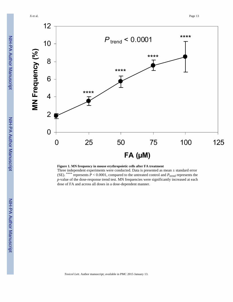
Figure 1. MN frequency in mouse erythropoietic cells after FA treatmentThree independent experiments were conducted. Data is presented as mean ± standard error(SE). **** represents P < 0.0001, compared to the untreated control and Ptrend represents thep-value of the dose-response trend test. MN frequencies were significantly increased at eachdose of FA and across all doses in a dose-dependent manner.
Ji et al. Page 13
Toxicol Lett. Author manuscript; available in PMC 2015 January 13.
NIH
-PA Author Manuscript
NIH
-PA Author Manuscript
NIH
-PA Author Manuscript

Figure 2. Human EPC expansion from peripheral bloodThree experiments on three blood donors were conducted. A. Ratio of EPCs relative to day 0during the course of the culture. Expansion began at day 6 and increased by ~12-fold by day10. B. Expression levels (%) of erythroid markers after expansion (day 10). EPCs expanded~100-fold by day 10. Data is presented as mean ± standard error (SE).
Ji et al. Page 14
Toxicol Lett. Author manuscript; available in PMC 2015 January 13.
NIH
-PA Author Manuscript
NIH
-PA Author Manuscript
NIH
-PA Author Manuscript

Figure 3. Human EPC expansion after FA treatmentSix experiments on five blood donors were conducted. A. Ratio of EPCs relative to vehiclecontrol on day 10. *** and **** represent P < 0.001 and P < 0.0001, respectively, comparedto the untreated control and Ptrend represents the p-value of the dose-response trend test. Thenumber of EPCs generated as a percentage of untreated control were significantly reducedby 100 and 150 µM FA and across the dose-range in a dose-dependent manner. B.Expression levels (%) of erythroid markers after expansion (day 10). Levels were unchangedby FA. Data is presented as mean ± SE.
Ji et al. Page 15
Toxicol Lett. Author manuscript; available in PMC 2015 January 13.
NIH
-PA Author Manuscript
NIH
-PA Author Manuscript
NIH
-PA Author Manuscript

Figure 4. Cell cycle distribution of the expanded human EPCs after FA treatmentFour experiments on three blood donors were conducted. Data is presented as mean ± SE. A.Cell cycle distribution. At 100 µM FA, the percentage of cells in G0 and G1 phase wassignificantly decreased (44.1%) compared to that of the untreated control (48.9%, P < 0.05)and the percentage of cells in G2 and M phase was significantly increased (21.5%)compared to that of the untreated control (17.9%, P < 0.05). The proportion of cells in Sphase was not significantly changed by FA treatment. B. Proliferation index. The index wassignificantly increased at 100 µM FA (0.54) compared to the untreated control (0.49, P <
Ji et al. Page 16
Toxicol Lett. Author manuscript; available in PMC 2015 January 13.
NIH
-PA Author Manuscript
NIH
-PA Author Manuscript
NIH
-PA Author Manuscript

0.05). * represents P < 0.05, compared to the untreated control and Ptrend represents the p-value of the dose-response trend test.
Ji et al. Page 17
Toxicol Lett. Author manuscript; available in PMC 2015 January 13.
NIH
-PA Author Manuscript
NIH
-PA Author Manuscript
NIH
-PA Author Manuscript

NIH
-PA Author Manuscript
NIH
-PA Author Manuscript
NIH
-PA Author Manuscript
Ji et al. Page 18
Tabl
e 1
Ane
uplo
idie
s of
chr
omos
omes
7 a
nd 8
in th
e ex
pand
ed E
PCs
afte
r FA
trea
tmen
t a
FA
Cel
l #M
onos
omy
Tri
som
yM
onos
omie
sT
riso
mie
sSt
ruct
ural
(µM
)So
rted
78
78
7 &
87
& 8
CA
e
077
3360
(0.
78)b
52 (
0.67
)1
(0.0
1)1
(0.0
1)11
2 (1
.45)
2 (0
.03)
3 (0
.04)
5074
9474
(0.
99)
71 (
0.95
)c4
(0.0
5)6
(0.0
8)c
145
(1.9
3)d
10 (
0.13
)d4
(0.0
5)
100
6871
46 (
0.67
)39
(0.
57)
1 (0
.01)
5 (0
.07)
85 (
1.24
)6
(0.0
9)4
(0.0
6)
a Four
exp
erim
ents
on
four
blo
od d
onor
s w
ere
cond
ucte
d.
b Dat
a ar
e pr
esen
ted
as n
(%
).
c an
d d re
pres
ent P
< 0
.1 a
nd P
< 0
.05,
res
pect
ivel
y, c
ompa
red
to u
ntre
ated
con
trol
s.
e CA
: Chr
omos
ome
aber
ratio
ns
Toxicol Lett. Author manuscript; available in PMC 2015 January 13.