Embed Size (px)
Citation preview

This article was downloaded by: [Universitätsbibliothek Bern]On: 10 June 2015, At: 01:14Publisher: Taylor & FrancisInforma Ltd Registered in England and Wales Registered Number: 1072954 Registered office: Mortimer House,37-41 Mortimer Street, London W1T 3JH, UK
Click for updates
Biotechnology & Biotechnological EquipmentPublication details, including instructions for authors and subscription information:http://www.tandfonline.com/loi/tbeq20
Prognostic value of matrix metalloproteinases in oralsquamous cell carcinomaGeorgi Misheva, Elitsa Deliverskab, Ruslan Hlushchuka, Nikolay Velinova, Daniel Aebersoldc,Felix Weinsteina & Valentin Djonova
a Institute of Anatomy, University of Bern, Bern, Switzerlandb Department of Oral and Maxillofacial Surgery, Faculty of Dental Medicine, MedicalUniversity of Sofia, Sofia, Bulgariac Department of Radiation Oncology, Inselspital, Bern University Hospital, Bern, SwitzerlandPublished online: 20 Nov 2014.
To cite this article: Georgi Mishev, Elitsa Deliverska, Ruslan Hlushchuk, Nikolay Velinov, Daniel Aebersold, Felix Weinstein& Valentin Djonov (2014) Prognostic value of matrix metalloproteinases in oral squamous cell carcinoma, Biotechnology &Biotechnological Equipment, 28:6, 1138-1149, DOI: 10.1080/13102818.2014.967510
To link to this article: http://dx.doi.org/10.1080/13102818.2014.967510
PLEASE SCROLL DOWN FOR ARTICLE
Taylor & Francis makes every effort to ensure the accuracy of all the information (the “Content”) contained inthe publications on our platform. Taylor & Francis, our agents, and our licensors make no representations orwarranties whatsoever as to the accuracy, completeness, or suitability for any purpose of the Content. Versionsof published Taylor & Francis and Routledge Open articles and Taylor & Francis and Routledge Open Selectarticles posted to institutional or subject repositories or any other third-party website are without warrantyfrom Taylor & Francis of any kind, either expressed or implied, including, but not limited to, warranties ofmerchantability, fitness for a particular purpose, or non-infringement. Any opinions and views expressed in thisarticle are the opinions and views of the authors, and are not the views of or endorsed by Taylor & Francis. Theaccuracy of the Content should not be relied upon and should be independently verified with primary sourcesof information. Taylor & Francis shall not be liable for any losses, actions, claims, proceedings, demands,costs, expenses, damages, and other liabilities whatsoever or howsoever caused arising directly or indirectly inconnection with, in relation to or arising out of the use of the Content. This article may be used for research, teaching, and private study purposes. Terms & Conditions of access anduse can be found at http://www.tandfonline.com/page/terms-and-conditions It is essential that you check the license status of any given Open and Open Select article to confirmconditions of access and use.
source: http://boris.unibe.ch/69258/ | downloaded: 13.3.2017

ARTICLE; MEDICAL BIOTECHNOLOGY
Prognostic value of matrix metalloproteinases in oral squamous cell carcinoma
Georgi Misheva*, Elitsa Deliverskab, Ruslan Hlushchuka, Nikolay Velinova, Daniel Aebersoldc, Felix Weinsteina and
Valentin Djonova
aInstitute of Anatomy, University of Bern, Bern, Switzerland; bDepartment of Oral and Maxillofacial Surgery, Faculty of DentalMedicine, Medical University of Sofia, Sofia, Bulgaria; cDepartment of Radiation Oncology, Inselspital, Bern University Hospital, Bern,Switzerland
(Received 23 November 2013; accepted 26 May 2014)
The aim of this study was to investigate whether there is a correlation between the expressions of four matrixmetalloproteinases (MMPs): MMP-2, MMP-7, MMP-9 and MMP-13, and the TNM (tumour�node�metastasis) stages oforal squamous cell carcinoma (OSCC); and to explore the implication of these MMPs in OSCC dissemination. Samplesfrom 61 patients diagnosed with oropharyngeal tumour were studied by immunohistochemistry against MMP-2, MMP-7,MMP-9 and MMP-13. The assessment of immunoreactivity was semi-quantitative. The results showed that MMP-2 andMMP-9 had similar expression patterns in the tumour cells with no changes in the immunoreactivity during tumourprogression. MMP-9 always had the highest expression, whereas that of MMP-2 was moderate. MMP-7 showed asignificant decrease in expression levels during tumour evolution. MMP-13 had constant expression levels within stage T2and T3, but showed a remarkable decline in immunoreactivity in stage T4. No significant differences in the MMPsimmunoreactivity between tumour cells and stroma were observed. Although strong evidence for the application of MMPsas reliable predictive markers for node metastasis was not acquired, we believe that combining patients’ MMPs expressionintensity and clinical features may improve the diagnosis and prognosis. Strong evidence for the application of MMPs asreliable predictive markers for node metastasis was not acquired. Application of MMPs as prognostic indicators for themalignancy potential of OSCC might be considered in every case of tumour examination. We believe that combiningpatients’ MMPs expression intensity and clinical features may improve the process of making diagnosis and prognosis.
Keywords: matrix metalloproteinase; oropharyngeal squamous cell carcinoma; tumour progression
Introduction
Squamous-cell carcinoma (SCC) is a highly aggressive
and malignant form of tumour, with a very poor progno-
sis. At least 90% of all oral malignancies are of this kind.
In the world-incidence ranking of cancers, it holds the 8th
position.[1] oral squamous cell carcinoma (OSCC) can
invade the adjacent tissue and spreads fairly rapidly. It
has a notorious tendency to invade the regional lymph
nodes, probably at a very early stage of tumourogenesis.
Consequently, well-nigh 50% of the patients who undergo
therapy develop distant metastases, which binds them to
an uninterrupted course of treatment until death inter-
venes.[2�5] The localization of the primary tumour plays
a crucial role in its dissemination, close anatomic relation-
ships with lymphatic and blood vessels being a prerequi-
site for early metastases.
The metastatic potential of OSCC depends upon its
ability to digest the extracellular matrix (ECM), to pene-
trate the basement membrane (BM), to initiate tumour
angiogenesis,[6�9] and to invade the adjacent tissues and
vessels. The BM, which separates the epithelium from the
mesenchymal tissue, is the first barrier against invasion.
Degradation of the ECM and the BM requires the partici-
pation of matrix metalloproteinases (MMPs). Most of
these enzymes are involved in common physiological pro-
cesses, such as the proliferation, differentiation and apo-
ptosis of cells, angiogenesis, and the morphogenesis and
repair of bodily tissues.[10�16] In the early 1980s, Liotta
et al. [17] identified proteolysis, and, specifically, the
digestion of type-IV collagen, to be essential for the inva-
siveness of melanomas. Formerly, MMPs were deemed to
be expressed exclusively within tumour cells, but this
belief has since been refuted. Their up-regulation therein
is now known to be triggered by the host’s stromal cells.
[18,19] It has been postulated that the invasion potential
of OSCCs is conferred by their ability to utilize MMPs
that are produced by the host’s stromal cells.[20�22] Yor-
ioka et al. [23] have suggested that tumour cells either
stimulate the liberation of MMPs from the host’s stromal
cells, thereby favouring the proteolytic degradation of the
ECM or themselves synthesize these proteinases.
Although some authors argue that the zymographic detec-
tion of the gelatinolytic activity of MMPs is more repre-
sentative than their immunohistochemical mapping, Ikebe
*Corresponding author. Email: [email protected]
� 2014 The Author(s). Published by Taylor & Francis.
This is an Open Access article distributed under the terms of the Creative Commons Attribution License http://creativecommons.org/licenses/by/3.0/, which permits unre-
stricted use, distribution, and reproduction in any medium, provided the original work is properly cited. The moral rights of the named author(s) have been asserted
Biotechnology & Biotechnological Equipment, 2014
Vol. 28, No. 6, 1138�1149, http://dx.doi.org/10.1080/13102818.2014.967510
Dow
nloa
ded
by [
Uni
vers
itäts
bibl
ioth
ek B
ern]
at 0
1:14
10
June
201
5

et al. [24] have demonstrated the existence of a significant
correlation between the results obtained using these two
methods.
To date, 25 members of the MMP family have been
identified. They are classified according to their amino-
acid-sequence homology either as soluble enzymes (colla-
genases, gelatinases, stromelysins and an elastinase), or as
membrane-associated ones.[15] Most of the MMPs are
secreted in an inactive form, which is activated pericellu-
larly or extracellularly by serine proteases such as trypsin,
plasmin or neutrophil elastase. MMPs are down-regulated
by endogenous tissue inhibitors of metalloproteinases
(TIMPs).[11,25] The role of MMPs in tumourigenesis and
angiogenesis has been confirmed by many authors during
the past few decades. The latter process is essential for the
delivery of nutrients to the proliferating cancer cells and
for furnishing the structural support for tumour expansion.
[26,27] MMPs do not act synergically during tumourigen-
esis: the up-regulation of MMP-2 and MMP-9 is associ-
ated with the degradation of the ECM and the BM, and
with an increase in tumour aggressiveness; the down-reg-
ulation of MMP-13 is associated with a worsening of the
disease prognosis; and that of MMP-7, with an increase in
tumour invasiveness.
MMPs have also been demonstrated to be involved in
the progression of tumours and in their metastatic dissem-
ination. Given the important role played by this family of
enzymes in degrading the BM and the ECM, their partici-
pation in the propagation of tumour cells is perhaps not
surprising. Selected MMPs have been implicated in angio-
genesis, a process without which a tumour cannot undergo
the neovascularization that is crucial for its nourishment
and structural support.[3,26,28�31] Certain members of
the MMP family are the only proteinases that are known
to be capable of cleaving the collagen types of which the
BM (type IV) and the ECM (types I and III) are com-
posed,[32] MMP-2 and MMP-9 degrade type-IV
[21,33,34] and MMP-13, types I to IV [35]. MMP-7
degrades fibronectin, tenascin and b4 integrin, which play
a crucial role in the adhesion and migration of cells during
tumourigenesis.[12,30,31,36,37]
The aim of this study was to define the role played by
MMP-2, MMP-7, MMP-9 and MMP-13 in the progres-
sion and invasion of OSCC and to ascertain whether they
could serve as reliable markers of these phenomena. To
this end, we mapped immunohistochemically their
expression profiles within tumour samples that were
derived from 61 patients with OSCC, and correlated
these data with histological findings that were classified
according to Bryne’s malignancy-grading system. The
distribution of MMPs within OSCCs was mapped immu-
nohistochemically because this technique (1) permits a
direct correlation with morphological data, and (2) can
be performed on paraffin-embedded tissue sections that
are routinely produced for diagnostic purposes.[38]
Hence, the technique could be performed on a regular
basis even in hospitals.
Materials and methods
This study was approved by the Regional Board of Medi-
cal Ethics.
Tumour samples were derived from 61 patients who
had been diagnosed as suffering from OSCC and who had
been treated at Inselspital in Bern. Paraffin-embedded
samples were made available to us by the Department of
Pathology. Information about clinicopathological covari-
ables was gleaned from the medical records of the
patients. The patients were administered a median total
dose of 74 Gy (range: 54�80.5 Gy), which was delivered
in daily fractions, five times a week for five to eight
weeks. Ninety-seven per cent of the patients were exposed
to at least 66 Gy.[39] The primary tumour and lymph-
node metastases were evaluated separately. After treat-
ment, all patients were clinically examined and imaged on
a regular basis. Fifty-five patients were observed until the
time of death, the median follow-up period being
2.6 years.
Of the 61 patients, 44 were male and 17 female. Their
age ranged from 41 to 85 years. Fifty-two of the patients
smoked at least one cigarette per day, and 52 drank an
alcoholic beverage at least once a week (Table 1).
The patients were classified according to the Union for
International Cancer Control TNM system (UICC-TNM)
of 1997. Clinicopathologically, the tumours were graded
on a scale from I to IV. According to the TNM system, 55
of the patients were at stage T3/4. In 45 of the patients, at
least one lymph node was affected and 50 manifested dis-
tant metastasis. In 47 of the patients, the tumour was
graded histologically as I, II, and in the remaining 14,
as III.
Immunohistochemistry
Three-micrometre-thick sections of the paraffin-embed-
ded tumour samples were transferred to gelatinized
micro-slides and air-dried overnight at 37 �C. They were
deparaffinized in xylene (three changes of medium) and
rehydrated in ethanol (100% through 30%). They were
then washed three times in distilled water and twice in
Tris-buffered saline (TBS) [50 mmol/L Tris/HCL (pH
7.4), containing 100 mmol/L sodium chloride]. To block
non-specific binding, the sections were incubated in TBS
containing 1% casein (SIGMA 8654) for 10 min. They
were then exposed to primary antibodies against MMP-2,
MMP-7, MMP-9, MMP-13, a-smooth-muscle actin (anti-
a-SMA) (A-2547, Sigma-Aldrich-Chemie, Gmbh),
diluted 1:200 in TBS, and rabbit anti-fibronectin (F 3648
Sigma-Aldrich-Chemie, Gmbh), diluted 1:200 in TBS.
Biotechnology & Biotechnological Equipment 1139
Dow
nloa
ded
by [
Uni
vers
itäts
bibl
ioth
ek B
ern]
at 0
1:14
10
June
201
5
The latter two antibodies were applied to identify stromal-
muscle cells and fibroblasts, respectively.
Primary antibodies
The following primary antibodies were used: mouse anti-
MMP-2 (Ab-4) VC2 (Neo Markers, Cat# MS-806-R7,
Lot# 806R107, ready to use); rabbit polyclonal anti-
MMP-7 (Ab-4) (OncogeneTM, Cat# PC492, Lot#
D18417-1) at a 1:100 dilution in Antibody-Diluent; mouse
anti-MMP 7 Ab-1 (ID2) (NeoMarkers, Cat# MS-813-R7,
Lot# 813R111, ready to use); rabbit anti-MMP-9 (Neo-
Markers, Cat# RB-1539-R7, Lot# 1539R303, ready to
use, ON �4 �C); mouse anti-MMP-13 Ab-2 (ID3)
(OncogeneTM, Cat# MS-826-R7, Lot# 826R909; ready to
use, ON �4 �C).The sections were then rinsed three times in TBS
before incubating with the secondary antibodies for
45 min at ambient temperature.
Secondary antibodies
The following secondary antibodies were used: Biotin-
anti-Mouse Ig (DAKO EO 433) at a 1:200 dilution in
TBS; Biotin-anti-Rabbit Ig (DAKO EO 353) at a 1:200
dilution in Antibody-Diluent; Biotin-anti-Rabbit Ig
(DAKO EO 353) at a 1:200 dilution in TBS; Biotin-anti-
Mouse Ig (DAKO EO 433) at a 1:200 dilution in TBS.
They were again rinsed three times in TBS and then
treated with horseradish-peroxidase�streptavidin com-
plex (P355, DAKO) likewise for 45 min at ambient tem-
perature. The reaction product was visualized by exposing
the slides to 3-amino-9-ethylcarbazole (Sigma). The
specimens were then mounted in Aquatext (Merck). Tis-
sue samples that were incubated with non-immune serum
served as negative controls.
Semi-quantitative evaluation of immunoreactivity for
MMPs
Immunoreactivity for MMPs in each tumour sample was
assessed in a blinded fashion and independently by two
persons who had no prior knowledge of the clinical fea-
tures. The intensity of the immunostaining reaction and
the percentage of positively stained cells within the entire
section area were graded as follows: (0): negative (no
immunoreactivity); (1): minimal staining (<10% of the
section area); (2): moderate staining (10%�50% of the
section area); (3): strong staining (>50% of the section
area).
For convenient description of the results, the intensity
of the immunostaining reaction was subdivided into two
broad categories: weak (grades 1 and 2) and strong
(grades 3 and 4). Immunoreactivity was statistically corre-
lated with clinical parameters, using Fisher’s exact test.
The invasion front was evaluated histologically
according to Bryne’s malignancy grading system,[40]
which yields a better prognostic value for SCCs [41] than
the conventional one. Four morphological parameters
were individually evaluated at the invasion front: (i)
degree of keratinization; (ii) tumour structure (pattern of
invasion); (iii) nuclear polymorphism; and (iv) the host’s
cellular inflammatory response. For each parameter, an
average of four fields were evaluated under a light micro-
scope at a final magnification of £100.
Each parameter (i�iv) was graded on a scale from 1 to
4. Degree of keratinization: (1) highly keratinized (>50%
of the cells); (2) moderately keratinized (20%�50% of
the cells); (3) minimally keratinized (5%�20% of the
cells); (4) no keratinization (0%�5% of the cells).
Tumour structure (pattern of invasion): (1) advancing,
well-delineated borders (solid sheets); (2) infiltrating solid
cords, bands and/or strands; (3) small groups or cords of
infiltrating cells (n < 15); (4) marked and widespread dis-
sociation into small groups of cells and/or into single cells
(n >15). Nuclear polymorphism: (1) minimal (mature
cells: >75%); (2) moderate (mature cells: 50%�75%);
(3) abundant (mature cells: 25%�50%); and (4) extreme
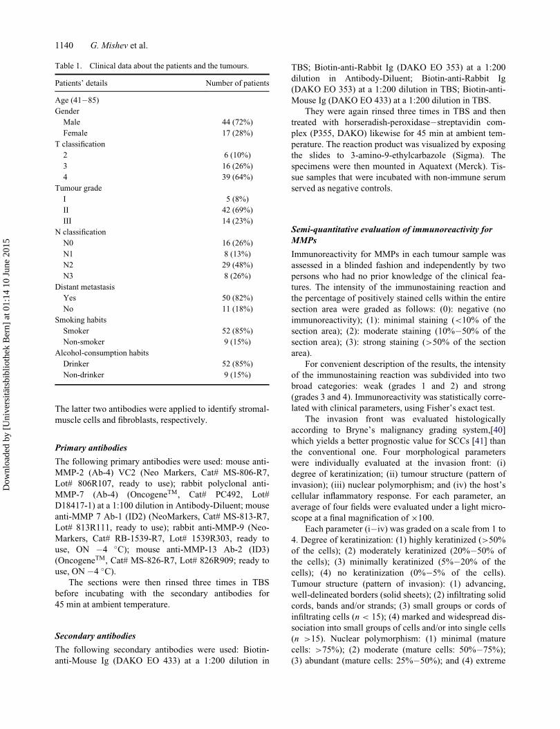
Table 1. Clinical data about the patients and the tumours.
Patients’ details Number of patients
Age (41�85)
Gender
Male 44 (72%)
Female 17 (28%)
T classification
2 6 (10%)
3 16 (26%)
4 39 (64%)
Tumour grade
I 5 (8%)
II 42 (69%)
III 14 (23%)
N classification
N0 16 (26%)
N1 8 (13%)
N2 29 (48%)
N3 8 (26%)
Distant metastasis
Yes 50 (82%)
No 11 (18%)
Smoking habits
Smoker 52 (85%)
Non-smoker 9 (15%)
Alcohol-consumption habits
Drinker 52 (85%)
Non-drinker 9 (15%)
1140 G. Mishev et al.
Dow
nloa
ded
by [
Uni
vers
itäts
bibl
ioth
ek B
ern]
at 0
1:14
10
June
201
5

(mature cells: 0%�25%). Host’s cellular inflammatory
response: (1) marked; (2) moderate; (3) slight; (4) none.
Quantitative evaluation of immunoreactivity for MMPs
To quantify the expression pattern of MMPs at the inva-
sive front, tissue immunoreactivity (Tiss) and the relative
number of immunopositive cells (Rn) were computed
within the 284 investigated fields. Initially, the number of
fields with the same numerical combination of Tiss and
Rn was calculated. Then, the number of different score
combinations for Tiss and Rn (according to Bryne’s grad-
ing system) was recorded.
Results and discussion
The expression patterns of MMP-2, MMP-7, MMP-9 and
MMP-13 in the tumour samples, both within the tumour
cells (intratumoural) and within the intervening and sur-
rounding stroma, were mapped immunohistochemically.
Immunoreactivity was graded semi-quantitatively, and
the data were correlated with histological findings
that were classified according to Bryne’s malignancy-
grading system, as well as with clinicopathological
features, namely the stage of the tumour and lymph-node
involvement.
MMPs expression patterns
Our results showed that in approximately 90% of the
tumour samples (from 61 patients with OSCC), the immu-
nostaining for MMP-2, MMP-7 and MMP-9 was classi-
fied as strong and these findings accord with those
published by other authors.[38,42] Nevertheless, the
intensity distributions of intratumoural and stromal stain-
ing were likewise comparable.
MMP-2
Intratumoural staining for MMP-2 (Figure 1) was graded
as weak in 18% of the samples (11/61) and as strong in
82% (50/61). Within the intervening and surrounding
Figure 1. Immunoreactivity for MMP-2 (a and b): immunopositive vessels (arrows); St D stroma; Tu D tumour. Correlation betweenMMP-2 expression intensity and tumour stage (c); lymph-node involvement (d).
Biotechnology & Biotechnological Equipment 1141
Dow
nloa
ded
by [
Uni
vers
itäts
bibl
ioth
ek B
ern]
at 0
1:14
10
June
201
5

stroma (Figure 1), immunostaining for MMP-2 was weak
in 23% of the samples (14/61) and strong in 77% (47/61).
In patients with no lymph-node involvement (16/61),
inratumoural staining for MMP-2 was weak in 25% of the
cases (4/16) and strong in the other 75% (12/16). In
patients with lymph-node involvement (45/61), intratu-
moural staining for MMP-2 was weak in 16% of the cases
(7/45) and strong in the other 84% (38/45) (Table 2).
Although Kato et al. [20] propose the use of MMP-2
as a predictive marker for tumour progression and the
early invasion of lymph nodes, according to our immunos-
taining data, the expression of MMP-2 was not closely
associated with the stage of the tumour (Figure 1). In 62%
of our patients with lymph-node involvement, immunos-
taining for MMP-2 was strong, but in 20% of those with
no lymph-node involvement, immunostaining for MMP-2
was likewise strong (Figure 1, Table 2). This suggests that
in patients with no lymph-node involvement at the time of
treatment, the process of dissemination had most probably
already begun. Therefore, on the basis of our data, we do
not consider MMP-2 to be a reliable predictive marker of
tumour invasiveness in OSCC, but other authors have
reported the existence of a positive correlation between
the expression of MMP-2, lymph-node recurrence and a
worsening of the survival rate.[43�48]
MMP-7
Intratumoural staining for MMP-7 (Figure 2) was graded
as weak in 3% of the samples (2/16) and as strong in 97%
(59/61). Within the intervening and surrounding stroma
(Figure 2), immunostaining for MMP-7 was weak in 8%
of the samples (5/61) and strong in 92% (56/61). In
patients with no lymph-node involvement (16/61), intratu-
moural staining for MMP-7 was weak in 6% of the cases
(1/16) and strong in the other 94% (15/16). In patients
with lymph-node involvement (45/61), intratumoural
staining for MMP-7 was weak in 2% of the cases (1/45)
and strong in the other 98% (44/45) (Table 3).
These results show the immunostaining for MMP-7 to
be closely correlated with the stage of the tumour: strong
staining decreased, and moderate staining increased with
an advance in the stage of the tumour. That is, overall, the
expression of MMP-7 declined with the progression of
OSCC. There was a correlation between the expression of
MMP-7 and lymph-node involvement. In 72% of the
patients with lymph-node involvement, immunostaining
for MMP-7 was strong, and likewise, immunostaining for
MMP-7 was also strong in 24% of those with no lymph-
node involvement (Figure 2, Table 3). These data do not
fully accord with published findings; for example, Impola
et al. [9] observed no correlation between immunoreactiv-
ity for MPP-7 and the stage of OSCC. However, the same
authors have since reported the existence of a link
between the expression of MMP-7, an inhibition of angio-
genesis and a retardation of tumour growth.[8,49] Mimori
et al. [37] have suggested that MMP-7 is involved either
directly in tumour growth, or, indirectly, by activating the
epidermal growth factor. In the light of this suggestion
our data are interesting, as they indicate that MMP-7
could possibly serve as a predictive marker of tumour
invasiveness in OSCC. Further studies on a larger cohort
would, of course, be needed to prove this correlation.
MMP-9
Intratumoural staining for MMP-9 (Figure 3) was graded
as weak in 2% of the samples (1/61) and as strong in 98%
(60/61). Within the intervening and surrounding stroma
(Figure 3), immunostaining for MMP-9 was weak in 16%
of the samples (10/61) and strong in 84% (51/61). In
patients with no lymph-node involvement (16/61),
Table 2. Intensity of distribution of Tu and stromal staining for MMP-2.
MMP-2
Intensity of Tu staining Intensity of stromal staining
Intensity gradeWeak Strong Weak Strong
Tu stage 0 1 2 3 0 1 2 3
Tu2 0 2 2 2 0 1 3 2
Tu3 0 2 8 7 0 3 8 6
Tu4 0 7 15 16 0 10 17 11
Total 11 (18%) 50 (82%) 14 (23%) 47 (77%)
Node (¡)neg. 0 4 7 5 0 4 8 4
Total 4 (7%) 12 (20%) 4 (7%) 12 (20%)
Node (C)pos. 0 7 18 20 0 10 20 15
Total 7 (11%) 38 (62%) 10 (16%) 35 (57%)
Note: Tu � Tumour.Node (¡) neg.� Lymph-node involvement.Node (C)pos. � Lymph-node involvement.
1142 G. Mishev et al.
Dow
nloa
ded
by [
Uni
vers
itäts
bibl
ioth
ek B
ern]
at 0
1:14
10
June
201
5

intratumoural staining for MMP-9 was strong in all cases
(16/16). In patients with lymph-node involvement
(45/61), intratumoural staining was weak in 2% of the
cases (1/45) and strong in the other 98% (44/45) (Figure 3,
Table 4).
MMP-9 has been detected in the plasma of patients
with carcinomas and has been proposed as a good marker
of head and neck SCC.[16] Our data, however, revealed
no evidence of a correlation between immunostaining for
MMP-9 and the stage of the tumour (Figure 3). Indeed,
Figure 2. Immunoreactivity for MMP-7 (a and b): St D stroma; Tu D tumour. Correlation between MMP-7 expression intensity andtumour stage (c); lymph-node involvement (d).
Table 3. Irrespective of the intensity distribution of stromal staining for MMP-7 corresponded to the intratumoural pattern.
MMP-7
Intensity of Tu staining Intensity of stromal staining
Intensity gradeWeak Strong Weak Strong
Tu stage 0 1 2 3 0 1 2 3
Tu2 0 0 1 5 0 0 4 2
Tu3 0 0 6 11 0 3 13 1
Tu4 0 2 16 20 0 2 29 7
Total 2 (2%) 59 (98%) 5 (8%) 56 (92%)
Node(�)neg. 0 1 5 10 0 3 12 1
Total 1 (2%) 15 (24%) 3 (5%) 13 (21%)
Node(C)pos. 0 1 18 26 0 2 34 9
Total 1 (2%) 44 (72%) 2 (3%) 43 (71%)
Biotechnology & Biotechnological Equipment 1143
Dow
nloa
ded
by [
Uni
vers
itäts
bibl
ioth
ek B
ern]
at 0
1:14
10
June
201
5

the incidence of both strong and moderate staining
remained remarkably constant with the progression of
OSCC. Katayama et al.[50] have reported the expression
of MMP-9 to be significantly higher in patients with
regional lymph-node or distant metastasis than in those
without such. Our data that in 68% of the patients with
lymph-node involvement immunostaining for MMP-9
was strong (Figure 3, Table 4) support these findings; but
there are also reports that question the reliability of MMP-
9 as a marker of head and neck SCCs.[34]
Figure 3. Immunoreactivity for MMP-9 (a and b): keratinized pearl (asterisk); St D stroma; Tu D tumour. Correlation between MMP-9expression intensity and tumour stage (c); lymph-node involvement (d).
Table 4. Irrespective of lymph-node involvement, the intensity distribution of stromal staining for MMP-9 corresponded to the intratu-moural pattern.
MMP-9
Intensity of Tu staining Intensity of stromal staining
Intensity gradeWeak Strong Weak Strong
Tu stage 0 1 2 3 0 1 2 3
Tu2 0 0 2 4 0 2 2 2
Tu3 0 0 6 11 0 0 14 3
Tu4 0 1 11 26 2 6 28 2
Total 1 (2%) 60 (98%) 10 (16%) 51 (84%)
Node(¡) neg. 0 0 7 9 0 3 11 2
Total 0 (0%) 16 (26%) 3 (5%) 13 (21%)
Node(C) pos. 0 1 12 32 2 5 33 5
Total 1 (2%) 44 (72%) 7 (11%) 38 (63%)
1144 G. Mishev et al.
Dow
nloa
ded
by [
Uni
vers
itäts
bibl
ioth
ek B
ern]
at 0
1:14
10
June
201
5

MMP-13
MMP-13 is a potent collagenase, which is rarely
expressed in normal tissue, but which is often up-regu-
lated when a rapid turnover of the ECM is required, as
during the local invasion and growth of a malignant
tumour.[34,51] MMP-13 is expressed at high levels in
SCCs of the head and neck.[52�54] In our study, the
intratumoural staining for MMP-13 (Figure 4) was graded
as weak in 33% of the samples (19/57) and as strong in
67% (38/57). Within the intervening and surrounding
stroma (Figure 4), immunostaining for MMP-13 was
weak in 39% of the samples (22/57) and strong in 61%
(35/57). In patients with no lymph-node involvement (16/
57), intratumoural staining for MMP-13 was weak in 31%
of the analysed negative samples (5/16) and strong in the
other 69% (11/16). In patients with lymph-node involve-
ment (41/57), intratumoural staining for MMP-13 was
weak in 34% of the analysed positive samples (14/41) and
strong in the other 66% (27/41) (Figure 4, Table 5).
Our data revealed an obvious correlation between
immunoreactivity for MMP-13 and tumour progression
only for strong staining, which decreased, and for minimal
staining, which increased (Figure 4). Overall, the expres-
sion of MMP-13 declined with the progression of OSSC.
The existing data regarding the diagnostic/prognostic
value of MMP-13 are conflicting. Whilst Brinckerhoff
et al. [52] have reported the existence of a positive corre-
lation between the expression of MMP-13 and lymph-
node involvement, Gottschlich et al. [53] have observed
no such relationship. As compared with the other three
MMPs investigated by us, strong staining for MMP-13
was observed in a higher proportion of patients with
lymph-node involvement than in those without it. How-
ever, for the other MMPs investigated, although lymph-
node involvement was associated with a high incidence of
strong staining, no correlation existed between lymph-
node involvement, strong immunostaining and the stage
of the tumour.
Figure 4. Immunoreactivity for MMP-13 (a and b): St D stroma; Tu D tumour. Correlation between MMP-13 expression intensity andtumour stage (c); lymph-node involvement (d). N.B.: For the analyses of MMP-13 a sufficiency of tissue was available from only 57 ofthe 61 patients.
Biotechnology & Biotechnological Equipment 1145
Dow
nloa
ded
by [
Uni
vers
itäts
bibl
ioth
ek B
ern]
at 0
1:14
10
June
201
5

MMPs and OSCC invasion
As a next step in our study, the invasion front of each
OSCC was evaluated histologically according to Bryne’s
malignancy-grading system. Four parameters were
assessed: (1) degree of keratinization; (2) nuclear poly-
morphism; (3) tumour structure (pattern of invasion); and
(4) host’s cellular inflammatory response. These findings
were then correlated with the immunostaining data for
each MMP.
Degree of keratinization
Between 80% and 87% of the samples were given a grade
of 4, i.e. no keratinization (0%�5% of the cells). In
70%�80% of these grade 4 cases, intratumoural staining
for MMP-2, MMP-7, MMP-9 and MMP-13 was strong
(Figure 5(a)). Highly keratinized regions (grade 1) did not
stain strongly for any of these MMPs.
Structure (pattern of invasion)
According to the pattern of invasion, between 75% and
85% of the samples were given a grade of 1 (advancing)
or 2 (infiltrating solid cords, etc.). In 75%�85% of these
grade 1 and grade 2 cases, intratumoural staining for
MMP-2, MMP-7, MMP-9 and MMP-13 was strong
(Figure 5(b)). Generally, the presence of single or small
groups of tumour cells within the stroma is a sign of high
malignancy and poor prognosis.[4] In our experiments,
this structural feature (grade 4 in Figure 5(b)) was associ-
ated with weak staining for MMP-7 and MMP-13.
Nuclear polymorphism
With regard to nuclear polymorphism, between 85% and
95% of the samples were given a grade of 1 (minimal) or
2 (moderate). In 80%�90% of these grade 1 and grade 2
cases, intratumoural staining for MMP-2, NNP-7, MMP-9
and MMP-13 was strong (Figure 5(c)). An extremely high
degree of nuclear polymorphism (mature cells:
0%�25%), which occurred within regions where no BMs
were evident, was likewise associated with minimal
immunostaining for MMP-7 and MMP-13 (grade 4 in
Figure 5(c)). Thus, MMP-7 and MMP-13 appeared to be
absent from areas of diffuse invasion and high
malignancy.
Host’s cellular inflammatory response
The host’s cellular immune response to a tumour reflects
its potency. In our study, however, no consistent trend in
this parameter was observed for any of the tested MMPs
(Figure 5(d)). Grades 1 (marked), 2 (moderate) and 4
(none) were each observed in 15%�20% of the samples,
and grade 3 (slight), in 37%. Weak intratumoural staining
for MMP-2, MMP-7, MMP-9 and MMP-13 was not asso-
ciated with any of the grades of the host’s cellular inflam-
matory response.
Final remarks
Our results that MMP-7 and MMP-13 were absent from
areas of diffuse invasion and high malignancy, in good
correlation with Bryne’s malignancy grading of tumour
structure and nuclear polymorphism, suggest that together
these two MMPs might be useful as predictive markers of
invasiveness in OSCC. This observation might possibly
be considered to be associated with the mechanisms of
action of these MMPs. MMP-7 cleaves plasminogen to
angiostatin and type-XVIII collagen to endostatin �events which lead to an inhibition of angiogenesis and
possibly to a retardation of tumour growth.[49] It could be
speculated that an increase in the substrate activity of
MMP-7 possibly accounts for the overall decrease in the
Table 5. Irrespective of lymph-node involvement, the intensity distribution of stromal staining for MMP-13 corresponded to the intra-tumoural pattern.
MMP-13
Intensity of Tu staining Intensity of stromal staining
Intensity gradeWeak Strong Weak Strong
Tu stage 0 1 2 3 0 1 2 3
Tu2 0 0 3 3 0 2 3 1
Tu3 1 3 3 7 0 4 5 5
Tu4 2 13 13 9 0 16 16 5
Total 19 (33%) 38 (67%) 22 (39%) 35 (61%)
Node(¡) neg. 2 3 6 5 0 6 5 5
Total 5 (9%) 11 (19%) 6 (11%) 10 (18%)
Node(C) pos. 1 13 13 14 0 16 19 6
Total 14 (25%) 27 (47%) 16 (28%) 25 (44%)
1146 G. Mishev et al.
Dow
nloa
ded
by [
Uni
vers
itäts
bibl
ioth
ek B
ern]
at 0
1:14
10
June
201
5

expression of this enzyme with an advance in the stage of
the tumour. As the malignancy potential of the tumour
increases, the ability of MMP-7 to retard its invasion
declines considerably. The marked decline in the expres-
sion of MMP-13 that was associated with advanced stages
of tumour invasion could reflect the fact that the BM has
already been digested.
Together with TNM-staging of the patient, immuno-
histochemistry for MMP-7 and MMP-13 could facilitate
diagnosis and an assessment of the prognosis. On the
other hand, the fairly constant levels of expression of
MMP-2 and MMP-9 during the evolution of OSCCs, and
the lack of correlation between their patterns of immunos-
taining and Byrne’s malignancy grading of either tumour
structure or nuclear polymorphism, indicate that they are
not likely to be considered suitable markers for the inva-
sion potential of OSCCs. We believe that testing for
MMPs in patients with OSCC could help to define the
roles of MMPs in oncogenesis, ultimately leading to tar-
geted therapy and an improvement in outcome.
Conclusions
Our data revealed MMP-7 and MMP-13 to be down-regu-
lated in highly invasive and malignant areas of OSCCs.
Their patterns of immunostaining, unlike those of MMP-2
and MMP-9, are very likely to afford an indication of
the invasion potential of the tumour. Our findings indicate
that MMP-7 and MMP-13 are reliable markers of the
invasion potential of OSCC, whereas MMPs 2 and 9 are
not. The patterns of immunostaining of MMP-7 and
MMP-13 correlated well with Bryne’s malignancy grad-
ing of tumour structure and nuclear polymorphism,
thereby indicating that these MMPs can be relevant in
gauging the malignancy potential of OSCC. Together
with TNM-staging of the patient, immunohistochemistry
for MMP-7 and MMP-13 could facilitate diagnosis and an
assessment of the prognosis. We believe that testing for
MMPs in patients with OSCC could help to define their
roles in oncogenesis, ultimately leading to targeted ther-
apy and an improvement in outcome.
Figure 5. Distribution of MMP expression intensity (%) according to keratinization grades (a), structure grades (b), nuclear polymor-phism grades (c) and host reaction grades (d).
Biotechnology & Biotechnological Equipment 1147
Dow
nloa
ded
by [
Uni
vers
itäts
bibl
ioth
ek B
ern]
at 0
1:14
10
June
201
5

Funding
This study was supported by University of Bern, Institute ofAnatomy, Bern, Switzerland.
References
[1] Massano J, Regateiro FS, Januario G, Ferreira A. Oralsquamous cell carcinoma: review of prognostic and predic-tive factors. Oral Surg Oral Med Oral Pathol Oral RadiolEndod. 2006;102:67�76.
[2] Carlos de Vicente J, Junquera Guti�errez LM, Zapatero AH,Fresno Forcelledo MF, Hernandez-Vallejo G, L�opezArranz JS. Prognostic significance of p53 expression inoral squamous cell carcinoma without neck node metasta-ses. Head Neck. 2004;26:22�30.
[3] Guerra MF, Campo FJ, Gias LN, Perez JS. Rim versus sag-ittal mandibulectomy for the treatment of squamous cellcarcinoma: two types of mandibular preservation. HeadNeck. 2003;25:982�989.
[4] Lo WL, Kao SY, Chi LY, Wong YK, Chang RC. Out-comes of oral squamous cell carcinoma in Taiwan aftersurgical therapy: factors affecting survival. J Oral Maxillo-fac Surg. 2003;61:751�758.
[5] Nguyen TV, Yueh B. Weight loss predicts mortality afterrecurrent oral cavity and oropharyngeal carcinomas. Can-cer. 2002;95:553�562.
[6] Aebersold DM, Beer KT, Laissue J, Djonov V, GreinerRH. Intratumoral microvessel density predicts local treat-ment failure of radically irradiated squamous cell cancer ofthe oropharynx. Int J Radiat Oncol Biol Phys.2000;48:17�25.
[7] Goldberg G. Human fibroblasts collagenase: complete pri-mary structure and homology to an oncogene transforma-tion-induced rat protein. J Biol Chem. 1986;261(14):6600�6605.
[8] Impola U, Cuccuru MA, Masala MV, Jeskanen L, CottoniF, Saarialho-Kere U. Preliminary communication: matrixmetalloproteinases in Kaposi’s sarcoma. Br J Dermatol.2003;149:905�907.
[9] Impola U, Toriseva M, Suomela S, Jeskanen L, Hieta N,Jahkola T, Grenman R. Matrix metalloproteinase-19 isexpressed by proliferating epithelium but disappears withneoplastic dedifferentiation. Int J Cancer.2003;103:709�716.
[10] Jayade BV, Bhat K, Patil BR, Nayak R, Sant A. Histologi-cal significance of p53 gene expression in squamous cellcarcinoma of the buccal mucosa. J Maxillofac Oral Surg.2009;8(3):205�210.
[11] Kleiner DE, Stetler-Stevenson WG. Matrix metalloprotei-nases and metastasis. Cancer Chemother Pharmacol.1999;43(Suppl):S42�S51.
[12] Luukkaa M, Vihinen P, Kronqvist P, Vahlberg T, Pyrh€onenS, K€ah€ari V. Association between high collagenase-3expression levels and poor prognosis in patients with headand neck cancer. Head Neck. 2006;28:225�234.
[13] Matrisian LM. Epidermal growth factor and oncogenesinduce transcription of the same cellular mRNA in ratfibroblasts. EMBO J. 1985;4(6):1435�1440.
[14] Matrisian LM. The matrix-degrading metalloproteinases.Bioessays. 1992;14(7):455�463.
[15] Murray GI. Matrix metalloproteinases: a multifunctionalgroup of molecules. J Pathol. 2001;195:135�137.
[16] Ranuncolo SM, Matos E, Loria D, Vilensky M, Rojo R,Bal de Kier Joff�e E, In�es Puricelli L. Circulating 92-kilo-
dalton matrix metalloproteinase (MMP-9) activity isenhanced in the euglobulin plasma fraction of head andneck squamous cell carcinoma. Cancer.2002;94:1483�1491.
[17] Liotta LA, Tryggvason K, Garbisa S, Hart I, Foltz CM,Shafie S. Metastatic potential correlates with enzymaticdegradation of basement membrane collagen. Nature.1980;284:67�68.
[18] Basset P, Bellocq JP, Wolf C, Stoll I, Hutin P, LimacherJM, Podhajcer OL, Chenard MP, Rio MC, Chambon P. Anovel metalloproteinase gene specifically expressed instromal cells of breast carcinomas. Nature.1990;348:699�704.
[19] Nebeshima K. Partial sequencing and characterization ofthe tumor cell-derived collagenase stimulatory factor. ArchBiochem Biophys. 1991;285(1):90�96.
[20] Kato K, Hara A, Kuno T, Kitaori N, Huilan Z, Mori H.Matrix metalloproteinases 2 and 9 in oral squamous cellcarcinomas: manifestation and localization of their activ-ity. J Cancer Res Clin Oncol. 2005;131:340�346.
[21] Kurahara S, Shinohara M, Ikebe T, Nakamura S, Beppu M,Hiraki A. Expression of MMPS, MT-MMP, and TIMPs insquamous cell carcinoma of the oral cavity: correlationswith tumor invasion and metastasis. Head Neck.1999;21:627�638.
[22] Thomas GT, Lewis MP, Speight PM. Matrix metalloprotei-nases and oral cancer. Oral Oncol. 1999;35:227�233.
[23] Yorioka CW, Coletta RD, Alves F, Nishimoto IN, Kowal-ski LP, Graner E. Matrix metalloproteinase-2 and -9 activi-ties correlate with the disease-free survival of oralsquamous cell carcinoma patients. Int J Oncol.2002;20:189�194.
[24] Ikebe T, Shinohara M, Takeuchi H, Beppu M, Kurahara S.Gelatinolytic activity of matrix metalloproteinase in tumortissues correlates with the invasiveness of oral cancer. ClinExp Metastasis 1999;17:315�323.
[25] Stetler-Stevenson WG. Matrix metalloproteinases in angio-genesis: a moving target for therapeutic intervention. J ClinInvestig. 1999;103:1237�1241.
[26] Aebersold DM. [Angiogenesis as prognostic factor inmalignant tumors]. Ther Umsch. 1998;55:462�463.German.
[27] Burri PH, Hlushchuk R, Djonov V. Intussusceptive angio-genesis: its emergence, its characteristics, and its signifi-cance. Dev Dyn. 2004;231:474�488.
[28] Djonov V, Cresto N, Aebersold DM, Burri PH, AltermattHJ, Hiristic M. Tumor cell specific expression of MMP-2correlates with tumor vascularisation in breast cancer. Int JOncol. 2002;21:25�30.
[29] Djonov V, Hogger K, Sedlacek R, Laissue J, Draeger A.MMP-19: cellular localization of a novel metalloproteinasewithin normal breast tissue and mammary gland tumours. JPathol. 2001;195:147�155.
[30] Rautava J, Luukkaa M, Heikinheimo K, Alin J, GrenmanR, Happonen RP. Squamous cell carcinomas arising fromdifferent types of oral epithelia differ in their tumor andpatient characteristics and survival. Oral Oncol. 2007;43(9):911�919.
[31] Tang Y, Nakada MT, Kesavan P, McCabe F, Millar H,Rafferty P. Extracellular matrix metalloproteinase inducerstimulates tumour angiogenesis by elevating vascularendothelial cell growth factor and matrix metalloprotei-nases. Cancer Res. 2005;65:3193�3199.
[32] Yurchenco PD, Schittny JC. Molecular architecture ofbasement membranes. FASEB J. 1990;4:1577�1590.
1148 G. Mishev et al.
Dow
nloa
ded
by [
Uni
vers
itäts
bibl
ioth
ek B
ern]
at 0
1:14
10
June
201
5

[33] Imanishi Y, Fujii M, Tokumaru Y, Tomita T, Kanke M.Clinical significance of expression of membrane type 1matrix metalloproteinase and matrix metalloproteinase-2in human head and neck squamous cell carcinoma. HumPathol. 2000;31:895�904.
[34] Patel BP, Shah PM, Rawal UM, Desai AA, Shah SV,Rawal RM, Patel PS. Activation of MMP-2 and MMP-9 inpatients with oral squamous cell carcinoma. J Surg Oncol.2005;90:81�88.
[35] Chiang WC, Wong YK, Lin SC, Chang KW, Liu CJ.Increase of MMP-13 expression in multi-stage oral carci-nogenesis and epigallocatechin-3-gallate suppress MMP-13 expression. Oral Dis. 2006;12(1):27�33.
[36] Impola U, Jeskanen L, Ravanti L, Syrj€anen S, BaldurssonB, K€ah€ari VM, Saarialho-Kere U. Expression of matrixmetalloproteinase (MMP)-7 and MMP-13 and loss ofMMP-19 and p16 are associated with malignant progres-sion in chronic wounds. Br J Dermatol. 2005;152:720�726.
[37] Mimori K, Yamashita K, Ohta M, Yoshinaga K, IshikawaK, Ishii H. Coexpression of matrix metalloproteinase-7(MMP-7) and epidermal growth factor (EGF) receptor incolorectal cancer: an EGF receptor tyrosine kinase inhibi-tor is effective against MMP-7-expressing cancer cells.Clin Cancer Res. 2004;10:8243�8249.
[38] Franchi A, Santucci M, Masini E, Sardi I, Paglierani M,Gallo O. Expression of matrix metalloproteinase 1, matrixmetalloproteinase 2, and matrix metalloproteinase 9 incarcinoma of the head and neck. Cancer. 2002;95:1902�1910.
[39] Aebersold DM, Landt O, Berthou S, Gruber G, Beer KT,Greiner RH, Zimmer Y. Prevalence and clinical impact ofMet Y1253D-activating point mutation in radiotherapy-treated squamous cell cancer of the oropharynx. Oncogene.2003;22:8519�8523.
[40] Bryne M, Boysen M, Alfsen CG, Abeler VM, Sudbø J,Nesland JM, Kristensen GB, Piffko J, Bankfalvi A. Theinvasive front of carcinomas: the most important area fortumour prognosis? Anticancer Res. 1998;18:4757�4764.
[41] Lim SC, Zhang S, Ishii G, Endoh Y, Kodama K, MiyamotoS, Hayashi R, Ebihara S, Cho JS, Ochiai A. Predictivemarkers for late cervical metastasis in stage I and II inva-sive squamous cell carcinoma of the oral tongue. Clin Can-cer Res. 2004;10:166�172.
[42] Vairaktaris E, Vassiliou S, Nkenke E, Serefoglou Z, DerkaS, Tsigris C, Vylliotis A. A metalloproteinase-9 polymor-phism which affects its expression is associated with
increased risk for oral squamous cell carcinoma. Eur JSurg Oncol. 2008;34(4):450�455.
[43] De Vicente JC, Recio OR, Pendas SL, Lopez-Arranz JS.Oral squamous cell carcinoma of the mandibular region: asurvival study. Head Neck. 2001;23:536�543.
[44] Garcia-Gomez JM, Vidal C, Vicente J, Marti-Bonmati L,Robles M. Medical decision support system for diagnosisof soft tissue tumors based on distributed architecture.Conf Proc IEEE Eng Med Biol Soc. 2004;5:3225�3228.
[45] Hong SD, Hong SP, Lee JI, Lim CY. Expression of matrixmetalloproteinase-2 and -9 in oral squamous cell carcino-mas with regard to the metastatic potential. Oral Oncol.2000;36:207�213.
[46] Kosukawa J. Expression of matrix metalloproteinase-2related to lymph node metastasis of oral squamous cell car-cinoma. Am J Clin Pathol. 1993;99(1):18�23.
[47] Tokumaru Y, Fujii M, Otani Y, Kameyama K, Imanishi Y,Igarashi N. Activation of matrix metalloproteinase-2 inhead and neck squamous cell carcinoma: studies of clinicalsamples and in vitro cell lines co-cultured with fibroblasts.Cancer Lett. 2000;150:15�21.
[48] Yoshizaki T, Sato H, Maruyama Y, Murono S, FurukawaM, Park C, Seiki. Increased expression of membrane type1-matrix metalloproteinase in head and neck carcinoma.Cancer. 1997;79:139�144.
[49] Impola U, Uitto VJ, Hietanen J, Hakkinen L, Zhang L, Lar-java H. Differential expression of matrilysin-1 (MMP-7),92 kD gelatinase (MMP-9), and metalloelastase (MMP-12)in oral verrucous and squamous cell cancer. J Pathol.2004;202:14�22.
[50] Katayama A, Bandoh N, Kishibe K, Takahara M, Ogino T,Nonaka S. Expressions of matrix metalloproteinases inearly-stage oral squamous cell carcinoma as predictiveindicators for tumor metastases and prognosis. Clin CancerRes. 2004;10:634�640.
[51] Vihinen P, Ala-aho R, Kahari VM. Matrix metalloprotei-nases as therapeutic targets in cancer. Curr Cancer DrugTargets. 2005;5:203�220.
[52] Brinckerhoff CE, Rutter JL, Benbow U. Interstitial collage-nases as markers of tumor progression. Clin Cancer Res.2000;6:4823�4830.
[53] Gottschlich S, Koch R, Gorogh T, Holtmeier C, HoffmannM, Rudert H, Maune S. [Collagenase 3 mRNA expressionin squamous epithelial carcinomas of the oropharynx].HNO. 2002;50:43�47. German.
[54] Werner JA, Rathcke IO, Mandic R. The role of matrix met-alloproteinases in squamous cell carcinomas of the headand neck. Clin Exp Metastasis. 2002;19:275�282.
Biotechnology & Biotechnological Equipment 1149
Dow
nloa
ded
by [
Uni
vers
itäts
bibl
ioth
ek B
ern]
at 0
1:14
10
June
201
5