Embed Size (px)
Citation preview
EEL DISEASES WORKSHOP, EAFP Conference SPLIT, 15th
Sept 2011
1
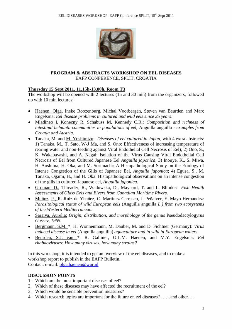
PROGRAM & ABSTRACTS WORKSHOP ON EEL DISEASES
EAFP CONFERENCE, SPLIT, CROATIA
Thursday 15 Sept 2011, 11.15h-13.00h, Room T3
The workshop will be opened with 2 lectures (15 and 30 min) from the organizers, followed
up with 10 min lectures:
Haenen, Olga, Ineke Roozenburg, Michal Voorbergen, Steven van Beurden and Marc
Engelsma: Eel disease problems in cultured and wild eels since 25 years.
Mladineo I, Konecny R, Schabuss M, Kennedy C.R.: Composition and richness of
intestinal helminth communities in populations of eel, Anguilla anguilla - examples from
Croatia and Austria.
Tanaka, M. and M. Yoshimizu: Diseases of eel cultured in Japan, with 4 extra abstracts:
1) Tanaka, M., T. Sato, W-J Ma, and S. Ono: Effectiveness of increasing temperature of
rearing water and non-feeding against Viral Endothelial Cell Necrosis of Eel); 2) Ono, S.,
K. Wakabayashi, and A. Nagai: Isolation of the Virus Causing Viral Endothelial Cell
Necrosis of Eel from Cultured Japanese Eel Anguilla japonica; 3) Inouye, K., S. Miwa,
H. Aoshima, H. Oka, and M. Sorimachi: A Histopathological Study on the Etiology of
Intense Congestion of the Gills of Japanese Eel, Anguilla japonica; 4) Egusa, S., M.
Tanaka, Ogami, H., and H. Oka: Histopathological observations on an intense congestion
of the gills in cultured Japanese eel, Anguilla japonica.
Groman, D., Threader, R., Wadowska, D., Maynard, T. and L. Blimke: Fish Health
Assessments of Glass Eels and Elvers from Canadian Maritime Rivers.
Muñoz, P., R. Ruiz de Ybañez, C. Martínez-Carrasco, J. Peñalver, E. Mayo-Hernández:
Parasitological status of wild European eels (Anguilla anguilla L.) from two ecosystems
of the Western Mediterranean.
Saraiva, Aurelia: Origin, distribution, and morphology of the genus Pseudodactylogyrus
Gussev, 1965.
Bergmann, S.M. *, H. Wonnenmann, M. Dauber, M. and D. Fichtner (Germany): Virus
induced disease in eel (Anguilla anguilla) aquaculture and in wild in European waters.
Beurden, S.J. van *, R. Galinier, O.L.M. Haenen, and M.Y. Engelsma: Eel
rhabdoviruses: How many viruses, how many strains?
In this workshop, it is intended to get an overview of the eel diseases, and to make a
workshop report to publish in the EAFP Bulletin.
Contact: e-mail: [email protected]
DISCUSSION POINTS
1. Which are the most important diseases of eel?
2. Which of these diseases may have affected the recruitment of the eel?
3. Which would be sensible prevention measures?
4. Which research topics are important for the future on eel diseases? ……and other….
EEL DISEASES WORKSHOP, EAFP Conference SPLIT, 15th
Sept 2011
2
EEL DISEASE PROBLEMS IN CULTURED AND WILD EELS SINCE 25 YEARS
Olga Haenen, Ineke Roozenburg, Michal Voorbergen, Steven van Beurden, Marc
Engelsma
Fish and Shellfish Diseases Laboratory, CVI Lelystad, The Netherlands;
e-mail: [email protected]
WELCOME to the EAFP Eel Diseases workshop!
The worldwide decline of the wild freshwater eel stocks is getting much attention. Reasons in
view for the decline are so far fisheries (overfishing), birds (predation), migration barriers,
and pollution, or a combination of these factors. The possible factor eel disease has not
received much attention.
Eels can have different diseases, caused by specific to nonspecific pathogens, like parasites,
bacteria en viruses. Some eel pathogens were transmitted by international transport, like
Anguillicoloides crassus, and viruses. Diseases may be lethal under stressful conditions and
high eel densities, like in eel farming. Wild eels may also show mortality under extreme
conditions.
Clinics of various diseases of wild and cultured eel are shown, based on 25 years diagnosis in
The Netherlands. Eels were infected with specific parasites (Anguillicoloides crassus,
Myxidium giardi, Trypanosoma), and common ectoparasites like Trichodina,
Ichthyophthirius multifiliis, Ichthyobodo, Chilodonella, (Pseudo)dactylogyrus, and
Gyrodactylus. Bacteria of eel were Vibrio vulnificus, and Edwardsiella tarda (both
potentially zoonotic), other Vibrio species, Aeromonas sobria, A.hydrophila, Pseudomonas
anguilliseptica, and Flavobacterium spp.. Viruses found were EVE, EVEX, and/or AngHV-
1(HVA). Apart from single infections, eels showed double or triple infections with
combinations of 1 or 2 viruses and 1 or 2 bacteria, often with parasites.
Would the factor “disease” be important in the decline of the wild eel populations? We found
44% of silver eels of the lower River Rhine positive for AngHV-1, with high % of A.crassus
& Trypanosoma. Our hypothesis is, that migrating, stressed silver eels, which are infected
with these parasites and AngHV-1, may get herpes viral disease during their spawning
migration, at permissive temperatures for the virus. The factor “disease” should be considered
as a possible serious factor in the decline of the eel stocks.
Eel farming is dependent on wild glass eels. Diseases thereby are transmitted to eel farms,
often closed systems. However, lately, farmed yellow eels are restocked into the wild, to
restore wild eel populations. This is dangerous for wild eels, as pathogens accumulate at eel
farms, and as for instance EVE (Eel Virus European) of farmed eel has not yet been found in
wild eels – these pathogens can be transmitted to wild eels without knowing the long term
effect.
EEL DISEASES WORKSHOP, EAFP Conference SPLIT, 15th
Sept 2011
3
COMPOSITION AND RICHNESS OF INTESTINAL HELMINTH COMMUNITIES
IN POPULATIONS OF EEL, ANGUILLA ANGUILLA –
EXAMPLES FROM CROATIA AND AUSTRIA
Mladineo I.1, Konecny R.
2,4, Schabuss M.
2, Kennedy C.R.
3
1Institute of Oceanography & Fisheries, Setaliste Ivana Mestrovica 63, 21000 Split, Croatia;
e-mail: [email protected] 2Department of Limnology, University of Vienna, Althanstrasse 14, A- 1090 Vienna, Austria;
e-mail: [email protected] 3 Department of Biological Sciences, University of Exeter, EX4 4PS, UK.
4 Environment Agency Austria, Department Surface Waters, Spittelauer Lände 5, 1090
Vienna, Austria.
Data from a long-term study of the intestinal helminth parasite community of eels, Anguilla
anguilla, stocked into the shallow eutrophic Neusiedler See, Austria, were collected from
1994 – 2009. 1044 eels from two sampling sites (Illmitz & South) were examined. The
parasite community showed characteristics similar to those in the natural eel populations in
Europe with only six species comprising the component community and a maximum
infracommunity richness of four species. Between 1994 and 2004, the intestinal parasite
community of the sampling site in Illmitz, which was originally dominated by
Acanthocephalus. lucii, changed. As levels of Acanthocephalus. anguillae increased to a
point at which it dominated the community, diversity increased whilst dominance of this
species decreased. By contrast, the community in the southern sampling site remained rather
constant with a continuously high infection level of A. anguillae and low abundance of A.
lucii. Both acanthocephalan species exhibited higher infestation levels in larger eels and in
different seasons of the year and the infestation parameters were significantly different
between the years of study. After 2004, A. lucii was not found in either of the two sampling
sites, which coincided with a drastic decline of its main final host Perca fluviatilis, whereas
A. anguillae infestation remained at similar levels as before. Changes and differences in the
fish communities of the two sampling sites and eel movements rather than interspecific
competition are discussed as possible explanations for the differences in the parasite
communities of the two sampling sites.
In Croatia, two sampling sites were chosen, one with typically brackish (Zrmanja) and the
other freshwater (Neretva). Eels were sampled every second month for two consecutive
years, showing structural and quantitative differences reflected by differences in salinity.
Component community numbered in total ten helminth/ copepod, three myxozoan and one
protozoan species. While A. crassus was more abundant in Zrmanja, Neretva population was
dominated by Ergasilus sieboldi and monogenean Pseudodactylogyrus sp. Seasonal
oscillations were observed in both eels populations.
EEL DISEASES WORKSHOP, EAFP Conference SPLIT, 15th
Sept 2011
4
DISEASE PROBLEMS OF EEL CULTURED IN JAPAN
Tanaka, M1 and M Yoshimizu
2*
1 Fuji Trout Farm, Shizuoka Prefectural Research Institute of Fishery, Inogashira,
Fujinomiya, Shizuoka 418-0108 Japan; e-mail: [email protected] 2 Laboratory of Biotechnology and Microbiology, Faculty of Fisheries Sciences, Hokkaido
University, Hakodate, Hokkaido 041-8611, Japan; e-mail: [email protected]
In Japan, eel culture began in 1879 in Fukagawa, Tokyo. Annual eel production in Japan has
gradually declined from 40,000 to 20,000 tons from 1999 to 2008, whereas imported eel has
been increased year by year from 1995. However, from 2000 there was a reduction from
130,000 to 50,000 tons. Despite no major change in production, Japanese eel culture industry
has been changing, from 1972. They are using a pond inside a green house, and water
temperature is kept around 28℃. Japanese eel culture industry seeks to produce a better
quality meat of cultured eel. These changes resulted in new disease problems, among the
disease issues, bacterial (Vibrio anguillarum, Pseudomonas anguilliseptica, Aeromonas
hydrophila, and atypical A. salmonicida), fungal (Saprolegnia diclina) and parasitic
(Heterosporis anguillarum) diseases decreased. Now, major diseases occurred in cultured eel
are viral (viral endothelial cell necrosis of eel; VECNE), bacterial (Edwardsiella tarda,
Flavobacterium columnare, gliding bacteria) and other diseases. Viral endothelial cell
necrosis of Japanese eel (VECNE) Anguilla japonica has been a serious problem of eel
culture in Japan. Histopathology of this disease, VEVNE was reported to be characterized by
intense congestion in central venous sinuses of gill filaments found in cultured eel. In gill
filaments of diseased fish, dilatation of the central venous sinuses was observed. In the liver,
hemorrhage and destruction of blood vessels were observed. Furthermore, hemorrhage in the
hematopoietic tissue and destruction of blood vessels and glomeruli were observed in the
kidney. Electron microscopy revealed hexagonal viral particles in the nuclei of the
degenerated endothelial cells. Each virion measured about 80 nm in diameter. The virion was
observed only in the degenerated endothelial cells. To isolate the causative virus, Japanese
eel endothelial cell (JEEC) line originating from vascular endothelial cells of Japanese eel
was established by Ono et al. (2003) and CPE with hypertrophied nuclei was found in JEEC
cells 7 days after inoculation. Virus particles of 75 nm in diameter were observed in the
hypertrophied nuclei. The isolated virus was DNA. It showed a tolerance up to 42℃. The
experimental infection by injecting the virus at 105.75
TClD50 into the abdominal cavity of
healthy eel produced congestion in the central venous sinus of gill lamella, with the
cumulative mortality being 60%. The virus was recovered from the gills, liver and kidney of
the experimentally infected fish. Effectiveness of increasing water temperature and non-
feeding against VECNE was evaluated. Cumulative mortalities of fish at 35℃ was as low as
that of 30℃. Rearing infected fish under the non-feeding condition further enhanced the
effect of treatment at 35℃. The effectiveness was dependent on rearing periods at the high
temperature; more than 3 days at 35℃ were needed to reduce mortality. Fish which survived
the primary challenge with virus at 35℃, showed high resistance to re-challenge. From these
results, the treatment of fish under the non-feeding condition at 35℃ is useful to control
VECEN.
EEL DISEASES WORKSHOP, EAFP Conference SPLIT, 15th
Sept 2011
5
Extra abstracts from Japan (not presented as lectures):
Fish Pathology, 43: 79-82, 2008
Effectiveness of increasing temperature of rearing water and non-feeding against Viral
Endothelial Cell Necrosis of Eel
Tanaka, M., T. Sato, W-J Ma, and S. Ono
Viral endothelial cell necrosis of eel (VECNE) of Japanese eel Anguilla japonica, caused by
an adenovirus (JEAdV), has been a serious problem of aquaculture industry in Japan. In the
present study, effectiveness of increasing water temperature and non-feeding against VECNE
was evaluated. Cumulative mortalities of fish intraperitoneally injected with 105.05
TC1D50/fish of JEAdV increased with elevating water temperature in the range between 20C
and 31C, but mortality at 35C was as low as that at 20C. Rearing infected fish under the non-
feeding condition further enhanced the effect of treatment at 35C. The effectiveness was
dependent on rearing periods at the high temperature; more than 3 days at 35C were needed
to reduce mortality. Fish, which survived the primary challenge with JEAdV at 35C, showed
high resistance to re-challenge with JEAdV. From these results, the treatment of fish under
the non- feeding condition at 35C is useful to control VECEN.
Fish Pathology, 42: 191-200, 2007
Isolation of the Virus Causing Viral Endothelial Cell Necrosis of Eel from Cultured
Japanese Eel Anguilla japonica
Ono, S., K. Wakabayashi, and A. Nagai
To isolate the causative virus for viral endothelial cell necrosis of eel (VECNE), we
established the cell line JEEC, originated from vascular endothelial cells of Japanese eel
Anguilla japonica. CPE with hypertrophied nuclei was found in JEEC inoculated with the
filtrate of homogenized gills of diseased fish 7 days after inoculation. In the hypertrophied
nuclei, icosahedral virus particles of about 75 nm in diameter were observed. The nucleic
acid in the isolated virus was DNA. The virus was tolerant to chloroform and pH 3.0. It
showed tolerance up to 42C. Thus,this virus was classified as an adenovirus. The
experimental infection by injecting the virus at 105.75
TClD50 into the abdominal cavity of
healthy eel produced congestion in the central venous sinus of gill lamella, which is the
typical sign of this disease, with the cumulative mortality being 60%. The virus was
recovered from the gills, liver and kidney of the experimentally infected fish. Therefore, the
virus of VECNE was identified as the causative agent.
Fish Pathology, 29: 35-41, 1994
A Histopathological Study on the Etiology of Intense Congestion of the Gills of Japanese
Eel, Anguilla japonica
Inouye, K., S. Miwa, H. Aoshima, H. Oka, and M. Sorimachi
Histopathology of the disease, which was reported to be characterized by intense congestion
in central venous sinuses of gill filaments found in cultured eel was studied with
spontaneously diseased fish and experimentally infected fish by light and electron
microscopy. Experimental infection produced similar histopathological changes to those
observed in the spontaneously diseased fish. In gill filaments of diseased fish, dilatation of
EEL DISEASES WORKSHOP, EAFP Conference SPLIT, 15th
Sept 2011
6
the central venous sinuses was observed. In the liver, hemorrhage in the parenchyma and
destruction of blood vessels were observed. Furthermore, hemorrhage in the hematopoietic
tissue and destruction of blood vessels and glomeruli were observed in the kidney. These
pathological changes were always accompanied by degeneration of endothelial cell nuclei of
b1ood vessels. The degeneration of the nuclei was characterized by swelling, intense staining
of the nuclear rim with hematoxylin, and a homogenous appearance of the nucleoplasm.
Electron microscopy revealed hexagonal viral particles in the nuclei of the degenerated
endothelial cells. Each virion measured about 80 nm in diameter. The virions were observed
only in the degenerated endothelial cells. These results suggest that the disease of cultured eel
is a systemic viral infection, which is characterized by the necrosis of endothelial cells of
b1ood vessels.
Fish Pathology 24: 51-56, 1989
Histopathological observations on an intense congestion of the gills in cultured Japanese
eel, Anguilla japonica
Egusa, S., M. Tanaka, Ogami, H., and H. Oka
A new disease of Anguilla japonica which is characterized by an intense congestion of the
gill filaments was histopathologically studied. In all the diseased eels studied the central
venous sinuses and all other venous vessels of the gill filaments were observed to be filled
with blood and markedly inflated. In part of the gill filaments it was observed that the blood
once filled the central venous sinuses and other venous vessels had partly or almost entirely
flowed out, leaving a marked dilatation of them. Destructive lesions were not observed in any
part of the gill vasculature. No evidence of obstruction was observed in any part of the
arterio-arterior vasculature. Arteriovenous anastomoses were observed to maintain the normal
structure. The intense congestion of the arterio-venous vasculature in discased eels is of
essentially unknown etiology, but some physiological disturbance in cardiovasculature
functions may be involved in the causation.
EEL DISEASES WORKSHOP, EAFP Conference SPLIT, 15th
Sept 2011
7
RESULTS OF FISH HEALTH ASSESSMENTS OF GLASS EELS, ANGUILLA
ROSTRATA, FROM CANADIAN MARITIME RIVERS FROM 2006-2010
D Groman1*
, R Threader2, D Wadowska
3, T Maynard
2 and L Blimke
4
1
Aquatic Diagnostic Services, Atlantic Veterinary College, University of Prince Edward
Island, 550 University Ave., Charlottetown, PEI C1A4P3 Canada. [email protected] 2 Ontario Power Generation, 2 Innovation Dr., Renfrew, ON K7V4H4 Canada.
[email protected], [email protected] 3
Electron Microscopy Laboratory, Atlantic Veterinary College, University of Prince Edward
Island, 550 University Ave., Charlottetown, PEI C1A4P3 Canada.
Kleinschimidt Associates, 35 Pratt St., Suite 201, Essex, CT 06426 USA.
Results of fish health assessments for newly captured glass eels (Anguilla rostrata) are
presented. Populations were segregated by river of capture, held in quarantine and
subsequently assessed for the following infectious disease: for viruses - VHSV, IPNV, ISAV,
IHNV, SVC and HVA; for bacteria - Aeromonas salmonicida, Yersinia ruckeri; and for
parasites Anguillicoloides crassus. Assessments completed in 2006 were on a single sample
lot of 225 eels. Those completed in 2007 were on 5 sample lots of 300 individuals. In 2008
on four additional sample lots with the glass eel numbers increased to 340. And, in 2009-10
on 5 sample lots of 340 eels, including bacteriology on each of the 17 virus pools per.
Results of virus isolation and molecular assays of pooled samples from all years were
negative for all viruses of interest. Bacteriological culture of the 2009-10 pooled samples
revealed that one lot held on surface water prior to sampling was harboring Yersinia ruckeri.
Parasitological examination revealed trophozoite stages of the ciliate Ichthyophthirius
multifiliis. Histopathological examination of individuals in all lots revealed necrotizing
hepatitis and associated an intranuclear microsporidian, morphologically consistent with a
Nucleospora sp. One Lot of older elvers from the estuary of the St. Mary’s River in
Guysbourgh County, Nova Scotia was found to be harbouring larval and pre-adult nematodes
in association with the wall and lumen of the gas bladder. These nematodes were
morphologically consistent with Anguillicoloides crassus. In addition, this lot was infected
with an as yet identified Myxosporidian infection of the urethra and urinary bladder.
EEL DISEASES WORKSHOP, EAFP Conference SPLIT, 15th
Sept 2011
8
PARASITOLOGICAL STATUS OF WILD EUROPEAN EELS (Anguilla anguilla L.)
FROM TWO ECOSYSTEMS OF THE WESTERN MEDITERRANEAN
P. Muñoz1, R. Ruiz de Ybañez
1, C. Martínez-Carrasco
1, J. Peñalver
2, E. Mayo-
Hernández1
1 Dpto. Sanidad Animal. Universidad de Murcia, Murcia, Spain; e-mail: [email protected]
2 Dirección General de Ganadería y Pesca, Comunidad Autónoma de la Región de Murcia,
Murcia, Spain.
Parasitological status of wild European eels from two different ecosystems in Western
Mediterranean Sea, an oligohaline (up to 2 g/l) coastal lagoon (the Albufera Lake in
Valencia, ALB) and a hypersaline (43-46.5 g/l) coastal lagoon (the Mar Menor lagoon, MM
in Murcia) has been evaluated. A total of 48 eels from ALB and 406 from MM have been
sampled between years 2008 and 2011.
Samples of swim bladder, intestine, gill and skin were processed for parasitological studies.
Additionally, an artificial digestion method was optimized for the detection, isolation and
counting of larval stages (second stage L2, third stage L3 and fourth stage L4) of the
nematode Anguillicoloides crassus in the swimbladder wall.
Myxidium giardi, Eimeria anguillae and two cestoda species (Bothriocephalus claviceps and
Proteocephalus macrocephalus), were detected in the intestine of ALB eels. Myxidium giardi
was also observed in gills from ALB eels. Eimeria anguillae and three trematoda species
(Deropristis inflata, Lecithochirium spp and Bucephalus spp.) were isolated from the
intestine of MM eels.
A. crassus prevalence detected in eels from ALB (82%) was much higher than prevalence
previously reported in wild eels from this ecosystem (Esteve and Alcaide, 2009). This could
be probably due to the fact that the A. crassus infection rates rapidly increase in the first few
years following its appearance and then stabilize at a prevalence of around 60-70%. On the
contrary, a very low A. crassus prevalence (3%) was detected in eels from MM, which could
be due either to the recent arrival of this exotic nematode in this environment or to the lack of
adequate intermediate hosts. The artificial digestion method used allows a rapid, precise and
easy quantification of L2. This was the most prevalent larval stage in infected eels from MM,
suggesting that it is a chronic infection. In the absence of re-infection, L2 persist in the final
host until they are eliminated into the environment in faeces or when eels die. A similar
pattern has previously been described for other nematodes.
The results obtained show differences in parasitological status of eels from the two
ecosystems studied, suggesting that the presence of pathogens in eels is highly influenced by
the environmental conditions of each type of coastal lagoon habitats.
Esteve C., Alcaide E. (2009) Aquaculture, 289, 143-149.
EEL DISEASES WORKSHOP, EAFP Conference SPLIT, 15th
Sept 2011
9
ORIGIN, DISTRIBUTION AND MORPHOLOGY OF THE GENUS
PSEUDODACTYLOGYRUS GUSSEV, 1965
A. Saraiva1,2
1Faculdade de Ciências, Universidade do Porto, Porto, Portugal, e-mail: [email protected]
2CIIMAR/CIMAR-LA, Interdisciplinary Centre of Marine and Environmental Research, U.P.,
Porto, Portugal
The first references to the monogenean eel parasites Pseudodactylogyrus bini (Kikuchi, 1929)
Gussev 1965 and Pseudodactylogyrus anguillae (Yin et Sproston, 1948) Gussev 1965 come
from Asiatic countries, mainly from Japan and China. At that time Kikuchi included the
monogenean he found in the genus Dactylogyrus Diesing, 1850 while Yin and Sproston
referred to the species he detected as belonging to the genus Neodactylogyus Price, 1938. It
was only in 1965 that Gussev studying the Australian eel monogeneans consider they were
different enough to create a new genus that he proposed to be Pseudodactylogyrus.
In1977 Golovin was the first to record these parasites in Russia and in 1984 Molnár reported
them from Hungary. Since then these parasites were reported in many European countries. In
Portugal these parasites were reported for the first time in 1989 by Saraiva & Chubb. These
parasites were observed for the first time in North America (Canada) by Cone and
Marcogliese in 1995. The last new geographical records of these parasites were from South
Africa (Christison & Baker, 2007) and Island of Reunion located in the Indian Ocean (Sasal
et al., 2008).
In the genus Pseudodactylogyrus the haptor is ventral, the anchors point ventrally and the
connecting bar is ventral to the anchors. The marginal hooks are of the larval type. There is
only one prostatic reservoir in this genus. The shape and the size of the haptor and of the
anchors are the main distinguish features of P. bini from P. anguillae.
In the last decades it was stated a significant decrease of glass eel in the European coast. In
what extent eel diseases including that ones of parasitological ethyology namely
Pseudodactylogyrosis are responsible for this reduction it is not known, and should be
discussed in this workshop.
EEL DISEASES WORKSHOP, EAFP Conference SPLIT, 15th
Sept 2011
10
VIRUS INDUCED DISEASE IN EEL (ANGUILLA ANGUILLA) AQUACULTURE
AND IN WILD IN EUROPEAN WATERS
S.M. Bergmann*1, H. Wonnenmann
2, M. Dauber
1, M. and D. Fichtner
1
1Friedrich-Loeffler-Institut (FLI), German Reference Laboratory for Fish Diseases, Federal
Research Institute for Animal Health, Insel Riems,Germany;
e-mail: [email protected] 2Federal State Laboratory of Schleswig-Holstein, Neumünster, Germany
Over the last 20 years not only the costs for elvers from European eel (A. anguilla) exploded
but also the importance of aquaculture, also for eel, in general. Due to different viruses,
massive losses were observed in European eel aquaculture but also in the wild.
In primarily investigations the eel herpesvirus (herpesvirus anguillae, HVA) was found to be
one of the main threats for both aquacultured and wild eel. Statewide investigations,
especially in the German Federal State Schleswig-Holstein, by a newly established real-time
PCR have shown that HVA is widespread in the wild. Investigation on farm level came to the
same conclusions. The majority of eel farms are latently infected by HVA.
Virological and serological tools for the detection and confirmation of different eel viruses
were designed, established and widely used in diagnostics of viral eel diseases by the German
Reference Laboratory for Fish Diseases
On farm level mainly HVA, birnavirus (type Sp), picornavirus and reovirus but also different
rhabdoviruses (EVEX, SVCV-like, Perch Rhabdovirus-like) were detected by PCR, RT-PCR,
real-time PCR, electron microscopy. After cell cultivation onto EK-1 cells, the isolates were
characterized and identified by immunofluorescence assay (IFAT) using polyclonal and
monoclonal antibodies.
With those reagents, also assays like serum neutralisation or antibody ELISA have been
established: The tests were also used for virus or antibody detection in freshly caught elvers
which were tested directly without re-laying.
It could be shown in some experiments that especially elvers may carry different viruses
latently without any clinical signs. After metamorphosis from glass eels over elvers into
silver eels those latently or persistently present viruses may cause a disease with high losses.
To combat viral diseases in eel, pilot projects on farm level with immunization against HVA
and birnavirus were successfully performed. After immersion of inactivated viral antigen
preparation losses in elvers due to these viruses could be reduced from 90 to 10%.
EEL DISEASES WORKSHOP, EAFP Conference SPLIT, 15th
Sept 2011
11
EEL RHABDOVIRUSES: HOW MANY VIRUSES, HOW MANY STRAINS?
S.J. van Beurden*1,2
, R. Galinier3, O.L.M. Haenen
1, and M.Y. Engelsma
1
1Central Veterinary Institute, part of Wageningen UR, Lelystad, The Netherlands;
e-mail: [email protected] 2Faculty of Veterinary Medicine, Utrecht University, Utrecht, The Netherlands.
3Laboratoire Ecologie et Evolution des Interactions (2EI) UMR 5244 CNRS-UPVD,
Perpignan, France.
Several rhabdoviruses infecting freshwater eels of the genus Anguilla have been described.
Infection with eel rhabdoviruses is not necessarily accompanied by clinical signs, but under
certain conditions a severe hemorrhagic disease with significant mortality may develop. The
first rhabdoviruses from eel were described by Sano et al. in Japan in the mid-1970s, namely
Eel virus American (EVA) and Eel virus European X (EVEX). Further characterization in the
early-1980s showed that these viruses are highly similar in morphological, serological and
biochemical characteristics, and probably represent two strains of the same eel vesiculovirus.
Other eel rhabdoviruses have been described and partially characterized since. Most
importantly five rhabdovirus isolates were found in elvers from the Loire estuary (Castric et
al., 1984). Three of these isolates, namely B44, C30, and D13, are similar or closely related to
EVEX, two other isolates (B12 and C26) are considered to be eel novirhabdoviruses. In 1992,
another eel rhabdovirus was found in Japanese eel, associated with a dermatitis. This
rhabdovirus appeared to be serologically similar to EVEX (Kobayashi & Miyazaki, 1996).
Our molecular characterization of partial sequences from the RNA polymerase or L gene
from EVEX and EVA isolates confirms the existence of two lineages with 91.5% sequence
identity over a 2040 bp fragment. Recently, the complete genome sequence of EVEX isolate
CVI-NL 153311 has been determined. EVEX has a genome of 11,806 bp in length, which
encodes for the five classical structural proteins, plus an overlapping putative open reading
frame in the phosphoprotein. Based on phylogenetic analyses of gene and genejunction
sequences, EVEX has tentatively been placed in the genus Vesiculovirus of the family
Rhabdoviridae, most closely related to trout rhabdovirus. Future research should focus on the
molecular characterization of the eel vesiculovirus strain EVA and the eel novirhabdovirus
isolates.