Embed Size (px)
Citation preview
lable at ScienceDirect
Progress in Biophysics and Molecular Biology 104 (2011) 2e21
Contents lists avai
Progress in Biophysics and Molecular Biology
journal homepage: www.elsevier .com/locate/pbiomolbio
Review
Cardiac cell modelling: Observations from the heart of the cardiacphysiome project
Martin Fink a,1,3, Steven A. Niederer b,1, Elizabeth M. Cherry c, Flavio H. Fenton c,Jussi T. Koivumäki d,4, Gunnar Seemann e, Rüdiger Thul f, Henggui Zhang g, Frank B. Sachse h,2,Dan Beard i,2, Edmund J. Crampin j,2, Nicolas P. Smith b,*,2
aDepartment of Physiology, Anatomy and Genetics, University of Oxford, OX1 3JP, United KingdombComputing Laboratory, University of Oxford, OX1 3QD, United KingdomcDepartment of Biomedical Sciences, Cornell University, Ithaca, NY 14850, USAdDepartment of Physics, University of Oulu & Biocenter Oulu, PO Box 3000, 90014 Oulun yliopisto, Finlande Institute of Biomedical Engineering, Karlsruhe Institute of Technology, 76131 Karlsruhe, Germanyf School of Mathematical Sciences, University of Nottingham, NG7 2RD, United KingdomgBiological Physics Group, School of Physics and Astronomy, The University of Manchester, Manchester M13 9PL, UKhNora Eccles Harrison Cardiovascular Research and Training Institute and Bioengineering Department, University of Utah, Salt Lake City, UT 84112, USAiBiotechnology and Bioengineering Center, Dept. of Physiology, Medical College of Wisconsin, Milwaukee, WI, USAjAuckland Bioengineering Institute and Department of Engineering Science, The University of Auckland, Private Bag 92019, Auckland 1142, New Zealand
a r t i c l e i n f o
Article history:Available online 18 March 2010
Keywords:CardiacCellularPhysiomeElectrophysiologyMathematical modelling
* Corresponding author. Tel.: þ44 (0)1865 610 669E-mail address: [email protected] (N.P. S
1 Joint first authors.2 Joint senior authors.3 Present address: Modeling and Simulation, Novar4 Present address: A. I. Virtanen Institute for Molec
0079-6107/$ e see front matter � 2010 Elsevier Ltd.doi:10.1016/j.pbiomolbio.2010.03.002
a b s t r a c t
In this manuscript we review the state of cardiac cell modelling in the context of international initiativessuch as the IUPS Physiome and Virtual Physiological Human Projects, which aim to integrate compu-tational models across scales and physics. In particular we focus on the relationship between experi-mental data and model parameterisation across a range of model types and cellular physiologicalsystems. Finally, in the context of parameter identification and model reuse within the Cardiac Physiome,we suggest some future priority areas for this field.
� 2010 Elsevier Ltd. All rights reserved.
Contents
1. Introduction . . . . . . . . . . . . . . . . . . . . . . . . . . . . . . . . . . . . . . . . . . . . . . . . . . . . . . . . . . . . . . . . . . . . . . . . . . . . . . . . . . . . . . . . . . . . . . . . . . . . . . . . . . . . . . . . . . . . . . . . .32. Experimental preparations and techniques . . . . . . . . . . . . . . . . . . . . . . . . . . . . . . . . . . . . . . . . . . . . . . . . . . . . . . . . . . . . . . . . . . . . . . . . . . . . . . . . . . . . . . . . . . . . . . .4
2.1. Experimental preparations . . . . . . . . . . . . . . . . . . . . . . . . . . . . . . . . . . . . . . . . . . . . . . . . . . . . . . . . . . . . . . . . . . . . . . . . . . . . . . . . . . . . . . . . . . . . . . . . . . . . . . 42.2. Experimental techniques . . . . . . . . . . . . . . . . . . . . . . . . . . . . . . . . . . . . . . . . . . . . . . . . . . . . . . . . . . . . . . . . . . . . . . . . . . . . . . . . . . . . . . . . . . . . . . . . . . . . . . . 5
2.2.1. Protein structure and expression . . . . . . . . . . . . . . . . . . . . . . . . . . . . . . . . . . . . . . . . . . . . . . . . . . . . . . . . . . . . . . . . . . . . . . . . . . . . . . . . . . . . . . . . . 52.2.2. Sub-cellular and cell architecture . . . . . . . . . . . . . . . . . . . . . . . . . . . . . . . . . . . . . . . . . . . . . . . . . . . . . . . . . . . . . . . . . . . . . . . . . . . . . . . . . . . . . . . . 52.2.3. Electrophysiology . . . . . . . . . . . . . . . . . . . . . . . . . . . . . . . . . . . . . . . . . . . . . . . . . . . . . . . . . . . . . . . . . . . . . . . . . . . . . . . . . . . . . . . . . . . . . . . . . . . . . . 5
2.3. Current experimental challenges and limitations . . . . . . . . . . . . . . . . . . . . . . . . . . . . . . . . . . . . . . . . . . . . . . . . . . . . . . . . . . . . . . . . . . . . . . . . . . . . . . . . . . 63. Model component development . . . . . . . . . . . . . . . . . . . . . . . . . . . . . . . . . . . . . . . . . . . . . . . . . . . . . . . . . . . . . . . . . . . . . . . . . . . . . . . . . . . . . . . . . . . . . . . . . . . . . . . .6
3.1. Models for ion channels . . . . . . . . . . . . . . . . . . . . . . . . . . . . . . . . . . . . . . . . . . . . . . . . . . . . . . . . . . . . . . . . . . . . . . . . . . . . . . . . . . . . . . . . . . . . . . . . . . . . . . . . 63.2. Models of ion pumps and exchangers . . . . . . . . . . . . . . . . . . . . . . . . . . . . . . . . . . . . . . . . . . . . . . . . . . . . . . . . . . . . . . . . . . . . . . . . . . . . . . . . . . . . . . . . . . . . 7
4. Review of existing models . . . . . . . . . . . . . . . . . . . . . . . . . . . . . . . . . . . . . . . . . . . . . . . . . . . . . . . . . . . . . . . . . . . . . . . . . . . . . . . . . . . . . . . . . . . . . . . . . . . . . . . . . . . .74.1. Overview of mammalian electrophysiology models . . . . . . . . . . . . . . . . . . . . . . . . . . . . . . . . . . . . . . . . . . . . . . . . . . . . . . . . . . . . . . . . . . . . . . . . . . . . . . . . 84.2. Electrophysiology models of human ventricular myocytes . . . . . . . . . . . . . . . . . . . . . . . . . . . . . . . . . . . . . . . . . . . . . . . . . . . . . . . . . . . . . . . . . . . . . . . . . . 9
.mith).
tis Pharma AG, 4002 Basel, Switzerland.ular Sciences, University of Eastern Finland, P.O. Box 1627, 70211 Kuopio, Finland.
All rights reserved.
M. Fink et al. / Progress in Biophysics and Molecular Biology 104 (2011) 2e21 3
4.3. Models of cell contraction . . . . . . . . . . . . . . . . . . . . . . . . . . . . . . . . . . . . . . . . . . . . . . . . . . . . . . . . . . . . . . . . . . . . . . . . . . . . . . . . . . . . . . . . . . . . . . . . . . . . . . 94.4. Models of calcium signalling in cardiac cells . . . . . . . . . . . . . . . . . . . . . . . . . . . . . . . . . . . . . . . . . . . . . . . . . . . . . . . . . . . . . . . . . . . . . . . . . . . . . . . . . . . . . 114.5. Reduced electrophysiology models . . . . . . . . . . . . . . . . . . . . . . . . . . . . . . . . . . . . . . . . . . . . . . . . . . . . . . . . . . . . . . . . . . . . . . . . . . . . . . . . . . . . . . . . . . . . . 12
5. Model identificationdmodel selection . . . . . . . . . . . . . . . . . . . . . . . . . . . . . . . . . . . . . . . . . . . . . . . . . . . . . . . . . . . . . . . . . . . . . . . . . . . . . . . . . . . . . . . . . . . . . . . . .135.1. Parameterisation, data availability and local minima . . . . . . . . . . . . . . . . . . . . . . . . . . . . . . . . . . . . . . . . . . . . . . . . . . . . . . . . . . . . . . . . . . . . . . . . . . . . . . 135.2. Sensitivity of parameters and their confidence interval . . . . . . . . . . . . . . . . . . . . . . . . . . . . . . . . . . . . . . . . . . . . . . . . . . . . . . . . . . . . . . . . . . . . . . . . . . . . 145.3. Validation with distinct data, identification of limitations and possible extrapolation . . . . . . . . . . . . . . . . . . . . . . . . . . . . . . . . . . . . . . . . . . . . . . . . . 14
6. Discussion . . . . . . . . . . . . . . . . . . . . . . . . . . . . . . . . . . . . . . . . . . . . . . . . . . . . . . . . . . . . . . . . . . . . . . . . . . . . . . . . . . . . . . . . . . . . . . . . . . . . . . . . . . . . . . . . . . . . . . . . . .156.1. Initiative 1: Application of tools for optimal model identification . . . . . . . . . . . . . . . . . . . . . . . . . . . . . . . . . . . . . . . . . . . . . . . . . . . . . . . . . . . . . . . . . . . 166.2. Initiative 2: Reconciling model complexity with data availability and modelling goals . . . . . . . . . . . . . . . . . . . . . . . . . . . . . . . . . . . . . . . . . . . . . . . . 166.3. Initiative 3: Establishment of a repository of experimental data connected with models . . . . . . . . . . . . . . . . . . . . . . . . . . . . . . . . . . . . . . . . . . . . . . . 17Acknowledgements . . . . . . . . . . . . . . . . . . . . . . . . . . . . . . . . . . . . . . . . . . . . . . . . . . . . . . . . . . . . . . . . . . . . . . . . . . . . . . . . . . . . . . . . . . . . . . . . . . . . . . . . . . . . . . . . . 17References . . . . . . . . . . . . . . . . . . . . . . . . . . . . . . . . . . . . . . . . . . . . . . . . . . . . . . . . . . . . . . . . . . . . . . . . . . . . . . . . . . . . . . . . . . . . . . . . . . . . . . . . . . . . . . . . . . . . . . . . . 17
1. Introduction
The Physiome Project is a worldwide effort that aims to producean integrative computational framework with which to understandeukaryotic and human physiology (Crampin et al., 2004; Hunterand Borg, 2003). Formalised under the banners of the Interna-tional Union of Physiological Sciences (IUPS) (Hunter, 2004) andVirtual Physiological Human (VPH) (Viceconti et al., 2008) initia-tives, the approach and philosophy are common to a large numberof computational physiology research efforts.
Multiscale heart modelling is arguably further advanced thanany other organ system; in particular the modelling of cardiaccontraction, electrical excitation and metabolism at the cellularscale provides perhaps the most sophisticated examples ofmodelling frameworks currently available. Most notably, within theclass of cell and tissue models dealing with cardiac electrophysi-ology, multiple data sets and functional knowledge have beenrationalised and incorporated into single-cell modelling frame-works. These frameworks have in turn provided the ability toanalyse the effects of the multiple coupled mechanisms whichunderpin the emergent behaviour of the heart (Rudy, 2000).
The inherently modular structure of cell physiology has beencentral to the progression of this cellular modelling work andperhaps the Physiome concept itself. Modelling the channels andtransporters which regulate ionic flux across cellular membranes asindividual components provides a set of discrete sub-models. Thesesub-models are often phenomenological at the scale of theirbiochemical regulation (described by kinetic schemes which arefitted to data). However, by coupling sub-models describing regu-lation of ionic fluxes within and between sub-spaces (e.g. buffers,channels and exchangers), whole cell models can be assembled,capturing the electrophysiological organisation of the cell. Thismodular structure, both in vivo and in silico, provides the potentialto uncouple these physiological components and to facilitate thedevelopment and assembly of the sub-models which describethem. Specifically, new cell models can be created through intro-ducing new components into existing cellular modelling frame-works or by assembling existing components into a new cellularbiophysical structure (e.g. Terkildsen et al., 2008; Cortassa et al.,2003). The cardiac cellular modelling literature has many exam-ples of model development through both processes, often incombination, i.e. individual components are replaced (e.g. Flaimet al., 2006) and frameworks are altered (e.g. Campbell et al.,2009). The success of this approach is evident in the expandingnumber of cardiac models with increasing focus on representingspecific tissue types, animal species and physiological functions;many of these are reviewed below.
The relative maturity of cardiac cell modelling has meant thatdevelopments in this field have shaped many of the ideas funda-mental to the philosophy of the Physiome, in particular, usingbiophysical models for integrating data from multiple sources, thedevelopment of multiscale models through coupling existingmodel components and using interfaces that represent specificbiophysical mechanisms. Through the development of communitystandards for unambiguously representing and curating models,we now have the ability to rapidly and accurately assemble andcouple functional representations of physiological processes.Through this framework, we gain in turn the capacity to embedmultiple layers of knowledge to reveal insights and account foremergent or otherwise unanticipated effects. There are a number ofexamples of such insights in the cardiac field where modelling haspredicted phenomena which was subsequently confirmed experi-mentally. The earliest example in cardiac cell modelling is arguablyfrom the pioneeringmodel work of Noble (1962) who identified theenergy-saving properties of the inward potassium rectifier current.Another example is the theoretical prediction of complex patternsof excitation in cardiac tissue by stimuli and the related mecha-nisms of make and break in defibrillation (Sepulveda et al., 1989;Roth and Wikswo, 1986; Roth, 1995), which preceded experi-mental verification (Wikswo et al., 1995; Nikolski et al., 2002). Oneof the most important discoveries that changed how cardiacarrhythmias were understood was the discovery of functionalreentrant waves (spiral waves), which again were demonstratedfirst using cardiac cell models (Courtemanche and Winfree, 1991;Holden and Panfilov, 1991) before being shown experimentallyusing optical mapping techniques (Davidenko et al., 1992). Mostrecently electro-mechanical models have been applied to predictspatial heterogeneity (Campbell et al., 2008) before experimentaldata provided direct supporting evidence (Stelzer et al., 2008).
However, the relative maturity of computational modelling ofcardiac cellular function means that, in addition to being ina unique position to illustrate the power of the Physiome approach,it also highlights most tangibly some of the most significant currentchallenges. In our experience, many of these issues also relate to thereuse of model components (Niederer et al., 2009). Specifically,while markup languages such as CellML (Garny et al., 2008) andSBML (Hucka et al., 2004) allow models to be reused with fidelity,there is no clear way for the assumptions and data on whichindividual model components are based to also be inherited. Thismeans there is potential for existing sub-models to be embedded innew frameworks which aim to simulate a dynamic range inap-propriate for the initial parameterisation of the model component.It also means that errors that were made when an existingsub-model was developed, for example in the kinetic scheme or

M. Fink et al. / Progress in Biophysics and Molecular Biology 104 (2011) 2e214
parameter fit of a channel or exchanger, are carried through ‘as is’when building a new cell model. Furthermore, because in some cellmodels practitioners have determined parameters by fitting thewhole cell model to emergent measures of cell function, such asaction potentials or tension transients, errors that this introduces insub-models are propagated when components of these models arereused. In cases where the emergent behaviour is not sensitive toerror in a particular sub-component this is not an important issue.However, in at least some cases this will not be true and thesensitivity of a particular physiological property to parametererrors will need to be considered during model development. Thisis, apparently, rarely done at present.
Many modelling papers do not provide any details about themethod used for determining the model parameters; some revealthat the fit was done by hand. This may indicate a lack of softwaretools to guide modellers in this task. More drastically, physiologicalconclusions have been drawn from models that apparently do notprovide a good fit to the experimental data (e.g., Fink and Noble,2009). Another problem is that the experimental data ultimatelyunderlying the parameterisation of whole cell models do notalways comprise a consistent data set, i.e., the data have beencollected from different cell types and species, and under variousexperimental conditions, including substantial variations intemperature. Any model derived from, and able to fit all these dataat once would only describe some mean, relatively nonspecificbehaviour. It is important to note that data may not always beconsistent between, or evenwithin, experimental laboratories and/or preparations. However, species and temperature consistency is,arguably, a fundamental step in enabling models to representphysiological function quantitatively and mechanistically. In fact,different models of cell electrophysiology for the same species andregion of the heart tend to show different behaviour in terms ofresponses to channel block and in emergent properties like rateadaptation andmemory (e.g. Nygren et al., 2001; ten Tusscher et al.,2006; Cherry and Fenton, 2007; Bueno-Orovio et al., 2008; Cherryet al., 2008).
Finally, the parameter identification problem is itself also a keyissue in managing the link between cellular model parameters andexperimental data. Specifically, the challenge is in determining andquantifying the ability to uniquely extract a parameter value fromavailable experimental data. By reusing model components thisprocess of determining parameters from data can be avoided,although it is unclear to what extent parameter values for differenttypes of cells and across species remain the same. However, withincardiac cell models, inheritance of model components has recentlybeen shown to span multiple generations of models meaning thatthese components, and thus also the models that contain them, areoften based at least in part on data collected many years before themodel was published (Niederer et al., 2009). For example, oldermodels routinely relied onmulticellular preparation data, which, inmany cases, have now been superseded by isolated cell data.Furthermore, species and/or temperature-specific data may nothave been available for earlier models but is now more readilyavailable. Through incautious model reuse, parameters derivedfrom data which are now superseded can find their way into newlypublished models. With new experiments producing an increasingquality and quantity of measurement data, this represents anopportunity lost.
Our goals for this manuscript are multiple. For the reader new tothe subject, in Section 2 we outline the sources and issues ofexperimental data used to parameterise equations representingspecific cellular processes, described in Section 3. In Section 4 wereview different classes of cardiac cell models before, in Section 5,discussing research issues relating to model identification andparameterisation and the need, arising from this, to robustly link
model parameters and data in the form of standardised tests.Finally in Section 6 we conclude with some proposals for potentialfuture initiatives for the field.
2. Experimental preparations and techniques
As outlined above, models of cardiac cells and sub-cellularcomponents are based on numerous experimental studies carriedout on various types of preparations. Data from these studies areused for guiding the design of model structure as well as forparameterisation of model components and validation. This sectiongives an introductory overview on major types of experimentalpreparations and measurement techniques. Attention is paidprimarily to preparations and techniques at the cellular, sub-cellularand molecular levels that are currently providing the major datasource for cardiac cell modelling. Furthermore, we identify experi-mental challenges and limitations of experimental approaches thatare relevant for cardiac cell modelling with a particular emphasis onnormal and diseased human cells.
2.1. Experimental preparations
A major decision in the design of experimental studies is theselection of an appropriate preparation. This selection has impor-tant implications for the interpretation of data from the experi-mental study and also the application of the data in modelling.Across the range of scales from protein-expression systems throughto cell-membrane patches and isolated cells to tissue sections,established types of preparations have been developed. Mostpreparations for cardiac studies are harvested from excised animalhearts. Human preparations are commonly obtained by biopsy andfrom unused heart transplants.
At the protein level several expression systems have beendeveloped to study the biochemistry and function of proteins. Thesystems allow for expressing exogenous proteins in cell types suchas frog oocytes, human embryonic kidney (HEK) cell lines andimmortalised monkey kidney cells (COS). Protein expression iscaused by injection of mRNA into oocytes. The mRNA can bemodified by site-directed mutagenesis, which allows for charac-terisation of the effects of mutations. A feature of expressionsystems is the commonly high level of protein expression, whichallows one not only to ignore the effects of native proteins(expressed at lower levels), but also to increase the signal-to-noiseratio of measurement data. Thus, expression systems facilitatemeasurements of signals that are not apparent in native cells and/or cannot be distinguished from other signals. An example is themeasurement of gating currents of ion channels (Armstrong andBezanilla, 1973), which have amplitudes smaller than 1% of theion current passing through the channels. A limitation of expres-sion systems is that the protein is not expressed in its nativeenvironment, which would include auxiliary proteins andmicroenvironment.
Membrane patches can be extracted from many cell types usingtechniques developed by Neher and Sakmann (2007). Thesemicroscopic patches provide the basis for a number of measure-ment approaches to studying the electrophysiology of ion channels,transporters and exchangers (see next section) and the character-isation of related membrane proteins and signalling pathways.Membrane patches are commonly extracted from isolated cells,which constitute themajor preparation for studies of the propertiesof single cells. An established method to obtain isolated cell prep-arations is based on dissociating cells from tissue by collagenases(enzymes that degrade collagen). These preparations of isolatedcells are used, for instance, for recording electrical currents andvoltages through cell membranes. Isolated cells are also a standard
M. Fink et al. / Progress in Biophysics and Molecular Biology 104 (2011) 2e21 5
preparation for microscopic imaging studies of cell architecture andfunction, e.g. intracellular calcium concentration and membranevoltage. A problemwith isolated-cell preparations is the removal ofintercellular coupling and coupling to the extracellular matrix,which are well-established modulators of cell function. To addressthis issue, recent modifications of cell-isolation protocols have beendeveloped to provide for preparations of isolated myocyte pairs(Zaniboni et al., 2003). This type of preparation provides a valuableresource for studies of intercellular coupling and the involvedproteins, in particular connexins.
Various preparations at tissue level have been developed forcardiac studies. Established tissue preparations include trabeculae,papillary muscle, stripes of atrial tissue and ventricular wedges.Depending on preparation size and experimental protocols, main-taining viability of these preparations necessitates specificconsideration. While the application of tissue preparations haslimitations in their ability to provide information at the cellular andsub-cellular level, the preparations can offer a more native envi-ronment for the study of various processes at this level. Otherpreparation types are based on cell culture and tissue engineering.Commonly, a selection of isolated cells from neonatal animals isapplied. A specific feature of this preparation is its long-termavailability, which allows for monitoring of developmental andremodelling phenomena.
2.2. Experimental techniques
2.2.1. Protein structure and expressionStructures of proteins at sub-nanometre resolution can be
determined with X-ray crystallography and NMR spectroscopy(Doyle et al., 1998; Opella et al., 2002; Jiang et al., 2003). Structuralmodels relevant to the modelling in this review include those forion channels, actin and myosin. Homology modelling allows theconstruction of models of related proteins based on known struc-tures. The underlying assumptions are that structures areconserved and amino acid sequences are similar in related proteins.Structural models can provide guidance for biophysically motivateddesign of models. Structural models are also used in moleculardynamics simulations, for instance predicting the binding of drugsto ion channels. The relationship of these atom-level modellingapproaches to sub-cellular and cellular modelling is further dis-cussed in Section 3.1.
Detection of protein expression and qualitative assessment ofexpression levels are commonly carried out with the western blotmethod (Kurien and Scofield, 2006). The method can be applied toall the types of experimental preparation outlined above. Themethod requires antibodies specific to the protein of interest. Majorapplications of the method include testing whether proteins areexpressed in a specific cell, determining whether protein expres-sion is altered, for instance in disease, and providing for qualitativerelationships between expression levels in different cell or tissuetypes. Limitations of the western blot method are associated withits qualitative character and the specificity of antibodies.
2.2.2. Sub-cellular and cell architectureOptical and electron microscopy are the major techniques used
to gain insights into sub-cellular and cell architecture. Someexamples of applying these techniques to sub-cellular architectureof myocytes are given in Savio-Galimberti et al. (2008), Hayashiet al. (2009), and Asghari et al. (2009). A major differencebetween the two techniques is their image resolution. Resolution ofelectron microscopy is in the sub-nanometre range whereas theresolution of optical microscopy is in the sub-micrometre range(Bolte and Cordelières, 2006). However, novel developments indi-cate that significant improvements can be achieved for optical
microscopy (Baddeley et al., 2009; Soeller et al., 2009). While bothtechniques were initially limited to two-dimensional imaging,confocal microscopy and wide-field microscopy together withdeconvolution methods and 3D electron microscopy now alsofacilitate volumetric characterisation of preparations. The obtainedmicroscopic data provide a structural basis for cellular modellingin a similar manner to how magnetic resonance imaging andcomputed tomography provide image data for macroscopicmodelling of the heart. Commonly, image processing and visual-isation methods are necessary to produce models of cells and sub-cellular structure.
Methods such as confocal microscopy commonly take advan-tage of fluorescent dyes conjugated to antibodies for proteins orother markers. Confocal imaging systems allow the application andimaging of various dyes simultaneously, e.g. to study co-localisationof proteins and enrich functional data with structural information.A general advantage of optical microscopy is that it allows forfunctional imaging, e.g. of transmembrane voltage, intracellularcalcium and pH, and can be applied to in vivo preparations(Goldman and Spector, 2005). Recent technological advances incatheter-based confocal microscopy and local dye delivery promisein vivo imaging of cells inside heart tissue in situ at sub-micrometreresolution (Lasher et al., 2009).
2.2.3. ElectrophysiologyMethodologies for electrophysiological characterisation of
cardiac preparations at the cellular and sub-cellular levelscomprise patch-clamp techniques on isolated membranes andcells, recordings of membrane voltages of cells in tissue usingpiercing micro-electrodes, and microscopic imaging with func-tional dyes. Patch-clamp techniques as introduced above are theprimary approach for studying membranes and their associatedproteins (Hamill et al., 1981; Sakmann and Neher, 1984; Neher andSakmann, 2007). The techniques are based on micropipettes,which are used for extraction of patches from cell or organellemembranes as well as serving as electrodes for the application andmeasurement of voltages across the membrane and currentsthrough it. The techniques allow for measurement of currents ofsingle ion channels, demonstrating their stochastic behaviour.Whole-cell recordings can be based on a similar approach to thatfor patch-clamping. The recordings involve the contribution ofvarious channels, exchangers and transporters over the wholemembrane. Both patch-clamp techniques and whole-cell record-ings also play an important role in the characterisation of cellsfrom protein expression systems.
Standard protocols for characterisation of electrophysiology ofmembranes and proteins include voltage stepping, e.g. to assessactivation, deactivation and inactivation of ion channels. Actionpotential clamps have been used for characterisation of thecontribution of specific currents during an action potential (deHassand Vogel, 1989) and for studying intracellular calcium dynamics(Chudin et al., 1999).
Recordings using micro-electrodes piercing through cellmembranes provide information about the membrane voltages ofa single cell that can remain coupled to other cells in a tissuepreparation. One of the main features of this approach is that it ispossible to obtain action potentials from cells that are stillcoupled. Similarly to the preparations discussed above, the tissuepreparations can remain viable for hours. For example, superfusedrabbit papillary muscle exhibits only minor alterations of elec-trophysiological properties during the first 8 h after dissection(unpublished data, experimental conditions detailed in McNaryet al., 2008). This robustness allows a significant amount of dataacquisition through a wide range of experimental conditions usingprotocols that may take several hours (Cherry and Fenton, 2007).
M. Fink et al. / Progress in Biophysics and Molecular Biology 104 (2011) 2e216
Commonly, properties of sub-cellular components cannot bemeasured with this technique.
Various fluorescent dyes have been developed for imaging ofmembrane voltages and calcium concentrations in cells and cellcompartments (Fluhler et al., 1985; Nilius et al., 1985; Matiukaset al., 2006). Voltage-sensitive dyes reside in the membrane andreport changes in the membrane voltage by changes in theiremitted light spectra (Rosenbaum and Jalife, 2001; Efimov et al.,2004). This method has allowed the measurement of variousmembrane properties such as action potential duration and adap-tation to cycle lengths for thewhole epicardium, especially with thedevelopment of panoramic mapping techniques (Qu et al., 2007a,b;Rogers et al., 2007) as well as for many species ranging over a broadscale from the very small (mouse) to the very large (human(Rosenbaum and Jalife, 2001; Nanthakumar et al., 2007) and horse(Fenton et al., 2008). A major application of calcium dyes is thecharacterisation of mechanisms underlying excitationecontractioncoupling in normal and diseased myocytes (Cheng et al., 1993;Litwin et al., 2000; Izu et al., 2001; Blatter et al., 2003; Izu et al.,2006; Goldhaber and Bridge, 2009). Commonly, these functionaldyes are used for imaging with confocal microscopy. The imagingapproach promises to provide detail on the spatio-temporalheterogeneity of functions such as intracellular calcium transients.Current limitations include reduced temporal resolution incomparison to electrical measurements, issues with the toxicity ofdyes and their effects on cell physiology.
2.3. Current experimental challenges and limitations
There are various limitations to experimental preparations,techniques and data. These limitations should be accounted for inmodel development and application. A major limitation is thedeterioration of most of the previously described experimentalpreparations. Deterioration can affect both structure and functionof preparations. Deterioration can occur over time scales that canbe close to or even smaller than the time scale of experimental dataacquisition. Furthermore, isolation of cells and tissues (see above)can accelerate deterioration and cause further damage, therebyaffecting normal structure and function. Isolation of cells and tissuealso removes various environmental stimuli, such asmechanics andhormones, which are known to modulate electrophysiology.Development of reliable experimental preparations remainsa challenge, in particular for studies of long-term developmentaland disease processes.
In general, data measured with the previously describedexperimental techniques are affected by various measurementartefacts, for instance noise from various sources intrinsic orextrinsic to the experimental preparation. The frequency responseproperties of the measurement system, discretisation of temporaland spatial signals, and processing of data such as filtering canintroduce further artefacts. These artefacts can complicate dataanalysis and interpretation. Other limitations of experimental dataare related to the separability of signals and extraction of signalsfrom the component of interest. For instance, voltage-step proto-cols modulate the electrophysiology of various components ina myocyte. Application of blockers and activators, pipette and bathsolutions with appropriate ion concentrations and specificallydesigned experimental protocols can attenuate this problem.However, their application can also affect the electrophysiology ofthe component of interest.
Application of data in modelling studies necessitates variousselection and design decisions. Detailed knowledge of experi-mental preparations, techniques and measurement conditions isnecessary for parameterisation and integration of models.Measurement conditions that are requisite, for example in the
development of ion channel models, include temperature, pH andion concentrations in the intra- and extracellular solutions.
The focus of most cardiac experimental studies is on the char-acterisation of basic physiological mechanisms in animals. Experi-mental preparations of disease and aging in various species havebeen developed, but the data available from those preparations ismuch less comprehensive in comparison to the amount and detailof physiological data. The extent of human data of cellular and sub-cellular process, in particular in disease and aging, is even smaller.These deficits mean that even state-of-the-art cellular and sub-cellular human models are still based on a significant amount ofanimal data.
3. Model component development
Ideally, model design and parameters would be derived fromwell-known and tested biophysical principles from a smaller scalethan themodel scaledin the case of ion-channelmodels this wouldmean deriving model design and parameters from moleculardynamic simulations of the ion-channel protein. As this is currentlyimpossible, it is necessary to develop hybrid models (connectingmicroscale with mesoscale), i.e. models that are constructedpartially from knowledge of the molecular structure (see Section2.2.1), but fitted to data at the same scale, for example gatingcurrents, single channel measurements or whole cell current data(see Section 2.2.3). For models of contraction and intracellularcalcium dynamics not only the protein functions but also theirlocalisation is important. For calcium models in particular it isessential to have knowledge about the localisation of calcium storesand release units from data on sub-cellular structures in addition tothe information on molecular structures and electrophysiologicaldata (see Section 2.2.2).
3.1. Models for ion channels
The major approach for quantitative description of ion channelsand other proteins is Markov chain-type models. This approachallows description of memory-less processes, i.e. only the presentstate of a process is known and used for the calculation of subse-quent states.
A Markov model consists of a set of states and a further set ofequations describing the transitions between these states. Thestates can be considered as discrete configurations of proteins, forinstance, the open and closed state of an ion channel. Transitionsbetween states can be dependent on various quantities includingvoltage, temperature and drug concentrations. For example,a simple two-state Markov model is defined as:
with the states C and O. The transitions between these states aredescribed by the rate coefficients a and b. Consideration of a pop-ulation of such channels allows a continuum limit to be taken inwhich the time evolution of population state occupancy probabil-ities, and in particular the open probability p0, can be described byordinary differential equations (ODEs)dleading, for example, to theHodgkineHuxley type models originally developed to describe thegating of sodium and potassium channels in the squid giant axonfrom experimental data.
Knowledge of protein function and structure is commonlyapplied in Markov-model design to determine the set of states andthe regulation and rate of transitions between each state. Themethods to gain this knowledge are outlined in Section 2.2.1.
M. Fink et al. / Progress in Biophysics and Molecular Biology 104 (2011) 2e21 7
For instance, the tetrameric structure of Shaker potassium chan-nels, with four independently acting voltage sensors, motivated thedesign of a 15-state model of gating currents (Zagotta and Aldrich,1990). Structural considerations can also limit the number of freeparameters, e.g. the number of rate coefficients in the mentionedmodel of gating currents. Further constraints may arise from ther-modynamic considerations leading, for example to imposition ofa detailed balance on the kinetic parameters. However, the tensionbetween the complexity of a model framework required to repli-cate biophysical structure and the ability to define parameters fromexperimental data is arguably most apparent in the use of Markovmodels. Specifically, motivated by knowledge of protein structure,Markov models can consist of a large number of states and tran-sitions, which may be excessive from an information theoreticalpoint of view. For example, the transient outward Kþ current Ito hasbeen modelled using ten-state Markov models for each of the twocomponents (IKv4.3 and IKv1.4), leading to a total of 20 states todescribe Ito (Iyer et al., 2004).
A further modelling step is commonly necessary to connectMarkov models of protein dynamics with functional or measureddata. For example, a model of ion fluxes through the channel pore isnecessary to describe the currents measured in voltage clampexperiments. To a first approximation, Ohm’s law can be used tocalculate the fluxes caused by voltage and concentration gradients(Hodgkin and Huxley, 1952):
IX ¼ NpogXðVm � EXÞwith IX the ion current of ion type X, N the number of channels inthe membrane, po the open probability, gX the maximal conduc-tance of a single channel, Vm the transmembrane voltage and EX theNernst potential of ion type X. Depending on channel and ion type,another description of current flow might be more appropriate, i.e.the Goldman, Hodgkin and Katz current model (Hille, 2001):
IX ¼ NpoPXz2XF2Vm
RTgi½X�i�go½X�oexpð � zXFVm=RTÞ
1� expð � zXFVm=RTÞ ðVm � EXÞ
with the membrane permeability PX, the ion valence zX, the intra-cellular and extracellular ion concentration [X]i and [X]o, respec-tively, with the corresponding partition coefficients gi and go, thetemperature T, the gas constant R, and the Faraday constant F.
A further way to describe the current through an ion channel isto consider the energy barrier of the channel pore. A pore has oneor more internal binding sites at which the ion has to bind. Acommon approximation of the energy barrier approach is given by(Hille, 2001):
IX ¼ NpogX
1þ l expðbVmÞ ðVm � EXÞ
with the constants b and l defining the voltage sensitivity andgradient of the barrier function respectively. This assumes that thepore is immediately in a steady state.
3.2. Models of ion pumps and exchangers
Ion pumps are often described in terms of binding processes,where the transported ion binds to the transporter protein. Thecurrent Ip in the kinetic models of the ion pump has the charac-teristics (DeFelice, 1997):
Ip ¼ NIp;X;maxðVmÞY
Psites;i
with Ip,X,max as themaximal current through the transporter and theprobability of having occupied sites Psites described by Hille (2001):
Psites ¼ 11þ �
Km;X=½X��n
with the equilibrium constant of the reaction Km,X of ion Xdescribing the concentration at half saturation, the concentration ofthe ion [X] and the number n of ions that can bind to the receptor.
Ionic exchangers use the energy available in one given ion’selectrochemical gradient to transport another ion against itsgradient. Since ionic exchangers can work in both forward andbackward mode, the total exchanger current Ix is given by thedifference in the currents for the forward reaction Ix,forw and for thebackward reaction Ix,back (Hille, 2001):
Ix ¼ Ix;forw � Ix;back
Both current components follow a description of binding processes.Since two ion types, each with two binding sites are involved, thecurrent components are:
Ix;m ¼ NFkmðVÞPsites;XoPsites;Yi
�1� Psites;Xi
��1� Psites;Yo
�
with m being either forward or backward mode, the forward andbackward reaction rate kforw and kback, respectively, and the prob-abilities of having occupied entrance sites Psites for the ion types Xand Y for either intracellular i or extracellular o binding sites. Asimplification to this equation can be made at low concentrationsby neglecting the saturation effect of one ion type or by assuminga balance relation between the forward and backward reactions,leading to only one reaction rate.
More detailed kinetic schemes for transporters involvedescription of the full enzymatic cycle and transition rates betweenstates of the transporter. Ion pumps, inwhich the free energy of ATPhydrolysis is coupled to ion movement, are similarly described, andare constrained by thermodynamic considerations on the freeenergy transduction between ion transport and ATP hydrolysis.Such models may be simplified on the basis of rapid bindingassumptions and steady state approximations where appropriate(Smith and Crampin, 2004).
4. Review of existing models
Electrophysiological cell models integrate descriptions of ionfluxes through various ion channels, transporters and exchangers,to describe cellular properties such as transients in transmembranevoltage (the action potential) and intracellular calcium. Data sour-ces for the development of cell models and their componentsinclude electrophysiological studies of ion channels and isolatedcells as well as imaging studies of structure and function atmolecular, cellular and sub-cellular scales. Models of cardiac cellfunction have focused on the key physiological processes under-lying generation of the action potential and calcium signallingleading to contraction. Other aspects of cell function, such as keyregulatory signalling pathways (including a- and b-adrenergicsignalling pathways: Cooling et al., 2007; Saucerman et al., 2003),and cellular energy metabolism (Beard and Kushmerick, 2009)have also received considerable attention, but will not be addressedin detail here. Although many of the most basic biophysicalmechanisms underlying electrophysiology, calcium handling andforce development of cardiac cells are largely understood, modelsdeveloped to study these functions vary significantly in complexityand their level of abstraction. Furthermore, although the field ismaking rapid progress in detailed identification and character-isation of the basic components involved in these functions, thereremain significant differences between models developed toexplain ostensibly the same set of phenomena. In this section we
M. Fink et al. / Progress in Biophysics and Molecular Biology 104 (2011) 2e218
illustrate this issue with an introduction to a wide range of detailedmodels of the electrophysiology of mammalian cardiac cells, fol-lowed by a more detailed discussion of human ventricular cells. Wewill also introduce models of cell contraction and calcium signal-ling. These models exhibit a large diversity in their approaches todescribing cellular properties. Finally, in this section we discussapproaches for model reduction.
4.1. Overview of mammalian electrophysiology models
Many models of cell electrophysiology for different species andregions of the heart have been developed over the last severaldecades (for a review, see Fenton and Cherry, 2008). Along withhuman ventricular models, which are discussed in detail below,ventricular models have been developed for guinea pig (Luo andRudy, 1991; Nordin, 1993; Luo and Rudy, 1994; Noble et al., 1998;Faber and Rudy, 2000; Matsuoka et al., 2003), canine (Winslowet al., 1999; Fox et al., 2002; Greenstein and Winslow, 2002; Caboand Boyden, 2003; Hund and Rudy, 2004; Flaim et al., 2006),rabbit (Puglisi and Bers, 2001; Shannon et al., 2004; Mahajan et al.,2008), rat (Pandit et al., 2001), andmouse (Bondarenko et al., 2004;Wang and Sobie, 2008; Niederer and Smith, 2007). The existence ofspecies-specific ventricular models is a significant improvementover earlier, more generic ventricular models and recognises thedifferences in current contributions across species. However,despite these advances, having multiple models for the samespecies can lead to confusion over cellular properties andmechanisms.
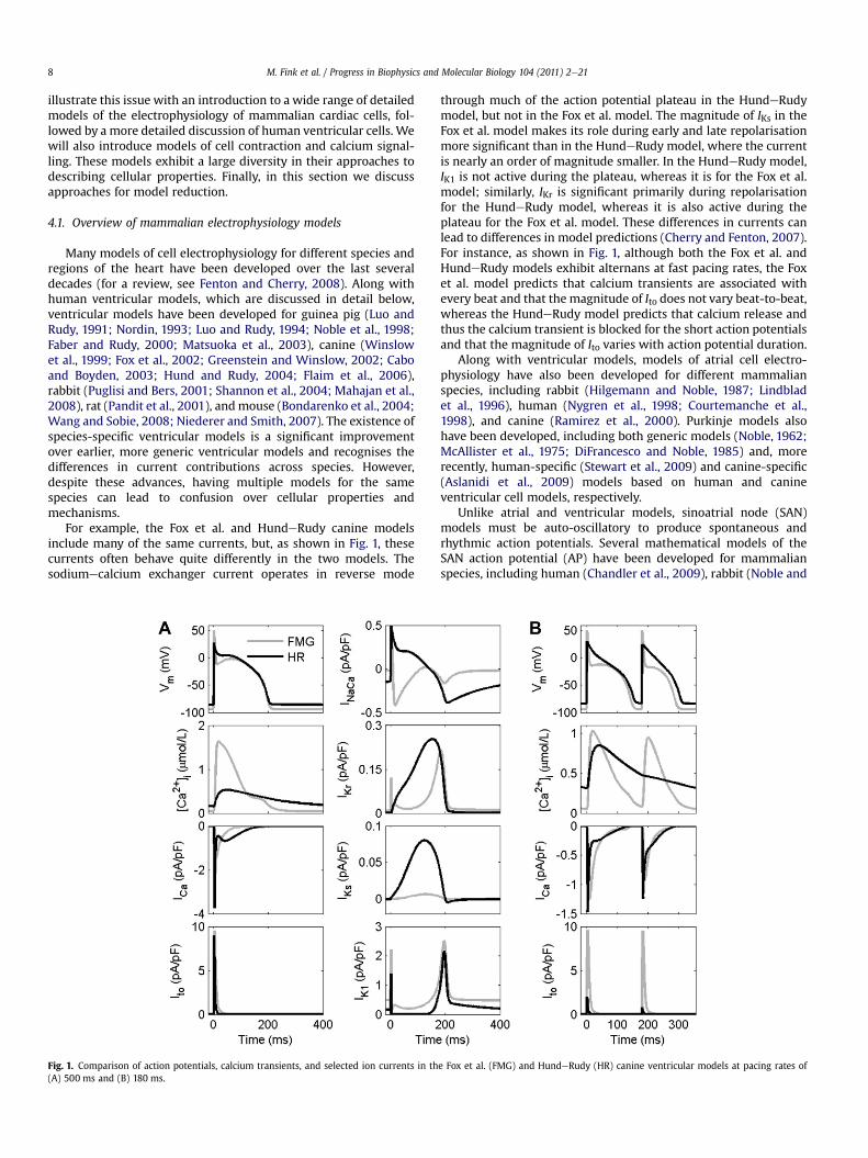
For example, the Fox et al. and HundeRudy canine modelsinclude many of the same currents, but, as shown in Fig. 1, thesecurrents often behave quite differently in the two models. Thesodiumecalcium exchanger current operates in reverse mode
Fig. 1. Comparison of action potentials, calcium transients, and selected ion currents in th(A) 500 ms and (B) 180 ms.
through much of the action potential plateau in the HundeRudymodel, but not in the Fox et al. model. The magnitude of IKs in theFox et al. model makes its role during early and late repolarisationmore significant than in the HundeRudy model, where the currentis nearly an order of magnitude smaller. In the HundeRudy model,IK1 is not active during the plateau, whereas it is for the Fox et al.model; similarly, IKr is significant primarily during repolarisationfor the HundeRudy model, whereas it is also active during theplateau for the Fox et al. model. These differences in currents canlead to differences in model predictions (Cherry and Fenton, 2007).For instance, as shown in Fig. 1, although both the Fox et al. andHundeRudy models exhibit alternans at fast pacing rates, the Foxet al. model predicts that calcium transients are associated withevery beat and that the magnitude of Ito does not vary beat-to-beat,whereas the HundeRudy model predicts that calcium release andthus the calcium transient is blocked for the short action potentialsand that the magnitude of Ito varies with action potential duration.
Along with ventricular models, models of atrial cell electro-physiology have also been developed for different mammalianspecies, including rabbit (Hilgemann and Noble, 1987; Lindbladet al., 1996), human (Nygren et al., 1998; Courtemanche et al.,1998), and canine (Ramirez et al., 2000). Purkinje models alsohave been developed, including both generic models (Noble, 1962;McAllister et al., 1975; DiFrancesco and Noble, 1985) and, morerecently, human-specific (Stewart et al., 2009) and canine-specific(Aslanidi et al., 2009) models based on human and canineventricular cell models, respectively.
Unlike atrial and ventricular models, sinoatrial node (SAN)models must be auto-oscillatory to produce spontaneous andrhythmic action potentials. Several mathematical models of theSAN action potential (AP) have been developed for mammalianspecies, including human (Chandler et al., 2009), rabbit (Noble and
e Fox et al. (FMG) and HundeRudy (HR) canine ventricular models at pacing rates of
M. Fink et al. / Progress in Biophysics and Molecular Biology 104 (2011) 2e21 9
Noble, 1984; Demir et al., 1994; Dokos et al., 1996; Zhang et al.,2000; Boyett et al., 2001; Kurata et al., 2002) and guinea pig(Sarai et al., 2003). Comparing the dynamical behaviour of variousSAN cell models, in particular the functional role of each individualionic current in generating pacemaking APs, it can be seen thatthese models show consistency in some respects, but diversity inothers. In all cases, SAN models are able to reproduce periodic andauto-rhythmic action potentials and the experimentally observeddepressive modulations by the surrounding atrium due to elec-trotonic interactions. However, models have implemented differentsubsets of ion channel currents, reflecting the limitations ofexperimental data and knowledge about SAN electrical propertiesat the time when these models were developed. As a consequence,the functional role of each individual current in these models maybe different. Even in the most recently developed modelsdsuch asthose by Zhang et al. (2000) and Kurata et al. (2002) discussedbelowdalthough the functional role of somemajor ionic currents isqualitatively similar, significant quantitative differences remain, asshown by differences in the sensitivities of the two models tomodel parameters (Kharche et al., 2009).
While SAN modelling has a lengthy history (Wilders et al., 1991;Demir et al., 1994; Dokos et al., 1996; Wilders, 2007), this reviewwill focus on recently introduced SAN models. Zhang et al. (2000)developed a family of SAN cell models that successfully repro-duce the differences in the electrical properties of ‘central’ and‘peripheral’ SAN cells. Recent advances in experimental electro-physiology have determined the electrical heterogeneity across theSAN and the surrounding atriumdcells in the SAN presentpronounced regional differences in their electrical APs and associ-ated properties of ion channels (Boyett et al., 2000). Experimentaldata obtained from different regions of the SAN show gradientvariations of electrical APs from the centre to the periphery of theSAN. Correlations between current density and cell size for severalionic currents have been reported that match the correlationbetween the characteristics of APs and the cell size (Boyett et al.,2000).
At the same time, based on previous SANmodels (Wilders et al.,1991; Demir et al. 1994; Dokos et al., 1996; Zhang et al., 2000),Kurata et al. (2002) developed an improved model for a primarySAN pacemaking cell. This model presented new formulations forthe voltage- and calcium-dependent inactivation kinetics of ICaLand intracellular calcium regulation and buffering. Later on, thisKurata et al. model was modified to account for the heterogeneousproperties of the SAN (Kurata et al., 2008), following the approachof the Zhang et al. (2000) model. Obviously, model development forthe electrical AP of SAN cells has been an evolutionary process,which, like the previously described development of ventricularcell models, closely follows experimental progress. With increas-ingly advanced experimental techniques emerging, more detailedelectrical properties for the SAN will be unveiled, warrantingdevelopment of new or improved models for the SAN electricalaction potential in the future.
4.2. Electrophysiology models of human ventricular myocytes
Recently, several models have been developed to describe theorigins and form of the human ventricular myocyte action poten-tial, an important step towards development of multiscale modelsof the human heart, and the potential for patient-specific model-ling, one of the major ambitions of computational modelling of theheart and the physiome in general. Modelling of human ventricularcells startedwith a study by Priebe and Beuckelmann (1998), aimedat understanding the effects of electrophysiological alterations inheart failure. In this study, models were developed to describenormal and failing human myocytes with similar approaches to
those previously used for cardiac cells of other species. The basis fortheir modelling was the LuoeRudy phase-2 model of guinea pigventricular myocytes, which was parameterised with dataincluding transients of transmembrane voltage, ion fluxes andconcentrations measured in normal and diseased humanmyocytes.A major achievement of this modelling study was the identificationof relationships between disease-associated alterations at the levelof transmembrane proteins and features of the action potential ofhuman myocytes. More recent applications include integrationwith models of cellular contraction and tissue electrophysiology(Sachse et al., 2003; Niederer and Smith, 2007; Niederer andSmith, 2009).
In 2004, two further models of human ventricular cells wereintroduced, the Iyer et al. (2004) and ten Tusscher et al. (2004)models. Both provide significantly more detail on ion channelfunction and compartmentalisation of ion handing within the cell.The increase in detail (and complexity) reflects new insights inchannel function and the availability of novel measurement datafrom human cells and ion channels. The Iyer et al. model describesthe electrophysiology of subepicardial cells, applying Markovianmodels for most channels. In contrast, ten Tusscher et al. modelledsubendocardial, midwall and subepicardial cells, commonly usingHodgkineHuxley type models. A revision of this model, publishedin 2006, includes more detail on intracellular calcium handling.Detailed comparisons of these human ventricular models,including ion currents, action potential shapes and durations, rateadaptation, and other properties, have been performed to assessthe similarities and differences of the models (ten Tusscher et al.,2006; Bueno-Orovio et al., 2008).
The Priebe and Beuckelmann (1998), Iyer et al. (2004) and tenTusscher et al. (2006) models exhibit significant differences in thereconstruction of transmembrane voltages, ion currents andconcentration during an action potential. These differences canpartially be explained by the differences in the experimental dataused to characterise these models (Niederer et al., 2009), the use ofdifferent model structures and quality of fit.
Fig. 2 demonstrates the disparity in model predictions for themembrane potential, calcium transient and IKr channel current. Thesignificant differences in these metrics of cardiac function haveramifications for the application and reuse of these models.Specifically the different action potential morphologies and Ca2þ
transients will cause significant changes in the predicted conduc-tion velocities and action potential durations in tissue-scale modelsand active tension generation in electromechanics models. In silicostudies of drug interactions and mutations may show effects thatare model-dependent. Experiments where these models areembedded in tissue simulations may have different conclusions,and linking thesemodels tomodels of contractionwill have a majorimpact on the resulting tension transient. While it is important torecognise that some differences betweenmodels are to be expectedgiven biological variability and the scarcity of human data, theproblem arising with the current models is that the source of thevariability is poorly characterised, hindering appropriate reuse andcontinued development.
4.3. Models of cell contraction
The modelling frameworks described above outline thecomponents of cardiac electrophysiology models. While thedevelopment of computational cell-based representations ofcardiac contraction has paralleled cardiac electrophysiologydevelopments, these models have, in general, lagged in complexity.More recently, detailed biophysical models of the inner workings ofthe sarcomere have emerged. These models are now capable offacilitating the investigation of the mechanisms responsible for
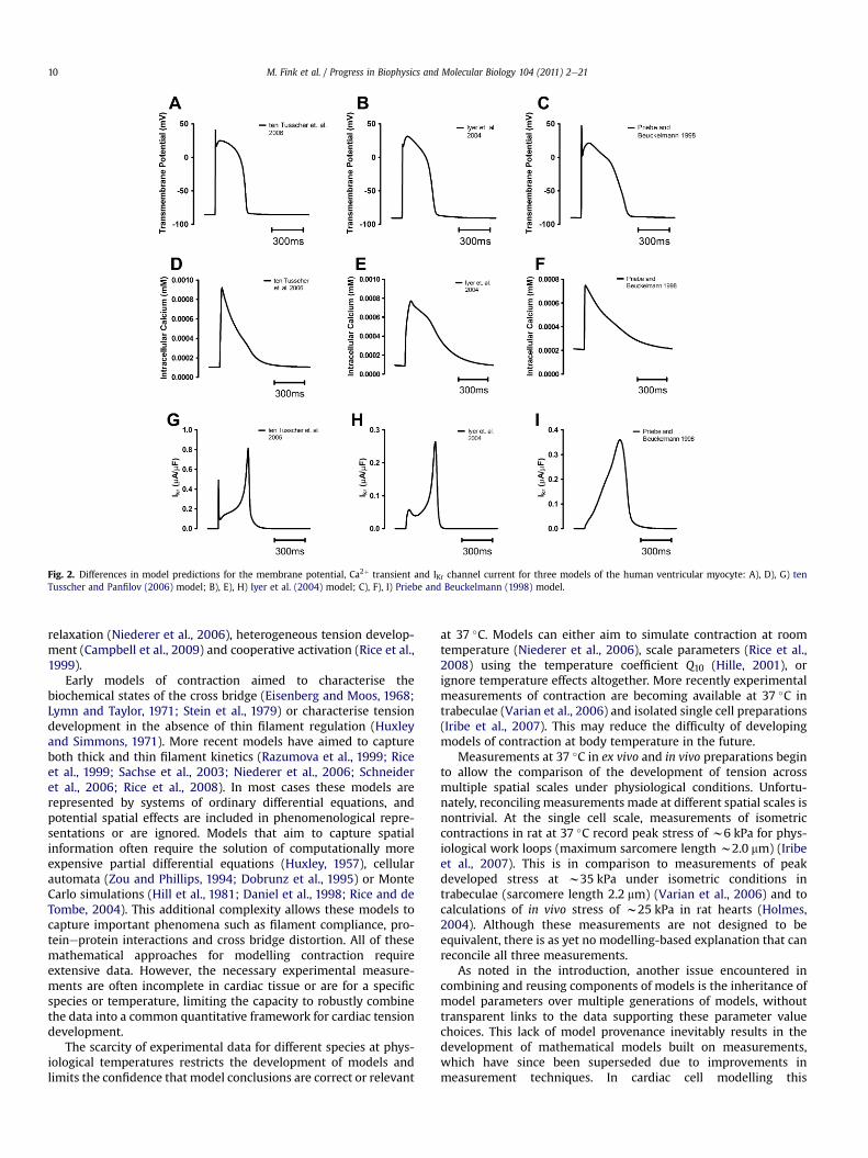
Fig. 2. Differences in model predictions for the membrane potential, Ca2þ transient and IKr channel current for three models of the human ventricular myocyte: A), D), G) tenTusscher and Panfilov (2006) model; B), E), H) Iyer et al. (2004) model; C), F), I) Priebe and Beuckelmann (1998) model.
M. Fink et al. / Progress in Biophysics and Molecular Biology 104 (2011) 2e2110
relaxation (Niederer et al., 2006), heterogeneous tension develop-ment (Campbell et al., 2009) and cooperative activation (Rice et al.,1999).
Early models of contraction aimed to characterise thebiochemical states of the cross bridge (Eisenberg and Moos, 1968;Lymn and Taylor, 1971; Stein et al., 1979) or characterise tensiondevelopment in the absence of thin filament regulation (Huxleyand Simmons, 1971). More recent models have aimed to captureboth thick and thin filament kinetics (Razumova et al., 1999; Riceet al., 1999; Sachse et al., 2003; Niederer et al., 2006; Schneideret al., 2006; Rice et al., 2008). In most cases these models arerepresented by systems of ordinary differential equations, andpotential spatial effects are included in phenomenological repre-sentations or are ignored. Models that aim to capture spatialinformation often require the solution of computationally moreexpensive partial differential equations (Huxley, 1957), cellularautomata (Zou and Phillips, 1994; Dobrunz et al., 1995) or MonteCarlo simulations (Hill et al., 1981; Daniel et al., 1998; Rice and deTombe, 2004). This additional complexity allows these models tocapture important phenomena such as filament compliance, pro-teineprotein interactions and cross bridge distortion. All of thesemathematical approaches for modelling contraction requireextensive data. However, the necessary experimental measure-ments are often incomplete in cardiac tissue or are for a specificspecies or temperature, limiting the capacity to robustly combinethe data into a common quantitative framework for cardiac tensiondevelopment.
The scarcity of experimental data for different species at phys-iological temperatures restricts the development of models andlimits the confidence that model conclusions are correct or relevant
at 37 �C. Models can either aim to simulate contraction at roomtemperature (Niederer et al., 2006), scale parameters (Rice et al.,2008) using the temperature coefficient Q10 (Hille, 2001), orignore temperature effects altogether. More recently experimentalmeasurements of contraction are becoming available at 37 �C intrabeculae (Varian et al., 2006) and isolated single cell preparations(Iribe et al., 2007). This may reduce the difficulty of developingmodels of contraction at body temperature in the future.
Measurements at 37 �C in ex vivo and in vivo preparations beginto allow the comparison of the development of tension acrossmultiple spatial scales under physiological conditions. Unfortu-nately, reconciling measurements made at different spatial scales isnontrivial. At the single cell scale, measurements of isometriccontractions in rat at 37 �C record peak stress of w6 kPa for phys-iological work loops (maximum sarcomere length w2.0 mm) (Iribeet al., 2007). This is in comparison to measurements of peakdeveloped stress at w35 kPa under isometric conditions intrabeculae (sarcomere length 2.2 mm) (Varian et al., 2006) and tocalculations of in vivo stress of w25 kPa in rat hearts (Holmes,2004). Although these measurements are not designed to beequivalent, there is as yet no modelling-based explanation that canreconcile all three measurements.
As noted in the introduction, another issue encountered incombining and reusing components of models is the inheritance ofmodel parameters over multiple generations of models, withouttransparent links to the data supporting these parameter valuechoices. This lack of model provenance inevitably results in thedevelopment of mathematical models built on measurements,which have since been superseded due to improvements inmeasurement techniques. In cardiac cell modelling this
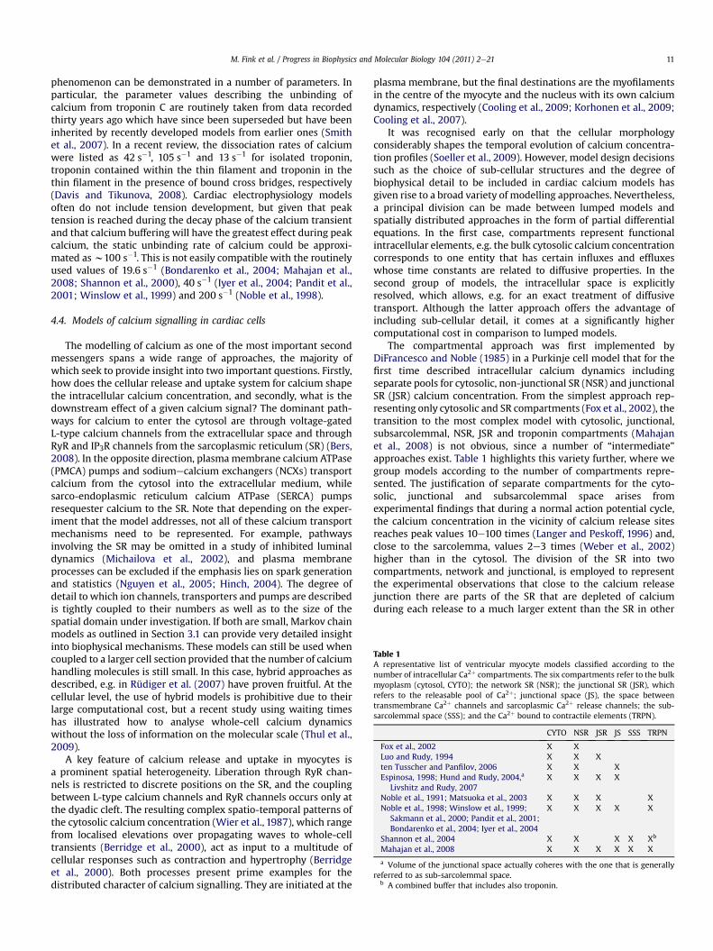
Table 1A representative list of ventricular myocyte models classified according to thenumber of intracellular Ca2þ compartments. The six compartments refer to the bulkmyoplasm (cytosol, CYTO); the network SR (NSR); the junctional SR (JSR), whichrefers to the releasable pool of Ca2þ; junctional space (JS), the space betweentransmembrane Ca2þ channels and sarcoplasmic Ca2þ release channels; the sub-sarcolemmal space (SSS); and the Ca2þ bound to contractile elements (TRPN).
CYTO NSR JSR JS SSS TRPN
Fox et al., 2002 X XLuo and Rudy, 1994 X X Xten Tusscher and Panfilov, 2006 X X XEspinosa, 1998; Hund and Rudy, 2004,a
Livshitz and Rudy, 2007X X X X
Noble et al., 1991; Matsuoka et al., 2003 X X X XNoble et al., 1998; Winslow et al., 1999;
Sakmann et al., 2000; Pandit et al., 2001;Bondarenko et al., 2004; Iyer et al., 2004
X X X X X
Shannon et al., 2004 X X X X Xb
Mahajan et al., 2008 X X X X X X
a Volume of the junctional space actually coheres with the one that is generallyreferred to as sub-sarcolemmal space.
b A combined buffer that includes also troponin.
M. Fink et al. / Progress in Biophysics and Molecular Biology 104 (2011) 2e21 11
phenomenon can be demonstrated in a number of parameters. Inparticular, the parameter values describing the unbinding ofcalcium from troponin C are routinely taken from data recordedthirty years ago which have since been superseded but have beeninherited by recently developed models from earlier ones (Smithet al., 2007). In a recent review, the dissociation rates of calciumwere listed as 42 s�1, 105 s�1 and 13 s�1 for isolated troponin,troponin contained within the thin filament and troponin in thethin filament in the presence of bound cross bridges, respectively(Davis and Tikunova, 2008). Cardiac electrophysiology modelsoften do not include tension development, but given that peaktension is reached during the decay phase of the calcium transientand that calcium buffering will have the greatest effect during peakcalcium, the static unbinding rate of calcium could be approxi-mated asw100 s�1. This is not easily compatible with the routinelyused values of 19.6 s�1 (Bondarenko et al., 2004; Mahajan et al.,2008; Shannon et al., 2000), 40 s�1 (Iyer et al., 2004; Pandit et al.,2001; Winslow et al., 1999) and 200 s�1 (Noble et al., 1998).
4.4. Models of calcium signalling in cardiac cells
The modelling of calcium as one of the most important secondmessengers spans a wide range of approaches, the majority ofwhich seek to provide insight into two important questions. Firstly,how does the cellular release and uptake system for calcium shapethe intracellular calcium concentration, and secondly, what is thedownstream effect of a given calcium signal? The dominant path-ways for calcium to enter the cytosol are through voltage-gatedL-type calcium channels from the extracellular space and throughRyR and IP3R channels from the sarcoplasmic reticulum (SR) (Bers,2008). In the opposite direction, plasmamembrane calcium ATPase(PMCA) pumps and sodiumecalcium exchangers (NCXs) transportcalcium from the cytosol into the extracellular medium, whilesarco-endoplasmic reticulum calcium ATPase (SERCA) pumpsresequester calcium to the SR. Note that depending on the exper-iment that the model addresses, not all of these calcium transportmechanisms need to be represented. For example, pathwaysinvolving the SR may be omitted in a study of inhibited luminaldynamics (Michailova et al., 2002), and plasma membraneprocesses can be excluded if the emphasis lies on spark generationand statistics (Nguyen et al., 2005; Hinch, 2004). The degree ofdetail to which ion channels, transporters and pumps are describedis tightly coupled to their numbers as well as to the size of thespatial domain under investigation. If both are small, Markov chainmodels as outlined in Section 3.1 can provide very detailed insightinto biophysical mechanisms. These models can still be used whencoupled to a larger cell section provided that the number of calciumhandling molecules is still small. In this case, hybrid approaches asdescribed, e.g. in Rüdiger et al. (2007) have proven fruitful. At thecellular level, the use of hybrid models is prohibitive due to theirlarge computational cost, but a recent study using waiting timeshas illustrated how to analyse whole-cell calcium dynamicswithout the loss of information on the molecular scale (Thul et al.,2009).
A key feature of calcium release and uptake in myocytes isa prominent spatial heterogeneity. Liberation through RyR chan-nels is restricted to discrete positions on the SR, and the couplingbetween L-type calcium channels and RyR channels occurs only atthe dyadic cleft. The resulting complex spatio-temporal patterns ofthe cytosolic calcium concentration (Wier et al., 1987), which rangefrom localised elevations over propagating waves to whole-celltransients (Berridge et al., 2000), act as input to a multitude ofcellular responses such as contraction and hypertrophy (Berridgeet al., 2000). Both processes present prime examples for thedistributed character of calcium signalling. They are initiated at the
plasma membrane, but the final destinations are the myofilamentsin the centre of the myocyte and the nucleus with its own calciumdynamics, respectively (Cooling et al., 2009; Korhonen et al., 2009;Cooling et al., 2007).
It was recognised early on that the cellular morphologyconsiderably shapes the temporal evolution of calcium concentra-tion profiles (Soeller et al., 2009). However, model design decisionssuch as the choice of sub-cellular structures and the degree ofbiophysical detail to be included in cardiac calcium models hasgiven rise to a broad variety of modelling approaches. Nevertheless,a principal division can be made between lumped models andspatially distributed approaches in the form of partial differentialequations. In the first case, compartments represent functionalintracellular elements, e.g. the bulk cytosolic calcium concentrationcorresponds to one entity that has certain influxes and effluxeswhose time constants are related to diffusive properties. In thesecond group of models, the intracellular space is explicitlyresolved, which allows, e.g. for an exact treatment of diffusivetransport. Although the latter approach offers the advantage ofincluding sub-cellular detail, it comes at a significantly highercomputational cost in comparison to lumped models.
The compartmental approach was first implemented byDiFrancesco and Noble (1985) in a Purkinje cell model that for thefirst time described intracellular calcium dynamics includingseparate pools for cytosolic, non-junctional SR (NSR) and junctionalSR (JSR) calcium concentration. From the simplest approach rep-resenting only cytosolic and SR compartments (Fox et al., 2002), thetransition to the most complex model with cytosolic, junctional,subsarcolemmal, NSR, JSR and troponin compartments (Mahajanet al., 2008) is not obvious, since a number of “intermediate”approaches exist. Table 1 highlights this variety further, where wegroup models according to the number of compartments repre-sented. The justification of separate compartments for the cyto-solic, junctional and subsarcolemmal space arises fromexperimental findings that during a normal action potential cycle,the calcium concentration in the vicinity of calcium release sitesreaches peak values 10e100 times (Langer and Peskoff, 1996) and,close to the sarcolemma, values 2e3 times (Weber et al., 2002)higher than in the cytosol. The division of the SR into twocompartments, network and junctional, is employed to representthe experimental observations that close to the calcium releasejunction there are parts of the SR that are depleted of calciumduring each release to a much larger extent than the SR in other
M. Fink et al. / Progress in Biophysics and Molecular Biology 104 (2011) 2e2112
areas of the cell (Shannon et al., 2003). That is, the junctional SRcompartment refers to a releasable calcium pool, which is “sepa-rated” from the network SR due to a significant diffusion resistance(Wussling and Szymanski, 1986).
A mitochondrial calcium compartment is typically omitted fromcardiomyocyte models. This omission generally has been justifiedby findings that the role of mitochondria as a calcium sink or sourcein beat-to-beat regulation of the cytosolic calcium concentration israther small (Bassani et al., 1992). However, recent results fromvarious groups suggest that mitochondrial calcium uptake andrelease can have a significant effect on cellular calcium homeostasis(Duchen et al., 1998; Trollinger et al., 2000), see Dedkova andBlatter (2008) for a review. Thus, it appears that the addition ofa mitochondrial calcium compartment should be addressed infuture modelling studies. This intention is supported by recentdevelopments in the mathematical description of mitochondrialcalcium dynamics (Nguyen et al., 2007; Dash et al., 2009). Mostrecently Pásek et al. (Pásek et al., 2008a,b) have developedcompartmental models that explicitly include the transverse-axialtubular system. These models have been applied to assess theimportance of ion channel heterogeneity and restricted diffusionwithin the tubular system on physiological function.
The fundamental challenge of the compartmental approach isthe lack of corresponding distinct anatomical structures insidecells. Thus, there is no straightforward way of choosing theappropriate selection of compartments for a specific researchquestion. Furthermore, model validation always requires that thesecompartmental parameters are derived from experimental data bysome procedure, rather than by direct comparison with measureddata.
One of the motivations in developing models with an increasingnumber of compartments was the insight that the calciumconcentration in originally unrefined compartments was not asspatially homogeneous as initially assumed. Although thisapproach could be continued and more compartments introducedto account for a larger degree of cellular heterogeneity, an alter-native approach is to consider space- and time-dependentconcentrations within a single spatial domain. The calciumconcentration then exhibits large spatial gradients around releasesites upon calcium release, and decays to resting conditions inquiescent cells. As with the compartmentalised models, thecomplexity of partial differential equation-basedmodels for cardiaccalcium dynamics has increased over the years. Early works focusedon a one-dimensional representation of the cytosol and wereinstrumental in our understanding of calcium waves in cardiacmyocytes (Dupont and Goldbeter, 1994; Pearson and Ponce-Dawson, 1998; Keizer et al., 1998; Dawson et al., 1999). Morerecently, these models were extended to incorporate stochasticrelease and a dynamic SR (Coombes and Timofeeva, 2003; Coombeset al., 2004; Thul et al., 2008). While integrating additionalelements of calcium signalling into cardiac models has been ofongoing interest, a parallel development has been focusing on theimplementation of realistic cellular geometries. This has resulted inthree-dimensional simulations of calcium concentration profilesand has provided the first insights into the coupling between cellshape, ion channel and receptor distributions, and calcium signal-ling (Izu et al., 2001, 2006; Michailova et al., 2002; Means et al.,2006; Li et al., 2007; Li and Holden, 2009; Korhonen et al., 2009;Lu et al., 2009; and see Lemon, 2004 for an analytical study).Although consensus is emerging as regarding core components ofany cardiac calciummodel, details of some keymechanisms are stilldebated. There is no doubt that the dynamics in the SR plays a vitalrole, but some studies report fast luminal calcium diffusion (Wuand Bers, 2006), whereas others support a small diffusion coeffi-cient (Swietach et al., 2008). However, different ion mobility has
far-reaching consequences for wave propagation. Measurements ofthe geometry of the SR have revealed a highly tortuous structure,which in turn reduces the effective velocity of diffusional transport.Whether models need to explicitly incorporate the shape of the SRor whether an effective reduction of the diffusion coefficient assuggested by Olveczky and Verkman (1998) suffices is still an openquestion. The impact of mobile and stationary buffers on calciumsignals, both in themyoplasm and the SR, is now firmly established.Specifically, key properties such as the buffering capacity still lackfinal quantification (Trafford et al., 1999; Walden et al., 2009), andthe regulatory role of buffers such as calsequestrin has emergedonly recently (Györke et al., 2004; Chopra et al., 2007; Györke andTerentyev, 2008). Furthermore, as the calcium-sensitive fluorescentdyes (Section 2.2.3) added during experiments to measure calciumconcentration are themselves exogenous calcium buffers, carefulanalysis (often using models) of the measured fluorescence trace isrequired to quantify the calcium transient from the raw data. Thevariety of cardiac calcium models reflects the uncertainties that westill face with respect to both underlying mechanisms andparameter values, but continuous advances in theoretical andexperimental techniques will promote the convergence of allmodels towards a unified framework of cardiac calcium signalling.
4.5. Reduced electrophysiology models
To study cellular properties at a higher level than the specificformulations of individual ion channels, such as rate adaptation,memory and bifurcations, less biophysically detailed models alsocan be useful. Of course, one of the most important advantages ofreducedmodels is their increased computational tractability, whichcan permit larger, longer and more spatially resolved three-dimensional simulations. As discussed below, there are threedifferent approaches for reducing model complexity: simplifiedmodels, minimal models and discrete iterative maps.
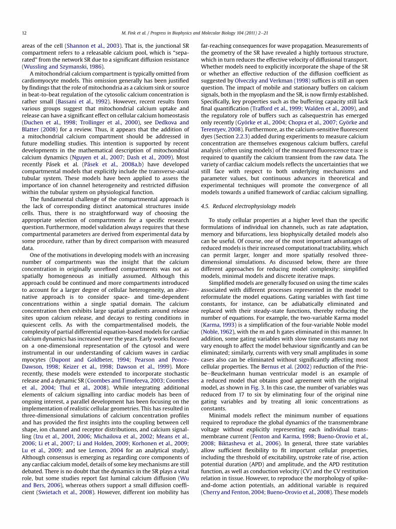
Simplifiedmodels are generally focused on using the time scalesassociated with different processes represented in the model toreformulate the model equations. Gating variables with fast timeconstants, for instance, can be adiabatically eliminated andreplaced with their steady-state functions, thereby reducing thenumber of equations. For example, the two-variable Karma model(Karma, 1993) is a simplification of the four-variable Noble model(Noble, 1962), with the m and h gates eliminated in this manner. Inaddition, some gating variables with slow time constants may notvary enough to affect the model behaviour significantly and can beeliminated; similarly, currents with very small amplitudes in somecases also can be eliminated without significantly affecting mostcellular properties. The Bernus et al. (2002) reduction of the Prie-beeBeuckelmann human ventricular model is an example ofa reduced model that obtains good agreement with the originalmodel, as shown in Fig. 3. In this case, the number of variables wasreduced from 17 to six by eliminating four of the original ninegating variables and by treating all ionic concentrations asconstants.
Minimal models reflect the minimum number of equationsrequired to reproduce the global dynamics of the transmembranevoltage without explicitly representing each individual trans-membrane current (Fenton and Karma, 1998; Bueno-Orovio et al.,2008; Biktasheva et al., 2006). In general, three state variablesallow sufficient flexibility to fit important cellular properties,including the threshold of excitability, upstroke rate of rise, actionpotential duration (APD) and amplitude, and the APD restitutionfunction, as well as conduction velocity (CV) and the CV restitutionrelation in tissue. However, to reproduce the morphology of spike-and-dome action potentials, an additional variable is required(Cherry and Fenton, 2004; Bueno-Orovio et al., 2008). Thesemodels
Fig. 3. Four action potentials for the PriebeeBeuckelmann (PB) model and for thereduced PB (rPB) model of Bernus et al. (2002). Cycle length is 430 ms for the first twobeats and 900 ms for the last two beats.
M. Fink et al. / Progress in Biophysics and Molecular Biology 104 (2011) 2e21 13
can be obtained by asymptotic approaches different from thestandard fast-slow reductions used in simplified models (Biktashevet al., 2008).
Because simplified and minimal models do not include detailedbiophysical descriptions of most cellular process, their applicationin, e.g. studies of specific effects like ion channel blockade ormutations, is less straightforward.. These models are intendedprimarily for use in tissue simulations where multicellular effectsare being investigated and computational efficiency becomesimportant. In some cases, simplified orminimalmodels can be usedto represent the overall membrane dynamics under variousconditions, including pharmaceutical agents or mutations, byfittingmodel parameters directly from experimental data, a processthat becomes faster and more straightforward when the number ofparameters is reduced significantly (often by an order of magni-tude) (Bueno-Orovio et al., 2008; Bernus et al., 2002). These modelvariations then can be simulated in tissue to study tissue-levelphenomena, including wave stability and dynamical bifurcations(see, for example, the paper in this issue).
Discrete iterative maps are used to study certain characteristicsof cell dynamics without tracking the membrane potential, gatingvariables, currents, and concentrations explicitly, but instead bydeveloping and using equations to compute quantities like APD orpeak calcium concentration as a function of these and otherdiscrete quantities. They can be as simple as iterative functions thatdescribe rate adaptation from standard experimental APD restitu-tion curves (Nolasco and Dahlen,1968; Guevara et al., 1984) or fromcell models (Cain and Schaeffer, 2006; Schaeffer et al., 2007, 2008).Slightly more complex maps can be created to describe thedynamics of intracellular calcium alone (Eisner et al., 2000) orcombined with the membrane potential (Qu et al., 2007a,b) tostudy the dynamics and types of bifurcations during alternans.
5. Model identificationdmodel selection
Depending on the amount of available data and the knowledgeof underlying mechanisms, the described models can be more orless complex. For example the fast sodium channel has beendescribed using between two and 21 states and nine and 171parameters (Hodgkin and Huxley, 1952; Kahlig et al., 2006,respectively). The necessary and sufficient complexity of the model
design depends on themodelling goal. For example the informationcontent in voltage-clamp steps for voltage-gated ion channelsprovides approximately 15 parameters (see Fink and Noble, 2009).Fig. 4 shows that the optimised voltage-clamp step protocol pre-sented in Fink and Noble (2009) provides more information thana normal action potential clamp, indicating that it should be suffi-cient for a model to fit data from the optimised voltage clamp stepprotocol if the goal is to replicate whole cell phenomena.
With regards to the computational aspects determined by themodel design, it has been shown that stiffness is more stringentthan the number of ordinary differential equations regarding theslowing down of single cell computations (e.g. Fink and Noble,2009). Stiffness can be eliminated by separation of time scales,for instance by asymptotic approximations (e.g. Hinch et al., 2004).Principal component analysis or sensitivity analysis can be used forremoval of parameters or model parts to which the output isinsensitive (e.g. Fink and Noble, 2009, Bernus et al., 2002; Schmidtet al., 2008). Lately, a similar approach of invariant manifoldreductions for Markov models has been used (Keener, 2009).
The final decision concerning appropriatemodel designmust bemade using validation of the models using the full range of avail-able experimental data outlined in Section 3. However, so far thegeneral lack of ability to fit the parameter values has made itimpossible to compare the validity of different model designs. Thisis because differences in their predictive power could have comefrom the framework of model states and transitions or from thequality of the parameter fit, e.g. the error between model outputand experimental data.
5.1. Parameterisation, data availability and local minima
Experimental design can be used to determine the experimentalprotocols necessary for estimation of the model parameters.However, it still remains difficult to find the right amount of datafor obtaining a full action potential model using data from singlecell experiments. Boundaries on conductances and maximum ratescan be given from experimental data, but to find the optimalparameter set, local identifiability of the parameters is necessary atleast, whilst still avoiding overdetermination (Dokos and Lovell,2004).
Table 2 shows the parameter identifiability results using the tenTusscher model (ten Tusscher and Panfilov, 2006). The voltagetraces for one action potential using patch clamp recordings wouldonly provide 4 conductances (for iNa, iCaL, iKto, and iKr or iKs) out of 12(neglecting background currents). Adding measurements of intra-cellular calcium for the same cell would provide only two moreparameters (for NCX and SERCA). Having the complete data fora S1S2 protocol would provide eight parameters for voltage onlyand 10 for additional data on intracellular calcium. Only bymeasuring the amount of calcium in the dyadic cleft would all12 parameters be obtained. Another possibility giving full identi-fiability would be to have voltage and intracellular calciummeasurements for two different extracellular potassium concen-trations (see Table 2)
In addition to the problem of finding the right amount of data,the nonlinearity of the minimisation problem and the associatednumerical problems make finding an optimal fitting algorithmdifficult. Furthermore, the number of local minima typicallybecomes larger and more problematic as model complexity isincreased to include dozens of variables and hundreds of parame-ters. The availability of simple methods like the Simplex and Lev-enbergeMarquardt algorithms provided in Matlab� or a recentlydeveloped CellML-based tool (Hui et al., 2007) and their limitedneed for significant computational resources would make themgood candidates but their ability to find global minima is very
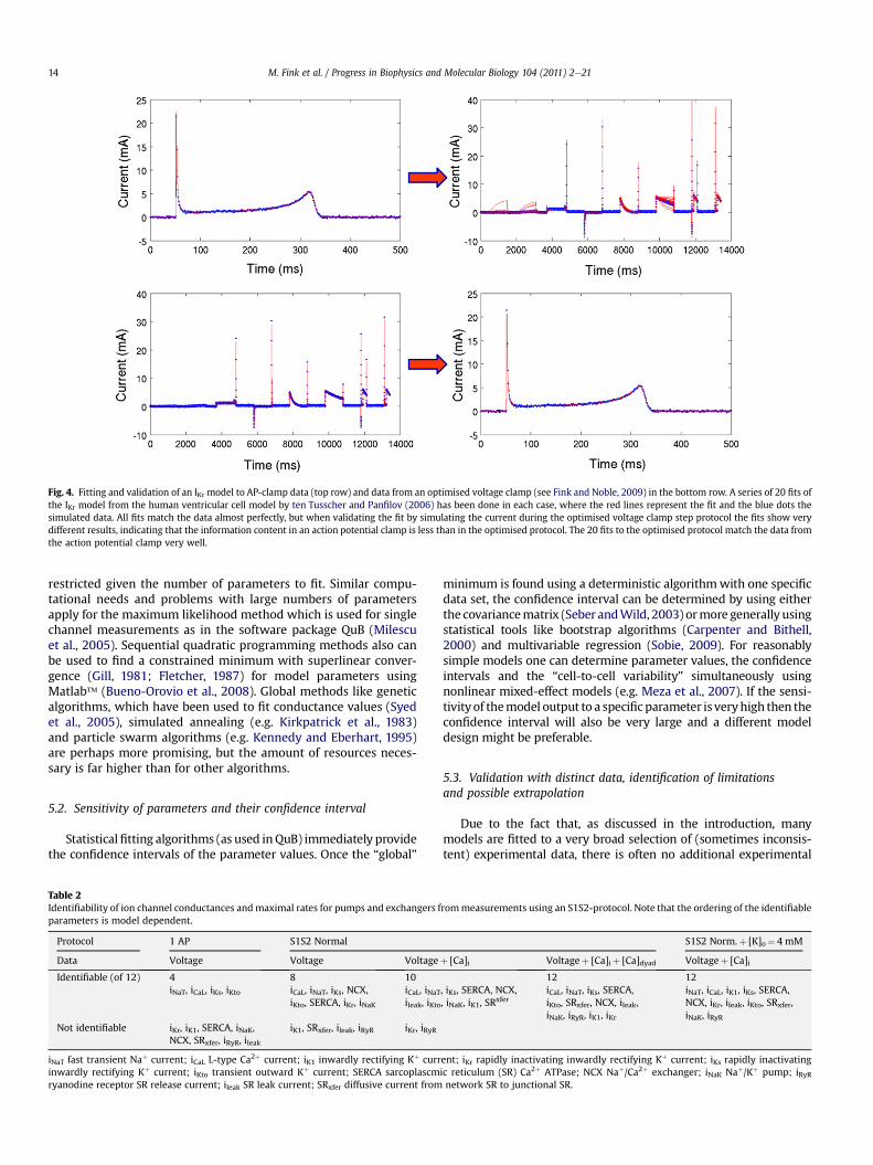
Fig. 4. Fitting and validation of an IKr model to AP-clamp data (top row) and data from an optimised voltage clamp (see Fink and Noble, 2009) in the bottom row. A series of 20 fits ofthe IKr model from the human ventricular cell model by ten Tusscher and Panfilov (2006) has been done in each case, where the red lines represent the fit and the blue dots thesimulated data. All fits match the data almost perfectly, but when validating the fit by simulating the current during the optimised voltage clamp step protocol the fits show verydifferent results, indicating that the information content in an action potential clamp is less than in the optimised protocol. The 20 fits to the optimised protocol match the data fromthe action potential clamp very well.
M. Fink et al. / Progress in Biophysics and Molecular Biology 104 (2011) 2e2114
restricted given the number of parameters to fit. Similar compu-tational needs and problems with large numbers of parametersapply for the maximum likelihood method which is used for singlechannel measurements as in the software package QuB (Milescuet al., 2005). Sequential quadratic programming methods also canbe used to find a constrained minimum with superlinear conver-gence (Gill, 1981; Fletcher, 1987) for model parameters usingMatlab� (Bueno-Orovio et al., 2008). Global methods like geneticalgorithms, which have been used to fit conductance values (Syedet al., 2005), simulated annealing (e.g. Kirkpatrick et al., 1983)and particle swarm algorithms (e.g. Kennedy and Eberhart, 1995)are perhaps more promising, but the amount of resources neces-sary is far higher than for other algorithms.
5.2. Sensitivity of parameters and their confidence interval
Statisticalfitting algorithms (as used inQuB) immediately providethe confidence intervals of the parameter values. Once the “global”
Table 2Identifiability of ion channel conductances andmaximal rates for pumps and exchangers fparameters is model dependent.
Protocol 1 AP S1S2 Normal
Data Voltage Voltage Voltage
Identifiable (of 12) 4 8 10iNaT, iCaL, iKs, iKto iCaL, iNaT, iKs, NCX,
iKto, SERCA, iKr, iNaKiCaL, iNaTileak, iKto
Not identifiable iKr, iK1, SERCA, iNaK,NCX, SRxfer, iRyR, ileak
iK1, SRxfer, ileak, iRyR iKr, iRyR
iNaT fast transient Naþ current; iCaL L-type Ca2þ current; iK1 inwardly rectifying Kþ currinwardly rectifying Kþ current; iKto transient outward Kþ current; SERCA sarcoplascmryanodine receptor SR release current; ileak SR leak current; SRxfer diffusive current from
minimum is found using a deterministic algorithmwith one specificdata set, the confidence interval can be determined by using eitherthe covariancematrix (Seber andWild, 2003) ormore generally usingstatistical tools like bootstrap algorithms (Carpenter and Bithell,2000) and multivariable regression (Sobie, 2009). For reasonablysimple models one can determine parameter values, the confidenceintervals and the “cell-to-cell variability” simultaneously usingnonlinear mixed-effect models (e.g. Meza et al., 2007). If the sensi-tivity of themodel output to a specific parameter is veryhigh then theconfidence interval will also be very large and a different modeldesign might be preferable.
5.3. Validation with distinct data, identification of limitationsand possible extrapolation
Due to the fact that, as discussed in the introduction, manymodels are fitted to a very broad selection of (sometimes inconsis-tent) experimental data, there is often no additional experimental
rommeasurements using an S1S2-protocol. Note that the ordering of the identifiable
S1S2 Norm.þ [K]o¼ 4 mM
þ [Ca]i Voltageþ [Ca]iþ [Ca]dyad Voltageþ [Ca]i
12 12, iKs, SERCA, NCX,, iNaK, iK1, SRxfer
iCaL, iNaT, iKs, SERCA,iKto, SRxfer, NCX, ileak,iNaK, iRyR, iK1, iKr
iNaT, iCaL, iK1, iKs, SERCA,NCX, iKr, ileak, iKto, SRxfer,iNaK, iRyR
ent; iKr rapidly inactivating inwardly rectifying Kþ current; iKs rapidly inactivatingic reticulum (SR) Ca2þ ATPase; NCX Naþ/Ca2þ exchanger; iNaK Naþ/Kþ pump; iRyRnetwork SR to junctional SR.
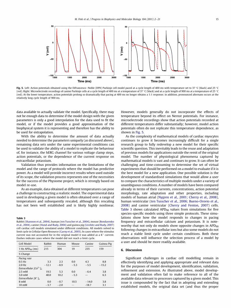
Fig. 5. Left: Action potentials obtained using the DiFrancescoeNoble (DFN) Purkinje cell model paced at a cycle length of 400 ms with temperature set to 37 �C (black) and 25 �C(red). Right: Microelectrode recordings of canine Purkinje cells at a cycle length of 400 ms at a temperature of 37 �C (black) and at a cycle length of 900 ms at a temperature of 25 �C(red). At the lower temperature, action potentials prolong so dramatically that pacing at 400 ms no longer obtains a 1:1 response; in addition, pronounced alternans occurs at therelatively long cycle length of 900 ms.
M. Fink et al. / Progress in Biophysics and Molecular Biology 104 (2011) 2e21 15
data available to actually validate the model. Specifically, there maynot be enough data to determine if the model design with the givenparameters is only a good interpolation for the data used to fit themodel, or if the model provides a good approximation of thebiophysical system it is representing and therefore has the ability tobe used for extrapolation.
With the ability to determine the amount of data actuallyneeded to determine the parameters uniquely (as discussed above),remaining data sets under the same experimental conditions canbe used to validate the ability of a model to replicate the behaviourof, for instance, the hERG channel for various voltage clamp steps,action potentials, or the dependence of the current response onextracellular potassium.
Validation thus provides information on the limitations of themodel and the range of protocols where the model has predictivepower. As a model will provide incorrect results when used outsideof its scope, the validation process represents one of the necessitiesfor the success of the Physiome project, which is strongly based onmodel re-use.
As an example, data obtained at different temperatures can posea challenge to constructing a realistic model. The experimental dataused in developing a given model is often obtained over a range oftemperatures and subsequently rescaled, although this rescalinghas not been well established and is likely highly nonlinear.
Table 3Rabbit (Shannon et al., 2004), human (tenTusscher et al., 2004), mouse (Bondarenkoet al., 2004), canine (Hund and Rudy, 2004) and guinea pig (Livshitz and Rudy, 2007)cell cardiac cell models simulated under different conditions. All models solved tolimit cycle in Cellular Open Resource (Garny et al., 2003). In cases where the stimuluscurrent was not accounted for in the original model it was added as a Kþ current.Dashes indicate cases where the model did not reach a limit cycle.
Cell Model Rabbit Human Mouse Canine Guinea Pig
1 Hz APD90 (ms) 215 304 19 235 159
% Change
Pacing rate0.5 Hz 3.3 2.3 0.0 4.3 8.82.0 Hz �12.1 �8.9 �5.3 �5.5 �15.2Extracellular [Ca2þ]o2.5 mM 19.5 5.3 0.0 �6.4 3.83.0 mM 40.0 10.2 �5.3 d 6.3Extracellular [Kþ]o8 mM 0.0 �0.7 10.5 �14.0 3.810 mM �3.7 �2.0 d -18.7 3.1
However, models generally do not incorporate the effects oftemperature beyond its effect on Nernst potentials. For instance,microelectrode recordings show that action potentials recorded atdifferent temperatures differ substantially; however, model actionpotentials often do not replicate this temperature dependence, asshown in Fig. 5.
As the complexity of mathematical models of cardiac myocytescontinues to grow it becomes increasingly difficult for a singleresearch group to fully redevelop a new model for their specificscientific question. This inevitably leads to the reuse and adaptationof previous models for applications outside the remit of the originalmodel. The number of physiological phenomena captured bymathematical models is vast and continues to grow. It can often bechallenging and time-consuming to determine the set of virtualexperiments that should be performed on amodel to evaluate if it isthe best model for a new application. One possible solution is thedevelopment of standardised simulations that would allow a userto compare the characteristics of multiple models under a variety ofunambiguous conditions. A number of models have been comparedalready in terms of their currents, concentrations, action potentialmorphology, rate adaptation and other properties, includingmodels of human atrial (Nygren et al., 2001; Cherry et al., 2008),human ventricular (ten Tusscher et al., 2006; Bueno-Orovio et al.,2008) and canine ventricular (Cherry and Fenton, 2007) cells.Table 3 shows calculated APD90 values from simulations for fivespecies-specific models using three simple protocols. These simu-lations show how the model responds to changes in pacingfrequency and extracellular calcium and potassium. It is note-worthy that not only do models show opposite changes in APD90following changes in extracellular ions but also somemodels do notreach a stable limit cycle under certain conditions. Both theseobservations will influence the selection process of a model bya user and should be more readily available.
6. Discussion
Significant challenges in cardiac cell modelling remain ineffectively identifying and applying appropriate and relevant datafor the purposes of model development, identification, validation,refinement and extension. As illustrated above, model develop-ment and validation often fail to make reference to all of theavailable data relevant to processes captured by a given model. Thisissue is compounded by the fact that in adopting and extendingestablished models, the original data set (and thus the proper
M. Fink et al. / Progress in Biophysics and Molecular Biology 104 (2011) 2e2116
context) on which a model is based is often lost or ignored (andthus the model context is corrupted). Moreover, as models growmore complex and diverse, we are increasingly faced withdisagreements (in terms of their predicted qualitative or quanti-tative behaviour) between models purporting to simulate the samesystem (Nygren et al., 2001; ten Tusscher et al., 2006; Cherry andFenton, 2007; Wilders, 2007; Bueno-Orovio et al., 2008; Cherryet al., 2008; Cherry and Evans, 2008). Assuming that disagreeingmodels are constructed with appropriate care and expertise, thisphenomenon must be a consequence of disagreement in either theunderlying data or the assumptions used to build the models.Ideally in such cases, models should provide a vehicle to elucidateand reconcile the source of apparent disagreement.
In response to these challenges, we propose a number ofinitiatives to help establish broader, deeper, and more transparentconnections between models and experimental data than aretypically associated with currently used practices. Specifically, wepropose that the cardiac cell modelling community cooperate in:(1) applying state-of-the-art tools for model identification, sensi-tivity analysis and optimal design of experiments for model iden-tification and parameterisation; (2) appropriately reconciling thecomplexity of models with the availability of data and the goals ofa particular model; and (3) building a database of experimentaldata associated with cardiac cell modelling.
5 http://www.cellml.org/tools6 http://sbml.org/SBML_Software_Guide7 http://vcell.org/
6.1. Initiative 1: Application of tools for optimal model identification
Identification of a complex model (that is, sensitive and reli-able determination of appropriate parameter values) and valida-tion of the basic model framework require comparison to a richset of experimental data. In essence, it is necessary to designproper experimental protocols and optimal data samplingschedules for minimising the error and covariance in parameterestimates (Banga and Balsa-Canto, 2008). Such design requiresinitial models to simulate potential experiments, which should bedesigned to refine the models. Thus the process requires not onlyiteration between experiment and simulation, but coordination inusing models to formally design the best possible experimentsand using experiments to formally identify the best possiblemodels.
Adopting optimal design approaches will require the develop-ment of appropriate software to facilitate the process. There existnumerous software environments for model simulation, such asCOR and PCEnv (Garny et al., 2008), some of which incorporatetools for parameter estimation and sensitivity analysis. Forexample, the JSim package incorporates sophisticated optimisationroutines for parameter estimation as well as the ability to auto-matically calculate sensitivity functions and generate multiplesimulations by looping through ranges of parameter values. TheQuB (Milescu et al., 2005) and Neuron (Brette et al., 2007) packagesprovide the ability to simulate and fit models of ion channels orcells. Yet none of these packages provide the means to systemati-cally interrogate parameter identifiability. Furthermore, few toolsexist for designing experiments for optimal model identification.Existing tools for optimal experimental design are targeted topharmacokinetic experiments and simulation (Foracchia et al.,2004) and not easily ported to cardiac cell modelling andexperiments.
Thus the opportunity (and need) exists for much progress indeveloping tools in this area. The approaches outlined in Section 5of this paper will become part of the standard tool set of the cardiaccell physiology community only when easy-to-use and reliablesoftware for experimental design, parameter estimation and anal-ysis of sensitivity and covariance is available.
6.2. Initiative 2: Reconciling model complexity with dataavailability and modelling goals
Models with more degrees of freedom typically provide closerfits to data, but at the expense of the ability to predict (or be vali-dated against) new data. Model complexity is also at issue whenrelating the level of detail included in models to the goals of themodelling study. The problem of reconciling model complexitywith the data available to parameterise the model and with thedesired purpose of the model development is rarely directlyaddressed by the Physiome modelling community. Demonstrationof appropriateness of model complexity and parameterisationshould be a key requirement in the modelling process.
An advantage of biophysical modelling, wherein the mathe-matical description of a kinetic scheme or other process is con-strained by known biophysical, chemical, structural orthermodynamic considerations, is that it restricts the form thata model for a given biological process may take. Tools and algo-rithms are available to assist with modelling within this paradigm(see, for example software available under the CellML,5 SBML6 andVirtual Cell7 community websites), including algorithms to imple-ment the KingeAltmaneHill approach to deriving steady state rateequations for biochemical cycles and for deriving and simulatingmulti-compartment multi-species biochemical reaction systems(Cornish-Bowden, 1977; Qi et al., 2009; Vanlier et al., 2009).
Models developed using such approaches may contain a largenumber of parameters. Standard approaches for model reductionare based around simplifying assumptions such as rapid equilib-rium steps in which specific reactions or bindingeunbinding stepsare assumed to take place on a faster time scale than other cellularprocesses of interest (see, e.g. Tran et al., 2009), or quasi-steadystate assumptions (classically used in the derivation of theMichaeliseMenten expression for the rate of enzyme-catalysedreactions). Such simplification strategies produce models withfewer degrees of freedom, but which retain the underlyingbiophysical and thermodynamic principles on which they wereconstructed (Smith and Crampin, 2004).
Different modelling assumptions, simplification strategies oralternate views on mechanism give rise to alternative models fora given cellular process. Selection between such competing modelscan be made on the basis of a parsimony principle (see, e.g.Burnham and Anderson, 2002); formalised in approaches such asAkaike’s Information Criterion (Akaike, 1974), and Rissanen’sMinimum Description Length (Rissanen, 1978). Other methodswhich are also proving very useful in this area include Markov-chain Monte Carlo (MCMC) methods, using, for example, singlechannel data to determine model structure and parameterisation(e.g. Gin et al., 2009).
One further approach useful here is in assessing parametersensitivitydthat is, on which parameters in a model is the behav-iour of interest strongly dependent. Measures of global parametersensitivity based on sampling of local sensitivities (for example theMorris method: see Cooling et al., 2007) can be useful in indicatingthose parameters which are most important to determine experi-mentally. We note, however, that in a recent analysis of parametersensitivity in models of a range of biological networks Gutenkunstet al. (2007) identified parameter ‘sloppiness’dimplying thatdespite being ill-constrained, the precise determination of at leastsome parameters may not be essential, while the behaviour of thesystem depends only on a few ‘stiff’ parameter combinations.
M. Fink et al. / Progress in Biophysics and Molecular Biology 104 (2011) 2e21 17
Thus the aims of the Physiome initiative, including model reuse,are best served when models of appropriate complexity (suitablefor their intended purpose) are developed in line with biophysicalprinciples, with parameter identifiably and appropriate parame-terisation considered. We argue that the onus should be on authorsof models to demonstrate during the publication process thata model is not overly complex for its intended purpose.
6.3. Initiative 3: Establishment of a repository of experimentaldata connected with models
As models grow in sophistication and complexity, it is necessarythat the scope of the data on which they rest grows commensu-rately. Yet the ability to revise, reuse, extend and combine estab-lished cardiac cell models has rapidly outpaced the access to theoriginal data on which cardiac cell models are based. Thus wepropose to develop an open community-driven cardiac cell physi-ology data repository.
There exist a number of well-established repositories forexperimentally obtained data. Notably, the Worldwide ProteinDatabank (which grew out of the Protein Databank, established in1971 at Brookhaven National Laboratory), is an internationalresource to archive and disseminate biomolecular structure data(Berman et al., 2003). Similarly, public repositories exist for genesequence data (e.g. the NCBI Genome database), RNA microarraydata (e.g. ArrayExpress (Brazma et al., 2003), Gene ExpressionOmnibus (Edgar et al., 2002) and the Center for Information BiologyGene Expression Database (Ikeo et al., 2003)) and proteomic data(e.g. PRIDE, PeptideAtlas, Global Proteome Machine Database,Tranche and Human Proteinpedia).
While for sequence and structural data the possible formats arerelatively simple and straightforward to standardise, RNA expres-sion and proteomic data cannot be separated from the experi-mental andmeasurement context. Thus as wemove up in scale andcomplexity, the databases are necessarily more sophisticated interms of their scope and flexibility. The RNA and proteomic data-base/repository efforts rely on minimal information standardsMIAME (Brazma et al., 2001) and MIAPE (Taylor et al., 2007), whichhelp to define the scope of what information is required tounambiguously describe a given data set.
It would also be highly desirable to include experimental data atthe cellular level that would include detailed information aboutcardiac action potentials over a broad range of stimulation rates.Such information could include the rate dependence of restingmembrane potential, action potential duration, action potentialmorphology, action potential amplitude, upstroke velocity dV/dtmax, and the threshold of excitation for both isolated cells andsingle cells in multicellular preparations. Detailed information onthese properties could provide valuable constraints and additionalvalidation data for cell models (Cherry and Fenton, 2007; Bueno-Orovio et al., 2008).
It is not practically possible to develop an analogous set ofstandards to cover the breadth of cardiac cell physiology experi-ments. Yet this fact should not justify a failure to move forward ondeveloping an open repository forwidely disseminating data for usein any number of research applications, including model develop-ment and analysis. Only when the original data are available will itbe possible to properly validate model components reused and/orextended for new applications. Such a community-driven datarepository would provide a means for preserving associated dataalong with cardiac cell models. Furthermore, it would help makeconflicts between data sets and conflicts between models and dataapparent and transparent, inspiring further investigations andprogress on resolving the conflicts. Thus we believe that such anopen data repository will be a crucial resource for the field. It is
inevitable that such an initiativemust have the engagement of, if notalso be driven by, the experimental community, who will berequired to populate such a repository. To provide scientific incen-tive for such curationwork themodelling community must, in turn,provide the tools necessary to gain new insights not possible fromintuitive analysis of the data alone. It is meeting this challenge thatwill be fundamental to the success of the Physiome vision in the areaof cardiac cellular modelling in the future.
Acknowledgements
This manuscript is the product of discussions at the CardiacPhysiome Program hosted at, and supported by, the Newton Insti-tute Cambridge United Kingdom. NPS would like to acknowledgesupport from the United Kingdom Engineering and PhysicalSciences Research Council (FP/F059361/1), Biotechnology and Bio-logical Sciences Research Council (BB/F01080X/1) and the EuropeanCommission (FP7-ICT-2007-224495: euHeart). FBS acknowledgessupport by the Nora Eccles Treadwell Foundation. This publicationis based on work (by EJC) supported in part by Award No KUK-C1-013-04, made by King Abdullah University of Science andTechnology (KAUST). The authors also extend thanks to Liz Gre-sham for her proofreading of the manuscript.
References
Akaike, H., 1974. A new look at the statistical model identification. IEEE Trans.Automat. Contr. 19, 716e723.
Armstrong, C.M., Bezanilla, F., 1973. Currents related to movement of the gatingparticles of the sodium channels. Nature 242 (5398), 459e461.
Asghari, P., Schulson, M., Scriven, D.R., Martens, G., Moore, E.D., 3 June, 2009. Axialtubules of rat ventricular myocytes form multiple junctions with the sarco-plasmic reticulum. Biophys. J. 96 (11), 4651e4660.
Aslanidi, O.V., Stewart, P., Boyett, M.R., Zhang, H., 2009. Optimal velocity and safetyof discontinuous conduction through the heterogeneous Purkinje-ventricularjunction. Biophys. J. 97, 20e39.
Baddeley, D., Jayasinghe, I.D., Cremer, C., Cannell, M.B., Soeller, C., 2009. Light-induced dark states of organic fluochromes enable 30 nm resolution imaging instandard media. Biophys. J. 96 (2), L22eL24.
Banga, J.R., Balsa-Canto, E., 2008. Parameter estimation and optimal experimentaldesign. Essays Biochem. 45, 195e209.
Bassani, R.A., Bassani, J.W., Bers, D.M., 1992. Mitochondrial and sarcolemmal Ca2þ
transport reduce [Ca2þ]i during caffeine contractures in rabbit cardiac myo-cytes. J. Physiol. 453, 591e608.
Beard, D.A., Kushmerick, M.J., 2009. Strong inference for systems biology. PLoSComput. Biol. 5 (8), e1000459.
Berman, H., Henrick, K., Nakamura, H., 2003. Announcing the worldwide ProteinData Bank. Nat. Struct. Biol. 10, 980.
Bernus, O., Wilders, R., Zemlin, C.W., Verschelde, H., Panfilov, A.V., 2002.A computationally efficient electrophysiological model of human ventricularcells. Am. J. Physiol. Heart Circ. Physiol. 282 (6), H2296eH2308.
Berridge, M., Lipp, P., Bootmann, M., 2000. The versatility and universality ofcalcium signaling. Nat. Rev. Mol. Cell Biol. 1 (1), 11e21.
Bers, D., 2008. Calcium cycling and signaling in cardiac myocytes. Annu. Rev.Physiol. 70, 23e49.
Blatter, L.A., Kockskämper, J., Sheehan, K.A., Zima, A.V., Hüser, J., Lipsius, S.L., 2003.Local calcium gradients during excitationecontraction coupling and alternansin atrial myocytes. J. Physiol. 546, 19e31.
Biktashev, V.N., Suckley, R., Elkin, Y.E., Simitev, R.D., 2008. Asymptotic analysis andanalytical solutions of a model of cardiac excitation. Bull. Math. Biol. 70 (2),517e554.
Biktasheva, I.V., Simitev, R.D., Suckley, R., Biktashev, V.N., 2006. Asymptoticproperties of mathematical models of excitability. Philos. Transact. A Math.Phys. Eng. Sci. 364 (1842), 1283e1298.
Bolte, S., Cordelières, F.P., 2006. A guided tour into subcellular colocalizationanalysis in light microscopy. J. Microsc. 224 (3), 213e232.
Bondarenko, V.E., Szigeti, G.P., Bett, G.C.L., Kim, S.-J., Rasmusson, R.L., 2004.Computer model of action potential of mouse ventricular myocytes. Am.J. Physiol. Heart Circ. Physiol. 287, H1378eH1403.
Boyett, M.R., Zhang, H., Garny, A., Holden, A.V., 2001. Control of the pacemakeractivity of the sinoatrial node by intracellular Ca2þ. Experiments and modelling.Phil. Trans. R. Soc. Lond. A 359, 1091e1110.
Boyett, M.R., Honjo, H., Kodama, I., 2000. The sinoatrial node, a heterogeneouspacemaker structure. Cardiovasc. Res., 47658e47687.
Brazma, A., Hingamp, P., Quackenbush, J., Sherlock, G., Spellman, P., Stoeckert, C.,Aach, J., Ansorge, W., Ball, C.A., Causton, H.C., Gaasterland, T., Glenisson, P.,Holstege, F.C., Kim, I.F., Markowitz, V., Matese, J.C., Parkinson, H., Robinson, A.,
M. Fink et al. / Progress in Biophysics and Molecular Biology 104 (2011) 2e2118
Sarkans, U., Schulze-Kremer, S., Stewart, J., Taylor, R., Vilo, J., Vingron, M., 2001.Minimum information about a microarray experiment (MIAME)dtowardstandards for microarray data. Nat. Genet. 29, 365e371.
Brazma, A., Parkinson, H., Sarkans, U., Shojatalab, M., Vilo, J., Abeygunawardena, N.,Holloway, E., Kapushesky, M., Kemmeren, P., Lara, G.G., Oezcimen, A., Rocca-Serra, P., Sansone, S.A., 2003. ArrayExpressda public repository for microarraygene expression data at the EBI. Nucleic Acids Res. 31, 68e71.
Brette, R., Rudolph, M., Carnevale, T., Hines, M., Beeman, D., Bower, J.M.,Diesmann, M., Goodman, P.H., Harris, F.C.J., Zirpe, M., Natschläger, T.,Pecevski, D., Ermentrout, B., Djurfeldt, M., Lansner, A., Rochel, O., Vieville, T.,Muller, E., Davison, A., El Boustani, S., Destexhe, A., 2007. Simulation ofnetworks of spiking neurons: a review of tools and strategies. J. Comput.Neurosci. 23, 349e398.
Bueno-Orovio, A., Cherry, E.M., Fenton, F.H., 2008. Minimal model for humanventricular action potentials in tissue. J. Theor. Biol. 253 (3), 544e560.
Burnham, K.P., Anderson, D.R., 2002. Model Selection and Multimodel Inference:a Practical-Theoretic Approach, second ed. Springer-Verlag, ISBN 0-387-95364-7.
Cabo, C., Boyden, P.A., 2003. Electrical remodeling of the epicardial border zone inthe canine infarcted heart: a computational analysis. Am. J. Physiol. Heart Circ.Physiol. 284, H372eH384.
Cain, J.W., Schaeffer, D.G., 2006. Two-term asymptotic approximation of a cardiacrestitution curve. SIAM Rev. Soc. Ind. Appl. Math. 48 (3), 537e546.
Campbell, S.G., Flaim, S.N., Leem, C.H., McCulloch, A.D., 2008. Mechanisms oftransmurally varying myocyte electromechanics in an integrated computationalmodel. Phil. Trans. A Math. Phys. Eng. Sci. 366 (1879), 3361e3380.
Campbell, S.G., Howard, E., Aguado-Sierra, J., Coppola, B.A., Omens, J.H., Mulligan, L.J.,McCulloch, A.D., Kerckhoffs, R.C., 2009. Effect of transmurally heterogeneousmyocyte excitation-contraction coupling on canine left ventricular electro-mechanics. Exp. Physiol. 94 (5), 541e552.
Carpenter, J., Bithell, J., May 15, 2000. Bootstrap confidence intervals: when, which,what? A practical guide for medical statisticians. Stat. Med. 19 (9), 1141e1164.
Chandler, N.J., Greener, I.D., Tellez, J.O., Inada, S., Musa, H., Molenaar, P.,Difrancesco, D., Baruscotti, M., Longhi, R., Anderson, R.H., Billeter, R., Sharma, V.,Sigg, D.C., Boyett, M.R., Dobrzynski, H., 2009. Molecular architecture of thehuman sinus node: insights into the function of the cardiac pacemaker.Circulation 19 (12), 1562e1575.
Cheng, H., Lederer, W.J., Cannell, M.B., 1993. Calcium sparks: elementary eventsunderlying excitation-contraction coupling in heart muscle. Science 262 (5134),740e744.
Cherry, E.M., Evans, S.J., 2008. Properties of two human atrial cell models intissue: restitution, memory, propagation, and reentry. J. Theor. Biol. 254,674e690.
Cherry, E.M., Fenton, F.H., 2004. Suppression of alternans and conduction blocksdespite steep APD restitution: electrotonic, memory, and conduction velocityrestitution effects. Am. J. Physiol. Heart Circ. Physiol. 286, H2332eH2341.
Cherry, E.M., Fenton, F.H., 2007. A tale of two dogs: analyzing two models of canineventricular electrophysiology. Am. J. Physiol. Heart Circ. Physiol. 292, H43eH55.
Cherry, E.M., Hastings, H.M., Evans, S.J., 2008. Dynamics of human atrial cellmodels: restitution, memory, and intracellular calcium dynamics in single cells.Prog. Biophys. Mol. Biol. 98, 24e37.
Chopra, N., Kannankeril, P., Yang, T., Hlaing, T., Holinstat, I., Ettensohn, K., Pfeifer, K.,Akin, B., Jones, L., Franzini-Armstrong, C., Knollmann, B., 2007. Modest reduc-tions of cardiac calsequestrin increase sarcoplasmic reticulum Ca2þ leak inde-pendent of luminal Ca2þ and trigger ventricular arrhythmias in mice. Circ.Res.May 15 (6), 617e626.
Chudin, E., Goldhaber, J., Garfinkel, A., Weiss, J., Kogan, B., 1999. Intracellular Ca2þ
dynamics and the stability of ventricular tachycardia. Biophys. J. 77, 2930e2941.Cooling, M., Hunter, P.J., Crampin, E.J., 2007. Modelling hypertrophic IP3 transients
in the cardiac myocyte. Biophys. J. 93, 3421e3433.Cooling, M., Hunter, P., Crampin, E.J., 2009. Sensitivity of NFAT cycling to cytosolic
calcium concentration: implications for hypertrophic signals in cardiac myo-cytes. Biophys. J. 96 (6), 2095e2104.
Coombes, S., Hinch, R., Timofeeva, Y., 2004. Receptors, sparks and waves in a fire-ediffuseefire framework for calcium release. Prog. Biophys. Mol. Biol. 85,197e219.
Coombes, S., Timofeeva, Y., 2003. Sparks and waves in a stochastic fireediffuseefiremodel of Ca2þ release. Phys. Rev. E: Stat., Nonlinear, Soft Matter Phys. 68,021915.
Cornish-Bowden, A., 1977. An automatic method for deriving steady state rateequations. Biochem. J. 165 (1), 55e59.
Cortassa, S., Aon, M.A., Marbán, E., Winslow, R.L., O’Rourke, B., 2003. An integratedmodel of cardiac mitochondrial energy metabolism and calcium dynamics.Biophys. J. 84 (4), 2734e2755.
Courtemanche, M., Ramirez, R.J., Nattel, S., 1998. Ionic mechanisms underlyinghuman atrial action potential properties: insights from a mathematical model.Am. J. Physiol. 275, H301eH321.
Courtemanche, M., Winfree, A.T., 1991. Re-entrant rotating waves in a Bee-lereReuter based model of two-dimensional cardiac electrical activity. Int. J.Bifur. Chaos 1, 431e444.
Crampin, E.J., Halstead, M., Hunter, P.J., Nielsen, P.F., Noble, D., Smith, N.P.,Tawhai, M.H., 2004. Computational physiology and the physiome project. Exp.Physiol. 89 (1), 1e26.
Daniel, T.L., Trimble, A.C., Chase, P.B., 1998. Compliant realignment of binding sitesin muscle: transient behavior and mechanical tuning. Biophys. J. 74, 1611e1621.
Dash, R.K., Qiand, F., Beard, D.A., 2009. A biophysically based mathematical modelfor the kinetics of mitochondrial calcium uniporter. Biophys. J. 96 (4),1318e1332.
Davidenko, J.M., Pertsov, A.V., Salomonsz, R., Baxter, W., Jalife, J., 1992. Stationaryand drifting spiral waves of excitation in isolated cardiac muscle. Nature 355,349e351.
Davis, J.P., Tikunova, S.B., 2008. Ca2þ exchange with troponin C and cardiac muscledynamics. Cardiovasc. Res. 77, 619e626.
Dawson, S.P., Keizer, J., Pearson, J.E., 1999. Fireediffuseefire model of dynamics ofintracellular calcium waves. Proc. Natl. Acad. Sci. U.S.A. 96 (11), 6060e6063.
Dedkova, E.N., Blatter, L.A., 2008. Mitochondrial Ca2þ and the heart. Cell Calcium 44(1), 77e91.
DeFelice, L.D., 1997. Electrical Properties of the Cells. Patch Clamp for Biologists.Plenum Publishing Corporation.
deHass, V., Vogel, W., 1989. Sodium and potassium currents recorded during anaction potential. Eur. Biophys. J. 17 (1), 49e51.
Demir, S.S., Clark, J.W., Murphey, C.R., Giles, W.R., 1994. A mathematical model ofa rabbit sinoatrial node cell. Am. J. Physiol. 266, C832eC852.
DiFrancesco, D., Noble, D., 1985. A model of cardiac electrical activity incorporatingionic pumps and concentration changes. Philos. Trans. R. Soc. Lond. B, Biol. Sci.307, 353e398.
Dobrunz, L.E., Backx, P.H., Yue, D.T., 1995. Steady-state [Ca2þ]ieforce relationship inintact twitching cardiac muscle: direct evidence for modulation by isoproter-enol and EMD 53998. Biophys. J. 69, 189e201.
Dokos, S., Celler, B., Lovell, N., 1996. Ion currents underlying sinoatrial node pace-maker activity: a new single cell mathematical model. J. Theor. Biol. 181,245e272.
Dokos, S., Lovell, N.H., 2004. Parameter estimation in cardiac ionic models. Prog.Biophys. Mol. Biol. 85, 407e431.
Doyle, D.A., Morais Cabral, J., Pfuetzner, R.A., Kuo, A., Gulbis, J.M., Cohen, S.L.,Chait, B.T., MacKinnon, R., 3, 1998. The structure of the potassium channel:molecular basis of Kþ conduction and selectivity. Science 280 (5360), 69e77.
Duchen, M.R., Leyssens, A., Crompton, M., 1998. Transient mitochondrial depolar-izations reflect focal sarcoplasmic reticular calcium release in single rat car-diomyocytes. J. Cell Biol. 142 (4), 975e988.
Dupont, G., Goldbeter, A., 1994. Properties of intracellular Ca2þ waves generated bya model based on Ca2þ-induced Ca2þ release. Biophys. J. 67 (6), 2191e2204.
Edgar, R., Domrachev, M., Lash, A.E., 2002. Gene expression omnibus: NCBI geneexpression and hybridization array data repository. Nucleic Acids Res. 30,207e210.
Efimov, I.R., Nikolski, V.P., Salama, G., 2004. Optical imaging of the heart. Circ Res.95, 21e33.
Eisenberg, E., Moos, C., 1968. Adenosinetriphosphatase activity of acto-heavy mero-myosin. Kinetic analysis of actin activation. Biochemistry 7, 1486e1489.
Eisner, D.A., Choi, H.S., Díaz, M.E., O’Neill, S.C., Trafford, A.W., 2000. Integrativeanalysis of calcium cycling in cardiac muscle. Circ. Res. 87 (12), 1087e1094.
Espinosa, L., 1998. L’Echange Naþ/Ca2þ dans l’Hypertrophie Ventriculaire D’Altitudechez le Rat: Etude Electrophysiologique et Utilisation du Modele “Oxsoft Heart”.PhD thesis, Leon.
Faber, G.M., Rudy, Y., 2000. Action potential and contractility changes in [Naþ](i)overloaded cardiac myocytes: a simulation study. Biophys. J. 78, 2392e2404.
Fenton, F., Karma, A., 1998. Vortex dynamics in three-dimensional continuousmyocardium with fiber rotation: filament instability and fibrillation. Chaos 8(1), 20e47.
Fenton, F.H., Cherry, E.M., 2008. Models of cardiac cell. Scholarpedia 3, 1868.Fenton, F.H., Cherry, E.M., Kornreich, B.G., 2008. Termination of equine atrial
fibrillation by quinidine: an optical mapping study. J. Vet. Cardiol. 10,87e103.
Fink, M., Noble, D., 2009. Markov models for ion channels: versatility vs. identifi-ability and speed. Philos. Trans. R. Soc. Lond. A 367, 2161e2179.
Flaim, S.N., Giles, W.R., McCulloch, A.D., 2006. Contributions of sustained INa andIKv43 to transmural heterogeneity of early repolarization and arrhythmo-genesis in canine left ventricular myocytes. Am. J. Physiol. Heart Circ. Physiol.291, H2617eH2629.
Fletcher, R., 1987. Practical Methods of Optimization. Wiley, New York.Fluhler, E., Burnham, V.G., Loew, L.M., 1985. Spectra, membrane binding, and
potentiometric responses of new charge shift probe. Biochemistry 24 (21),5749e5755.
Foracchia, M., Hooker, A., Vicini, P., Ruggeri, A., 2004. POPED, a software for optimalexperimental design in population kinetics. Comput. Methods ProgramsBiomed. 74, 29e46.
Fox, J.J., McHarg, J.L., Gilmour, R.F., 2002. Ionic mechanism of electrical alternans.Am. J. Physiol. Heart Circ. Physiol. 282 (2), H516eH530.
Garny, A., Kohl, P., Noble, D., 2003. Cellular open resource (COR): a public CellMLbased environment for modeling biological function. Int. J. Bifurcat. Chaos 12,3579e3590.
Garny, A., Nickerson, D.P., Cooper, J., Weber dos Santos, R., Miller, A.K., McKeever, S.,Nielsen, P.M., Hunter, P.J., 2008. CellML and associated tools and techniques.Philos. Transact. A Math. Phys. Eng. Sci. 366 (1878), 3017e3043.
Gill, P.E., 1981. Practical Optimization. Academic Press, London.Gin, E., Falcke, M., Wagner, L.E., Yule, D.I., Sneyd, J., 2009. Markov chain Monte Carlo
fitting of single-channel data from inositol trisphosphate receptors. J. Theor.Biol. 257, 460e474.
Goldman, R.D., Spector, D.L., 2005. Live Cell Imaging, a Laboratory Manual. ColdSpring Harbor Laboratory Press, Cold Spring Harbor, New York.
M. Fink et al. / Progress in Biophysics and Molecular Biology 104 (2011) 2e21 19
Goldhaber, J.I., Bridge, J.H., 2009. Loss of intracellular and intercellular synchrony ofcalcium release in systolic heart failure. Circ. Heart Fail. 2 (3), 157e159.
Greenstein, J.L., Winslow, R.L., 2002. An integrative model of the cardiac ventricularmyocyte incorporating local control of Ca2þ release. Biophys. J. 83, 2918e2945.
Guevara, M.R., Ward, G., Shrier, A., Glass, L., 1984. Electrical alternans and period-doubling bifurcations. Comput. Cardiol. 167e170.
Gutenkunst, R.N., Waterfall, J.J., Casey, F.P., Brown, K.S., Myers, C.R., Sethna, J.P., Oct2007. Universally sloppy parameter sensitivities in systems biology models.PLoS Comput. Biol. 3 (10), 1871e1878.
Györke, I., Hester, N., Jones, L.R., Györke, S., 2004. The role of calsequestrin, triadin,and junctin in conferring cardiac ryanodine receptor responsiveness to luminalcalcium. Biophys. J. 86 (4), 2121e2128.
Györke, S., Terentyev, D., 2008. Modulation of ryanodine receptor by luminalcalcium and accessory proteins in health and cardiac disease. Cardiovasc. Res.77 (2), 245e255.
Hamill, O.P., Marty, A., Neher, E., Sakmann, B., Sigworth, F.J., 1981. Improved patch-clamp techniques for high-resolution current recording from cells and cell-freemembrane patches. Pflugers Arch. 391 (2), 85e100.
Hayashi, T., Martone, M.E., Zeyun, Y., Thor, A., Doi, M., Holst, M.J., Ellisman, M.H.,Hoshijima, M., 2009. Three-dimensional electron microscopy reveals newdetails of membrane systems for Ca2þ signaling in the heart. J. Cell Sci. 122,1005e1013.
Hilgemann, D.W., Noble, D., 1987. Excitationecontraction coupling and extracellularcalcium transients in rabbit atrium: reconstruction of basic cellular mecha-nisms. Proc. R. Soc. Lond. B230, 163e205.
Hill, T.L., Eisenberg, E., Chalovich, J.M., 1981. Theoretical models for cooperativesteady-state ATPase activity of myosin subfragment-1 on regulated actin. Bio-phys. J. 35, 99e112.
Hille, B., 2001. Ion Channels of Excitable Membranes, third ed. Sinauer Associates,Sunderland, MA.
Hinch, R., 2004. A mathematical analysis of the generation and termination ofcalcium sparks. Biophys. J. 86 (3), 1293e1307.
Hinch, R., Greenstein, J.L., Tanskanen, A.J., Xu, L., Winslow, R.L., 2004. A simplifiedlocal control model of calcium-induced calcium release in cardiac ventricularmyocytes. Biophys. J. 87, 3723e3736.
Hodgkin, A.L., Huxley, A.F., 1952. A quantitative description of membrane currentand its application to conduction and excitation in nerve. J. Physiol. 117,500e544.
Holden, A.V., Panfilov, A.V., 1991. Spatiotemporal chaos in a model of cardiac elec-trical activity. Int. J. Bifurcation Chaos 1, 219e225.
Holmes, J.W., 2004. Candidate mechanical stimuli for hypertrophy during volumeoverload. J. Appl. Physiol. 97, 1453e1460.
Hui, B.B.B., Dokos, S., Lovell, N.H., 2007. Parameter identifiability of cardiac ionicmodels using a novel CellML least squares optimization tool. In: ConferenceProceedings: Annual International Conference of the IEEE Engineering inMedicine and Biology Society, 2007, pp. 5307e5310.
Hucka, M., Finney, A., Bornstein, B.J., Keating, S.M., Shapiro, B.E., Matthews, J.,Kovitz, B.L., Schilstra, M.J., Funahashi, A., Doyle, J.C., Kitano, H., 2004. Evolvinga lingua franca and associated software infrastructure for computationalsystems biology: the Systems Biology Markup Language (SBML) project. Syst.Biol. (Stevenage) 1, 41e53.
Hund, T.J., Rudy, Y., 2004. Rate dependence and regulation of action potential andcalcium transient in a canine cardiac ventricular cell model. Circulation 110,3168e3174.
Hunter, P.J., 2004. The IUPS Physiome Project: a framework for computationalphysiology. Prog. Biophys. Mol. Biol. 85, 551e569.
Hunter, P.J., Borg, T.K., 2003. Integration from proteins to organs: the PhysiomeProject. Nat. Rev. Mol. Cell Biol. 4, 237e243.
Huxley, A.F., 1957. Muscle structure and theories of contraction. Prog. Biophys. Mol.Biol. 7, 255.
Huxley, A.F., Simmons, R.M., 1971. Proposed mechanism of force generation instriated muscle. Nature 233, 533.
Ikeo, K., Ishi-i, J., Tamura, T., Gojobori, T., Tateno, Y., 2003. CIBEX: center for infor-mation biology gene expression database. C.R. Biol. 326, 1079e1082.
Iribe, G., Helmes, M., Kohl, P., 2007. Force-length relations in isolated intact car-diomyocytes subjected to dynamic changes in mechanical load. Am. J. Physiol.Heart Circ. Physiol. 292, H1487eH1497.
Iyer, V., Mazhari, R., Winslow, R.L., 2004. A computational model of the human left-ventricular epicardial myocyte. Biophys. J. 87, 1507e1525.
Izu, L.T., Means, S.A., Shadid, J.N., Chen-Izu, Y., Balke, C.W., 2006. Interplay of rya-nodine receptor distribution and calcium dynamics. Biophys. J. 91 (1), 95e112.
Izu, L.T., Wier, W.G., Balke, C.W., 2001. Evolution of cardiac calcium waves fromstochastic calcium sparks. Biophys. J. 80 (1), 103e120.
Jiang, Y., Lee, A., Chen, J., Ruta, V., Cadene, M., Chait, B.T., MacKinnon, R., 1, 2003.X-ray structure of a voltage-dependent Kþ channel. Nature 423 (6935), 33e41.
Kahlig, K.M., Misra, S.N., George Jr., A.L., 2006. Impaired inactivation gate stabili-zation predicts increased persistent current for an epilepsy-associated SCN1Amutation. J. Neurosci. 26 (43), 10958e10966.
Karma, A., 1993. Spiral breakup in model equations of action potential propagationin cardiac tissue. Phys. Rev. Lett. 71, 1103e1106.
Keener, J.P., 2009. Invariant manifold reductions for Markovian ion channeldynamics. J. Math. Biol. 58 (3), 447e457.
Keizer, J., Smith, G.D., Ponce-Dawson, S., Pearson, J.E., 1998. Saltatory propagation ofCa2þ waves by Ca2þ sparks. Biophys. J. 75 (2), 595e600.
Kennedy, J., Eberhart, R., 1995. Particle swarm optimization. In: Proc. IEEE Int. Conf.on Neural Networks, Piscataway, NJ, pp. 1942e1948.
Kharche, S., Lüdtke, N., Panzeri, S., Zhang, H., 2009. A global sensitivity index forbiophysically detailed cardiac cell models: a computational approach. LNCS5528, 366e375.
Kirkpatrick, S., Gelatt, C.D., Vecchi, M.P., 1983. Optimization by simulated annealing.Sci. New Ser. 220 (4598), 671e680.
Korhonen, T., Hanninen, S., Tavi, P., 2009. Model of excitationecontraction couplingof rat neonatal ventricular myocytes. Biophys. J. 96 (3), 1189e1209.
Kurata, Y., Hisatome, I., Imanishi, S., Shibamoto, T., 2002. Dynamical description ofsinoatrial node pacemaking: improved mathematical model for primarypacemaker cell. Am. J. Physiol. Heart Circ. Physiol. 283, H2074eH2101.
Kurata, Y., Matsuda, H., Hisatome, I., Shibamoto, T., 2008. Regional difference indynamical property of sinoatrial node pacemaking: role of Naþ channel current.Biophys. J. 95, 951e977.
Kurien, B.T., Scofield, R.H., 2006. Western blotting. Methods 38 (4), 283e293.Langer, G.A., Peskoff, A., 1996. Calcium concentration and movement in the diadic
cleft space of the cardiac ventricular cell. Biophys. J. 70 (3), 1169e1182.Lasher, R.A., Hitchcock, R.W., Sachse, F.B., 2009. Towards modeling of cardiac micro-
structure with catheter-based confocal microscopy: a novel approach for dyedelivery and tissue characterization. IEEE Trans. Med. Imag. 28 (8), 1156e1164.
Lemon, G., 2004. Fire-diffuse-fire calcium waves in confined intracellular spaces.Bull. Math. Biol. 66, 65e90.
Li, P., Holden, A., 2009. Intracellular Ca2þ nonlinear wave behaviours in a threedimensional ventricular cell model. Physica D 238 (11e12), 992e999.
Li, P., Lancaster, M., Holden, A., 2007. A three dimensional ventricular e-cell (3dve-cell) with stochastic intracellular Ca2þ handling. In: Functional Imaging andModeling of the Heart. Lecture Notes in Computer Science. Springer, Berlin/Heidelberg, pp. 180e189.
Lindblad, D.S., Murphey, C.R., Clark, J.W., Giles, W.R., 1996. A model of the actionpotential and underlying membrane currents in a rabbit atrial cell. Am. J.Physiol. 271, H1666eH1696.
Litwin, S.E., Zhang, D., Bridge, J.H., 2000. Dyssynchronous Ca(2þ) sparks in myocytesfrom infarcted hearts. Circ Res. 87 (11), 1040e1047.
Livshitz, L.M., Rudy, Y., 2007. Regulation of Ca2þ and electrical alternans in cardiacmyocytes: role of CAMKII and repolarizing currents. Am. J. Physiol. 292,H2854eH2866.
Lu, S., Michailova, A., Saucerman, J., Cheng, Y., Yu, Z., Kaiser, T., Li, W., Bank, R.,Holst, M., McCammon, J., Hayashi, T., Hoshijima, M., Arzberger, P., McCulloch, A.,2009. Multiscale modeling in rodent ventricular myocytes. IEEE Eng. Med. Biol.Mag. 28 (2), 46e57.
Luo, C.H., Rudy, Y., 1991. A model of the ventricular cardiac action potential.Depolarization, repolarization, and their interaction. Circ. Res. 68, 1501e1526.
Luo, C.H., Rudy, Y., 1994. A dynamic model of the cardiac ventricular actionpotential. I. Simulations of ionic currents and concentration changes. Circ. Res.74, 1071e1096.
Lymn, R.W., Taylor, E.W., 1971. Mechanism of adenosine triphosphate hydrolysis byactomyosin. Biochemistry 10, 4617.
Mahajan, A., Shiferaw, Y., Sato, D., Baher, A., Olcese, R., Xie, L.-H., Yang, M.-J.,Chen, P.-S., Restrepo, J.G., Karma, A., Garfinkel, A., Qu, Z., Weiss, J.N., 2008. Arabbit ventricular action potential model replicating cardiac dynamics at rapidheart rates. Biophys. J. 94 (2), 392e410.
Matiukas, A., Mitrea, B.G., Pertsov, A.M., Wuskell, J.P., Wei, M.D., Watras, J.,Millard, A.C., Loew, L.M., 2006. New near-infrared optical probes of cardiacelectrical activity. Am. J. Physiol. Heart Circ. Physiol. 290 (6), H2633eH2643.
Matsuoka, S., Sarai, N., Kuratomi, S., Ono, K., Noma, A., 2003. Role of individual ioniccurrent systems in ventricular cells hypothesized by a model study. Jpn. J.Physiol. 53, 105e123.
McAllister, R.E., Noble, D., Tsien, R.W., 1975. Reconstruction of the electrical activityof cardiac Purkinje fibres. J. Physiol. 251, 1e59.
McNary, T.G., Sohn, K., Taccardi, B., Sachse, F.B., 2008. Experimental and computa-tional studies of strain-conduction velocity relationships in cardiac tissue. Prog.Biophys. Mol. Biol. 97 (2e3), 383e400.
Means, S., Smith, A.J., Shepherd, J., Shadid, J., Fowler, J., Wojcikiewicz, R.J.H.,Mazel, T., Smith, G.D., Wilson, B.S., 2006. Reaction diffusion modeling of calciumdynamics with realistic ER geometry. Biophys. J. 91 (2), 537e557.
Meza, C., Jaffrezic, F., Foulley, J.L., 2007. REML estimation of variance parameters innon linear mixed effects models using the SAEM algorithm. Biom. J. 49, 1e13.
Michailova, A., DelPrincipe, F., Egger, M., Niggli, E., 2002. Spatiotemporal features ofCa2þ buffering and diffusion in atrial cardiac myocytes with inhibited sarco-plasmic reticulum. Biophys. J. 83, 3134e3151.
Milescu, L.S., Akk, G., Sachs, F., 2005. Maximum likelihood estimation of ion channelkinetics from macroscopic currents. Biophys. J. 88 (4), 2494e2515.
Nanthakumar, K., Jalife, J., Massé, S., Downar, E., Pop, M., Asta, J., Ross, H., Rao, V.,Mironov, S., Sevaptsidis, E., Rogers, J., Wright, G., Dhopeshwarkar, R., 2007.Optical mapping of Langendorff-perfused human hearts: establishing a modelfor the study of ventricular fibrillation in humans. Am. J. Physiol. Heart Circ.Physiol. 293, H875eH880.
Neher, E., Sakmann, B., 2007. Single-channel Recording. Springer, ISBN 030644870X.Nguyen, M.-H.T., Dudycha, S.J., Jafri, M.S., 2007. Effect of Ca2þ on cardiac mito-
chondrial energy production is modulated by Naþ and Hþ dynamics. Am. J.Physiol. Cell Physiol. 292, C2004eC2020.
Nguyen, V., Mathias, R., Smith, G.D., 2005. A stochastic automata networkdescriptor for Markov chain models of instantaneously coupled intracellularCa2þ channels. Bull. Math. Biol. 67, 393e432.
M. Fink et al. / Progress in Biophysics and Molecular Biology 104 (2011) 2e2120
Niederer, S.A., Fink, M., Noble, D., Smith, N.P., 2009. A meta-analysis of cardiacelectrophysiology computational models. Exp. Physiol. 94 (5), 486e495.
Niederer, S.A., Hunter, P.J., Smith, N.P., 2006. A quantitative analysis of cardiacmyocyte relaxation: a simulation study. Biophys. J. 90, 1697e1722.
Niederer, S.A., Smith, N.P., 2007. A mathematical model of the slow force responseto stretch in rat ventricular myocytes. Biophys. J. 92, 4030e4044.
Niederer, S.A., Smith, N.P., 2009. The role of the FrankeStarling Law in the trans-duction of cellular work to whole organ pump function: a computationalmodeling analysis. PLoS Comput. Biol. 5 (4), e1000371.
Nikolski, V.P., Sambelashvili, A.T., Efimov, I.R., 2002. Am. J. Phys. Heart Circ. Physiol.282, H565eH575.
Nilius, B., Hess, P., Lansman, J.B., Tsien, R.W., 1985. A novel type of cardiac calciumchannel in ventricular cells. Nature 316 (6027), 443e446.
Noble, D., Noble, S.J., 1984. A model of sino-atrial node electrical activity based ona modification of the DiFrancescoeNoble (1984) equations. Proc. R. Soc. Lond. BBiol. Sci. 222, 295e304.
Noble, D., 1962. A modification of the HodgkineHuxley equations applicable toPurkinje fibre action and pace-maker potentials. J. Physiol. 160, 317e352.
Noble, D., Noble, S.J., Bett, G.C., Earm, Y.E., Ho, W.K., So, I.K., 1991. The role ofsodiumecalcium exchange during the cardiac action potential. Ann. Biom. J.N.Y. Acad. Sci. 639, 334e353.
Noble, D., Varghese, A., Kohl, P., Noble, P., 1998. Improved guinea-pig ventricular cellmodel incorporating a diadic space, I-Kr and I-Ks, and length- and tension-dependent processes. Can. J. Cardiol. 14, 123e134.
Nolasco, J.B., Dahlen, R.W., 1968. A graphic method for the study of alternation incardiac action potentials. J. Appl. Physiol. 25, 191e196.
Nordin, C., 1993. Computer model of membrane current and intracellular Ca2þ flux inthe isolated guinea pig ventricular myocyte. Am. J. Physiol. 265, H2117eH2136.
Nygren, A., Fiset, C., Firek, L., Clark, J.W., Lindblad, D.S., Clark, R.B., Giles, W.R., 1998.Mathematical model of an adult human atrial cell: the role of Kþ currents inrepolarization. Circ. Res. 82, 63e81.
Nygren, A., Leon, L.J., Giles, W.R., 2001. Simulations of the human atrial actionpotential. Philos. Transact. A Math. Phys. Eng. Sci. 359, 1111e1125.
Olveczky, B.P., Verkman, A.S., 1998. Monte Carlo analysis of obstructed diffusion inthree dimensions: application to molecular diffusion in organelles. Biophys. J.74 (5), 2722e2730.
Opella, S.J., Nevzorov, A., Mesleh, M.F., Marassi, F.M., 2002. Biochem. Structure deter-mination of membrane proteins by NMR spectroscopy. Cell Biol. 80 (5), 597e604.
Pandit, S.V., Clark, R.B., Giles, W.R., Demir, S.S., 2001. A mathematical model ofaction potential heterogeneity in adult rat left ventricular myocytes. Biophys. J.81, 3029e3051.
Pásek, M., Brette, F., Nelson, A., Pearce, C., Qaiser, A., Christe, G., Orchard, C.H., 2008a.Quantification of t-tubule area and protein distribution in rat cardiac ventric-ular myocytes. Prog. Biophys. Mol. Biol. 96 (1e3), 244e257.
Pásek, M., Simurda, J., Orchard, C.H., Christé, G., 2008b. A model of the guinea-pigventricular cardiac myocyte incorporating a transverse-axial tubular system.Prog. Biophys. Mol. Biol. 96 (1e3), 258e280.
Pearson, J.E., Ponce-Dawson, S., 1998. Crisis on skid row. Phys. A 257 (1e4), 141e148.Priebe, L., Beuckelmann, D.J., 1998. Simulation study of cellular electric properties in
heart failure. Circ. Res. 82, 1206e1223.Puglisi, J.L., Bers, D.M., 2001. LabHEART: an interactive computer model of rabbit
ventricular myocyte ion channels and Ca transport. Am. J. Physiol. Cell Physiol.281, C2049eC2060.
Qi, F., Dash, R.K., Han, Y., Beard, D.A., 2009. Generating rate equations for complexenzyme systems by a computer-assisted systematic method. BMC Bioinform. 10,238.
Qu, Z., Shiferaw, Y., Weiss, J.N., 2007a. Nonlinear dynamics of cardiac excita-tionecontraction coupling: an iterated map study. Phys. Rev. E: Stat., Nonlinear,Soft Matter Phys. 75, 011927.
Qu, F., Ripplinger, C.M., Nikolski, V.P., Grimm, C., Efimov, I.R., 2007b. Three-dimensional panoramic imaging of cardiac arrhythmias in rabbit heart.J. Biomed. Opt. 12, 044109.
Ramirez, R.J., Nattel, S., Courtemanche, M., 2000. Mathematical analysis of canineatrial action potentials: rate, regional factors, and electrical remodeling. Am.J. Physiol. Heart Circ. Physiol. 279, H1767eH1785.
Razumova, M.V., Bukatina, A.E., Campbell, K.B., 1999. Stiffnessedistortion sarcomeremodel for muscle simulation. J. Appl. Physiol. 87, 1861e1876.
Rice, J.J., de Tombe, P.P., 2004. Approaches to modeling crossbridges and calcium-dependent activation in cardiac muscle. Prog. Biophys. Mol. Biol. 85, 179e195.
Rice, J.J., Wang, F., Bers, D.M., de Tombe, P.P., 2008. Approximate model of cooper-ative activation and crossbridge cycling in cardiac muscle using ordinarydifferential equations. Biophys. J. 95, 2368e2390.
Rice, J.J., Winslow, R.L., Hunter, W.C., 1999. Comparison of putative cooperativemechanisms in cardiac muscle: length dependence and dynamic responses.Am. J. Physiol. 276, H1734eH1754.
Rogers, J.M., Walcott, G.P., Gladden, J.D., Melnick, S.B., Kay, M.W., 2007. Panoramicoptical mapping reveals continuous epicardial reentry during ventricularfibrillation in the isolated swine heart. Biophys. J. 92, 1090e1095.
Rosenbaum, D.S., Jalife, J., 2001. Optical Mapping of Cardiac Excitation andArrhythmias. Futura, New York.
Roth, B.J., Wikswo, J.P., 1986. A bidomain model of the extracellular potential andmagnetic field of cardiac tissue. IEEE Trans. Biomed. Eng. 3, 467e469.
Roth, B.J., 1995. A mathematical model of make and break electrical stimulation ofcardiac tissue by a unipolar anode or cathode. IEEE Trans. Biomed. Eng. 42,1174e1184.
Rissanen, J., 1978. Modeling by shortest data description. Automatica 14, 465e471.Rüdiger, S., Shuai, J.W., Huisinga, W., Nagaiah, C., Warnecke, G., Parker, I., Falcke, M.,
2007. Hybrid stochastic and deterministic simulations of calcium blips. Biophys.J. 93 (6), 1847e1857.
Rudy, Y., 2000. From genome to physiome: integrative models of cardiac excitation.Ann. Biomed. Eng. 28, 945e950.
Sachse, F.B., Glänzel, K.G., Seemann, G., 2003. Modeling of protein interactionsinvolved in cardiac tension development. Int. J. Bifurc. Chaos 13 (12),3561e3578.
Sakmann, B., Neher, E., 1984. Patch clamp techniques for studying ionic channels inexcitable membranes. Annu. Rev. Physiol. 46, 455e472.
Sakmann, B.F., Spindler, A.J., Bryant, S.M., Linz, K.W., Noble, D., 2000. Distribution ofa persistent sodium current across the ventricular wall in guinea pigs. Circ. Res.87 (10), 910e914.
Sarai, N., Matsuoka, S., Kuratomi, S., Ono, K., Noma, A., 2003. Role of individual ioniccurrent systems in the SA node hypothesized by a model study. Jpn. J. Physiol.53, 125e134.
Saucerman, J.J., Brunton, L.L., Michailova, A.P., McCulloch, A.D., 2003. Modelingbeta-adrenergic control of cardiac myocyte contractility in silico. J. Biol. Chem.278 (48), 47997e48003.
Savio-Galimberti, E., Frank, J., Inoue, M., Goldhaber, J.I., Cannell, M.B., Bridge, J.H.,Sachse, F.B., 2008. Novel features of the rabbit transverse tubular systemrevealed by quantitative analysis of three-dimensional reconstructions fromconfocal images. Biophys. J. 95 (4), 2053e2062.
Schaeffer, D.G., Cain, J.W., Gauthier, D.J., Kalb, S.S., Oliver, R.A., Tolkacheva, E.G.,Ying, W., Krassowska, W., 2007. An ionically based mapping model withmemory for cardiac restitution. Bull. Math. Biol. 69, 459e482.
Schaeffer, D.G., Ying, W., Zhao, X., 2008. Asymptotic approximation of an ionicmodel for cardiac restitution. Nonlinear Dyn. 51, 189e198.
Schmidt, H., Madsen, M.F., Dano, S., Cedersund, G., 2008. Complexity reduction ofbiochemical rate expressions. Bioinformatics 24 (6), 848e854.
Schneider, N.S., Shimayoshi, T., Amano, A., Matsuda, T., 2006. Mechanism of theFrankeStarling lawda simulation study with a novel cardiac muscle contractionmodel that includes titin and troponin. Int. J. Mol. Cell. Cardiol. 41, 522e536.
Seber, G.A.F., Wild, C.J., 2003. Nonlinear Regression. Wiley-Interscience, Hoboken, NJ.Sepulveda, N.G., Roth, B.J., Wikswo, J.P., 1989. Current injection into a two-dimen-
sional anisotropic bidomain. Biophys. J. 55, 987e999.Shannon, T.R., Ginsburg, K.S., Bers, D.M., 2000. Reverse mode of the sarcoplasmic
reticulum calcium pump and load-dependent cytosolic calcium decline involtage-clamped cardiac ventricular myocytes. Biophys. J. 78, 322e333.
Shannon, T.R., Guo, T., Bers, D.M., 2003. Ca2þ scraps: local depletions of free [Ca2þ]in cardiac sarcoplasmic reticulum during contractions leave substantial Ca2þ
reserve. Circ. Res. 93 (1), 40e45.Shannon, T.R., Wang, F., Puglisi, J., Weber, C., Bers, D.M., 2004. A mathematical
treatment of integrated Ca dynamics within the ventricular myocyte. Biophys. J.87, 3351e3371.
Smith, N.P., Crampin, E.J., 2004. Development of models of active ion transport forwhole-cell modelling: cardiac sodiumepotassium pump as a case study. Prog.Biophys. Mol. Biol. 85 (2e3), 387e405.
Smith, N.P., Crampin, E.J., Niederer, S.A., Bassingthwaighte, J.B., Beard, D.A., 2007.Computational biology of cardiac myocytes: proposed standards for the phys-iome. J. Exp. Biol. 210 (9), 1576e1583.
Sobie, E.A., 2009. Parameter sensitivity analysis in electrophysiological modelsusing multivariable regression. Biophys. J. 96, 1264e1274.
Soeller, C., Jayasinghe, I.D., Li, P., Holden, A.V., Cannell, M.B., 2009. Three-dimen-sional high-resolution imaging of cardiac proteins to construct models ofintracellular Ca2þ signalling in rat ventricular myocytes. Exp. Physiol. 94 (5),496e508.
Stein, L.A., Schwarz Jr., R.P., Chock, P.B., Eisenberg, E., 1979. Mechanism of actomy-osin adenosine triphosphatase. Evidence that adenosine 50-triphosphatehydrolysis can occur without dissociation of the actomyosin complex.Biochemistry 18, 3895e3909.
Stelzer, J.E., Norman, H.S., Chen, P.P., Patel, J.R., Moss, R.L., 2008. Transmural varia-tion in myosin heavy chain isoform expression modulates the timing ofmyocardial force generation in porcine left ventricle. J. Physiol. 586 (21),5203e5214.
Stewart, P., Aslanidi, O.V., Noble, D., Noble, P.J., Boyett, M.R., Zhang, H., 2009.Mathematical models of the electrical action potential of Purkinje fibre cells.Philos. Trans. A Math. Phys. Eng. Sci. 367, 2225e2255.
Swietach, P., Spitzer, K., Vaughan-Jones, R., 2008. Ca2þ-mobility in the sarcoplasmicreticulum of ventricular myocytes is low. Biophys. J. 95, 1412e1427.
Syed, Z., Vigmond, E., Leon, L., 2005. Suitability of genetic algorithm generatedmodels to simulate atrial fibrillation and Kþ channel blockades. In: ConferenceProceedings: Annual International Conference of the IEEE Engineering inMedicine and Biology Society, vol. 7, pp. 7087e7090.
Taylor, C.F., Paton, N.W., Lilley, K.S., Binz, P.A., Julian Jr., R.K., Jones, A.R., Zhu, W.,Apweiler, R., Aebersold, R., Deutsch, E.W., Dunn, M.J., Heck, A.J., Leitner, A.,Macht, M., Mann, M., Martens, L., Neubert, T.A., Patterson, S.D., Ping, P.,Seymour, S.L., Souda, P., Tsugita, A., Vandekerckhove, J., Vondriska, T.M.,Whitelegge, J.P., Wilkins, M.R., Xenarios, I., Yates 3rd, J.R., Hermjakob, H., 2007.The minimum information about a proteomics experiment (MIAPE). Nat. Bio-technol. 25, 887e893.
ten Tusscher, K.H.W.J., Bernus, O., Hren, R., Panfilov, A.V., 2006. Comparison ofelectrophysiological models for human ventricular cells and tissues. Prog. Bio-phys. Mol. Biol. 90, 326e345.
M. Fink et al. / Progress in Biophysics and Molecular Biology 104 (2011) 2e21 21
ten Tusscher, K.H.W.J., Noble, D., Noble, P.J., Panfilov, A.V., 2004. A model for humanventricular tissue. Am. J. Physiol. Heart Circ. Physiol. 286 (4), H1573eH1589.
ten Tusscher, K.H.W.J., Panfilov, A.V., 2006. Alternans and spiral breakup in a humanventricular tissue model. Am. J. Physiol. Heart Circ. Physiol. 291, H1088eH1100.
Thul, R., Smith, G., Coombes, S., 2008. A bidomain threshold model of propagatingcalcium waves. J. Math. Biol. 56 (4), 435e463.
Thul, R., Thurley, K., Falcke, M., 2009. Towards a predictive model of Ca2þpuffs.Chaos 19, 037108, doi:10.1063/1.3183809.
Trafford, A., Diaz, M., Eisner, D., 1999. A novel, rapid and reversible method tomeasure Ca buffering and time-course of total sarcoplasmic reticulum Cacontent in cardiac ventricular myocytes. Pflugers Arch. 437 (3), 501e503.
Tran, K., Smith, N.P., Loiselle, D.S., Crampin, E.J., 2009. A thermodynamic model ofthe cardiac sarcoplasmic/endoplasmic Ca2þ (SERCA) pump. Biophys. J. 96,2029e204217.
Terkildsen, J., Niederer, S.A., Hunter, P.J., Crampin, E.J., Smith, N.P., 2008. Usingphysiome standards to couple cellular functions for cardiac excita-tionecontraction. Exp. Physiol. 93 (7), 919e929.
Trollinger, D.R., Cascio, W.E., Lemasters, J.J., 2000. Mitochondrial calcium transientsin adult rabbit cardiac myocytes: inhibition by ruthenium red and artifactscaused by lysosomal loading of Ca2þ-indicating fluorophores. Biophys. J. 79 (1),39e50.
Vanlier, J., Wu, F., Qi, F., Vinnakota, K.C., Han, Y., Dash, R.K., Yang, F., Beard, D.A.,2009. BISEN: biochemical simulation environment. Bioinformatics 25 (6),836e837.
Varian, K.D., Raman, S., Janssen, P.M.L., 2006. Measurement of myofilament calciumsensitivity at physiological temperature in intact cardiac trabeculae. Am. J.Physiol. Heart Circ. Physiol. 290, H2092eH2097.
Viceconti, M., Clapworthy, G., Van Sint Jan, S., 2008. The Virtual Physiologicalhumanda European initiative for in silico human modelling. J. Physiol. Sci. 7,441e446.
Walden, A., Dibb, K., Trafford, A., 2009. Differences in intracellular calciumhomeostasis between atrial and ventricular myocytes. J. Mol. Cell. Cardiol. 46(4), 463e473.
Wang, L.J., Sobie, E.A., 2008. Mathematical model of the neonatal mouse ventricularaction potential. Am. J. Physiol. Heart. Circ. Physiol. 294, H2565eH2575.
Weber, C.R., Piacentino III, V., Ginsburg, K.S., Houser, S.R., Bers, D.M., 2002.NaþeCa2þ exchange current and submembrane [Ca2þ] during the cardiac actionpotential. Circ. Res. 90 (2), 182e189.
Wikswo, J.P., Lin, S.F., Abbas, R.A., 1995. Virtual electrodes in cardiac tissue: a commonmechanism for anodal and cathodal stimulation. Biophys. J. 69, 2195e2210.
Wier, W.G., Cannel, M.B., Berlin, J.R., Marban, E., Lederer, W.J., 1987. Cellular andsubcellular heterogeneity of Ca2þ in single heart cells revealed by fura-2.Science 235 (4786), 325e328.
Wilders, R., Jongsma, H.J., van Ginneken, A.C., 1991. Pacemaker activity of the rabbitsinoatrial node. A comparison of mathematical models. Biophys. J. 60,1202e1216.
Wilders, R., 2007. Computer modelling of the sinoatrial node. Med. Biol. Eng.Comput. 45, 189e207.
Winslow, R.L., Rice, J., Jafri, S., Marbán, E., O’Rourke, B., 1999. Mechanisms of alteredexcitation-contraction coupling in canine tachycardia-induced heart failure, II:model studies. Circ. Res. 84, 571e586.
Wu, X., Bers, D., 2006. Sarcoplasmic reticulum and nuclear envelope are one highlyinterconnectedCa2þ store throughout cardiacmyocyte. Circ. Res. 99 (3), 283e291.
Wussling, M., Szymanski, G., 1986. Simulation by two calcium store models ofmyocardial dynamic properties: potentiation, staircase, and biphasic tensiondevelopment. Gen. Physiol. Biophys. 5 (2), 135e152.
Zagotta, W.N., Aldrich, R.W., 1990. Voltage-dependent gating of Shaker A-typepotassium channels in Drosophila muscle. J. Gen. Physiol. 95 (1), 29e60.
Zaniboni, M., Rossini, A., Swietach, P., Banger, N., Spitzer, K.W., Vaughan-Jones, R.D.,2003. Proton permeation through the myocardial gap junction. Circ. Res. 93 (8),726e735.
Zhang, H., Holden, A.V., Kodama, I., Honjo, H., Lei, M., Varghese, T., Boyett, M.R.,2000. Mathematical models of action potentials in the periphery and center ofthe rabbit sinoatrial node. Am. J. Physiol. Heart Circ. Physiol. 279, H397eH421.
Zou, G., Phillips Jr., G.N., 1994. A cellular automaton model for the regulatorybehavior of muscle thin filaments. Biophys. J. 67, 11e28.


![Massachusetts Institute of Technologysunnyday.mit.edu/papers/tcas-intent.pdf · 2015. 12. 11. · 6^a`umo|dcxrpmoj K ^le g6iwjpe jpkKmdcfmo^avxk¦^w`u`u *jp` ¬\3]*jhiav 3jp`umWmÌ](https://img.pdfslide.net/doc/110x75/60d36baf4eda16383816b0d6/massachusetts-institute-of-2015-12-11-6aumodcxrpmoj-k-le-g6iwjpe-jpkkmdcfmoavxkwuu.jpg)