Embed Size (px)
Citation preview
Project 2: Protein Design
Part 4: Making a Better Bromodomain
This part is due on December 6th, 11:59 am.
Practical issues 1. Don’t panic. You got this.
2. Given our recent experiences with troublesome servers, we recommend that you install PyMol on your own
computers and run it from there. Specific instructions have been added to the PyMol Appendix.
3. Note that parts 4A and 4B have now been combined into the new and improved Part 4. After all, there’s no
need to increase both your writing and our grading.
Mutation In order to increase the specificity of our protein for its new peptide ligand, we need to mutate the binding
protein. In this part of the project, we’re going to use PyRosetta to estimate which mutations might lead to an
energetically beneficial alteration.
1. Use PyMOL and a reasonable distance of atoms from the ligand to determine which residues comprise the
binding site of your new ligand (the PyMOL command around can be used for this). Display these residues in
orange dot format, then render an image that includes the newly formatted binding site plus the original
rendition of the ligand and the rest of the protein (red dot-rendered ligand, green cartoon protein).
2. Use PyRosetta to redesign the sequence of these binding-pocket amino acids
a. You may go through several iterations when deciding which mutations to use, but in the end write your
code with only your final mutational decisions.
b. Hint: Your method for doing this should be very similar to your code in Part 3. Think about what sorts of
constraints you should apply to your repacking algorithm.
3. Visualize the optimized complex in PyMOL and print a ray-traced image formatted in the red-orange-green
format we just described.
Evaluating specificity Using the structures we’ve manipulated in the previous sections, we can now use PyRosetta and thermodynamic
principles to determine how our ligand and protein alterations affected specificity.
1. Use PyRosetta to compute the potential energies needed to complete the thermodynamic cycle calculation
you outlined in Part 2.
a. Hint: You already computed two of the required energies to the previous two parts. (Congratulations!)
Make sure you repack your individual proteins when necessary and explain why you have to do so in your
report.
b. Remember that packing is stochastic, so you should expect variation and path-dependence in each energy
calculation. We won’t be grading you on whether your energy levels here match whatever you said they
were in Part 3. Re-calculation is a great way to go.
2. Using the cycle outlined in Part 2, calculate
1
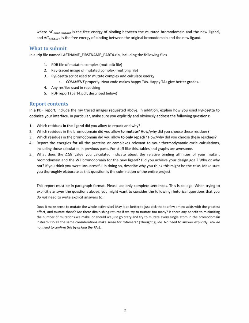
where is the free energy of binding between the mutated bromodomain and the new ligand,
and is the free energy of binding between the original bromodomain and the new ligand.
What to submit In a .zip file named LASTNAME_FIRSTNAME_PART4.zip, including the following files
1. PDB file of mutated complex (mut.pdb file)
2. Ray-traced image of mutated complex (mut.png file)
3. PyRosetta script used to mutate complex and calculate energy
a. COMMENT properly. Neat code makes happy TAs. Happy TAs give better grades.
4. Any resfiles used in repacking
5. PDF report (part4.pdf, described below)
Report contents In a PDF report, include the ray traced images requested above. In addition, explain how you used PyRosetta to
optimize your interface. In particular, make sure you explicitly and obviously address the following questions:
1. Which residues in the ligand did you allow to repack and why?
2. Which residues in the bromodomain did you allow to mutate? How/why did you choose these residues?
3. Which residues in the bromodomain did you allow to only repack? How/why did you choose these residues?
4. Report the energies for all the proteins or complexes relevant to your thermodynamic cycle calculations,
including those calculated in previous parts. For stuff like this, tables and graphs are awesome.
5. What does the ΔΔG value you calculated indicate about the relative binding affinities of your mutant
bromodomain and the WT bromodomain for the new ligand? Did you achieve your design goal? Why or why
not? If you think you were unsuccessful in doing so, describe why you think this might be the case. Make sure
you thoroughly elaborate as this question is the culmination of the entire project.
This report must be in paragraph format. Please use only complete sentences. This is college. When trying to
explicitly answer the questions above, you might want to consider the following rhetorical questions that you
do not need to write explicit answers to:
Does it make sense to mutate the whole active site? May it be better to just pick the top few amino acids with the greatest
effect, and mutate those? Are there diminishing returns if we try to mutate too many? Is there any benefit to minimizing
the number of mutations we make, or should we just go crazy and try to mutate every single atom in the bromodomain
instead? Do all the same considerations make sense for rotamers? [Thought guide. No need to answer explicitly. You do
not need to confirm this by asking the TAs].
2

Appendix A: Basic PyMOL
[New!] Getting PyMol 1. Register for an educational-use-only PyMol license
a. http://pymol.org/edu/ 2. You will receive an email with a username, password, and a link to the site where you can download
PyMol. 3. Share and enjoy!
a. Google that phrase, if you didn’t get the irony.
Getting structures for PyMol [no new stuff from here on] PyMOL is a viewer for macromolecular structures, typically derived from x-ray crystallography or NMR experiments. These structures are typically obtained from the RCSB Protein Data Bank (www.pdb.org) and come in pdb format. The full PyMOL manual is available at http://pymol.sourceforge.net/newman/userman.pdf, and is relatively thorough. Most PyMOL commands are available either through drop-down menus or a command line. Here I'll typically address the way to do things via drop-down menus. Let's start by obtaining a pdb file to play with. First download the pdb file 1YY8 (all pdb entries are named with a 4-character code). To do so, use the following instructions:
1. Open chrome in your nomachine client by clicking on the chrome icon in the lower left hand corner.
2. Download the relevant PDB structure. This should place the file in your Downloads fold. You most likely want to move it into the directory /home/yournamehere. To do so, use the graphical file
interface provided by the third button in the bottom left of your screen. Then open it with PyMOL. A mass of sticks should appear on the screen. You can change your viewpoint by left-clicking and dragging to rotate the object. If you are on a laptop, one-button mode is probably easiest for viewing. Select this from the mouse menu. If you have a 3-button mouse, select 3-button viewing.
Here are some basic mouse actions (these are also noted in the lower left corner of the viewing window).
Desired Action 1-button mode 3-button mode zoom in and out control and drag up/down Right click and drag translate the object in the alt and drag Center button and drag viewing plane PyMOL will show the sequence of the protein if you select 'sequence' under the Display menu on the command line window. You can then select particular residues by clicking on the letter in the sequence at the top of the viewer window. You can also select residues by simply clicking on them in the viewer. The atoms will have pink dots on them to indicate they are selected. You can modify the properties of whatever you have currently selected by using the drop down menus to the right of '(sele)' in the right column. Another way to select things is via the command line. The syntax is select <name>, <category> <identifier>
<name> is what you want to name your new selection. <category> can be any of:
resn - residue type res - residue number name - atom in amino acid: N, C, O, Ca, Cb, etc. symbol - element name chain - chain A, B, C, etc. ss - secondary structure type: 'h', 's' or 'l'
3
Selection criteria (in the form of <category> <identifier>) can be linked together by boolean operators: and, or, and not. When you create a new selection, it will show up in the right hand panel and you can access the drop down menus to manipulate only the atoms in that selection. For example, type select chainA, chain A. This way, you can modify only chain A of the molecule. For example, on “all,” click on H and choose everything, then chainA, click S and “cartoon” to show the cartoon version of only chain A. In addition to creating a new selection, the above syntax for selecting parts of the structure is generally used throughout pymol. For example, color red, res 40 will color residue 40 red.
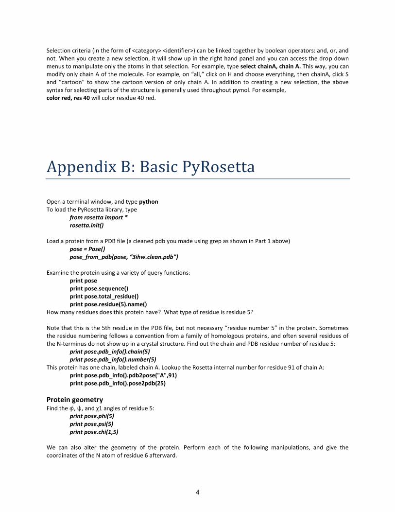
Appendix B: Basic PyRosetta
Open a terminal window, and type python To load the PyRosetta library, type
from rosetta import * rosetta.init()
Load a protein from a PDB file (a cleaned pdb you made using grep as shown in Part 1 above)
pose = Pose() pose_from_pdb(pose, “3ihw.clean.pdb”)
Examine the protein using a variety of query functions:
print pose print pose.sequence() print pose.total_residue() print pose.residue(5).name()
How many residues does this protein have? What type of residue is residue 5? Note that this is the 5th residue in the PDB file, but not necessary “residue number 5” in the protein. Sometimes the residue numbering follows a convention from a family of homologous proteins, and often several residues of the N-terminus do not show up in a crystal structure. Find out the chain and PDB residue number of residue 5:
print pose.pdb_info().chain(5) print pose.pdb_info().number(5)
This protein has one chain, labeled chain A. Lookup the Rosetta internal number for residue 91 of chain A: print pose.pdb_info().pdb2pose("A",91) print pose.pdb_info().pose2pdb(25)
Protein geometry Find the , ψ, and χ1 angles of residue 5:
print pose.phi(5) print pose.psi(5) print pose.chi(1,5)
We can also alter the geometry of the protein. Perform each of the following manipulations, and give the coordinates of the N atom of residue 6 afterward.
4
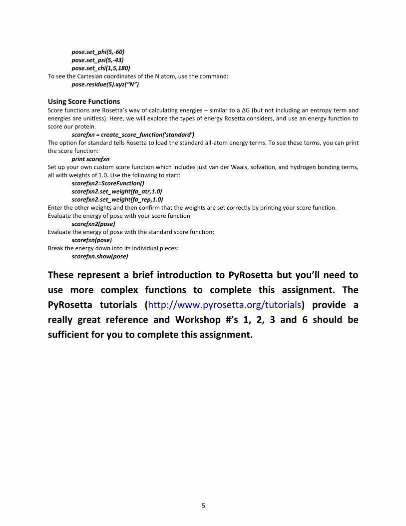
pose.set_phi(5,-60) pose.set_psi(5,-43) pose.set_chi(1,5,180)
To see the Cartesian coordinates of the N atom, use the command: pose.residue(5).xyz(“N”)
Using Score Functions Score functions are Rosetta’s way of calculating energies – similar to a ΔG (but not including an entropy term and energies are unitless). Here, we will explore the types of energy Rosetta considers, and use an energy function to score our protein.
scorefxn = create_score_function('standard') The option for standard tells Rosetta to load the standard all-atom energy terms. To see these terms, you can print the score function:
print scorefxn Set up your own custom score function which includes just van der Waals, solvation, and hydrogen bonding terms, all with weights of 1.0. Use the following to start:
scorefxn2=ScoreFunction() scorefxn2.set_weight(fa_atr,1.0) scorefxn2.set_weight(fa_rep,1.0)
Enter the other weights and then confirm that the weights are set correctly by printing your score function. Evaluate the energy of pose with your score function
scorefxn2(pose) Evaluate the energy of pose with the standard score function:
scorefxn(pose) Break the energy down into its individual pieces:
scorefxn.show(pose)
These represent a brief introduction to PyRosetta but you’ll need to
use more complex functions to complete this assignment. The
PyRosetta tutorials (http://www.pyrosetta.org/tutorials) provide a
really great reference and Workshop #’s 1, 2, 3 and 6 should be
sufficient for you to complete this assignment.
5
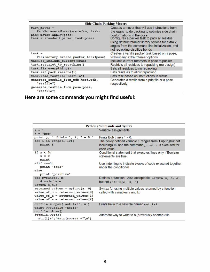
Here are some commands you might find useful:
6
MIT OpenCourseWarehttp://ocw.mit.edu
20.320 Analysis of Biomolecular and Cellular SystemsFall 2012
For information about citing these materials or our Terms of Use, visit: http://ocw.mit.edu/terms.