Embed Size (px)
Citation preview
Projections of the Pontine Nuclei to theCochlear Nucleus in Rats
MATTHIAS OHLROGGE,1 JOHN R. DOUCET,1AND DAVID K. RYUGO1,2*
1Departments of Otolaryngology-Head and Neck Surgery, Center for Hearing Sciences,Johns Hopkins University School of Medicine, Baltimore, Maryland 21205
2Department of Neuroscience, Center for Hearing Sciences, Johns Hopkins UniversitySchool of Medicine, Baltimore, Maryland 21205
ABSTRACTIn the cochlear nucleus, there is a magnocellular core of neurons whose axons form the
ascending auditory pathways. Surrounding this core is a thin shell of microneurons called thegranule cell domain (GCD). The GCD receives auditory and nonauditory inputs and projectsin turn to the dorsal cochlear nucleus, thus appearing to serve as a central locus forintegrating polysensory information and descending feedback. Nevertheless, the source ofmany of these inputs and the nature of the synaptic connections are relatively unknown. Weused the retrograde tracer Fast Blue to demonstrate that a major projection arises from thecontralateral pontine nuclei (PN) to the GCD. The projecting cells are more densely locatedin the ventral and rostral parts of the PN. They also are clustered into a lateral and a medialgroup. Injections of anterograde tracers into the PN labeled mossy fibers in the contralateralGCD. The terminals are confined to those parts of the GCD immediately surrounding theventral cochlear nucleus. There is no PN projection to the dorsal cochlear nucleus. Theseendings have the form of bouton and mossy fiber endings as revealed by light and electronmicroscopy. The PN represent a key station between the cerebral and cerebellar cortices, sothe pontocochlear nucleus projection emerges as a significant source of highly processedinformation that is introduced into the early stages of the auditory pathway. The cerebro-pontocerebellar pathway may impart coordination and timing cues to the motor system. In ananalogous way, perhaps the cerebropontocochlear nucleus projection endows the auditorysystem with a timing mechanism for extracting temporal information. J. Comp. Neurol. 436:290–303, 2001. © 2001 Wiley-Liss, Inc.
Indexing terms: audition; granule cells; mossy fibers; synapse
The auditory system has been described with an orga-nizing principle in which a core part conducts the ascend-ing auditory information to cortex and faithfully repre-sents the acoustic stimulus (Graybiel, 1974; Casseday etal., 1976; Ryugo, 1976). This core is surrounded by a shellthat receives descending input from higher auditory cen-ters as well as input from nonauditory parts of the brain.Feedback control from higher auditory centers is trans-ferred through the shell region to the core (Mitani et al.,1983; Winer et al., 1998). Nonauditory information that isused to facilitate certain aspects of hearing is integratedby way of the shell (Ryugo, 1976; Huffman and Henson,1990). Although the nature of this core-shell concept hadusually been reserved for levels of the system above themidbrain, recent work has revealed that multimodal anddescending inputs to the auditory system begin as early asthe cochlear nucleus (CN; Itoh et al., 1987; Weinberg andRustioni, 1987; Wright and Ryugo, 1996). The CN exhibits
this basic core-shell structure, in which the anterior andposterior ventral cochlear nucleus (VCN) and the dorsalcochlear nucleus (DCN) represent the core. The shell com-prises the granule cell domain (GCD) overlying the VCNand separating VCN and DCN. To gain a better under-standing of this shell in CN function, a first step is toidentify the source of inputs to the GCD.
The synaptic endings of type I auditory nerve fibersterminate as part of the ascending auditory pathway in
Contract grant sponsor: NIH/NIDCD; Contract grant number: DC04395;Contract grant number: DC00232; Contract grant number: DC04505.
*Correspondence to: David K. Ryugo, Center for Hearing Sciences, JHUSchool of Medicine, 720 Rutland Avenue, Baltimore, MD 21205.E-mail: [email protected]
Received 28 September 2000; Revised 26 March 2001; Accepted 11 May2001
THE JOURNAL OF COMPARATIVE NEUROLOGY 436:290–303 (2001)
© 2001 WILEY-LISS, INC.
the core of the cochlear nucleus. These rapidly conducting,myelinated axons convey acoustic information to the brainvia the auditory nerve. In contrast, the shell representedby the GCD receives input from type II auditory nervefibers (Brown et al., 1988a; Hurd et al., 1999). Theseunmyelinated auditory nerve fibers most likely carry mod-ulating signals or information about the status of theorgan of Corti, because they originate from outer haircells. The GCD also receives inputs from multiple higherauditory centers, such as the superior olivary complex(Brown et al., 1988b), the inferior colliculus (Caicedo andHerbert, 1993), and the auditory cortex (Feliciano et al.,1995; Weedman and Ryugo, 1996). Ascending terminals inthe GCD arise from neurons of the spinal trigeminal nu-cleus (Itoh et al., 1987), the cuneate nucleus (Weinbergand Rustioni, 1987; Wright and Ryugo, 1996), and thevestibular nuclei (Burian and Goesttner, 1988). Granulecells influence the output of the DCN by way of parallelfibers contacting the apical dendrites of pyramidal cells(Mugnaini et al., 1980a; Manis, 1989; Saade et al., 1989;Young et al., 1995). Granule cell influence on VCN outputhas not been shown. In addition to the known inputs to theGCD, ultrastructural studies have revealed multiple ter-minals within the GCD whose origins have not been de-termined (Mugnaini et al., 1980b). Of these, there aremany so-called mossy fibers, some of which originate inthe cuneate nucleus (Wright and Ryugo, 1996). Mossyfibers have a characteristic appearance, being relativelylarge (10–15 mm in diameter), making asymmetric synap-tic contacts with several postsynaptic dendrites (Weed-man and Ryugo, 1996; Weedman et al., 1996), and con-taining round synaptic vesicles. They have been so namedbecause their morphology is similar to that of mossy fiberterminals in the cerebellar cortex.
The purpose of the present study was to determine theorigin of some of these mossy fibers. This task representsa step towards a better understanding of multimodal in-tegration at the level of the cochlear nucleus. Retrogradeand anterograde tracing techniques were used to reveal anovel projection to the GCD and to demonstrate that anumber of mossy fibers originate in the contralateral pon-tine nuclei (PN). Previously published observations on PNanatomy and physiology will be considered to developideas of possible functions of this pontocochlear nucleusprojection. Some of these results were presented in ab-stract form at the 23rd annual Midwinter Research Meet-ing of the Association for Research in Otolaryngology,February 20–24, 2000.
MATERIAL AND METHODS
Animals and animal preparation
Male Sprague Dawley rats (250–300 g) from CharlesRiver were used for all experiments. Animals were deeplyanesthetized by intraperitoneal injection of sodium pento-barbital (45 mg/kg body weight), and oral secretions wereblocked by 0.05 mg intramuscular injections of atropinesulfate. Surgery was performed only after animals wereareflexic to tail or paw pinches. All procedures were inaccordance with the guidelines and had the approval ofthe Animal Care and Use Committee of the Johns Hop-kins School of Medicine.
Retrograde experiments
An incision by scalpel was made to the skin on theposterior surface of the skull and the soft tissue scrapedaway. An opening in the skull was made using drill androngeurs, and the dura covering the cerebellum was cut.That part of the cerebellum overlying the left CN wasaspirated to expose the characteristic smooth and shinysurface of the DCN. Fast blue (Sigma, St. Louis, MO) wasinjected into the lateral region of the granule cell laminaseparating the VCN and DCN by direct visual controlusing an operating microscope. An injection of an aqueoussolution containing 3% Fast Blue (20–60 nl) was madethrough a glass micropipette (30 mm inner diameter; ID)using an oocyte digital microdispenser (Nanoject Variable;Drummond Scientific Co., Broomall, PA). Injections weredelivered at a rate of 1–5 nl/minute over a time period of20 minutes. At the end of the injection, the pipette was leftin place for an additional 5 minutes before withdrawal.The hole in the skull was filled with gelfoam, the incisionwas sutured, and the animals were allowed to recover.Four to six days later, animals were administered a lethalIP dose of sodium pentobarbital (100 mg/kg) and thentranscardially perfused with ;50 ml of 0.1 M phosphate-buffered saline, pH 7.4, followed by 300 ml of 10% forma-lin in the same buffer. After perfusion the brain wasremoved from the skull and allowed to postfix for 1 hour at4°C. The brain was then transferred into a 20% sucrosesolution in phosphate buffer, pH 7.4, overnight at 4°C. Thebrainstem was sectioned on the next day on a freezingmicrotome at a thickness of 50 mm. Every other sectionwas mounted in serial order on clean glass slides and airdried, then coverslipped in Krystalon mounting medium(Harleco; EM Science, Gibbstown, NJ). Labeled cells werelocated and photographed on the same day using a fluo-rescent microscope at a wavelength of 360 nm and acooled, three-chip RGB digital CCD camera (C5810;Hamamatsu) at 3100, 3200, or 3400 magnification.
Anterograde experiments
Anesthetized animals were positioned into a stereotaxicframe, the skin and soft tissue over the parietal bone werereflected, and a 2-mm-diameter hole was drilled in theskull. A glass pipette (30 mm ID), filled with a mixture of2.5% biotinylated Phaseolus vulgaris leucoagglutinin(PHA-L; Vector Laboratories, Burlingame, CA) and 2.5%PHA-L (Vector Laboratories) in phosphate buffer, pH 7.9,was lowered into the brain using a micromanipulator. Thepipette was positioned according to stereotaxic coordi-nates so that its tip was placed in the left PN. An injectionof 200 nl was made at a rate of 10 nl/minute using theDrummond microdispenser. Pipettes were left in place for5 minutes after injection. The skin was sutured, and theanimals were given time to recover. After 7–10 days, theanimals were again deeply anesthetized with a lethal doseof pentobarbital (100 mg/kg body weight) and then tran-scardially perfused with 20 ml of 0.1 M phosphate-buffered saline, pH 7.4, followed by 4% paraformaldehydein phosphate buffer, pH 7.4. The brain was removed fromthe skull and postfixed for 1 hour at 4°C. Then, the tissuewas embedded in gelatin-albumin mixture for stabilitywhile sectioning and cut into coronal sections (50 mmthickness for the CN or 75 mm for the pons) on a Vi-bratome. Sections containing the CN or PN were pro-
291PONTINE PROJECTIONS TO THE COCHLEAR NUCLEUS
cessed differently to optimize visualization of the injectionsite and the labeled fibers.
In our experiments, we observed that, after injections ofbiotinylated PHA-L into the pontine nuclei, we could notstain the injection site. It was possible, however, to locatethe tissue damage by the pipette tip, so we could identifythe location of the injection site but could not determineits size. We solved this problem by adding PHA-L to thetracer solution and visualizing the injection site usingantibodies and an immunohistochemical reaction with3,3-diaminobenzidine (DAB). Because we applied thetracer mixture by pressure, we infer that the injection siterecovered by the visualization of the DAB reaction productto PHA-L also represented the biotinylated PHA-L injec-tion site. We have no explanation for why the biotinylatedPHA-L injection site was not detectable in the PN, in thatthe same protocol revealed intense biotinylated PHA-Lstaining of axons and terminals in the CN. Reports on theproperties of PHA-L and biotinylated PHA-L suggest thatthe same tracer can behave differently when applied indifferent areas of the brain (Schofield, 1990). Our obser-vation might be due to this phenomenon.
CN sections were incubated overnight in avidin-biotin-peroxidase complex (ABC Elite; Vector Laboratories) in0.1 M phosphate-buffered saline, pH 7.4, at 4°C. On thenext day the sections were rinsed twice in phosphatebuffer, pH 7.4, and twice in 0.05 M cacodylate buffer, pH7.2. The label was developed with 0.0125% DAB, 0.25%nickel ammonium sulfate, and 0.35% imidazole in 0.05 Mcacodylate, pH 7.2. Sections were preincubated in DABsolution for 10 minutes without hydrogen peroxide andthen incubated in fresh DAB with 0.02% hydrogen perox-ide for an additional 15 minutes. Sections were mountedon gelatin-coated microscope slides, counterstained withcresyl violet, and coverslipped with Permount.
Sections containing the PN were processed using thefollowing procedures. Unless otherwise stated, all immu-nocytochemical processing was performed at room tem-perature in Tris-buffered saline (TBS; 0.05 M, pH 7.6).Sections were washed in TBS, pH 7.6, blocked in 5%nonfat dry milk for 2 hours, and incubated overnight inrabbit anti-PHA-L (Vector Laboratories) at a concentra-tion of 1:2,000. Membranes were permeabilized by theaddition of 0.1% Photo-Flo (Kodak) to both the primaryand the secondary antibody solutions. On the next day,sections were washed three times in TBS and incubated ingoat anti-rabbit immunoglobulin (IgG) secondary anti-body (1:50) in TBS for 2 hours. After three more washes in0.05 M TBS, pH 7.6, sections were treated with rabbitperoxidase antiperoxidase at 1:400 for 1 hour. Washes inTBS were followed by a 5–10 minute incubation in 0.05%DAB and 0.015% H2O2 in Tris buffer (0.05 M; pH 7.6).Sections were washed further, mounted on gelatin-coatedslides, air dried, counterstained in cresyl violet, and cov-erslipped.
Because the DAB reaction product is electron dense, wecould dissect specific regions containing labeled mossyfiber endings from the main tissue sections and reembedthe smaller pieces for ultrathin sectioning and analysiswith an electron microscope. Light microscopic maps ofthese smaller blocks were made for orientation purposes,using blood vessels and labeled structures as landmarks.Serial ultrathin sections were collected on Formvar-coatedslotted grids and photographed with a Jeol 100CX electronmicroscope.
Data analysis
For the retrograde studies, only rats with injection sitesrestricted to the CN were included (n 5 6). Verification ofthe injection site consisted of microscopic analysis of serialsections spanning the entire rostral caudal extent of theCN. Every other section was drawn through the CN usinga microscope and drawing tube (total magnification363.5). The perimeter of the GCD was outlined and itssurface area calculated (The Image Processing Tool Kit;Reindeer Games, Inc., Gainsville, FL). The area for eachsection was multiplied by section thickness (0.05 mm) toobtain GCD volume, the resulting value doubled to com-pensate for the missing intervening section and summedto estimate total GCD volume. Outlines were digitized andscaled to match the corresponding photomicrograph, andthe injection site visible in each section was copied to itscorresponding drawing. The area of overlap between in-jection site and GCD was calculated, and the total injec-tion site volume in the GCD was determined.
We also examined nearby adjacent structures, such asthe inferior cerebellar peduncle, trigeminal nerve root,vestibular nerve root and nuclei, and descending tract andnucleus of the trigeminal nerve. Included in this analysiswere various nuclei known to give rise to axons travelingin the nearby tracts, such as the inferior olive projectingthrough the inferior cerebellar peduncle and Scarpa’s gan-glion projecting through the vestibular nerve. In this waywe could be confident that there was not a “fibers ofpassage” contamination problem.
The distribution of retrogradely labeled cell bodies inthe PN was determined using photomontages of the PN.Each montage was constructed from multiple digitizedimages using standard software (Adobe Photoshop 5.0).The perimeters and regional boundaries of individual sec-tions were digitized and superimposed on the montages,and labeled cells were plotted directly on the drawing andcounted. Every labeled cell in the PN was counted andnoted for its position ipsilateral or contralateral to theinjection site. We used cell counts to determine the rela-tive projection with respect to side. We did not calculatetotal cell counts, because that value would necessarily bedetermined by the size of the injection site.
Analysis of anterogradely labeled fibers and terminalswas conducted using light and electron microscopy. Onlyanimals with injection sites verified to be within the PN(n 5 5) were used. Alternate sections were drawn throughthe entire PN using a microscope and drawing tube (totalmagnification 363.5). The PN borders were establishedby cytoarchitectonic criteria (Mihailoff et al., 1981) andscaled to matching micrographs containing the injectionsites, and injection sites were copied to the drawings.PN and injection volumes were calculated as describedabove.
DAB reaction product filled fibers and terminals in acontinuous manner. Labeled fibers were followed by lightmicroscopy through serial sections, away from the PN andinto the CN, where they terminated as large swellings inthe granule cell domain. Selected sections were outlinedand terminals were plotted on the drawn sections usingtissue landmarks and a drawing tube. Labeled structureswere photographed using a CCD camera at various levelsof magnification.
292 M. OHLROGGE ET AL.
RESULTS
Retrograde experiments
GCD injections. The presence of mossy fiber endingsin the GCD has been known for some time, but their originhas not. Fast Blue was injected into the granule cell lam-ina, the part of the GCD that separates the VCN and
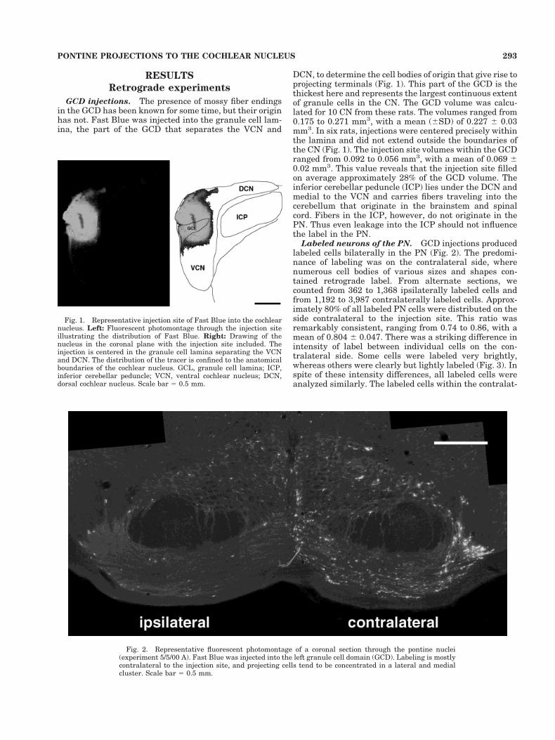
DCN, to determine the cell bodies of origin that give rise toprojecting terminals (Fig. 1). This part of the GCD is thethickest here and represents the largest continuous extentof granule cells in the CN. The GCD volume was calcu-lated for 10 CN from these rats. The volumes ranged from0.175 to 0.271 mm3, with a mean (6SD) of 0.227 6 0.03mm3. In six rats, injections were centered precisely withinthe lamina and did not extend outside the boundaries ofthe CN (Fig. 1). The injection site volumes within the GCDranged from 0.092 to 0.056 mm3, with a mean of 0.069 60.02 mm3. This value reveals that the injection site filledon average approximately 28% of the GCD volume. Theinferior cerebellar peduncle (ICP) lies under the DCN andmedial to the VCN and carries fibers traveling into thecerebellum that originate in the brainstem and spinalcord. Fibers in the ICP, however, do not originate in thePN. Thus even leakage into the ICP should not influencethe label in the PN.
Labeled neurons of the PN. GCD injections producedlabeled cells bilaterally in the PN (Fig. 2). The predomi-nance of labeling was on the contralateral side, wherenumerous cell bodies of various sizes and shapes con-tained retrograde label. From alternate sections, wecounted from 362 to 1,368 ipsilaterally labeled cells andfrom 1,192 to 3,987 contralaterally labeled cells. Approx-imately 80% of all labeled PN cells were distributed on theside contralateral to the injection site. This ratio wasremarkably consistent, ranging from 0.74 to 0.86, with amean of 0.804 6 0.047. There was a striking difference inintensity of label between individual cells on the con-tralateral side. Some cells were labeled very brightly,whereas others were clearly but lightly labeled (Fig. 3). Inspite of these intensity differences, all labeled cells wereanalyzed similarly. The labeled cells within the contralat-
Fig. 1. Representative injection site of Fast Blue into the cochlearnucleus. Left: Fluorescent photomontage through the injection siteillustrating the distribution of Fast Blue. Right: Drawing of thenucleus in the coronal plane with the injection site included. Theinjection is centered in the granule cell lamina separating the VCNand DCN. The distribution of the tracer is confined to the anatomicalboundaries of the cochlear nucleus. GCL, granule cell lamina; ICP,inferior cerebellar peduncle; VCN, ventral cochlear nucleus; DCN,dorsal cochlear nucleus. Scale bar 5 0.5 mm.
Fig. 2. Representative fluorescent photomontage of a coronal section through the pontine nuclei(experiment 5/5/00 A). Fast Blue was injected into the left granule cell domain (GCD). Labeling is mostlycontralateral to the injection site, and projecting cells tend to be concentrated in a lateral and medialcluster. Scale bar 5 0.5 mm.
293PONTINE PROJECTIONS TO THE COCHLEAR NUCLEUS
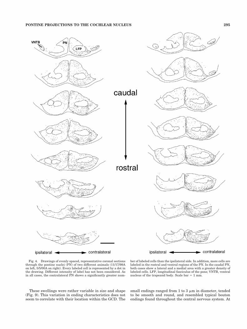
eral PN were not evenly distributed but exhibited distinctclustering in some areas. Labeled cells were distributedmore densely in the ventral parts of the PN than in thedorsal parts. In the rostral PN, cells seemed to be evenlydistributed throughout the PN; more caudally, labeledcells segregated into aggregations that could be followedthrough consecutive sections, forming elongated patchesor columns (Fig. 4). These columns were not consistentlyfound in any particular part of the PN except for a medialcluster.
The stained cells of the ipsilateral PN were less in-tensely, but more uniformly, stained. They did not exhibitany distinct clustering. In addition to the cells in the PN,there are labeled cells in other parts of the brain. Forexample, we observed labeled cells in the contralateralinferior colliculus and the superior olive bilaterally. Thoseprojections, however, have been described previously (see,e.g., Shore et al., 1991; Shore and Moore, 1998; Schofieldand Cant, 1999) and will not be discussed further hereexcept to say that they served as positive controls for ourmethods.
There has been discussion that, in the dorsal pontineregion, it is not always possible to distinguish betweengroups of cells that are part of the PN and groups thatrepresent the nucleus reticularis tegmenti pontis (NRTP;Mihailoff et al., 1981). PN neurons tend to have ovoidsomata, whereas NRTP neurons tend to be larger, withintensely basophilic somata. The dorsally located stainedcells in our experiments tended to be small, and we did notstain them with basophilic dyes. Because they were alsorelatively infrequent, for economy’s sake we grouped themwith the PN.
Anterograde experiments
The pontine nuclei. Results for the anterograde ex-periments were collected from 10 animals. Five animalshad injections confined to the PN that contained a mixtureof PHA-L and biotinylated PHA-L and provide the projec-
tion data. The PN represent an accumulation of neuronsmaking up the gray matter of the basal pons. Subdivisionsof this cell group have been described but for mostly de-scriptive rather than functional purposes (Mihailoff et al.,1981). These divisions include neuron clusters located me-dially, ventrally, and laterally to the cerebral peduncleand neurons lying adjacent to the peduncle defined as partof the peduncular nucleus. Neurons with cell bodies ofvarious sizes are distributed throughout the region with-out any particular order. The mean volume of the PN onone side was calculated as 1.81 6 0.2 mm3, with a range of1.58–2.01 mm3 (n 5 10). Resembling the cat, rabbit(Brodal and Jansen, 1946), and opossum (King et al.,1968), the rat does not exhibit any specific cytoarchitec-tonic features that characterize the major pontine subdi-visions (Mihailoff et al., 1981).
Running longitudinally through the pontine nuclei aretwo main fiber tracts, the longitudinal fasciculus of thepons (LFP; a continuation of cortical axons that make upthe cerebral peduncles) and the medial lemniscus. TheLFP carries efferent information to motor neurons in themedulla and spinal cord as part of the corticobulbar andcorticospinal tracts, respectively. No afferent fiber systemhas been described for the LFP so far. The medial lemnis-cus carries second-order sensory axons that arise from thedorsal column nuclei.
Pontine projections. In two cases (11/3/99 D and 11/3/99 B), the injection site was completely confined to thepontine nuclei (Figs. 5, 6). The limits of the PN weredrawn using the boundaries of areas with high cell den-sity, and the outlines were consistent with those of a ratstereotaxic atlas (Paxinos and Watson, 1982). In two othercases (9/29/99 C and 11/3/99 A), the injection site includedpart of the LFP. We found, however, that the results ofthese experiments were essentially identical to thosewhen the injection was anatomically limited to the PN.These showed labeled endings in the contralateral GCD ofthe CN, excluding the DCN, and no labeling was seen inthe magnocellular core regions of the CN. In one case(4/27/99 C), the injection site was on the dorsal border ofthe PN but produced labeling with the same pattern asthat from pure PN injections. The volume of the injectionsites contained within the PN ranged from 0.033 to 0.096mm3, with a mean of 0.063 6 0.03 mm3. The variation inamount and location of tracer injected in these experi-ments did not qualitatively change the nature or patternof label in the CN. Thus we inferred that the injectionsincluded in our database were limited to regions of the PNprojecting to the GCD.
In the five cases with injections in the PN, labeled fiberswere followed to the GCD, where they branched and dis-tributed terminals (Fig. 7). No label was found in the DCNor in the magnocellular core of the VCN. The fibers ap-peared to reach the CN by way of the intermediate acous-tic stria, where they proceeded to disperse into their sep-arate ways. That is, they did not distribute themselves asa discrete group within the anatomical boundaries of theCN. Rather, they ran as individuals through the granulecell lamina between VCN and DCN and continued into thelayer of granule cells that surround the dorsolateral sur-face of the VCN. There, they ran parallel to the VCNsurface. The labeled fibers were relatively thin, typicallybetween 0.5 and 1.0 mm in thickness. During their pas-sage, the fibers branched and gave rise to en passant andterminal swellings (Fig. 8).
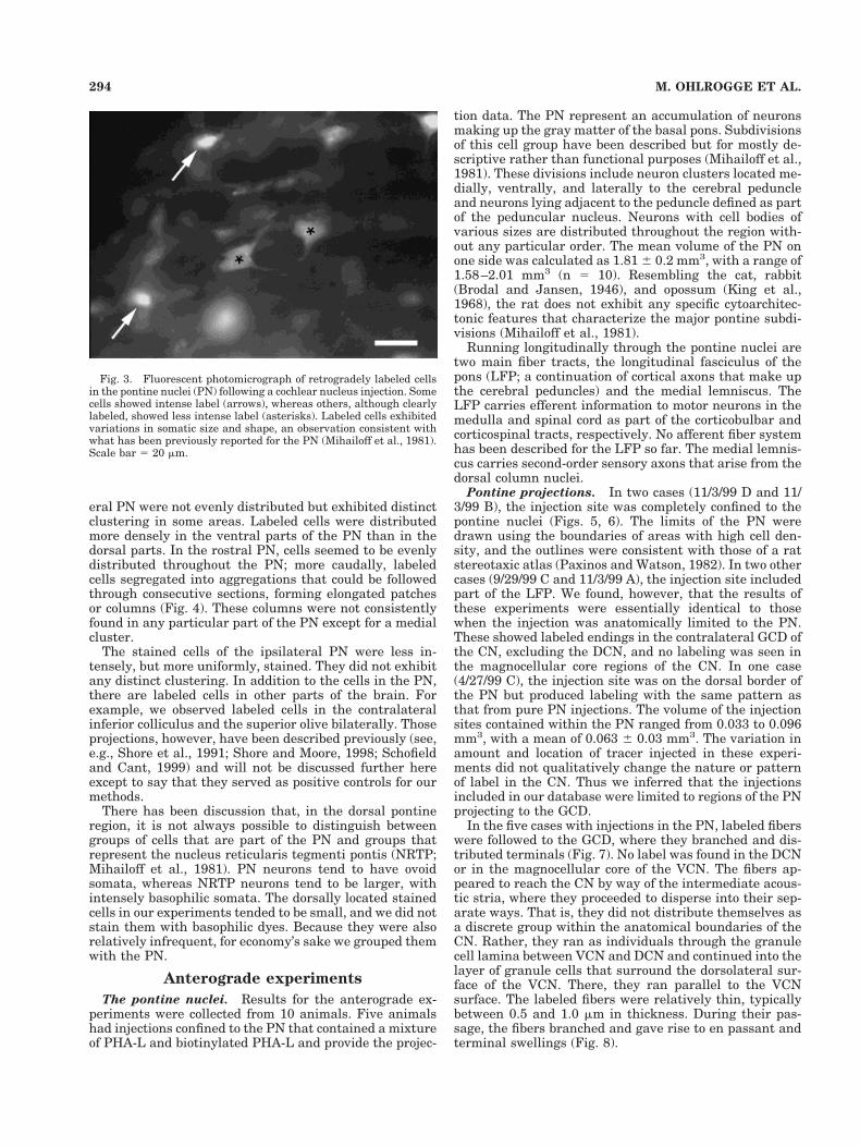
Fig. 3. Fluorescent photomicrograph of retrogradely labeled cellsin the pontine nuclei (PN) following a cochlear nucleus injection. Somecells showed intense label (arrows), whereas others, although clearlylabeled, showed less intense label (asterisks). Labeled cells exhibitedvariations in somatic size and shape, an observation consistent withwhat has been previously reported for the PN (Mihailoff et al., 1981).Scale bar 5 20 mm.
294 M. OHLROGGE ET AL.
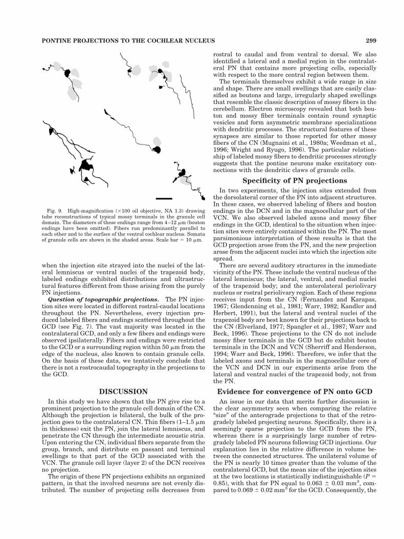
These swellings were rather variable in size and shape(Fig. 9). This variation in ending characteristics does notseem to correlate with their location within the GCD. The
small endings ranged from 1 to 3 mm in diameter, tendedto be smooth and round, and resembled typical boutonendings found throughout the central nervous system. At
Fig. 4. Drawings of evenly spaced, representative coronal sectionsthrough the pontine nuclei (PN) of two different animals (11/17/99Aon left, 5/5/00A on right). Every labeled cell is represented by a dot inthe drawing. Different intensity of label has not been considered. Asin all cases, the contralateral PN shows a significantly greater num-
ber of labeled cells than the ipsilateral side. In addition, more cells arelabeled in the rostral and ventral regions of the PN. In the caudal PN,both cases show a lateral and a medial area with a greater density oflabeled cells. LFP, longitudinal fasciculus of the pons; VNTB, ventralnucleus of the trapezoid body. Scale bar 5 1 mm.
295PONTINE PROJECTIONS TO THE COCHLEAR NUCLEUS
the other extreme, endings were relatively large (5–15 mmin diameter) and irregular in shape. These latter endingsclosely resembled the appearance of so-called mossy fibersof the cerebellum (see, e.g., Ramon y Cajal, 1909) andmossy fibers of the GCD that arise from the cuneate nu-cleus (Wright and Ryugo, 1996). In addition to being large,they had irregular edges and bleb-like protrusions, aroseas either en passant or terminal swellings, and were dis-tributed in regions with high density of granule cells. Theydid not appear to make contact with resident cell bodies.
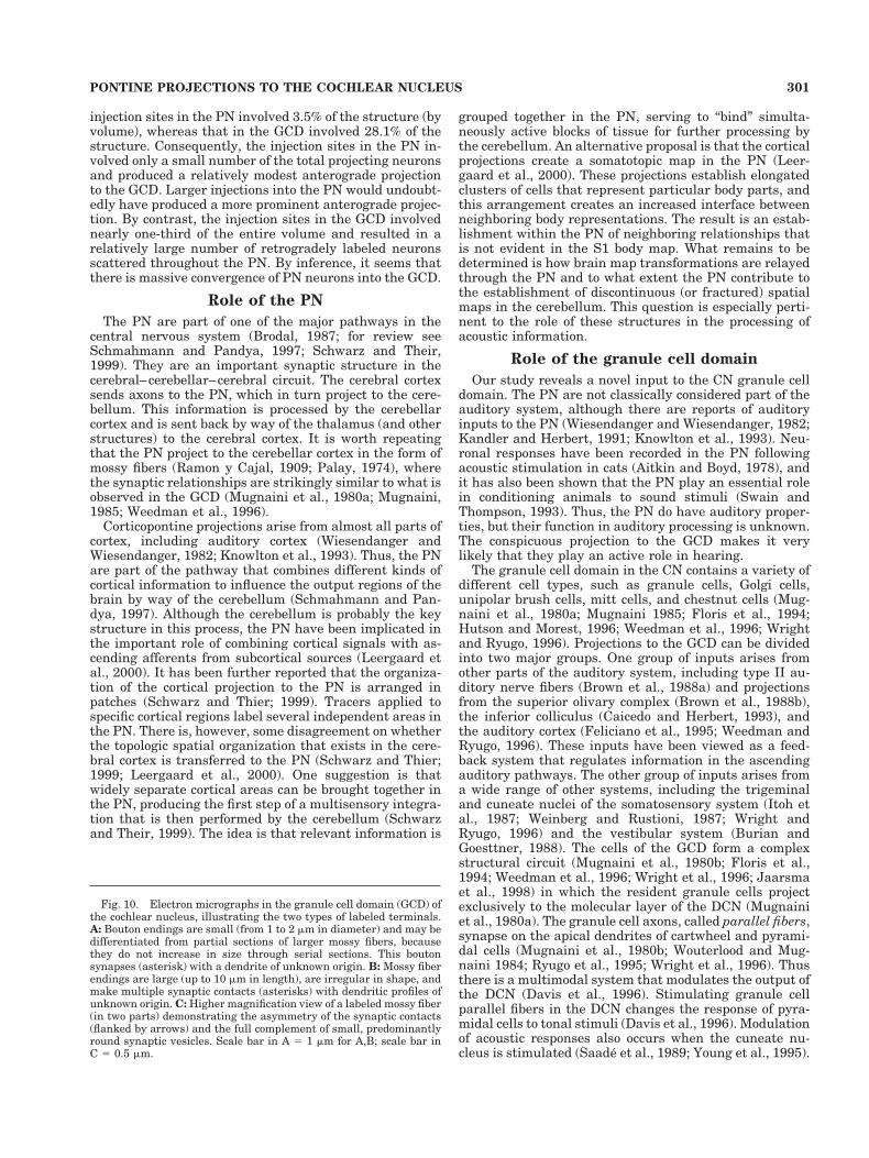
Ultrastructural characteristics. We examined anumber of large labeled endings from the GCD with theelectron microscope. Some of these endings were boutons(Fig. 10A), and others were mossy fibers (Fig. 10B). Bou-tons mostly contacted thin (,1.0 mm in diameter) den-dritic profiles. Mossy fibers formed an eccentric structureincompletely surrounded by dendritic and axonal pro-cesses. Consistent with the light microscopic appearance,the labeled mossy fiber endings exhibited considerablevariations in shape. The surrounding processes also var-ied. The labeled endings themselves exhibited the classicappearance of mossy fiber terminals in the CN, filled withround synaptic vesicles, numerous mitochondria, andprominent asymmetric synapses (Mugnaini et al., 1980a;Weedman et al., 1996; Wright and Ryugo, 1996). The pre-and postsynaptic densities are especially exaggerated bythe DAB reaction product, helping to highlight the synap-tic cleft. The labeled endings arising from the PN were
typically filled with small, round synaptic vesicles, and thepostsynaptic densities were either large and continuous(Fig. 10C) or segmented (Fig. 10B).
The dendrites contacted by labeled mossy fibers fromthe PN are not numerous, but there is a pattern to thesynaptic relationships. The number of postsynaptic den-drites surrounding the mossy fiber is inversely related toits profile diameter. That is, a single postsynaptic dendritetends to be large (1–2 mm in diameter). When there aretwo dendritic profiles, they are often close together andjust a bit thinner (0.75–1.5 mm in diameter) than thesingle profile, as though they were branches near the mainstalk. The thin dendritic profiles (0.3–0.8 mm in diameter)are typically separate from one another, resembling thedistal portions of the granule cell dendritic claw. In addi-tion, sometimes there are thin, finger-like dendritic pro-cesses that project into the mossy fiber, and these can bepostsynaptic. We infer that the pontine mossy fibers formsynaptic glomeruli with the distal dendrites of granulecells.
Injection sites outside the PN. In two cases that wererejected, the injection site included not only the PN butalso the adjacent lateral lemniscus or the ventral nucleus
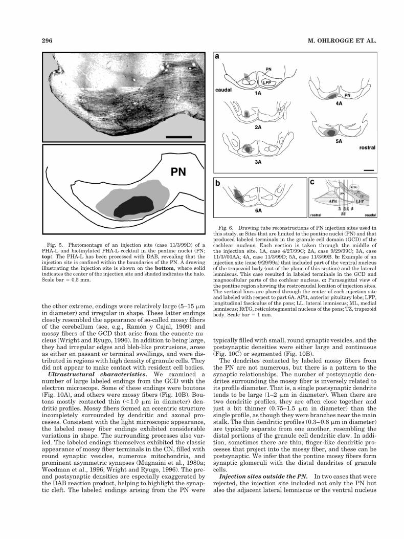
Fig. 5. Photomontage of an injection site (case 11/3/99D) of aPHA-L and biotinylated PHA-L cocktail in the pontine nuclei (PN;top). The PHA-L has been processed with DAB, revealing that theinjection site is confined within the boundaries of the PN. A drawingillustrating the injection site is shown on the bottom, where solidindicates the center of the injection site and shaded indicates the halo.Scale bar 5 0.5 mm.
Fig. 6. Drawing tube reconstructions of PN injection sites used inthis study. a: Sites that are limited to the pontine nuclei (PN) and thatproduced labeled terminals in the granule cell domain (GCD) of thecochlear nucleus. Each section is taken through the middle ofthe injection site. 1A, case 4/27/99C; 2A, case 9/29/99C; 3A, case11/3//00AA; 4A, case 11/3/99D; 5A, case 11/3/99B. b: Example of aninjection site (case 9/29/99a) that included part of the ventral nucleusof the trapezoid body (out of the plane of this section) and the laterallemniscus. This case resulted in labeled terminals in the GCD andmagnocellular parts of the cochlear nucleus. c: Parasagittal view ofthe pontine region showing the rostrocaudal location of injection sites.The vertical lines are placed through the center of each injection siteand labeled with respect to part 6A. APit, anterior pituitary lobe; LFP,longitudinal fasciculus of the pons; LL, lateral lemniscus; ML, mediallemniscus; RtTG, reticulotegmental nucleus of the pons; TZ, trapezoidbody. Scale bar 5 1 mm.
296 M. OHLROGGE ET AL.
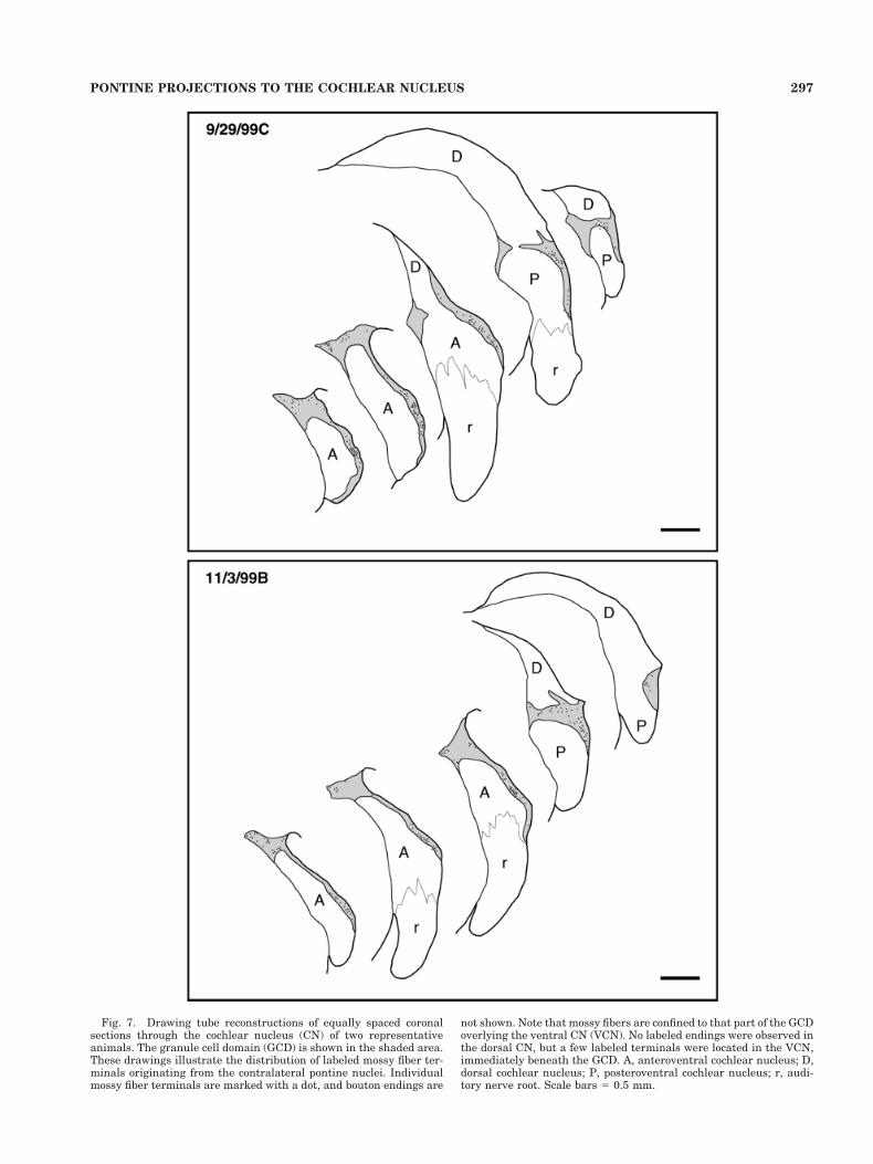
Fig. 7. Drawing tube reconstructions of equally spaced coronalsections through the cochlear nucleus (CN) of two representativeanimals. The granule cell domain (GCD) is shown in the shaded area.These drawings illustrate the distribution of labeled mossy fiber ter-minals originating from the contralateral pontine nuclei. Individualmossy fiber terminals are marked with a dot, and bouton endings are
not shown. Note that mossy fibers are confined to that part of the GCDoverlying the ventral CN (VCN). No labeled endings were observed inthe dorsal CN, but a few labeled terminals were located in the VCN,immediately beneath the GCD. A, anteroventral cochlear nucleus; D,dorsal cochlear nucleus; P, posteroventral cochlear nucleus; r, audi-tory nerve root. Scale bars 5 0.5 mm.
297PONTINE PROJECTIONS TO THE COCHLEAR NUCLEUS
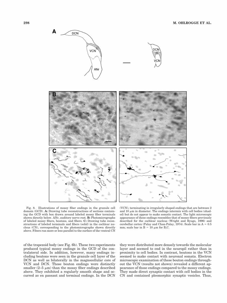
of the trapezoid body (see Fig. 6b). These two experimentsproduced typical mossy endings in the GCD of the con-tralateral side. In addition, however, many endings in-cluding boutons were seen in the granule cell layer of theDCN as well as bilaterally in the magnocellular core ofVCN and DCN. Those bouton endings were distinctlysmaller (2–5 mm) than the mossy fiber endings describedabove. They exhibited a regularly smooth shape and oc-curred as en passant and terminal endings. In the DCN
they were distributed more densely towards the molecularlayer and seemed to end in the neuropil rather than inproximity to cell bodies. In contrast, boutons in the VCNseemed to make contact with neuronal somata. Electronmicroscopic examination of these bouton endings through-out the VCN (results not shown) revealed a different ap-pearance of those endings compared to the mossy endings.They made direct synaptic contact with cell bodies in theCN and contained pleomorphic synaptic vesicles. Thus,
Fig. 8. Illustrations of mossy fiber endings in the granule celldomain (GCD). A: Drawing tube reconstructions of sections contain-ing the GCD with box drawn around labeled mossy fiber terminalsshown directly below. ANr, auditory nerve root. B: Photomicrographsof labeled mossy fibers, boutons, and fibers. C: Drawing tube recon-structions of labeled terminals and fibers (solid) in the cochlear nu-cleus (CN), corresponding to the photomicrographs shown directlyabove. Fibers run more or less parallel to the surface of the ventral CN
(VCN), terminating in irregularly shaped endings that are between 2and 10 mm in diameter. The endings intermix with cell bodies (shad-ed) but do not appear to make somatic contact. The light microscopicappearance of these endings resembles that of mossy fibers previouslydescribed for the cochlear nucleus (Wright and Ryugo, 1996) andcerebellar cortex (Palay and Chan-Palay, 1974). Scale bar in A 5 0.5mm; scale bar in B 5 10 mm for B,C.
298 M. OHLROGGE ET AL.
when the injection site strayed into the nuclei of the lat-eral lemniscus or ventral nuclei of the trapezoid body,labeled endings exhibited distributions and ultrastruc-tural features different from those arising from the purelyPN injections.
Question of topographic projections. The PN injec-tion sites were located in different rostral-caudal locationsthroughout the PN. Nevertheless, every injection pro-duced labeled fibers and endings scattered throughout theGCD (see Fig. 7). The vast majority was located in thecontralateral GCD, and only a few fibers and endings wereobserved ipsilaterally. Fibers and endings were restrictedto the GCD or a surrounding region within 50 mm from theedge of the nucleus, also known to contain granule cells.On the basis of these data, we tentatively conclude thatthere is not a rostrocaudal topography in the projections tothe GCD.
DISCUSSION
In this study we have shown that the PN give rise to aprominent projection to the granule cell domain of the CN.Although the projection is bilateral, the bulk of the pro-jection goes to the contralateral CN. Thin fibers (1–1.5 mmin thickness) exit the PN, join the lateral lemniscus, andpenetrate the CN through the intermediate acoustic stria.Upon entering the CN, individual fibers separate from thegroup, branch, and distribute en passant and terminalswellings to that part of the GCD associated with theVCN. The granule cell layer (layer 2) of the DCN receivesno projection.
The origin of these PN projections exhibits an organizedpattern, in that the involved neurons are not evenly dis-tributed. The number of projecting cells decreases from
rostral to caudal and from ventral to dorsal. We alsoidentified a lateral and a medial region in the contralat-eral PN that contains more projecting cells, especiallywith respect to the more central region between them.
The terminals themselves exhibit a wide range in sizeand shape. There are small swellings that are easily clas-sified as boutons and large, irregularly shaped swellingsthat resemble the classic description of mossy fibers in thecerebellum. Electron microscopy revealed that both bou-ton and mossy fiber terminals contain round synapticvesicles and form asymmetric membrane specializationswith dendritic processes. The structural features of thesesynapses are similar to those reported for other mossyfibers of the CN (Mugnaini et al., 1980a; Weedman et al.,1996; Wright and Ryugo, 1996). The particular relation-ship of labeled mossy fibers to dendritic processes stronglysuggests that the pontine neurons make excitatory con-nections with the dendritic claws of granule cells.
Specificity of PN projections
In two experiments, the injection sites extended fromthe dorsolateral corner of the PN into adjacent structures.In these cases, we observed labeling of fibers and boutonendings in the DCN and in the magnocellular part of theVCN. We also observed labeled axons and mossy fiberendings in the GCD, identical to the situation when injec-tion sites were entirely contained within the PN. The mostparsimonious interpretation of these results is that theGCD projection arose from the PN, and the new projectionarose from the adjacent nuclei into which the injection sitespread.
There are several auditory structures in the immediatevicinity of the PN. These include the ventral nucleus of thelateral lemniscus; the lateral, ventral, and medial nucleiof the trapezoid body; and the anterolateral periolivarynucleus or rostral periolivary region. Each of these regionsreceives input from the CN (Fernandez and Karapas,1967; Glendenning et al., 1981; Warr, 1982; Kandler andHerbert, 1991), but the lateral and ventral nuclei of thetrapezoid body are best known for their projections back tothe CN (Elverland, 1977; Spangler et al., 1987; Warr andBeck, 1996). These projections to the CN do not includemossy fiber terminals in the GCD but do exhibit boutonterminals in the DCN and VCN (Sherriff and Henderson,1994; Warr and Beck, 1996). Therefore, we infer that thelabeled axons and terminals in the magnocellular core ofthe VCN and DCN in our experiments arise from thelateral and ventral nuclei of the trapezoid body, not fromthe PN.
Evidence for convergence of PN onto GCD
An issue in our data that merits further discussion isthe clear asymmetry seen when comparing the relative“size” of the anterograde projections to that of the retro-gradely labeled projecting neurons. Specifically, there is aseemingly sparse projection to the GCD from the PN,whereas there is a surprisingly large number of retro-gradely labeled PN neurons following GCD injections. Ourexplanation lies in the relative difference in volume be-tween the connected structures. The unilateral volume ofthe PN is nearly 10 times greater than the volume of thecontralateral GCD, but the mean size of the injection sitesat the two locations is statistically indistinguishable (P 50.85), with that for PN equal to 0.063 6 0.03 mm3, com-pared to 0.069 6 0.02 mm3 for the GCD. Consequently, the
Fig. 9. High-magnification (3100 oil objective, NA 1.3) drawingtube reconstructions of typical mossy terminals in the granule celldomain. The diameters of these endings range from 4–12 mm (boutonendings have been omitted). Fibers run predominantly parallel toeach other and to the surface of the ventral cochlear nucleus. Somataof granule cells are shown in the shaded areas. Scale bar 5 10 mm.
299PONTINE PROJECTIONS TO THE COCHLEAR NUCLEUS
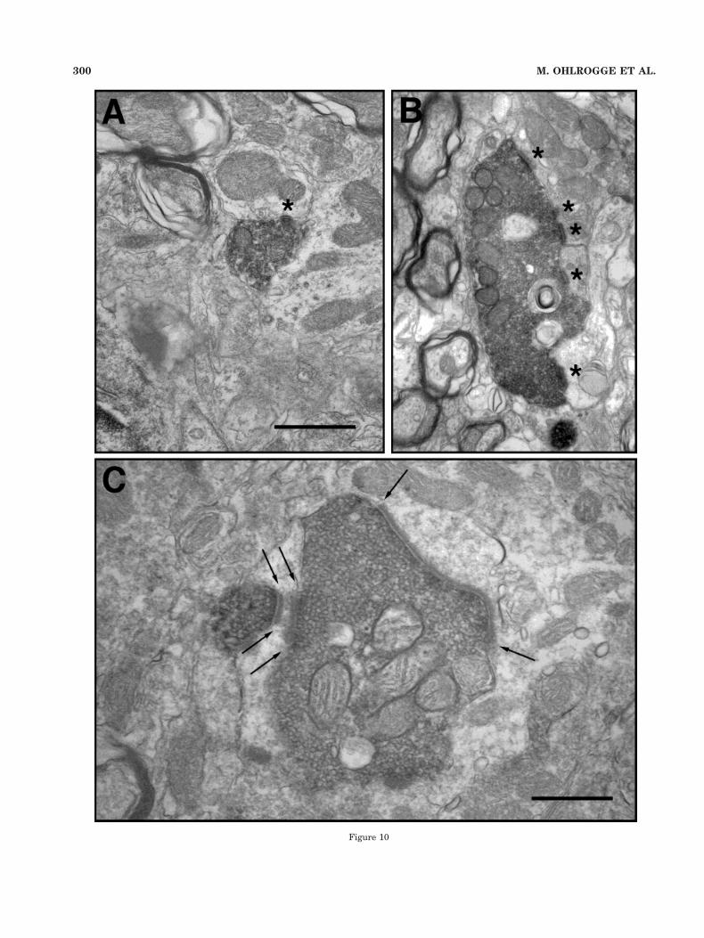
Figure 10
300 M. OHLROGGE ET AL.
injection sites in the PN involved 3.5% of the structure (byvolume), whereas that in the GCD involved 28.1% of thestructure. Consequently, the injection sites in the PN in-volved only a small number of the total projecting neuronsand produced a relatively modest anterograde projectionto the GCD. Larger injections into the PN would undoubt-edly have produced a more prominent anterograde projec-tion. By contrast, the injection sites in the GCD involvednearly one-third of the entire volume and resulted in arelatively large number of retrogradely labeled neuronsscattered throughout the PN. By inference, it seems thatthere is massive convergence of PN neurons into the GCD.
Role of the PN
The PN are part of one of the major pathways in thecentral nervous system (Brodal, 1987; for review seeSchmahmann and Pandya, 1997; Schwarz and Their,1999). They are an important synaptic structure in thecerebral–cerebellar–cerebral circuit. The cerebral cortexsends axons to the PN, which in turn project to the cere-bellum. This information is processed by the cerebellarcortex and is sent back by way of the thalamus (and otherstructures) to the cerebral cortex. It is worth repeatingthat the PN project to the cerebellar cortex in the form ofmossy fibers (Ramon y Cajal, 1909; Palay, 1974), wherethe synaptic relationships are strikingly similar to what isobserved in the GCD (Mugnaini et al., 1980a; Mugnaini,1985; Weedman et al., 1996).
Corticopontine projections arise from almost all parts ofcortex, including auditory cortex (Wiesendanger andWiesendanger, 1982; Knowlton et al., 1993). Thus, the PNare part of the pathway that combines different kinds ofcortical information to influence the output regions of thebrain by way of the cerebellum (Schmahmann and Pan-dya, 1997). Although the cerebellum is probably the keystructure in this process, the PN have been implicated inthe important role of combining cortical signals with as-cending afferents from subcortical sources (Leergaard etal., 2000). It has been further reported that the organiza-tion of the cortical projection to the PN is arranged inpatches (Schwarz and Thier; 1999). Tracers applied tospecific cortical regions label several independent areas inthe PN. There is, however, some disagreement on whetherthe topologic spatial organization that exists in the cere-bral cortex is transferred to the PN (Schwarz and Thier;1999; Leergaard et al., 2000). One suggestion is thatwidely separate cortical areas can be brought together inthe PN, producing the first step of a multisensory integra-tion that is then performed by the cerebellum (Schwarzand Their, 1999). The idea is that relevant information is
grouped together in the PN, serving to “bind” simulta-neously active blocks of tissue for further processing bythe cerebellum. An alternative proposal is that the corticalprojections create a somatotopic map in the PN (Leer-gaard et al., 2000). These projections establish elongatedclusters of cells that represent particular body parts, andthis arrangement creates an increased interface betweenneighboring body representations. The result is an estab-lishment within the PN of neighboring relationships thatis not evident in the S1 body map. What remains to bedetermined is how brain map transformations are relayedthrough the PN and to what extent the PN contribute tothe establishment of discontinuous (or fractured) spatialmaps in the cerebellum. This question is especially perti-nent to the role of these structures in the processing ofacoustic information.
Role of the granule cell domain
Our study reveals a novel input to the CN granule celldomain. The PN are not classically considered part of theauditory system, although there are reports of auditoryinputs to the PN (Wiesendanger and Wiesendanger, 1982;Kandler and Herbert, 1991; Knowlton et al., 1993). Neu-ronal responses have been recorded in the PN followingacoustic stimulation in cats (Aitkin and Boyd, 1978), andit has also been shown that the PN play an essential rolein conditioning animals to sound stimuli (Swain andThompson, 1993). Thus, the PN do have auditory proper-ties, but their function in auditory processing is unknown.The conspicuous projection to the GCD makes it verylikely that they play an active role in hearing.
The granule cell domain in the CN contains a variety ofdifferent cell types, such as granule cells, Golgi cells,unipolar brush cells, mitt cells, and chestnut cells (Mug-naini et al., 1980a; Mugnaini 1985; Floris et al., 1994;Hutson and Morest, 1996; Weedman et al., 1996; Wrightand Ryugo, 1996). Projections to the GCD can be dividedinto two major groups. One group of inputs arises fromother parts of the auditory system, including type II au-ditory nerve fibers (Brown et al., 1988a) and projectionsfrom the superior olivary complex (Brown et al., 1988b),the inferior colliculus (Caicedo and Herbert, 1993), andthe auditory cortex (Feliciano et al., 1995; Weedman andRyugo, 1996). These inputs have been viewed as a feed-back system that regulates information in the ascendingauditory pathways. The other group of inputs arises froma wide range of other systems, including the trigeminaland cuneate nuclei of the somatosensory system (Itoh etal., 1987; Weinberg and Rustioni, 1987; Wright andRyugo, 1996) and the vestibular system (Burian andGoesttner, 1988). The cells of the GCD form a complexstructural circuit (Mugnaini et al., 1980b; Floris et al.,1994; Weedman et al., 1996; Wright et al., 1996; Jaarsmaet al., 1998) in which the resident granule cells projectexclusively to the molecular layer of the DCN (Mugnainiet al., 1980a). The granule cell axons, called parallel fibers,synapse on the apical dendrites of cartwheel and pyrami-dal cells (Mugnaini et al., 1980b; Wouterlood and Mug-naini 1984; Ryugo et al., 1995; Wright et al., 1996). Thusthere is a multimodal system that modulates the output ofthe DCN (Davis et al., 1996). Stimulating granule cellparallel fibers in the DCN changes the response of pyra-midal cells to tonal stimuli (Davis et al., 1996). Modulationof acoustic responses also occurs when the cuneate nu-cleus is stimulated (Saade et al., 1989; Young et al., 1995).
Fig. 10. Electron micrographs in the granule cell domain (GCD) ofthe cochlear nucleus, illustrating the two types of labeled terminals.A: Bouton endings are small (from 1 to 2 mm in diameter) and may bedifferentiated from partial sections of larger mossy fibers, becausethey do not increase in size through serial sections. This boutonsynapses (asterisk) with a dendrite of unknown origin. B: Mossy fiberendings are large (up to 10 mm in length), are irregular in shape, andmake multiple synaptic contacts (asterisks) with dendritic profiles ofunknown origin. C: Higher magnification view of a labeled mossy fiber(in two parts) demonstrating the asymmetry of the synaptic contacts(flanked by arrows) and the full complement of small, predominantlyround synaptic vesicles. Scale bar in A 5 1 mm for A,B; scale bar inC 5 0.5 mm.
301PONTINE PROJECTIONS TO THE COCHLEAR NUCLEUS
Thus, the output of the DCN is clearly influenced byprocessing in the GCD.
The question arises of the role of these various inputs tothe auditory system. One idea is that input from thesomatosensory proprioceptors conveys information aboutpinna position, head position, and body movement, andthe vestibular inputs provide cues about head and bodymovement and position with respect to gravity. This kindof information would be important to distinguish betweena stationary body around a moving sound source and amoving body around a stationary sound source. One mightpredict that the establishment of a dynamic auditoryspace map would occur early in the auditory system. Thekinds of complex computations involved in such a dynamicspace map would necessarily involve a variety of auditoryand nonauditory systems. Interestingly, the PN is locatedbetween the cerebellum and the cerebral cortex and occu-pies a key synaptic position for higher order processing.Its projection to the GCD elevates the GCD as a primecandidate for the integration of multimodal signals in theascending auditory pathway.
DCN as a cerebellar folium
Several authors have indicated the similarities betweenthe cerebellum and the DCN (Mugnaini et al., 1980a,b;Lorente de No, 1981; Wouterlood and Mugnaini, 1984;Mugnaini and Morgan, 1987; Berrebi et al., 1990; Ryugoet al., 1995). Not only do both structures exhibit a similarcomposition of cell types but also both show input bymossy fibers to granule cells. The granule cells in turnform parallel fibers that run along a molecular layer tocontact the spines of apical dendrites of the resident neu-rons. Our study reveals another similarity between thecerebellum and the DCN in that incoming mossy fibersoriginate from the PN.
Do the PN provide a similar function in the pathwayfrom cortex via the PN to the DCN as they do in thepathway from cortex to the cerebellum? The idea of anintegrating function between corticopontine projectionsand the pontocochlear nucleus projection is supported bythe anatomy of the projections. The PN receive input fromalmost every region of the cerebral cortex (Wiesendangerand Wiesendanger, 1982). Other authors have describedinput to the PN from parts of the auditory systems, in-cluding the inferior colliculus (Burne et al., 1981) and CN(Kandler and Horst, 1991). Collectively, these data sug-gest that various auditory nuclei and auditory cortexproject to the lateral region of the PN. Terminals fromother sensory areas of cortex (visual, somatosensory) arealso located in the lateral region of the PN. In contrast,motor, premotor, frontal, and cingulate cortices target themedial regions of the PN. We found that the origin of thepontocochlear nucleus projection is divided into a lateraland a medial group in the PN. The lateral part of theprojection might carry information from sensory parts ofcortex, whereas the medial part carries signals from non-sensory parts of cortex.
Overall, the PN receive multimodal cortical informa-tion. This information is undoubtedly presented in a formthat is advantageous for further processing in either thecerebellum or the DCN, but it can only be speculated whatthis organization might be. For example, informationabout head and body position necessary to create an au-ditory space map is thought to be integrated into theauditory system at a low level (Goossens and van Opstal,
1999). The PN circuit, however, carries information thathas been highly processed by the cerebral cortex. Thisprocessing might include parts of the prefrontal and asso-ciation cortex, so our idea requires a function that is undercortical control. It could involve motion detection of asound source, which would require a constant update ofexpected changes in the auditory environment comparedto that caused by pinna, head, or body movement. A mis-match created by an unexpected change in the soundsource would alert the animal.
ACKNOWLEDGMENTS
The authors thank Tan Pongstaporn and Liana Rose fortheir expert technical assistance.
LITERATURE CITED
Aitkin LM, Boyd J. 1978. Acoustic input to the lateral pontine nuclei. HearRes 1:67–77.
Berrebi AS, Morgan JI, Mugnaini E. 1990. The Purkinje cell class mayextend beyond the cerebellum. J Neurocytol 19:643–654.
Brodal P. 1987. Organization of the cerebropontocerebellar connections asstudied with anterograde and retrograde transport of HRP-WGA in thecat. In: King JS, editor. New Concepts in Cerebellar Neurobiology. NewYork: Alan R. Liss, Inc. p 151–182.
Brodal A, Jansen J. 1946. The ponto-cerebellar projection in the rabbit andcat: experimental investigations. J Comp Neurol 84:31–118.
Brown MC, Berglund AM, Kiang NYS, Ryugo DK. 1988a. Central trajec-tories of type II spiral ganglion neurons. J Comp Neurol 278:581–590.
Brown MC, Liberman MC, Benson TE, Ryugo DK. 1988b. Brainstembranches from olivocochlear axons in cats and rodents. J Comp Neurol278:591–603.
Burian M, Goesttner W. 1988. Projection of primary vestibular afferentfibers to the cochlear nucleus in the guinea pig. Neurosci Lett 84:13–17.
Burne RA, Azizi SA, Mihailoff GA, Woodward DJ. 1981. The tectopontineprojection the the rat with comments on visual pathways to the basilarpons. J Comp Neurol 202:287–307.
Caicedo A, Herbert H. 1993. Topography of descending projections from theinferior colliculus to auditory brainstem nuclei in the rat. J CompNeurol 328:377–392.
Casseday HJ, Diamond IT, Harting JK. 1976. Auditory pathways to thecortex in Tupaia glis. J Comp Neurol 166:303–340.
Davis KA, Miller RL, Young ED. 1996. Effects of somatosensory andparallel-fiber stimulation on neurons in dorsal cochlear nucleus. J Neu-rophysiol 76:3012–3024.
Elverland HH. 1977. Descending connections between the superior olivaryand cochlear nuclear complexes in the cat studied by autoradiographicand horseradish peroxidase methods. Exp Brain Res 27:397–412.
Feliciano M, Saldana E, Mugnaini E. 1995. Direct projections from the ratprimary auditory neocortex to nucleus sagulum, paralemniscal regions,superior olivary complex, and cochlear nuclei. Aud Neurosci 1:287–308.
Fernandez C, Karapas F. 1967. The course and termination of the striae ofMonakow and Held in the cat. J Comp Neurol 131:371–386.
Floris A, Dino M, Jacobowitz DM, Mugnaini E. 1994. The unipolar brushcells of the rat cerebellar cortex and cochlear nucleus are calretinin-positive: a study by light and electron microscopic immunocytochemis-try. Anat Embryol 189:495–520.
Glendenning KK, Brunso-Bechtold JK, Thompson GC, Masterton RB.1981. Ascending auditory afferents to the nuclei of the lateral lemnis-cus. J Comp Neurol 197:673–703.
Goossens HH, van Opstal AJ. 1999. Influence of head position on thespatial representation of acoustic targets. J Neurophysiol 81:2720–2736.
Graybiel AM. 1974. Visuo-cerebellar and cerebello-visual connections in-volving the ventral lateral geniculate nucleus. Exp Brain Res 20:303–306.
Huffman RF, Henson OW Jr. 1990. The descending auditory pathway andacousticomotor systems: connections with the inferior colliculus. BrainRes Rev 15:295–323.
Hurd LB, Hutson KA, Morest DK. 1999. Cochlear nerve projections to the
302 M. OHLROGGE ET AL.
small cell shell of the cochlear nucleus: the neuroanatomy of extremelythin sensory axons. Synapse 33:83–117.
Hutson KA, Morest DK. 1996. Fine structure of the cell clusters in thecochlear nerve root: stellate, granule, and mitt cells offer insights intothe synaptic organization of local ciruit neurons. J Comp Neurol 371:397–414.
Itoh K, Kamiya H, Mitani A, Yasui Y, Takada M, Mizuno N. 1987. Directprojections from the dorsal column nuclei and the spinal trigeminalnuclei to the cochlear nuclei in the cat. Brain Res 400:145–150.
Jaarsma D, Dino MR, Ohishi H, Shigemoto R, Mugnaini E. 1998. Metabo-tropic glutamate receptors are associated with nonsynaptic append-ages of unipolar brush cells in rat cerebellar cortex and cochlear nu-clear complex. J Neurocytol 27:303–327.
Kandler K, Herbert H. 1991. Auditory projections from the cochlear nu-cleus to pontine and mesencephalic reticular nuclei in the rat. BrainRes 562:230–242.
King JS, Martin GF, Biggert TP. 1968. The basilar pontine gray of theopossum (Didelphis virginiana). I. Morphology. J Comp Neurol 133:439–445.
Knowlton BJ, Thompson JK, Thompson RF. 1993. Projections from theauditory cortex to the pontine nuclei in the rabbit. Behav Brain Res56:23–30.
Leergaard TB, Lyngstad KA, Thompson JH, Taeymans S, Vos BP, DeSchutter E, Bower JM, Bjaalie JG. 2000. Rat somatosensory cerebro-pontocerebellar pathways: spatial relationships of the somatotopic mapof the primary somatosensory cortex are preserved in a three-dimensional clustered pontine map. J Comp Neurol 422:246–266.
Lorente de No R. 1981. The Primary Acoustic Nuclei. New York: RavenPress.
Manis PB. 1989. Responses to parallel fiber stimulation in the guinea pigdorsal cochlear nucleus in vitro. J Neurophysiol 61:149–161.
Mihailoff GA, McArdle CB, Adams CE. 1981. The cytoarchitecture, cytol-ogy, and synaptic organization of the basilar pontine nuclei in the rat.I. Nissl and Golgi studies. J Comp Neurol 195:181–201.
Mitani A, Shimokouchi M, Nomura S. 1983. Effects of stimulation of theprimary auditory cortex upon colliculogeniculate neurons in the infe-rior colliculus of the cat. Neurosci Lett 42:185–189.
Mugnaini E. 1985. GABA neurons in the superficial layers of rat dorsalcochlear nucleus: light and electron microscopic immunocytochemistry.J Comp Neurol 235:537–570.
Mugnaini E, Morgan JI. 1987. The neuropeptide cerebellin is a marker fortwo similar neuronal circuits in rat brain. Proc Natl Acad Sci USA84:8692–8696.
Mugnaini E, Osen KK, Dahl AL, Friedrich VL Jr, Korte G. 1980a. Finestructure of granule cells and related interneurons (termed Golgi cells)in the cochlear nuclear complex of cat, rat, and mouse. J Neurocytol9:537–570.
Mugnaini E, Warr WB, Osen KK. 1980b. Distribution and light microscopicfeatures of granule cells in the cochlear nuclei of cat, rat, and mouse.J Comp Neurol 191:581–606.
Palay SL, Chan-Palay V. 1974. Cerebellar Cortex, Cytology, and Organi-zation. New York: Springer-Verlag.
Paxinos G, Watson C. 1982. The Rat Brain in Stereotaxic Coordinates.Sydney: Academic Press.
Ramon y Cajal R. 1909. Histologie du Systeme Nerveux de l’homme et desVertebres. Madrid: Instituto Ramon y Cajal.
Ryugo DK. 1976. An Attempt Towards an Integration of Structure andFunction in the Auditory System. Doctoral Dissertation, University ofCalifornia, Irvine, CA.
Ryugo DK, Pongstaporn T, Wright DD, Sharp AH. 1995. Inositol 1,4,5-trisphosphate receptors: immunocytochemical localization in the dorsalcochlear nucleus. J Comp Neurol 358:102–118.
Saade NE, Frangieh AS, Atweh SF, Jabbur SJ. 1989. Dorsal column inputto cochlear neurons in decerebrate-decerebellate cats. Brain Res 486:399–402.
Schmahmann JD, Pandya DN. 1997. The cerebrocerebellar system. Int RevNeurobiol 41:31–60.
Schofield BR. 1990. Uptake of Phaseolus vulgaris leucoagglutinin (PHA-L)by axons of passage. J Neurosci Methods 35:47–56.
Schofield BR, Cant NB. 1999. Descending auditory pathways: projectionsfrom the inferior colliculus contact superior olivary cells that projectbilaterally to the cochlear nuclei. J Comp Neurol 409:210–223.
Schwarz C, Thier P. 1999. Binding of signals relevant for action: towards ahypothesis of the functional role of the pontine nuclei. Trends Neurosci22:443–451.
Sherriff FE, Henderson Z. 1994. Cholinergic neurons in the ventral trap-ezoid nucleus project to the cochlear nuclei in the rat. Neuroscience58:627–633.
Shore SE, Moore JK. 1998. Sources of input to the cochlear granule cellregion in the guinea pig. Hear Res 116:33–42.
Shore SE, Helfert RH, Bledsoe SC Jr, Altschuler RA, Godfrey DA. 1991.Descending projections to the dorsal and ventral divisions of the co-chlear nucleus in guinea pig. Hear Res 52:255–268.
Spangler KM, Cant NB, Henkel CK, Farley GR, Warr WB. 1987. Descend-ing projections from the superior olivary complex to the cochlear nu-cleus of the cat. J Comp Neurol 259:452–465.
Swain RA, Thompson RF. 1993. In search of engrams. Ann NY Acad Sci702:27–39.
Warr WB. 1982. Parallel ascending pathways from the cochlear nucleus:Neuroanatomical evidence of functional specialization. In: Neff WD,editor. Contributions to sensory physiology. New York: AcademicPress. p 1–38.
Warr WB, Beck JE. 1996. Multiple projections from the ventral nucleus ofthe trapezoid body in the rat. Hear Res 93:83–101.
Weedman DL, Ryugo DK. 1996. Projections from auditory cortex to thecochlear nucleus in rats: synapses on granule cell dendrites. J CompNeurol 371:311–324.
Weedman DL, Pongstaporn T, Ryugo DK. 1996. Ultrastructural study ofthe granule cell domain of the cochlear nucleus in rats: mossy fiberendings and their targets. J Comp Neurol 369:345–360.
Weinberg RJ, Rustioni A. 1987. A cuneocochlear pathway in the rat.Neuroscience 20:209–219.
Wiesendanger R, Wiesendanger M. 1982. The corticopontine system in therat. II. The projection pattern. J Comp Neurol 208:227–38.
Winer JA, Larue DT, Diehl JJ, Hefti BJ. 1998. Auditory cortical projectionsto the cat inferior colliculus. J Comp Neurol 400:147–174.
Wouterlood FG, Mugnaini E. 1984. Cartwheel neurons of the dorsal co-chlear nucleus: a Golgi-electron microscopic study in rat. J Comp Neu-rol 227:136–157.
Wright DD, Ryugo DK. 1996. Mossy fiber projections from the cuneatenucleus to the cochlear nucleus in the rat. J Comp Neurol 365:159–172.
Wright DD, Blackstone CD, Huganir RL, Ryugo DK. 1996. Immunocyto-chemical localization of the mGluR1a metabotropic glutamate receptorin the dorsal cochlear nucleus. J Comp Neurol 364:729–745.
Young ED, Nelken I, Conley RA. 1995. Somatosensory effects on neuronsin dorsal cochlear nucleus. J Neurophysiol 73:743–765.
303PONTINE PROJECTIONS TO THE COCHLEAR NUCLEUS