Embed Size (px)
Citation preview

Original Article
Proliferation and mineralization ability of dental pulp cells derived fromprimary and permanent teeth
Zheng Guan1, Songtao Shi2, and Suttatip Kamolmatyakul1*
1 Department of Preventive Dentistry, Faculty of Dentistry,Prince of Songkla University, Hat Yai, Songkhla, 90112 Thailand.
2 University of Southern California, Center for Craniofacial Molecular Biology,Los Angeles, California, United States of America
Received 8 December 2010; Accepted 28 April 2011
Abstract
The aims of this study were to compare the proliferation and mineralization ability of CFU-F selected dental pulp cellsderived from primary and permanent teeth. Those cells were isolated by enzyme digestion and analyzed for their colony-forming capacity. The cell proliferation was measured by the MTT assay on day 1, day 7, and day14. Alizarin Red S stainingwas used to detect mineralized nodule formation of the cells on day 7, 14, 21, and 28. Proliferation of CFU-F selected pulpcells from primary teeth was significantly higher than that of CFU-F selected pulp cells from permanent teeth in all periods ofthe experiment. Upon cultured cells in mineralization inducing media, the mineralized nodules appeared as early as day 14 inCFU-F selected pulp cells from primary teeth and MG-63, whereas those of CFU-F selected pulp cells from permanent teethcan be found at day 21. On day 21 and day 28, the mineralized nodules of the CFU-F selected pulp cells from the primaryteeth group were more than those in the CFU-F selected pulp cells from the permanent teeth group. Mineralized noduleformation in the CFU-F selected pulp cells from the permanent teeth group appeared later and were less than those ofCFU-F selected pulp cells from primary teeth. However, mineralized nodules in CFU-F selected pulp cells from the permanentteeth group increased very fast after their appearance. Those results suggest that CFU-F selected pulp cells from primaryteeth had a higher proliferation rate and mineralization rate when compared to CFU-F selected pulp cells from permanentteeth.
Keywords: CFU-F (colony-forming efficiency), dental pulp cells, primary teeth, permanent teeth, proliferation, mineralization
Songklanakarin J. Sci. Technol.33 (2), 129-134, Mar. - Apr. 2011
1. Introduction
Recently, the research of tissue engineering has madegreat progress. Stem cells are key elements in tissue engineer-ing. Previous studies demonstrated that human dental pulpstem cells (DPSCs) (Gronthos et al., 2000) and those stemcells in human exfoliated deciduous teeth (SHED) (Miura etal., 2003) possess properties of high proliferative potential,
self-renewal capacity, and multi-lineage differentiation, likea novel adult stem cell. Liu et al. (2006) summarized theDPSCs isolation protocol and the prospect of DPSCs intissue regeneration. The SHED was also suggested as anideal source of stem cells for restoration of damaged toothstructure, bone regeneration, and therapy of neural tissueinjury or degenerative disease (Miura et al., 2003).
The golden standard in stem cell transplantation is theuse of an autologous stem cell source. Homologous stemcell transplantations can cause pathogen transmission andneed immunosuppression as long as any other tissue ororgan transplantation procedure (Graziano et al., 2008).
* Corresponding author.Email address: [email protected]
http://www.sjst.psu.ac.th

Z. Guan et al. / Songklanakarin J. Sci. Technol. 33 (2), 129-134, 2011130
DPSCs and SHED provide opportunity for patients use theirown stem cells, and those two cell lines can be obtainedfrom a unique organ; the tooth. A previous study showedthat the purified SHED possessed a higher proliferation ratethan purified DPSCs (Miura et al., 2003). The purified stemcells should be obtained from three or four different teethpulp tissues, and the purification step involved immuno-magnetic bead sorting including the use of several antigens,for example, STRO-1 and CD146+ (Shi and Gronthos, 2003).DPSCs are a heterogeneous population of postnatal stemcells akin to BMMSCs (Liu et al., 2006). In fact, DPSCs andSHED can be isolated by their ability to generate clonogenicadherent cell clusters when plated under the same growthconditions as described for BMSSCs (Shi et al., 2005). Forcolony-forming efficiency (CFU-F) of selected pulp cells,about 82 percent, were represented in STRO-1 positive,being 6-fold greater than in non–CFU-F selected pulp cells,whereas 96% of CFU-F cells are present in the CD146+ popu-lation, being 7-fold greater than in non–CFU-F selected pulpcells (Shi and Gronthos, 2003). In order to simplify theprocess and save costs of cell isolation procedure, CFU-Fselected pulp cells can possibly be used instead of purifiedSHED and DPSCs. Therefore, before these unique cellresources can be used for further research and potential cli-nical application, we need to find out which ones possessbetter functions according to their properties. This study isdesigned to observe the proliferation and mineralizationability of CFU-F selected pulp cells derived from humanprimary and permanent dental teeth pulp.
2. Materials and Methods
2.1 Cell culture
Primary and permanent teeth were collected under theapproved guideline of the Ethical Committee of Prince ofSongkla University (521.1.03/629. Human primary exfoliatedteeth were collected from 6 to 12 year old children (n=6).Informed consent was obtained from the parents. Permanentteeth were obtained from adult donors (< 29 years old, n=6).Impacted third molars and bicuspids were extracted due toorthodontic considerations (Hahn et al., 1989). All of theseteeth contained a normal healthy pulp and were confirmedby clinical and radiographic examination (Cohen et al., 1985).
Dental pulp cells were isolated as described byGronthos et al. (2000). Tooth surfaces were cleaned with70% alcohol and then cuts around the cementum-enameljunction were by using sterilized dental fissure burs toreveal the pulp chamber. The pulp tissue was gently sepa-rated and minced. The minced pulp tissues were digested ina mixture of 3 mg/ml collagenase type I and 4 mg/ml dispase(Dissolved in PBS, Sigma, St. Louis, Mo., U.S.A.) for 30-60min in a 37°C water-bath. Cell suspensions were obtained bypassing the digested tissues through a 70–m cell strainer(Becton/Dickinson, Franklin Lakes, N.J., U.S.A.). Single cellsuspensions were seeded in 100 mm culture plates (Nunc,
Denmark) containing DMEM (Life Technologies/GIBCOBRL) supplemented with 20% fetal bovine serum (FBS,Biochrom AG, Germany), 2 mM L-glutamine (Gibco Invitrogen,USA), 100 U/ml penicillin-G, 100 g/ml streptomycin, 50 U/mlmycostatin and 100 g/ml kanamycin, and maintained under5% CO2 at 37°C. Those cells were allowed to grow for 10 to12 days, and then were collected by assessing their colony-forming efficiency (CFU-F) (Shi et al., 2005). Cells aggregatedin groups of less than 50 cells were scratched from thebottom of the plate and removed by phosphate bufferedsaline (PBS). The other colonies (> 50 cells) were transferredto T-75 cultural flasks (TPP, Switzerland) and were culturedup to 70-80% confluence, and then they were passaged at1:3 ratios for experiment or storage. Cells at passage 3 wereused in the experiments.
2.2 Determined the cell proliferation by MTT assay
CFU-F selected pulp cells from primary teeth (n=6)and CFU-F selected pulp cells from permanent teeth (n=6)were seeded at the density of 3103 cells/well in 96-well-plates (Nunc). At least three wells without cells served as acontrol for the minimum absorbance. After 24 hours, 7 days,and 14 days, the cell proliferation was measured by a MTTassay modified from Mosmann et al. (1983). Absorbance ofthe colored solution was measured at wavelength 572 nm bya plate reader (Biotrak II, Amersham Biosciences). The vi-abilities of each cell line were calculated according to thefollowing formula (Machado et al., 2007):
Cell viability (%)= 100 × measurement of day 14/ measurement of day 1,= 100 × measurement of day 7/ measurement of day 1,= 100 × measurement of day 14/ measurement of day 7.Experiments were confirmed in triplicate.
2.3 Measured mineralized nodule formation by AlizarinRed S staining
In order to determine the differentiation ability ofDPSC and SHED, the cultural condition of mineralized noduleformation was established by Tsukamato et al. (1992). CFU-Fselected pulp cells from primary teeth, CFU-F selected pulpcells from permanent teeth, gingival fibroblast (served as thenegative control), and MG-63 (served as the positive control)were seeded at the density of 2104 cells /well in 24-well-plates (n=6). Those cells were cultured with normal growthmedia until reaching confluence. Then the conditioningmedia containing 1 ml DMEM supplemented with 10% FBS,10 mM -glycerophosphate (Sigma), 10–8 M dexamethasone(Sigma), 100 M L-ascorbic acid 2-phosphate (Sigma), 2 mML-glutamine, 100 U/ml penicillin-G, 100 g/ml streptomycin,50 U/ml mycostatin, and 100 g/ml kanamycin were added toeach well. The plates were cultured at 37°C in an incubatorsetting in a humidified atmosphere of 5% CO2. The cellswithin the normal media were set as the controls. The media

131Z. Guan et al. / Songklanakarin J. Sci. Technol. 33 (2), 129-134, 2011
were changed every two days. Detecting of mineralizationduring cell differentiation was performed at day 7, 14, 21, and28. The mineralized nodules were stained using Alizarin RedS (Nacalai Tesque Inc.) (Stanford et al., 1995) and observedunder a light microscope (Inverted Nikon TS 100E). Theresults were presented by descriptive analysis (Gay et al.,2007; Vitale-Brovarone et al., 2007; Huang et al., 2008).Experiments were confirmed in triplicate.
2.4 Data analysis
Statistical analyses were performed by using SPSSsoftware (Version 16.0, Standard Software Package Inc.,U.S.A.). The data of proliferation result was presented asthe mean ± SD. Differences among groups were analyzedusing the Student’s t-test. Significant differences were set at95% confidence. A mineralization study was presented bydescriptive analysis.
3. Results
3.1 Cell proliferation
As shown in Table 1 and Figure 1, the optical density(OD) volume is not more than 1 in CFU-F selected pulp cellsfrom the primary teeth group and the permanent teeth group,therefore those two cell lines were in exponential growthphase. There was no significant difference among the differ-ent cell groups on day 1. On day 7, the OD of CFU-F selectedpulp cells from primary teeth was found to be significantlyhigher than that of CFU-F selected pulp cells from permanentteeth. On day 14, the OD of CFU-F selected pulp cells fromprimary teeth was still markedly higher than that of CFU-Fselected pulp cells from permanent teeth. Moreover, thesetwo cell lines proliferated fast in the first 7 days, but theproliferation of those cells slowed down after 7 days. CFU-Fselected pulp cells from primary teeth proliferated signifi-cantly higher than CFU-F selected pulp cells from permanentteeth from 1 to 7 days and from 1 to 14 days. After day 7,CFU-F selected pulp cells from primary teeth proliferatedslower than CFU-F selected pulp cells from permanent teeth(Figure 2).
3.2 Mineralized nodule formation
There was no nodule formation on day 7 in all groups(Figure 3). On day 14, some mineralized nodules could befound in the test groups of CFU-F selected pulp cells fromprimary teeth and MG-63. In contrast, we could not find anymineralized nodules surrounding the cells in the test groupsof CFU-F selected pulp cells from permanent teeth andgingival fibroblasts (Figure 4). On day 21, the nodules werepresent in every test group. Many more mineralized nodulescould be observed both in the CFU-F selected pulp cellsfrom primary teeth and the MG-63 group compared to theCFU-F selected pulp cells from the permanent teeth group.
Figure 1. CFU-F selected dental pulp cells from primary and per-manent teeth proliferation detected by MTT assay: OD(A=572 nm) was expressed as a measure of cell prolifer-ation on days 1, 7 and 14, n=6. Error bars representmeans ± SD. Data was analyzed by Student’s t-test, andthe statistical significance was accepted at the 0.05 con-fidence level. (* P<0.01)
Figure 2. Cell viabilities of CFU-F selected dental pulp cells fromprimary and permanent teeth. Viabilities of CFU-F se-lected dental pulp cells increased from 1 to 7 days, 7 to 14days and 1 to 14 days were presented, n=6. Error barsrepresent means ± SD. Data was analyzed by Student’st-test, and the statistical significance was accepted at the0.05 confidence level. (* P<0.05)
Table 1. CFU-F selected dental pulp cells from primary andpermanent teeth proliferation detected by MTTassay.
SHED DPSC
Day 1 0.201±0.061 0.145±0.042Day 7 0.586±0.408* 0.308±0.116*Day 14 0.815±0.335* 0.495±0.182*
Data was analyzed by Student’s t-test, and the statistical sig-nificance was accepted at the 0.05 confidence level (*P<0.01).

Z. Guan et al. / Songklanakarin J. Sci. Technol. 33 (2), 129-134, 2011132
In addition, few mineralized nodules could be found in thegingival fibroblast group (Figure 5). On day 28, a greatnumber of mineralized nodules appeared in the test groups ofCFU-F selected pulp cells from permanent teeth and CFU-Fselected pulp cells from primary teeth, and mineralizednodules in the CFU-F selected pulp cells from primary teethgroups were still more than in the CFU-F selected pulp cellsfrom the permanent teeth group. However, the circumstancesfor miner-alized nodule formation in the MG-63 test groupwere not different from day 21 (Figure 6).
Figure 3. Mineralized nodule formation detected by Alizarin RedS staining on day 7. A. Gingival fibroblasts; B. MG-63;C. DPSC; D. SHED.
Figure 4. Mineralized nodule formation detected by Alizarin RedS staining on day 14. A. Gingival fibroblasts; B. MG-63;C. DPSC; D. SHED.
Figure 5. Mineralized nodule formation detected by Alizarin RedS staining on day 21. A. Gingival fibroblasts; B. MG-63;C. DPSC; D. SHED.
Figure 6. Mineralized nodule formation detected by Alizarin Red Sstaining on day 28.
4. Discussion and Conclusion
Langer and Vacanti (1993) reported that the mostcommon approach for engineering biological substitutes isbased on seeded cells, signal molecules, and polymer scaf-folds. The presence of the unique populations, DPSC andSHED, has been reported (Gronthos et al., 2000; Miura et al.,2003). Those two cell lines are capable of extensive prolifera-tion. Previous studies showed that the proliferation of SHEDis significantly higher than that of DPSC (Miura et al., 2003;
133Z. Guan et al. / Songklanakarin J. Sci. Technol. 33 (2), 129-134, 2011
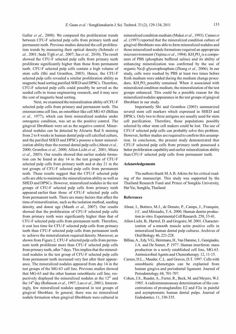
Galler et al., 2008). We compared the proliferation trendsbetween CFU-F selected pulp cells from primary teeth andpermanent teeth. Previous studies detected the cell prolifera-tion trends by measuring their optical density (Schmalz etal., 2001; Seda Ti li et al., 2007; Jue et al., 2010). The resultshowed the CFU-F selected pulp cells from primary teethproliferate significantly higher than those from permanentteeth. CFU-F selected pulp cells contain a high volume ofstem cells (Shi and Gronthos, 2003). Hence, the CFU-Fselected pulp cells revealed a similar proliferation ability asmagnetic bead sorting purified SHED and DPSCs. Therefore,CFU-F selected pulp cells could possibly be served as theseeded cells in tissue engineering research, and it may savethe cost of magnetic bead sorting.
Next, we examined the mineralization ability of CFU-Fselected pulp cells from primary and permanent teeth. Theosteosarcoma cell line, an osteoblast-like cell MG-63 (Billiauet al., 1977), which can form mineralized nodules underosteogenic condition, was set as the positive control. Thegingival fibroblasts were set as the negative control. Miner-alized nodules can be detected by Alizarin Red S stainingfrom 2 to 8 weeks in human dental pulp cell calcified culture,and the purified SHED and DPSCs possess a faster mineral-ization ability than the normal dental pulp cells (About et al.,2000; Gronthos et al., 2000; Alliot-Licht et al., 2001; Miuraet al., 2003). Our results showed that earlier nodule forma-tion can be found at day 14 in the test groups of CFU-Fselected pulp cells from primary teeth and at day 21 in thetest groups of CFU-F selected pulp cells from permanentteeth. These results suggest that the CFU-F selected pulpcells are able to maintain the mineralization ability as well asSHED and DPSCs. However, mineralized nodules in the testgroups of CFU-F selected pulp cells from primary teethappeared earlier than those of CFU-F selected pulp cellsfrom permanent teeth. There are many factors that affect thetime of mineralization, such as the isolation method, seedingdensity, and donor age (Mauth et al., 2007). Our resultsshowed that the proliferation of CFU-F selected pulp cellsfrom primary teeth were significantly higher than that ofCFU-F selected pulp cells from permanent teeth. Therefore,it cost less time for CFU-F selected pulp cells from primaryteeth than CFU-F selected pulp cells from permanent teethto achieve the mineralization required density. Moreover, asshown from Figure 2, CFU-F selected pulp cells from perma-nent teeth proliferate more than CFU-F selected pulp cellsfrom primary teeth, after 7 days. This implies that the mineral-ized nodules in the test group of CFU-F selected pulp cellsfrom permanent teeth increased very fast after their appear-ance. The mineralized nodules appeared from day 14 in thetest groups of the MG-63 cell line. Previous studies showedthat MG-63 and the other human osteoblastic cell line, res-pectively displayed the mineralized nodules at the 12th andthe 14th day (Robinson et al., 1997; Luo et al., 2001). Interest-ingly, few mineralized nodules appeared in test groups ofgingival fibroblast. In general, there was no mineralizednodule formation when gingival fibroblasts were cultured in
mineralized condition medium (Mukai et al., 1993). Cames etal. (1997) reported that the mineralized condition culture ofgingival fibroblasts was able to form mineralized nodules andthose mineralized nodule formations required an appropriatemicroenvironment (Yajima et al., 1984). KH2PO4 is a compo-nent of PBS (phosphate buffered saline) and its ability ofenhancing mineralization was confirmed by the use oforganic Na--glycerophosphate (Zhang et al., 2006). In ourstudy, cells were washed by PBS at least two times beforefresh medium were added during the medium change proce-dure. KH2PO4 possibly remained. When it associated withmineralized condition medium, the mineralization of the testgroups enhanced. This could be a possible reason for themineralized nodules appearance in the test groups of gingivalfibroblast in our study.
Importantly Shi and Gronthos (2003) summarizedseveral stem cell markers which expressed in SHED andDPSCs. Only two to three antigens are usually used for stemcell purification. Therefore, those populations possiblyselected by other stem cell makers could be lost. The use ofCFU-F selected pulp cells can probably solve this problem.However, further studies are required to confirm this assump-tion. In conclusion, the present study demonstrated thatCFU-F selected pulp cells from primary teeth possessed abetter proliferation capability and earlier mineralization abilitythan CFU-F selected pulp cells from permanent teeth.
Acknowledgements
The authors thank M.A.B. Atkins for his critical read-ing of the manuscript. This study was supported by theThailand Research Fund and Prince of Songkla University,Hat Yai, Songkla, Thailand
References
About, I., Bottero, M.J., de Denato, P., Camps, J., Franquin,J.C. and Mitsiadis, T.A. 2000. Human dentin produc-tion in vitro. Experimental Cell Research. 258, 33-41.
Alliot-Licht, B., Hurtrel, D. and Gregoire, M. 2001. Character-ization of a-smooth muscle actin positive cells inmineralized human dental pulp cultures. Archives ofOral Biology. 46, 221-228.
Billiau, A., Edy, V.G., Heremans, H., Van Damme, J., Georgiades,J.A. and De Somer, P. 1977. Human interferon: massproduction in a newly established cell line, MG-63.Antimicrobial Agents and Chemotherapy. 12, 11-15.
Carnes, D.L., Maeder, C.L. and Graves, D.T. 1997. Cells withosteoblastic phenotypes can be explanted fromhuman gingiva and periodontal ligament. Journal ofPeriodontology. 68, 701-707.
Cohen, J.S., Reader, A., Ferter, R., Beck, M. and Meyers, W.J.1985. A radioimmunoassay determination of the con-centrations of prostaglandins E2 and F2 in painfuland asymptomatic human dental pulps. Journal ofEndodontics. 11, 330-335.

Z. Guan et al. / Songklanakarin J. Sci. Technol. 33 (2), 129-134, 2011134
Galler, K.M., Cavender, A., Yuwono, V., Dong, H., Shi, S.,Schmalz, G., Hartgerink, J.D. and D’Souza, R.N. 2008.Self-assembling peptide amphiphile nanofibers as ascaffold for dental stem cells. Tissue Engineering PartA. 14, 2051-2058.
Gay, I.C., Chen, S. and MacDougall, M. 2007. Isolation andcharacterization of multipotent human periodontalligament stem cells. Orthodontics & CraniofacialResearch. 10, 149-160.
Graziano, A., d’Aquino, R., Laino, G. and Papaccio, G. 2008.Dental pulp stem cells: a promising tool for boneregeneration. Stem Cell Reviews. 4, 21-26.
Gronthos, S., Mankani, M., Brahim, J., Robey, P.G. and Shi,S. 2000. Postnatal human dental pulp stem cells(DPSCs) in vitro and in vivo. Proceedings of theNational Academy of Sciences U.S.A. 97, 13,625-13,630.
Hahn, C.L., Falkler, W.A. and Siegal, M.A. 1989. A study ofT & B cells in pulpal pathosis. Journal of Endodontics.15, 20-26.
Huang, A.H., Chen, Y.K., Lin, L.M., Shieh, T.Y. and Chan, A.W.2008. Isolation and characterization of dental pulpstem cells from a supernumerary tooth. Jounal of OralPathology Medicine. 37, 571-574.
Jue, S.S., Lee, W.Y., Kwon, Y.D., Kim, Y.R., Pae, A. and Lee,B. 2010. The effects of enamel matrix derivative on theproliferation and differentiation of human mesenchy-mal stem cells. Clinical Oral Implants Research. 21,741-746.
Langer, R. and Vacanti, J.P. 1993. Tissue engineering. Science.260, 920–926.
Liu, H., Gronthos, S. and Shi, S. 2006. Dental pulp stem cells.Methods in Enzymology. 419, 99-113.
Luo, X.H., Liao, E.Y. and Zhou, H.D. 2001. Differentiation andgene expression of human osteosarcoma cell lineMG-63. Hunan Yi Ke Da Xue Xue Bao. 26, 107-110.
Machado, C.B., Ventura, J.M., Lemos, A.F., Ferreira, J.M.,Leite, M.F. and Goes, A.M. 2007. 3D chitosan-gelatin-chondroitin porous scaffold improves osteogenic dif-ferentiation of mesenchymal stem cells. BiomedicalMaterials. 2, 124-131.
Mauth, C., Huwig, A., Graf-Hausner, U. and Roulet, J.-F.2007. Restorative application, for dental pulp therapy.Topic in tissue engineering. Eds 3: II Chapter 3.
Miura, M., Gronthos, S., Zhao, M., Lu, B., Fisher, L.W., Robey,P.G. and Shi, S. 2003. SHED: Stem cells from humanexfoliated deciduous teeth. Proceedings of theNational Academy of Sciences USA. 100, 5807-5812.
Mosmann, T. 1983. Rapid colorimetric assay for cellulargrowth and survival: application to proliferation andcytotoxicity assays. Journal of ImmunologicalMethods. 65, 55-63.
Mukai, M., Yoshimine, Y., Akamine, A. and Maeda, K. 1993.Bone-like nodules formed in vitro by rat periodontalligament cells. Cell and Tissue Research. 271, 453-460.
Robinson, J.A., Harris, S.A., Riggs, B.L. and Spelsberg, T.C.1997. Estrogen regulation of human osteoblastic cellproliferation and differentiation. Endocrinology. 138,2919-2927.
Stanford, C.M., Jacobson, P.A., Eanes, E.D., Lembke, L.A.and Midura, R.J. 1995. Rapidly forming apatiticmineral in an osteoblastic cell line (UMR 106-01 BSP).Journal of Biological Chemistry. 270, 9420-9428.
Schmalz, G., Schuster, U., Thonemann, B., Barth, M. andEsterbauer, S. 2001. Dentin barrier test with transfectedbovine pulp-derived cells. Journal of Endodontics.27, 96-102.
Seda Ti li, R., Karakeçili, A. and Gümüþderelio lu, M. 2007.In vitro characterization of chitosan scaffolds: influ-ence of composition and deacetylation degree. Jour-nal of Materials Science. Materials in Medicine. 18,1665-1674.
Shi, S., Bartold, P.M., Miura, M., Seo, B.M., Robey, P.G. andGronthos, S. 2005. The efficacy of mesenchymal stemcells to regenerate and repair dental structures. Or-thodontics & Craniofacial Research. 8, 191-199.
Shi, S. and Gronthos, S. 2003. Perivascular niche of postnatalmesenchymal stem cells in human bone marrow anddental pulp. Journal of Bone and Mineral Research.18, 696–704.
Tsukamoto, Y., Fukutani, S., Shin-Ike, T., Kubato, T., Sato,S., Suzuki, Y. and Mori, M. 1992. Mineralized noduleformation by human dental pulp-derived fibroblasts.Archives of Oral Biology. 37, 1045-1055.
Vitale-Brovarone, C., Verné, E., Robiglio, L., Appendino, P.,Bassi, F., Martinasso, G., Muzio, G. and Canuto, R.2007. Development of glass-ceramic scaffolds forbone tissue engineering: characterisation, prolifera-tion of human osteoblasts and nodule formation. ActaBiomaterialia. 3, 199-208
Yajima, T., Kumegawa, M. and Hiramatsu, M. 1984. Ectopicmineralization in fibroblast cultures. Archivum Histo-logicum Japonicum. 47, 43-45
Zhang, W., Walboomers, X.F., van Kuppevelt, T.H., Daamen,W.F., Bian, Z. and Jansen, J.A. 2006. The performanceof human dental pulp stem cells on different three-dimensional scaffold materials. Biomaterials. 27, 5658-5668.