Embed Size (px)
Citation preview
MAY 2014�CANCER DISCOVERY | OF1
Promising SINEs for Embargoing Nuclear–Cytoplasmic Export as an Anticancer Strategy David S.P. Tan , Philippe L. Bedard , John Kuruvilla , Lillian L. Siu , and Albiruni R. Abdul Razak
REVIEW
ABSTRACT In cancer cells, the nuclear–cytoplasmic transport machinery is frequently dis-
rupted, resulting in mislocalization and loss of function for many key regulatory
proteins. In this review, the mechanisms by which tumor cells co-opt the nuclear transport machinery
to facilitate carcinogenesis, cell survival, drug resistance, and tumor progression will be elucidated,
with a particular focus on the role of the nuclear–cytoplasmic export protein. The recent development
of a new generation of selective inhibitors of nuclear export (XPO1 antagonists) and how these novel
anticancer drugs may bring us closer to the implementation of this therapeutic strategy in the clinic
will be discussed.
Signifi cance: The nuclear transport mechanism is dysregulated in many malignancies and is associated
with dysfunction of many regulatory proteins. Targeting this mechanism as an anticancer strategy has
been compelling, and novel agents that selectively inhibit the nuclear export pathway have demon-
strated preliminary evidence of clinical effi cacy with an acceptable safety profi le. Cancer Discov; 4(5);
1–11. ©2014 AACR.
Authors’ Affi liation: Division of Medical Oncology and Hematology, Prin-cess Margaret Cancer Centre, University Health Network, University of Toronto, Toronto, Canada
Corresponding Author: Albiruni R. Abdul Razak, Princess Margaret Cancer Centre, WS33, 5-700, 610 University Avenue, Toronto, Ontario M5G 2M9, Canada. Phone: 416-946-4501, ext. 3428; Fax: 416-946-6546; E-mail: [email protected]
doi: 10.1158/2159-8290.CD-13-1005
©2014 American Association for Cancer Research.
INTRODUCTION Normal cellular function is dependent upon the appro-
priate localization of proteins within specifi c intracellular
compartments ( 1 ). There is growing evidence to suggest
that the nuclear–cytoplasmic transport machinery is fre-
quently disrupted by cancer cells, and that the subse-
quent mislocalization of key regulatory proteins facilitates
carcinogenesis, cell survival, drug resistance, and tumor
progression ( 2, 3 ). In this review, we will discuss the mecha-
nisms of nuclear transport dysfunction in tumor cells, with
a particular focus on the nuclear export apparatus as a
therapeutic target, and how the recent emergence of a new
generation of small-molecule selective inhibitors of nuclear
export (SINE), specifi cally exportin 1 (XPO1) inhibitors,
may provide a means to implement this therapeutic strat-
egy in cancer treatment.
NUCLEAR–CYTOPLASMIC TRANSPORT MECHANISMS The Nuclear Pore Complex Is a Gateway between the Nucleus and Cytoplasm
In the eukaryotic cell, critical cellular processes, such as
DNA synthesis, RNA transcription and translation, and pro-
tein processing, are carried out within distinct intracellular
compartments ( 1 ). Small molecules (<40 kD) can passively
diffuse across different compartments, but larger molecules,
including most proteins and RNAs, require transport by
signal-dependent and energy-dependent mechanisms ( 2 ). The
site of active transport between the nucleus and the cytoplasm
occurs at the nuclear pore complex (NPC), which is composed
of basic building blocks called nucleoporin. NPC is a large
macromolecular assembly that is embedded in the nuclear
envelope and mediates the exchange of >40-kD molecules that
pass between the nuclear and cytoplasmic compartments ( 3 ).
Nuclear transport processes are highly regulated, and there
is growing evidence to suggest that the ability to selectively
modulate the geographic location of macromolecules within
the cell is a key means to modulate cellular function ( 1 , 3 ).
Nuclear–Cytoplasmic Transport Receptors and Signals
The majority of nuclear–cytoplasmic transport receptors
belong to the karyopherin β family of proteins and act as car-
rier molecules that facilitate transit into (importins) and out
Research. on February 15, 2018. © 2014 American Association for Cancercancerdiscovery.aacrjournals.org Downloaded from
Published OnlineFirst April 17, 2014; DOI: 10.1158/2159-8290.CD-13-1005
OF2 | CANCER DISCOVERY�MAY 2014 www.aacrjournals.org
Tan et al.REVIEW
of (exportins) the nucleus via the NPC ( 4 ). Each karyopherin
transport receptor recognizes a unique group of cargo pro-
teins or RNA by the presence of either a nuclear localization
signal (NLS) or a nuclear export signal (NES) in the amino
acid sequence of the cargo protein, which directs them in and
out of the nucleus, respectively ( 4 ). Three classes of nuclear–
cytoplasmic transport signals have hitherto been identifi ed:
(i) the classical basic amino acid NLS sequences recognized
by a heterodimer composed of importins α and β ( 5 ); (ii) a
more complex NLS possessing N-terminal hydrophobic/basic
motif and C-terminal RX2-5PY motifs found in karyopherin
β cargo proteins, which directly binds to their specifi c carriers
( 6 ); and (iii) a hydrophobic leucine-rich NES recognized by
the ubiquitous transport receptor chromosome maintenance
protein 1 (CRM1, also known as XPO1; refs. 7, 8 ). Karyopherin
β proteins also bind weakly to phenylalanine–glycine (FG)
repeats in nucleoporins, thus targeting karyopherin β-cargo
complexes to the NPC for translocation. At present, there are
at least 19 karyopherin proteins identifi ed in humans ( 9 ).
Current evidence suggests that nuclear–cytoplasmic export
is mainly regulated by XPO1 in humans ( 3 ). XPO1 mediates
the nuclear export of a small subset of RNAs, as well as over
200 eukaryotic proteins that possess a canonical hydropho-
bic leucine-rich NES ( 9 ). In addition, XPO1 also seems to
regulate the nuclear export of some proteins [e.g., forkhead O
transcription factor 3a (FOXO3a) and histone deacetylases]
that form complexes with adapter molecules containing an
NES, such as 14-3-3 proteins, a family of basic proteins with
diverse physiologic functions that are also implicated in the
nuclear cytoplasmic shuttling of proteins ( 3 , 10 , 11 ).
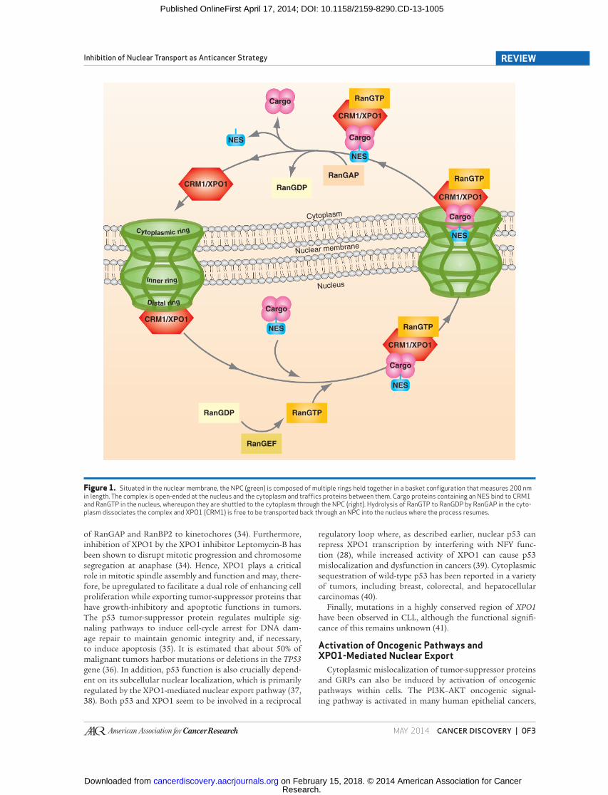
Nuclear Export Dynamics The direction of transport across the NPC is determined by
the concentration gradient of the small GTPase Ran. Levels
of RanGTP in the nucleus are approximately 100-fold greater
than those in the cytoplasm due to the location of the RanGTP
exchange factor (RanGEF or RCC1) within the nucleus. Con-
versely, in the cytoplasm, the majority of Ran is in the GDP-
bound form due to the cytoplasmic localization of (i) its
GTPase-activating protein (RanGAP), (ii) the RanGTP-binding
protein RanBP1, and (iii) homologous RanBP1 domains in
cytoplasmic nucleoporin NUP358 (also known as RanBP2;
refs. 3 , 12 ). Once XPO1 is bound to its cargo, it forms a com-
plex with RanGTP and exits the nucleus via the NPC ( 3 ). Upon
leaving the nucleus, RanGTP undergoes hydrolysis by RanGAP
and is converted to RanGDP, promoting dissociation of the
XPO1 complex, and the cargo is subsequently released into the
cytoplasm. Hence, interactions between transport receptors
and the Ran GTPase control bidirectional transport, as trans-
location is dependent on the different nucleotide-bound states
of Ran in the nucleus versus the cytoplasm ( 13, 14 ). A summary
of the nuclear export pathway is depicted in Fig. 1 .
XPO1-MEDIATED EXPORT AND CANCER Dysregulation of growth-regulatory proteins (GRP) and
functional inactivation of tumor-suppressor proteins are hall-
marks of oncogenesis ( 15 ). Given that many tumor-suppres-
sor proteins and GRPs perform their antineoplastic functions
within the nucleus, any mechanism that could potentially
enhance their nuclear export and cytoplasmic sequestration
would effectively result in their functional inactivation, lead-
ing to a protumorigenic cellular state. Known examples of
tumor-suppressor proteins and GRP sequestration within the
cytoplasm of cancer cells include BRCA1 and FOXO proteins
in breast cancer; RUNX3 in gastric cancer; p53 in breast,
ovarian, and colorectal cancers, retinoblastoma, and neuro-
blastoma; adenomatosis polyposis coli (APC) or β-catenin in
colon cancer; and BCR–ABL in chronic myelogenous leuke-
mia (CML; refs. 2, 3 , 16–21 ). The frequent observation of
molecular localization signal alterations and the cytoplasmic
mislocalization of these regulatory factors are consistent with
functional linkage of nuclear export to cancer.
Overexpression and Alteration of XPO1-Mediated Export in Cancer
Increased expression of XPO1 has been observed and cor-
related with poor prognosis or resistance to chemotherapy
in a number of solid (ovarian, pancreatic, brain, and cervical
cancers, osteosarcoma, and melanoma) and hematologic malig-
nancies [acute myeloid leukemia (AML), chronic lymphocytic
leukemia (CLL), multiple myeloma, and lymphoma; refs. 22–
27 ]. Importantly, this increased expression of XPO1 seems to
have functional signifi cance in cancer cells. A study by van der
Watt and colleagues ( 26 ) demonstrated that XPO1 is overex-
pressed in cervical cancer tissue and cell lines. In these tissue/
cell lines, the inhibition of XPO1 signifi cantly reduced cell pro-
liferation and induced apoptosis, while sparing the noncancer
cells. The exact mechanisms driving overexpression of XPO1 are
unknown, but recent evidence suggests that the nuclear factor
Y (NFY) and specifi city protein 1 (Sp1) transcription factors
may be responsible for XPO1 overexpression in cancer, and that
XPO1 transcription is inhibited when p53 interferes with NFY
function following DNA damage ( 28 ). Given that many tumor-
suppressor proteins and GRPs are exported by XPO1 in various
cancers ( Table 1 ), upregulation of XPO1-mediated export via
XPO1 overexpression may be a common mechanism of enhanc-
ing nuclear export in cancer ( 2 , 29 ).
Cancerous inhibitor of PP2A (CIP2A) is an oncoprotein
that inhibits protein serine/threonine phosphatase 2A (PP2A),
a protein distributed within the nucleus and cytoplasm that
controls the phosphorylation of numerous proteins involved
in cell signaling and regulates cell growth and apoptosis, and
has recently been identifi ed as a substrate of the XPO1 export
pathway ( 30 ). CIP2A also binds directly to c-Myc and inhibits
PP2A activity toward the c-Myc serine (Ser) 62 residue, thus
preventing proteolytic degradation of c-Myc ( 31 ). CIP2A has
also been shown to transform human cells when overexpressed
and to promote anchorage-independent cell growth and in
vivo tumor formation ( 31 ). CIP2A overexpression has been
observed in bladder, tongue, hepatocellular, and colon cancers,
non–small cell lung carcinoma, head and neck squamous cell
carcinoma, and CML ( 31, 32 ). Cytoplasmic overexpression of
CIP2A has also been associated with higher-grade, advanced-
stage, and poorer outcomes in serous ovarian cancer ( 33 ).
In addition to nuclear transport, XPO1 is also involved
in the regulation of mitotic checkpoints, spindle assembly,
and postmitotic nuclear envelope reassembly ( 34 ). XPO1
has been shown to localize to kinetochores, where it forms a
complex that is essential for RanGTP-dependent recruitment
Research. on February 15, 2018. © 2014 American Association for Cancercancerdiscovery.aacrjournals.org Downloaded from
Published OnlineFirst April 17, 2014; DOI: 10.1158/2159-8290.CD-13-1005
MAY 2014�CANCER DISCOVERY | OF3
Inhibition of Nuclear Transport as Anticancer Strategy REVIEW
of RanGAP and RanBP2 to kinetochores ( 34 ). Furthermore,
inhibition of XPO1 by the XPO1 inhibitor Leptomycin-B has
been shown to disrupt mitotic progression and chromosome
segregation at anaphase ( 34 ). Hence, XPO1 plays a critical
role in mitotic spindle assembly and function and may, there-
fore, be upregulated to facilitate a dual role of enhancing cell
proliferation while exporting tumor-suppressor proteins that
have growth-inhibitory and apoptotic functions in tumors.
The p53 tumor-suppressor protein regulates multiple sig-
naling pathways to induce cell-cycle arrest for DNA dam-
age repair to maintain genomic integrity and, if necessary,
to induce apoptosis ( 35 ). It is estimated that about 50% of
malignant tumors harbor mutations or deletions in the TP53
gene ( 36 ). In addition, p53 function is also crucially depend-
ent on its subcellular nuclear localization, which is primarily
regulated by the XPO1-mediated nuclear export pathway ( 37,
38 ). Both p53 and XPO1 seem to be involved in a reciprocal
regulatory loop where, as described earlier, nuclear p53 can
repress XPO1 transcription by interfering with NFY func-
tion ( 28 ), while increased activity of XPO1 can cause p53
mislocalization and dysfunction in cancers ( 39 ). Cytoplasmic
sequestration of wild-type p53 has been reported in a variety
of tumors, including breast, colorectal, and hepatocellular
carcinomas ( 40 ).
Finally, mutations in a highly conserved region of XPO1
have been observed in CLL, although the functional signifi -
cance of this remains unknown ( 41 ).
Activation of Oncogenic Pathways and XPO1-Mediated Nuclear Export
Cytoplasmic mislocalization of tumor-suppressor proteins
and GRPs can also be induced by activation of oncogenic
pathways within cells. The PI3K–AKT oncogenic signal-
ing pathway is activated in many human epithelial cancers,
Figure 1. Situated in the nuclear membrane, the NPC (green) is composed of multiple rings held together in a basket confi guration that measures 200 nm in length. The complex is open-ended at the nucleus and the cytoplasm and traffi cs proteins between them. Cargo proteins containing an NES bind to CRM1 and RanGTP in the nucleus, whereupon they are shuttled to the cytoplasm through the NPC (right). Hydrolysis of RanGTP to RanGDP by RanGAP in the cyto-plasm dissociates the complex and XPO1 (CRM1) is free to be transported back through an NPC into the nucleus where the process resumes.
Cargo
NES
NES
NES
NES
NES
RanGDP
Cytoplasm
Nucleus
Nuclear membrane
RanGAP
Cargo
Cargo
Cargo
Cargo
RanGTP
RanGTP
RanGTPRanGDP
RanGTP
RanGEF
CRM1/XPO1
CRM1/XPO1
CRM1/XPO1
CRM1/XPO1
CRM1/XPO1
Inner ring
Cytoplasmic ring
Distal ring
Research. on February 15, 2018. © 2014 American Association for Cancercancerdiscovery.aacrjournals.org Downloaded from
Published OnlineFirst April 17, 2014; DOI: 10.1158/2159-8290.CD-13-1005
OF4 | CANCER DISCOVERY�MAY 2014 www.aacrjournals.org
Tan et al.REVIEW
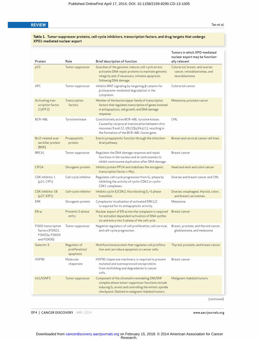
Table 1. Tumor-suppressor proteins, cell-cycle inhibitors, transcription factors, and drug targets that undergo XPO1-mediated nuclear export
Protein Role Brief description of function
Tumors in which XPO-mediated
nuclear export may be function-
ally relevant
p53 Tumor suppressor Guardian of the genome: induces cell-cycle arrest,
activates DNA repair proteins to maintain genomic
integrity and, if necessary, initiates apoptosis
following DNA damage.
Colorectal, breast, and ovarian
cancer, retinoblastomas, and
neuroblastoma
APC Tumor suppressor Inhibits WNT signaling by targeting β-catenin for
proteasome-mediated degradation in the
cytoplasm.
Colorectal cancer
Activating tran-
scription factor
2 (ATF2)
Transcription
factors
Member of the leucine zipper family of transcription
factors that regulates transcription of genes involved
in antiapoptosis, cell growth, and DNA damage
response.
Melanoma, prostate cancer
BCR–ABL Tyrosine kinase Constitutively active BCR–ABL tyrosine kinase.
Caused by reciprocal translocation between chro-
mosomes 9 and 22, t(9;22)(q34;q11), resulting in
the formation of the BCR–ABL fusion gene.
CML
Bcl2-related ovar-
ian killer protein
(BOK)
Proapoptotic
protein
Exerts proapoptotic function through the mitochon-
drial pathway.
Breast and cervical cancer cell lines
BRCA1 Tumor suppressor Regulates the DNA damage response and repair
functions in the nucleus and at centrosomes to
inhibit centrosome duplication after DNA damage.
Breast cancer
CIP2A Oncogenic protein Inhibits protein PP2A and stabilizes the oncogenic
transcription factor c-Myc.
Head and neck and colon cancer
CDK inhibitor 1
(p21, CIP1)
Cell-cycle inhibitor Regulates cell-cycle progression from G 1 -phase by
inhibiting the activity of cyclin-CDK2 or cyclin-
CDK1 complexes.
Ovarian and breast cancer and CML
CDK inhibitor 1B
(p27, KIP1)
Cell-cycle inhibitor Inhibits cyclin E/CDK2, thus blocking G 1 –S phase
transition.
Ovarian, esophageal, thyroid, colon,
and breast carcinomas
ERK Oncogenic protein Cytoplasmic localization of activated ERK1/2
is required for its antiapoptotic activity.
Melanoma
ER-α Prevents S-phase
entry
Nuclear export of ER-α into the cytoplasm is required
for estradiol-dependent activation of DNA synthe-
sis and entry into S-phase of the cell cycle.
Breast cancer
FOXO transcription
factors (FOXO1,
FOXO3a, FOXO4
and FOXO6)
Tumor suppressor Negative regulators of cell proliferation, cell survival,
and cell-cycle progression.
Breast, prostate, and thyroid cancer,
glioblastoma, and melanoma
Galectin-3 Regulator of
proliferation/
apoptosis
Multifunctional protein that regulates cell prolifera-
tion and can induce apoptosis in cancer cells.
Thyroid, prostate, and breast cancer
HSP90 Molecular
chaperone
HSP90 chaperone machinery is required to prevent
mutated and overexpressed oncoproteins
from misfolding and degradation in cancer
cells.
Breast cancer
InI1/hSNF5 Tumor suppressor Component of the chromatin remodeling SWI/SNF
complex whose tumor-suppressor functions include
inducing G 1 arrest and controlling the mitotic spindle
checkpoint. Deleted in malignant rhabdoid tumors.
Malignant rhabdoid tumors
(continued)
Research. on February 15, 2018. © 2014 American Association for Cancercancerdiscovery.aacrjournals.org Downloaded from
Published OnlineFirst April 17, 2014; DOI: 10.1158/2159-8290.CD-13-1005
MAY 2014�CANCER DISCOVERY | OF5
Inhibition of Nuclear Transport as Anticancer Strategy REVIEW
Protein Role Brief description of function
Tumors in which XPO-mediated
nuclear export may be function-
ally relevant
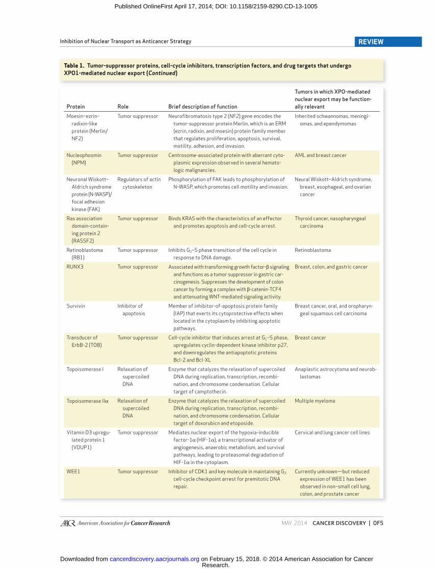
Moesin–ezrin–
radixin-like
protein (Merlin/
NF2)
Tumor suppressor Neurofi bromatosis type 2 ( NF2 ) gene encodes the
tumor-suppressor protein Merlin, which is an ERM
(ezrin, radixin, and moesin) protein family member
that regulates proliferation, apoptosis, survival,
motility, adhesion, and invasion.
Inherited schwannomas, meningi-
omas, and ependymomas
Nucleophosmin
(NPM)
Tumor suppressor Centrosome-associated protein with aberrant cyto-
plasmic expression observed in several hemato-
logic malignancies.
AML and breast cancer
Neuronal Wiskott–
Aldrich syndrome
protein (N-WASP)/
focal adhesion
kinase (FAK)
Regulators of actin
cytoskeleton
Phosphorylation of FAK leads to phosphorylation of
N-WASP, which promotes cell motility and invasion.
Neural Wiskott–Aldrich syndrome,
breast, esophageal, and ovarian
cancer
Ras association
domain-contain-
ing protein 2
(RASSF2)
Tumor suppressor Binds KRAS with the characteristics of an effector
and promotes apoptosis and cell-cycle arrest.
Thyroid cancer, nasopharyngeal
carcinoma
Retinoblastoma
(RB1)
Tumor suppressor Inhibits G 1 –S phase transition of the cell cycle in
response to DNA damage.
Retinoblastoma
RUNX3 Tumor suppressor Associated with transforming growth factor-β signaling
and functions as a tumor suppressor in gastric car-
cinogenesis. Suppresses the development of colon
cancer by forming a complex with β-catenin-TCF4
and attenuating WNT-mediated signaling activity.
Breast, colon, and gastric cancer
Survivin Inhibitor of
apoptosis
Member of inhibitor-of-apoptosis protein family
(IAP) that exerts its cytoprotective effects when
located in the cytoplasm by inhibiting apoptotic
pathways.
Breast cancer, oral, and oropharyn-
geal squamous cell carcinoma
Transducer of
ErbB-2 (TOB)
Tumor suppressor Cell-cycle inhibitor that induces arrest at G 1 –S phase,
upregulates cyclin-dependent kinase inhibitor p27,
and downregulates the antiapoptotic proteins
Bcl-2 and Bcl-XL
Breast cancer
Topoisomerase I Relaxation of
supercoiled
DNA
Enzyme that catalyzes the relaxation of supercoiled
DNA during replication, transcription, recombi-
nation, and chromosome condensation. Cellular
target of camptothecin.
Anaplastic astrocytoma and neurob-
lastomas
Topoisomerase IIα Relaxation of
supercoiled
DNA
Enzyme that catalyzes the relaxation of supercoiled
DNA during replication, transcription, recombi-
nation, and chromosome condensation. Cellular
target of doxorubicn and etoposide.
Multiple myeloma
Vitamin D3 upregu-
lated protein 1
(VDUP1)
Tumor suppressor Mediates nuclear export of the hypoxia-inducible
factor-1α (HIF-1α), a transcriptional activator of
angiogenesis, anaerobic metabolism, and survival
pathways, leading to proteasomal degradation of
HIF-1α in the cytoplasm.
Cervical and lung cancer cell lines
WEE1 Tumor suppressor Inhibitor of CDK1 and key molecule in maintaining G 2
cell-cycle checkpoint arrest for premitotic DNA
repair.
Currently unknown—but reduced
expression of WEE1 has been
observed in non–small cell lung,
colon, and prostate cancer
Table 1. Tumor-suppressor proteins, cell-cycle inhibitors, transcription factors, and drug targets that undergo XPO1-mediated nuclear export (Continued)
Research. on February 15, 2018. © 2014 American Association for Cancercancerdiscovery.aacrjournals.org Downloaded from
Published OnlineFirst April 17, 2014; DOI: 10.1158/2159-8290.CD-13-1005
OF6 | CANCER DISCOVERY�MAY 2014 www.aacrjournals.org
Tan et al.REVIEW
resulting in the upregulation of several downstream target
genes that inhibit apoptosis and promote cell proliferation
( 42 ). Several downstream PI3K–AKT signaling substrates
that negatively regulate cell proliferation, cell survival, and
cell-cycle progression have been shown to be functionally
inactivated because of cytoplasmic sequestration following
PI3K–AKT pathway activation, including the FOXO tran-
scription factors and the cell-cycle inhibitor p27 ( 11 , 43 ,
44 ). The lipid phosphatase PTEN negatively regulates the
PI3K–AKT pathway, and mutations in the PTEN gene have
frequently been found in a variety of cancers, including
glioblastoma multiforme, melanoma, renal cell carcinoma,
and prostate, ovarian, and endometrial cancers ( 45, 46 ). In
PTEN -null cancer cells, constitutive activation of AKT leads
to phosphorylation of FOXO transcription factors at mul-
tiple sites, thus preventing FOXO DNA binding and tran-
scriptional activity ( 47 ). Hyperphosphorylation of the FOXO
proteins also facilitates binding with 14-3-3, which then pro-
motes XPO1-mediated nuclear export of FOXO ( 11 ). Colon
cancer and renal cancer cell lines that do not express PTEN
show constitutive mislocalization and loss of function for
FOXO proteins, which in turn result in loss of regulation for
metabolism for many cell types ( 47 ).
The cell-cycle inhibitor p27 can also be sequestered in the
cytoplasm or exported from the nucleus by XPO1 via AKT
phosphorylation ( 48 ). Under normal conditions, p27 is local-
ized in the nucleus, where it acts as a cell-cycle inhibitor by
binding to and inhibiting the activity of cyclin-dependent
kinase 2 (CDK2), a serine/threonine protein kinase that pro-
motes G 1 –S phase cell-cycle progression ( 3 ). Phosphorylation
of the p27 NLS by AKT has been shown to impair nuclear
import with subsequent cytoplasmic sequestration of p27,
leaving unbound CDK2 within the nucleus free to facilitate
cell-cycle progression and tumorigenesis ( 48, 49 ). In addition,
RAS activation may lead to XPO1-mediated nuclear export of
p27 via phosphorylation of p27 at Ser10 by the PI3K–AKT
and RAF–MEK–ERK pathways ( 50, 51 ). Cytoplasmic localiza-
tion of p27 has been observed in esophageal, thyroid, colon,
and breast cancers ( 3 ). Wang and colleagues ( 52 ) recently
demonstrated that increased XPO1 and phosphorylated
Ser10 p27 (pSer10p27) expression levels were associated with
advanced-stage and high-grade epithelial ovarian cancer and
poorer overall survival. Knockdown of XPO1 and pSer10p27
expression levels also leads to cell-cycle arrest and inhibition
of cell proliferation in SKOV3 cells in vitro and in vivo ( 52 ).
The homologous recombination DNA repair proteins
BRCA1 and RAD51 have also been shown to be functionally
inactivated because of cytoplasmic sequestration following
PI3K–AKT pathway activation ( 20 ). Cytoplasmic localization
of BRCA1 has been observed in 60% of primary invasive ductal
breast cancers, with a high correlation between cytoplasmic
BRCA1 localization and AKT1 activity (based on increased
levels of phosphorylated AKT1) in the tumors ( 20 ). The exact
mechanism of AKT1-induced cytoplasmic sequestration of
BRCA1 is currently unknown, but nuclear export of BRCA1
via the XPO1 pathway has previously been described fol-
lowing DNA damage caused by ionizing radiation ( 53 ), and
XPO1 has also been implicated in the regulation of BRCA1
centrosomal localization, thus suggesting that AKT-induced
nuclear export of BRCA1 may be XPO1 mediated ( 54 ). The
relationship between AKT1 activity and homologous recom-
bination–related tumor-suppressor protein mislocalization
and function has recently been further highlighted by data
showing that BRCA1 defi ciency in cells may itself lead to
activation of AKT1 and impaired CHK1 nuclear localization
( 55 ). Impaired CHK1 nuclear import disrupts the interac-
tion between CHK1 and RAD51 in the nucleus, resulting in
homologous recombination repair defects ( 55 ).
Recent data suggest that the nuclear export of the estrogen
receptor α (ERα) into the cytoplasm is required for estrogen-
dependent activation of DNA synthesis and S-phase entry,
resulting in increased proliferative activity of breast cancer
cells ( 56, 57 ). Following estrogen stimulation, PI3K–AKT
phosphorylates FOXO1, also known as forkhead in rhab-
domyosarcoma (FKHR; ref. 57 ). In addition, estrogen also
activates the nonreceptor protooncogene SRC, resulting in
tyrosine phosphorylation of ERα, which in turn leads to
assembly of an ERα–FKHR complex within the nucleus and
its subsequent nuclear export via XPO1. The cytoplasmic
localization of the ER-α–FKHR leads to downregulation of
FKHR transcriptional activity and upregulation of cyclin D1
transcription, resulting in DNA synthesis and S-phase transi-
tion ( 56 ).
Other examples of intracellular mislocalization of tumor-
suppressor proteins and GRPs mediated by activation of
oncogenic pathways include the functional inactivation of
the RUNX3 tumor-suppressor protein by phosphorylation by
SRC kinase activity and subsequent cytoplasmic mislocaliza-
tion via XPO1-mediated nuclear export in gastric and breast
cancers, and aberrant activation of the WNT pathway in
more than 90% of colorectal cancers ( 17 , 19 ). The p53 tumor-
suppressor protein is also mislocalized. Hence, the develop-
ment and progression of cancer mediated by upregulation
of oncogenic signaling pathways (e.g., PI3K–AKT, SRC, and
WNT) and inactivation of tumor-suppressor proteins may be
partly underpinned by nuclear–cytoplasmic mislocalization
of key regulatory proteins.
XPO1-MEDIATED NUCLEAR EXPORT AND DRUG RESISTANCE IN CANCER
There is also accumulating evidence to suggest that XPO1
may mediate drug resistance in a variety of tumor types by
facilitating the nuclear export of various drug targets. These
include topoisomerase I α (topo1α) and topo2α, galectin-3,
and BCR–ABL ( 2 ).
The nuclear protein topo2α functions as a homodimer
that disentangles DNA and relieves the torsional stress in
supercoiled DNA caused by the process of DNA replica-
tion ( 58 ). Chemotherapeutic agents, such as doxorubicin
and etoposide, inhibit topo2α during DNA replication and
produce cleavable complexes, resulting in double-stranded
DNA breaks and cell death ( 59 ). It has been demonstrated
that for DNA damage to occur in this manner, topo2α must
be localized within the nucleus and in physical contact with
DNA ( 21 ). In myeloma cells, nuclear export of topo2α to
the cytoplasm via XPO1 has been associated with resistance
to topo2α inhibitors, and reversal of the topo2α inhibitor-
resistant phenotype can be induced by the XPO1 inhibi-
tors ratjadone C or Leptomycin-B or by siRNA-mediated
Research. on February 15, 2018. © 2014 American Association for Cancercancerdiscovery.aacrjournals.org Downloaded from
Published OnlineFirst April 17, 2014; DOI: 10.1158/2159-8290.CD-13-1005
MAY 2014�CANCER DISCOVERY | OF7
Inhibition of Nuclear Transport as Anticancer Strategy REVIEW
knockdown of XPO1 protein expression ( 21 ). Depletion of
XPO1 by siRNA or ratjadone C has been shown to result in
synergistic sensitivity of myeloma cells, obtained from bone
marrow aspirates of patients with multiple myeloma and
from human multiple myeloma cell lines, to doxorubicin and
etoposide ( 21 ).
Nuclear galectin-3 is a multifunctional protein that regu-
lates cell proliferation and can induce apoptosis in cancer
cells. The subcellular location of galectin-3 seems to be
tightly regulated by specifi c selective mechanisms via the
NPC depending on cell type, growth conditions, or neoplas-
tic transformation ( 60 ). Nuclear-to-cytoplasmic transloca-
tion of galectin-3 has been observed in prostate and breast
cancer cells following treatment with cytotoxic agents, with
subsequent inhibition of apoptosis in these cells ( 61, 62 ).
Treatment of the breast cancer cells by both cisplatin and
the nuclear export inhibitor Leptomycin-B has been shown
to induce nuclear retention of galectin-3 and apoptosis, thus
reversing cisplatin resistance ( 61 ).
The development of the BCR–ABL kinase inhibitor imat-
inib mesylate (Gleevec; Novartis) has revolutionized the
treatment of CML and has become its standard fi rst-line
treatment ( 2 , 16 ). Development of drug resistance is, however,
a common event when the disease progresses to its acceler-
ated or blast phase despite imatinib treatment. The main
mechanisms underlying the development of imatinib resist-
ance are point mutations in the kinase domain, BCR–ABL
amplifi cation and clonal evolution [i.e., the development of
additional nonrandom chromosomal abnormalities besides
the balanced translocation of t(9:22)(q34:q11.2) that results
in the Philadelphia chromosome; ref. 16 ]. The ABL protein
contains three putative NLSs and a single NES. In normal
cells, the ABL kinase functions in the cell nucleus and is
activated by DNA damage to induce the activation of the
p73 tumor-suppressor protein ( 63 ). In fact, the BCR–ABL
protein has been found to retain the apoptotic functions of
the wild-type ABL protein, and induces apoptosis in leuke-
mic cells when maintained in the nucleus using the export
inhibitor Leptomycin-B ( 64 ). Furthermore, inactivation of
BCR–ABL by imatinib partially restores nuclear localization
of the protein ( 2 ). A strategy of combined treatment with
imatinib and XPO1 inhibition by Leptomycin-B to effectively
trap BCR–ABL in the cell nucleus has been shown to induce
cell death in imatinib-resistant cells that displayed BCR–ABL
amplifi cation or signs of clonal evolution ( 16 ).
NUCLEAR EXPORT INHIBITORS AS ANTICANCER THERAPEUTICS
In view of the aforementioned evidence supporting the role
of nuclear transport dysregulation in maintaining the neo-
plastic phenotype and drug resistance, targeting the nuclear
transport mechanism as a therapeutic strategy has gained
interest within the oncology community in recent years. The
main focus of this strategy has been the targeted inhibition of
XPO1, with the hypothesis that inhibition of nuclear export
will result in forced nuclear retention and thereby activation of
several key tumor-suppressor proteins and GRPs, for example,
p53 pathway proteins, with consequent induction of cell-cycle
arrest and apoptosis in tumor cells while sparing normal cells.
Naturally Derived XPO1 Inhibitors Inhibition of XPO1 as a therapeutic strategy has been stud-
ied for almost two decades. The bacterial toxin Leptomycin-B
was the fi rst selective, irreversible XPO1 inhibitor that was
discovered and subsequently tested in a clinical trial ( 65 ).
Leptomycin-B has potent antitumor activity in vitro , but is
also toxic to normal cells ( 66 ). Despite marked toxicities
in animals, Leptomycin-B was administered intravenously
to patients with refractory solid tumors in a phase I study
by Newlands and colleagues ( 65 ). Toxicities included pro-
found gastrointestinal side effects, including nausea, vomit-
ing, watery diarrhea along with marked anorexia, fatigue/
asthenia, and malaise, whereas typical side effects of cytotoxic
agents such as neutropenia, mucositis, and alopecia were not
observed. Transient tumor marker responses in patients with
ovarian carcinoma and trophoblastic tumor and stable dis-
ease in a patient with refractory sarcoma were reported, but
the associated severe toxicities led to its development being
abandoned ( 65 ).
Leptomycin-B exerts its inhibitory effect on XPO1 by irre-
versibly alkylating the reactive site cysteine residue (Cys528;
ref. 29 ). Recently, it was demonstrated that upon binding
to XPO1, Leptomycin-B undergoes hydrolysis of the lactone
ring, which in turn enables persistent binding of the drug to
XPO1 ( 67 ). Alkylation of Cys528 prevents XPO1 from bind-
ing to the leucine-rich nuclear export sequence of the cargo
protein substrate, hence inhibiting the formation of the
XPO1–cargo–RanGTP export complex and effectively block-
ing nuclear export ( 3 , 29 ). There are now data suggesting that
the severe toxicities observed following treatment with Lepto-
mycin-B involve additional off-target effects caused by bind-
ing to cysteine proteases as well as the irreversible blockade
of all XPO1 functions per se ( 68 ). Therefore, a selective and
reversible inhibitor might possess potent anticancer activity
with an improved tolerability profi le. Most of the currently
available XPO1 inhibitors function by permanently modify-
ing the reactive site Cys528 and preventing XPO1 binding to
the NES of cargo proteins ( 2 ). Since the discovery of Lepto-
mycin-B, other naturally derived inhibitors have been identi-
fi ed, including the ratjadone analogs (A, B, C, and D) isolated
from Sorangium cellulosum , which have a similar chemical
structure to Leptomycin-B and use an identical molecular
mechanism to modify XPO1, but these compounds have not
been tested in vivo ( 2 ).
Synthetic XPO1 Inhibitors Kosan Biosciences Inc., before its acquisition by Bristol-
Myers Squibb, created derivatives of Leptomycin-B, includ-
ing KOS-2464, with better pharmacologic properties that
showed a much improved therapeutic index across multiple
tumor xenografts ( 69 ). Signifi cantly better tolerability and less
weight loss than with Leptomycin-B were observed in animals,
although transient anorexia remained ( 69 ). More recently, a
small-molecule reversible inhibitor of XPO1, CBS9106, which
induced cell-cycle arrest and apoptosis as a single agent in 60
different human cancer cell lines, including bladder, breast,
colon, and lung cancer, at submicromolar concentrations
has been described ( 70 ). Unlike the Leptomycin-B analogs,
CBS9106 binds to the Cys528 residue by forming a reversible
Research. on February 15, 2018. © 2014 American Association for Cancercancerdiscovery.aacrjournals.org Downloaded from
Published OnlineFirst April 17, 2014; DOI: 10.1158/2159-8290.CD-13-1005
OF8 | CANCER DISCOVERY�MAY 2014 www.aacrjournals.org
Tan et al.REVIEW
covalent bond with it and was found to inhibit tumor growth
in xenograft models without signifi cant weight loss or mortal-
ity ( 70 ). Both of these compounds have not progressed into
the clinic but are at least indicative that XPO1 can be inhibited
with an adequate therapeutic window.
Perhaps the compounds that herald the most promise for the
clinical utility of XPO1 inhibition are the new small-molecule
inhibitors of XPO1, also known as selective inhibitors of
nuclear export (SINE). These compounds share a similar struc-
ture with the N-azolylacrylate analog PKF050-638, a highly
specifi c and reversible small-molecule XPO1 inhibitor that was
originally developed to prevent nuclear export of the HIV-1 Rev
protein, an essential regulator of the HIV-1 mRNA expression
( 71 ). Karyopharm Therapeutics has developed a series of SINE
compounds [KPT-115, KPT-127, KPT-185, KPT-251, KPT-276,
KPT-330 (selinexor), and KPT-335 (verdinexor)] that have been
shown to inhibit XPO1-mediated export of p53, p73, RB1,
FOXO proteins, APC, IκB, NPM, topo2α, and survivin. Pre-
clinical studies of these compounds result in dose-dependent
cytotoxicity for a number of hematologic and solid tumors [i.e.,
CLL, AML, CML, multiple myeloma, non–Hodgkin lymphoma
(NHL), melanoma, and renal, breast, pancreatic, prostate, and
colorectal cancer cells; refs. 2 , 21 , 68 , 72–78 ]. These highly
specifi c inhibitors of XPO1 are water soluble, and they bind to
the reactive site Cys528 residue by forming a slowly reversible
covalent bond ( 79 ). SINEs have been shown to be selectively
cytotoxic for neoplastic cells with a half maximal effective
concentration (EC 50 ) of 10 to 1,000 nmol/L versus >5 to 20
μmol/L in nonneoplastic cell lines, and their cytotoxic effects
seem to be independent of known genetic alterations such as
BRAFV600E mutations in melanoma or TP53 , PI3K , AKT , and
BRCA1/2 mutation status in breast cancer cell lines ( 24 , 75 ).
Synergistic activity using KPT-185 in combination with SN38
and oxaliplatin in colorectal cancer cell lines and KPT-251
with chemotherapy in prostate cancer cell lines has also been
observed ( 72, 73 ). In murine and monkey pharmacokinetic
studies, KPT-185 showed limited bioavailability and systemic
exposure, whereas KPT-276 and selinexor showed >50% bio-
availability reaching C max >5 μmol/L following 10 mg/kg oral
administration of drug ( 24 ).
Preclinical in vitro and in vivo data have shown that both
hematologic and solid tumor cells are susceptible to single-
agent cytotoxicity caused by selinexor, consistent with its res-
toration of multiple tumor-suppressor and growth-regulatory
pathways, leading to the death of cancer cells ( 24 , 75 , 79 ). Inter-
estingly, SINE treatment also results in reduction in expres-
sion of multiple oncoproteins, including cMyc in multiple
myeloma, cKIT and FLT3 in AML, and cyclin D1 in mantle
cell lymphoma, as well as proteasome-mediated degradation
of the XPO1 protein itself. The major adverse event across all
tested species to date (mice, rats, dogs, and monkeys) is revers-
ible weight reduction accompanied by reduced food intake
without signifi cant vomiting or diarrhea ( 79 ). The improved
toxicity profi le of these newer XPO1 inhibitors relative to previ-
ous XPO1 inhibitors, such as Leptomycin-B, is thought to be
related to their exquisite specifi city for XPO1 with no detect-
able binding to other proteins, including the cysteine proteases,
believed to be the cause of poor tolerance to Leptomycin-B ( 68 ).
The SINE XPO1 inhibitors have also been investigated
in a comparative oncology setting, using dogs with newly
diagnosed and chemotherapy-refractory cancers ( 80 ). In one
study, the SINE compound verdinexor was administered in
17 dogs with NHL, mast cell tumor, or osteosarcoma. Com-
mon toxicities included anorexia, vomiting, and diarrhea.
Dose-limiting toxicities were anorexia and vomiting, which
occurred at 2 mg/kg twice weekly. Among 16 evaluable dogs,
2 achieved partial responses (both with NHL) and 12 others
achieved stable disease, indicating activity of XPO1 inhibi-
tors in large-animal models ( 80 ). Verdinexor is currently in
phase II studies in dogs with newly diagnosed or recurring
lymphoma.
Oral selinexor is currently being developed and evaluated
for tolerability and single-agent antitumor activity in phase
I clinical trials of patients with solid tumors (trial number
NCT01607892; http://clinicaltrials.gov , last accessed on Feb-
ruary 14, 2014), and the data from these studies have hitherto
been presented only in meeting abstracts. In these studies,
selinexor is administered orally at either 8 doses or 10 doses
in each 28-day cycle. The preliminary results of the phase I
solid-tumor study with the 10 doses per cycle were presented
recently. At the time of presentation, 2 of 3 patients had expe-
rienced dose-limiting toxicities at 40 mg/m 2 , with grade 3
anorexia with dehydration and fatigue in 1 patient, and grade
3 fatigue in another. Commonly observed nonhematologic
toxicities included fatigue, nausea, and anorexia that were
mostly low grade (grade 1–2), whereas the most common
hematologic toxicity was thrombocytopenia. Toxicities were
all reversible upon stopping the drug. Among the 39 patients
treated, at least 7 patients had disease stabilization of more
than 6 months, including patients who were treated at low
dose levels. Selinexor has a dose-proportional pharmacoki-
netic profi le and a half-life of 5 to 7 hours, and paired biop-
sies, done in 12 patients, showed increased nuclear retention
of a number of tumor-suppressor proteins, including FOXO1,
IκB, and p53, following treatment ( 81 ). The alternative dos-
ing regimen in which the drug is administered twice weekly
(i.e., 8 doses every 28 days) is currently being investigated in
patients with advanced solid tumors, and the maximum-
tolerated dose (MTD) has not been reached to date.
In the parallel phase I study in advanced, progressive
hematologic malignancies (NCT01607892), patients with
relapsed/refractory NHL, CLL, multiple myeloma, or AML
were dosed with oral selinexor (8–10 doses every 28 days;
refs. 82–84 ). The adverse-event profi le was similar to that
observed in the solid-tumor study, and MTD on the eight
doses per cycle schedule has not been reached to date. Dura-
ble responses and disease stabilization with single-agent
oral selinexor across all disease subtypes have been observed,
with some patients remaining on study for over 1 year. In
CML, recently published data have demonstrated that treat-
ment with selinexor results in apoptosis and impaired the
clonogenic potential of leukemic, but not normal, CD34(+)
progenitors ( 85 ). As a proof of mechanism, following expo-
sure to selinexor, nuclear accumulation of CIP2A, SET (an
oncoprotein that inhibits PP2A activity), IκBα (an inhibitor
of the NF-κB transcription factor), FOXO3a, p53, and p21,
with subsequent reactivation of PP2A within the cytoplasm
and reduction in BCR–ABL levels, was noted ( 85 ). Antileuke-
mic activity in a CML blast crisis mouse model was observed
following treatment with selinexor, and treatment of a single
Research. on February 15, 2018. © 2014 American Association for Cancercancerdiscovery.aacrjournals.org Downloaded from
Published OnlineFirst April 17, 2014; DOI: 10.1158/2159-8290.CD-13-1005
MAY 2014�CANCER DISCOVERY | OF9
Inhibition of Nuclear Transport as Anticancer Strategy REVIEW
patient in accelerated-phase CML with Selinexor also led
to a reduction in bone pain, splenomegaly, and immature
myeloid blasts in the peripheral blood ( 85 ).
CONCLUSIONS There is preclinical evidence to suggest that inhibition of
XPO1-mediated nuclear export leading to nuclear retention
and functional reactivation of tumor-suppressor proteins
and GRPs may be a viable therapeutic anticancer strategy.
Until recently, the development of this pharmacologic class
of compounds has been hampered by the narrow therapeutic
window of XPO1 inhibition ( 2 , 65 ). New-generation XPO1
inhibitors with improved preclinical effi cacy, pharmacoki-
netics, oral bioavailability, and reduced toxicities in animal
models have generated renewed interest in this approach and
are now being tested in early-phase clinical trials ( 79 ).
Notably, despite the encouraging early signals, and apart
from the obvious lack of published clinical toxicity and effi -
cacy data, there are crucial questions that remain unresolved
in the context of nuclear export inhibition as a therapeutic
strategy. These include the challenges of identifying potential
predictive biomarkers of effi cacy to optimize patient selec-
tion for these drugs, and the possible cumulative toxicities
of long-term nuclear export inhibition. Despite these reser-
vations, the emerging data from early-phase clinical studies
seem encouraging. In addition, given the noted synergistic
effects on cytotoxicity in neoplastic cells, the potential for
combining XPO1 inhibition with conventional chemother-
apy, such as topoisomerase inhibitors, is open for exploration
in the future ( 21 , 73 , 86 ). Moreover, combination strategies
with other targeted therapeutics may be another option for
consideration, and recent preclinical data showing that com-
bined XPO1 and BRAF inhibition leads to synergistic tumor
regression in BRAF -mutant melanoma suggest that this may
be a viable therapeutic approach as well ( 87 ).
Ultimately, it may be overly optimistic to expect that the
inhibition of nuclear–cytoplasmic transport will be an effec-
tive target in all cancer types, or that the therapeutic effi cacy of
this approach will not eventually be hampered by intrinsic and
acquired resistance mechanisms and/or toxicities engendered
by sustained physiologic inhibition of such transport in nor-
mal cells. In the context of drug resistance, it is worth noting
that the Cys528 residue, which seems to be the main target of
XPO1 inhibitors, is not part of an active site of XPO1 and also
seems to be nonessential for XPO1/CRM1 function. Mutant
forms of XPO1/CRM1 that carry a Cys528–Ser mutation can
substitute for wild-type CRM1 in mammalian cells, and Sac-
charomyces cerevisiae express a form of CRM1 that contains a
threonine at this position, instead of a cysteine. Human cells
that express CRM1 with the Cys528–Ser mutation have been
shown to be resistant to the effects of Leptomycin-B ( 88 ).
Nonetheless, the strategy of imposing a molecular embargo
on nuclear transport as a backbone for new avenues of cancer
therapy may be on the verge of becoming a clinical reality.
Disclosure of Potential Confl icts of Interest D.S.P. Tan and A.R.A. Razak have received commercial research
grants from Karyopharm Therapeutics. No potential confl icts of
interest were disclosed by the other authors.
Received December 20, 2013; revised March 6, 2014; accepted
March 14, 2014; published OnlineFirst April 17, 2014.
REFERENCES 1. Zaidi SK , Young DW , Javed A , Pratap J , Montecino M , van Wijnen A ,
et al. Nuclear microenvironments in biological control and cancer .
Nat Rev Cancer 2007 ; 7 : 454 – 63 .
2. Turner JG , Dawson J , Sullivan DM . Nuclear export of proteins and
drug resistance in cancer . Biochem Pharmacol 2012 ; 83 : 1021 – 32 .
3. Kau TR , Way JC , Silver PA . Nuclear transport and cancer: from
mechanism to intervention . Nat Rev Cancer 2004 ; 4 : 106 – 17 .
4. Cook A , Bono F , Jinek M , Conti E . Structural biology of nucleocyto-
plasmic transport . Annu Rev Biochem 2007 ; 76 : 647 – 71 .
5. Lange A , Mills RE , Lange CJ , Stewart M , Devine SE , Corbett AH . Clas-
sical nuclear localization signals: defi nition, function, and interaction
with importin alpha . J Biol Chem 2007 ; 282 : 5101 – 5 .
6. Lee BJ , Cansizoglu AE , Suel KE , Louis TH , Zhang Z , Chook YM . Rules
for nuclear localization sequence recognition by karyopherin beta 2 .
Cell 2006 ; 126 : 543 – 58 .
7. Dong X , Biswas A , Suel KE , Jackson LK , Martinez R , Gu H , et al.
Structural basis for leucine-rich nuclear export signal recognition by
CRM1 . Nature 2009 ; 458 : 1136 – 41 .
8. Kutay U , Guttinger S . Leucine-rich nuclear-export signals: born to be
weak . Trends Cell Biol 2005 ; 15 : 121 – 4 .
9. Xu D , Farmer A , Chook YM . Recognition of nuclear targeting signals
by Karyopherin-beta proteins . Curr Opin Struct Biol 2010 ; 20 : 782 – 90 .
10. Kao HY . Mechanism for nucleocytoplasmic shuttling of histone
deacetylase 7 . J Biol Chem 2001 ; 276 : 47496 – 507 .
11. Yang JY , Hung MC . A new fork for clinical application: targeting fork-
head transcription factors in cancer . Clin Cancer Res 2009 ; 15 : 752 – 7 .
12. Bischoff FR , Klebe C , Kretschmer J , Wittinghofer A , Ponstingl H .
RanGAP1 induces GTPase activity of nuclear Ras-related Ran . Proc
Natl Acad Sci U S A 1994 ; 91 : 2587 – 91 .
13. Izaurralde E , Kutay U , von Kobbe C , Mattaj IW , Gorlich D . The asym-
metric distribution of the constituents of the Ran system is essential
for transport into and out of the nucleus . EMBO J 1997 ; 16 : 6535 – 47 .
14. Marelli M , Dilworth DJ , Wozniak RW , Aitchison JD . The dynam-
ics of karyopherin-mediated nuclear transport . Biochem Cell Biol
2001 ; 79 : 603 – 12 .
15. Hanahan D , Weinberg RA . Hallmarks of cancer: the next generation .
Cell 2011 ; 144 : 646 – 74 .
16. Kancha RK , von Bubnoff N , Miething C , Peschel C , Gotze KS , Duyster J .
Imatinib and leptomycin B are effective in overcoming imatinib-resist-
ance due to Bcr-Abl amplifi cation and clonal evolution but not due to
Bcr-Abl kinase domain mutation . Haematologica 2008 ; 93 : 1718 – 22 .
17. Goh YM , Cinghu S , Hong ET , Lee YS , Kim JH , Jang JW , et al. Src
kinase phosphorylates RUNX3 at tyrosine residues and localizes the
protein in the cytoplasm . J Biol Chem 2010 ; 285 : 10122 – 9 .
18. Hu MC , Lee DF , Xia W , Golfman LS , Ou-Yang F , Yang JY , et al.
IkappaB kinase promotes tumorigenesis through inhibition of fork-
head FOXO3a . Cell 2004 ; 117 : 225 – 37 .
19. Ito K , Liu Q , Salto-Tellez M , Yano T , Tada K , Ida H , et al. RUNX3, a
novel tumor suppressor, is frequently inactivated in gastric cancer by
protein mislocalization . Cancer Res 2005 ; 65 : 7743 – 50 .
20. Plo I , Laulier C , Gauthier L , Lebrun F , Calvo F , Lopez BS . AKT1 inhib-
its homologous recombination by inducing cytoplasmic retention of
BRCA1 and RAD51 . Cancer Res 2008 ; 68 : 9404 – 12 .
21. Turner JG , Marchion DC , Dawson JL , Emmons MF , Hazlehurst LA ,
Washausen P , et al. Human multiple myeloma cells are sensitized
to topoisomerase II inhibitors by CRM1 inhibition . Cancer Res
2009 ; 69 : 6899 – 905 .
22. Noske A , Weichert W , Niesporek S , Röske A , Buckendahl A-C , Koch
I , et al. Expression of the nuclear export protein chromosomal region
maintenance/exportin 1/Xpo1 is a prognostic factor in human ovar-
ian cancer . Cancer 2008 ; 112 : 1733 – 43 .
23. Huang WY , Yue L , Qiu WS , Wang LW , Zhou XH , Sun YJ . Prognostic
value of CRM1 in pancreas cancer . Clin Invest Med 2009 ; 32 : E315 .
Research. on February 15, 2018. © 2014 American Association for Cancercancerdiscovery.aacrjournals.org Downloaded from
Published OnlineFirst April 17, 2014; DOI: 10.1158/2159-8290.CD-13-1005
OF10 | CANCER DISCOVERY�MAY 2014 www.aacrjournals.org
Tan et al.REVIEW
24. Lesinski GB , Yang J , Bill MA , Landesman Y , Shacham S , Kauffman M ,
et al. Effect of small inhibitors of nuclear export (SINE) on growth
inhibition and apoptosis of human melanoma cells . J Clin Oncol 30,
2012 (suppl; abstr e13549) .
25. Shen A , Wang Y , Zhao Y , Zou L , Sun L , Cheng C . Expression of CRM1
in human gliomas and its signifi cance in p27 expression and clinical
prognosis . Neurosurgery 2009 ; 65 : 153 – 9 .
26. van der Watt PJ , Maske CP , Hendricks DT , Parker MI , Denny L ,
Govender D , et al. The Karyopherin proteins, Crm1 and Karyopherin
beta1, are overexpressed in cervical cancer and are critical for cancer
cell survival and proliferation . Int J Cancer 2009 ; 124 : 1829 – 40 .
27. Yao Y , Dong Y , Lin F , Zhao H , Shen Z , Chen P , et al. The expression
of CRM1 is associated with prognosis in human osteosarcoma . Oncol
Rep 2009 ; 21 : 229 – 35 .
28. van der Watt PJ , Leaner VD . The nuclear exporter, Crm1, is regulated
by NFY and Sp1 in cancer cells and repressed by p53 in response to
DNA damage . Biochim Biophys Acta 2011 ; 1809 : 316 – 26 .
29. Nguyen KT , Holloway MP , Altura RA . The CRM1 nuclear export
protein in normal development and disease . Int J Biochem Mol Biol
2012 ; 3 : 137 – 51 .
30. Thakar K , Karaca S , Port SA , Urlaub H , Kehlenbach RH . Identifi ca-
tion of CRM1-dependent nuclear export cargos using quantitative
mass spectrometry . Mol Cell Proteomics 2013 ; 12 : 664 – 78 .
31. Junttila MR , Puustinen P , Niemela M , Ahola R , Arnold H , Bottzauw
T , et al. CIP2A inhibits PP2A in human malignancies . Cell 2007 ; 130 :
51 – 62 .
32. Khanna A , Pimanda JE , Westermarck J . Cancerous inhibitor of pro-
tein phosphatase 2A, an emerging human oncoprotein and a poten-
tial cancer therapy target . Cancer Res 2013 ; 73 : 6548 – 53 .
33. Bockelman C , Lassus H , Hemmes A , Leminen A , Westermarck J ,
Haglund C , et al. Prognostic role of CIP2A expression in serous ovar-
ian cancer . Br J Cancer 2011 ; 105 : 989 – 95 .
34. Arnaoutov A , Azuma Y , Ribbeck K , Joseph J , Boyarchuk Y , Karpova T ,
et al. Crm1 is a mitotic effector of Ran-GTP in somatic cells . Nat Cell
Biol 2005 ; 7 : 626 – 32 .
35. Vousden KH , Lane DP . p53 in health and disease . Nat Rev Mol Cell
Biol 2007 ; 8 : 275 – 83 .
36. Soussi T . TP53 mutations in human cancer: database reassessment
and prospects for the next decade . Adv Cancer Res 2011 ; 110 : 107 – 39 .
37. Cai X , Liu X . Inhibition of Thr-55 phosphorylation restores p53
nuclear localization and sensitizes cancer cells to DNA damage . Proc
Natl Acad Sci U S A 2008 ; 105 : 16958 – 63 .
38. Freedman DA , Levine AJ . Nuclear export is required for degradation
of endogenous p53 by MDM2 and human papillomavirus E6 . Mol
Cell Biol 1998 ; 18 : 7288 – 93 .
39. Stommel JM , Marchenko ND , Jimenez GS , Moll UM , Hope TJ , Wahl
GM . A leucine-rich nuclear export signal in the p53 tetramerization
domain: regulation of subcellular localization and p53 activity by
NES masking . EMBO J 1999 ; 18 : 1660 – 72 .
40. O’Brate A , Giannakakou P . The importance of p53 location: nuclear
or cytoplasmic zip code? Drug Resist Updat 2003 ; 6 : 313 – 22 .
41. Puente XS , Pinyol M , Quesada V , Conde L , Ordonez GR , Villamor
N , et al. Whole-genome sequencing identifi es recurrent mutations in
chronic lymphocytic leukaemia . Nature 2011 ; 475 : 101 – 5 .
42. Yap TA , Garrett MD , Walton MI , Raynaud F , de Bono JS , Workman
P . Targeting the PI3K–AKT–mTOR pathway: progress, pitfalls, and
promises . Curr Opin Pharmacol 2008 ; 8 : 393 – 412 .
43. Blain SW , Massague J . Breast cancer banishes p27 from nucleus . Nat
Med 2002 ; 8 : 1076 – 8 .
44. Vogt PK , Jiang H , Aoki M . Triple layer control: phosphorylation,
acetylation and ubiquitination of FOXO proteins . Cell Cycle 2005 ; 4 :
908 – 13 .
45. Zhang S , Yu D . PI(3)king apart PTEN’s role in cancer . Clin Cancer Res
2010 ; 16 : 4325 – 30 .
46. Burger RA , Brady MF , Bookman MA , Fleming GF , Monk BJ , Huang
H , et al. Incorporation of bevacizumab in the primary treatment of
ovarian cancer . N Engl J Med 2011 ; 365 : 2473 – 83 .
47. Nakamura N , Ramaswamy S , Vazquez F , Signoretti S , Loda M ,
Sellers WR . Forkhead transcription factors are critical effectors of
cell death and cell cycle arrest downstream of PTEN . Mol Cell Biol
2000 ; 20 : 8969 – 82 .
48. Shin I , Yakes FM , Rojo F , Shin NY , Bakin AV , Baselga J , et al. PKB/
Akt mediates cell-cycle progression by phosphorylation of p27(Kip1)
at threonine 157 and modulation of its cellular localization . Nat Med
2002 ; 8 : 1145 – 52 .
49. Chu IM , Hengst L , Slingerland JM . The Cdk inhibitor p27 in human
cancer: prognostic potential and relevance to anticancer therapy . Nat
Rev Cancer 2008 ; 8 : 253 – 67 .
50. Besson A , Gurian-West M , Chen X , Kelly-Spratt KS , Kemp CJ , Rob-
erts JM . A pathway in quiescent cells that controls p27Kip1 stability,
subcellular localization, and tumor suppression . Genes Dev 2006 ; 20 :
47 – 64 .
51. Fujita N , Sato S , Katayama K , Tsuruo T . Akt-dependent phospho-
rylation of p27Kip1 promotes binding to 14–3-3 and cytoplasmic
localization . J Biol Chem 2002 ; 277 : 28706 – 13 .
52. Wang Y , Xiang J , Ji F , Deng Y , Tang C , Yang S , et al. Knockdown of
CRM1 inhibits the nuclear export of p27(Kip1) phosphorylated at
serine 10 and plays a role in the pathogenesis of epithelial ovarian
cancer . Cancer Lett 2014 ; 343 : 6 – 13 .
53. Feng Z , Kachnic L , Zhang J , Powell SN , Xia F . DNA damage induces
p53-dependent BRCA1 nuclear export . J Biol Chem 2004 ; 279 :
28574 – 84 .
54. Brodie KM , Henderson BR . Characterization of BRCA1 protein tar-
geting, dynamics, and function at the centrosome: a role for the
nuclear export signal, CRM1, and Aurora A kinase . J Biol Chem 2012 ;
287 : 7701 – 16 .
55. Jia Y , Song W , Zhang F , Yan J , Yang Q . Akt1 inhibits homologous
recombination in Brca1-defi cient cells by blocking the Chk1-Rad51
pathway . Oncogene 2013 ; 32 : 1943 – 9 .
56. Castoria G , Giovannelli P , Lombardi M , De Rosa C , Giraldi T , de
Falco A , et al. Tyrosine phosphorylation of estradiol receptor by Src
regulates its hormone-dependent nuclear export and cell cycle pro-
gression in breast cancer cells . Oncogene 2012 ; 31 : 4868 – 77 .
57. Lombardi M , Castoria G , Migliaccio A , Barone MV , Di Stasio R ,
Ciociola A , et al. Hormone-dependent nuclear export of estradiol
receptor and DNA synthesis in breast cancer cells . J Cell Biol
2008 ; 182 : 327 – 40 .
58. Robinson MJ , Martin BA , Gootz TD , McGuirk PR , Moynihan M ,
Sutcliffe JA , et al. Effects of quinolone derivatives on eukaryotic
topoisomerase II. A novel mechanism for enhancement of enzyme-
mediated DNA cleavage . J Biol Chem 1991 ; 266 : 14585 – 92 .
59. Robinson MJ , Osheroff N . Effects of antineoplastic drugs on the
post-strand-passage DNA cleavage/religation equilibrium of topoi-
somerase II . Biochemistry 1991 ; 30 : 1807 – 13 .
60. Fukumori T , Kanayama H , Raz A . The role of galectin-3 in cancer
drug resistance . Drug Resist Updat 2007 ; 10 : 101 – 8 .
61. Takenaka Y , Fukumori T , Yoshii T , Oka N , Inohara H , Kim HR , et al.
Nuclear export of phosphorylated galectin-3 regulates its antiapop-
totic activity in response to chemotherapeutic drugs . Mol Cell Biol
2004 ; 24 : 4395 – 406 .
62. Fukumori T , Oka N , Takenaka Y , Nangia-Makker P , Elsamman E ,
Kasai T , et al. Galectin-3 regulates mitochondrial stability and antia-
poptotic function in response to anticancer drug in prostate cancer .
Cancer Res 2006 ; 66 : 3114 – 9 .
63. Gong JG , Costanzo A , Yang HQ , Melino G , Kaelin WG Jr , Levrero M ,
et al. The tyrosine kinase c-Abl regulates p73 in apoptotic response to
cisplatin-induced DNA damage . Nature 1999 ; 399 : 806 – 9 .
64. Vigneri P , Wang JY . Induction of apoptosis in chronic myelogenous
leukemia cells through nuclear entrapment of BCR-ABL tyrosine
kinase . Nat Med 2001 ; 7 : 228 – 34 .
65. Newlands ES , Rustin GJ , Brampton MH . Phase I trial of elactocin .
Br J Cancer 1996 ; 74 : 648 – 9 .
66. Kau TR , Schroeder F , Ramaswamy S , Wojciechowski CL , Zhao JJ ,
Roberts TM , et al. A chemical genetic screen identifi es inhibitors of
regulated nuclear export of a forkhead transcription factor in PTEN-
defi cient tumor cells . Cancer Cell 2003 ; 4 : 463 – 76 .
67. Sun Q , Carrasco YP , Hu Y , Guo X , Mirzaei H , Macmillan J ,
et al. Nuclear export inhibition through covalent conjugation and
Research. on February 15, 2018. © 2014 American Association for Cancercancerdiscovery.aacrjournals.org Downloaded from
Published OnlineFirst April 17, 2014; DOI: 10.1158/2159-8290.CD-13-1005
MAY 2014�CANCER DISCOVERY | OF11
Inhibition of Nuclear Transport as Anticancer Strategy REVIEW
hydrolysis of Leptomycin B by CRM1 . Proc Natl Acad Sci U S A
2013 ; 110 : 1303 – 8 .
68. Lapalombella R , Sun Q , Williams K , Tangeman L , Jha S , Zhong
Y , et al. Selective inhibitors of nuclear export (SINE) show that
CRM1/XPO1 is a target in chronic lymphocytic leukemia . Blood
2012 ; 120 : 4621 – 34 .
69. Mutka SC , Yang WQ , Dong SD , Ward SL , Craig DA , Timmermans
PB , et al. Identifi cation of nuclear export inhibitors with potent anti-
cancer activity in vivo . Cancer Res 2009 ; 69 : 510 – 7 .
70. Sakakibara K , Saito N , Sato T , Suzuki A , Hasegawa Y , Friedman JM ,
et al. CBS9106 is a novel reversible oral CRM1 inhibitor with CRM1
degrading activity . Blood 2011 ; 118 : 3922 – 31 .
71. Daelemans D , Afonina E , Nilsson J , Werner G , Kjems J , De Clercq
E , et al. A synthetic HIV-1 Rev inhibitor interfering with the
CRM1-mediated nuclear export . Proc Natl Acad Sci U S A 2002 ; 99 :
14440 – 5 .
72. Azmi AS , Kauffman M , McCauley D , Shacham S , Mohammad RM .
Novel small-molecule CRM-1 inhibitor for GI cancer therapy . J Clin
Oncol 30, 2012 (suppl 4; abstr 245) .
73. Chung HW , Salas Fragomeni RA , Shacham S , Kauffman M , Cusack
JC . Effect of inhibition of nuclear protein export using a novel CRM1
inhibitor on the apoptotic response to SN38 in preclinical colon can-
cer models . J Clin Oncol 30, 2012 (suppl 4; abstr 609) .
74. Inoue H , Kauffman M , Shacham S , Landesman Y , Weiss RH . Evalua-
tion of selective inhibitors of nuclear export (SINE) CRM1 inhibitors
for the treatment of renal cell carcinoma (RCC) . J Clin Oncol 30, 2012
(suppl; abstr 4634) .
75. McCauley D , Landesman Y , Senapedis W , Kashyap T , Saint-Martin J-R ,
Plamondon L , et al. Preclinical evaluation of selective inhibitors of
nuclear export (SINE) in basal-like breast cancer (BLBC) . J Clin Oncol
30, 2012 (suppl; abstr 1055) .
76. Shacham S , Barnard S , Kisseberth W , Ito D , Jensen K , Borgotti A ,
et al. Results of a phase I dose escalation study of the novel, oral
CRM1 selective inhibitor of nuclear export (SINE) KPT-335 in dogs
with spontaneous non-Hodgkin’s lymphomas (NHL) [abstract] . In:
Proceedings of the 54th ASH Annual Meeting and Exposition; 2012
Dec 8–11 ; Atlanta, GA. Abstract nr 161 .
77. Shacham S , Gravina GL , Ricevuto E , Mancini A , Chin L , Shechter S ,
et al. Preclinical evaluation of selective inhibitors of nuclear export
(SINE) CRM1 (XPO1) inhibitors in prostate cancer (PrCa) . J Clin
Oncol 30, 2012 (suppl; abstr e15200) .
78. Shacham S , Kauffman M , Sandanayaka V , Draetta G , Shechter S ,
Williams J , et al. Preclinical development of small-molecule CRM1
inhibitors as novel therapy for the treatment of colorectal cancer
(CRC) . J Clin Oncol 29, 2011 (suppl 4; abstr 430) .
79. Etchin J , Sun Q , Kentsis A , Farmer A , Zhang ZC , Sanda T , et al.
Antileukemic activity of nuclear export inhibitors that spare normal
hematopoietic cells . Leukemia 2013 ; 27 : 66 – 74 .
80. London CA , Bernabe LF , Barnard S , Kisseberth WC , Borgatti A ,
Henson M , et al. Preclinical evaluation of the novel, orally bio-
available Selective Inhibitor of Nuclear Export (SINE) KPT-335 in
spontaneous canine cancer: results of a phase I study . PLoS ONE
2014 ; 9 : e87585 .
81. Razak A , Sorensen M , Mahipal M , Shacham S . First-in-class, fi rst-in-
human phase I trial of KPT-330, a selective inhibitor of nuclear export
(SINE) in patients with advanced solid tumors . J Clin Oncol 31, 2013
(suppl; abstr 2505) .
82. Chen C , Gutierrez M , de Nully Brown P , Gabrail N, Baz R, Reece DE,
et al. A fi rst-in-class oral selective inhibitor of nuclear export (SINE)
XPO1/CRM1 antagonist in patients (pts) with relapsed/refractory
multiple myeloma (MM) or Waldenstrom’s macroglobulinemia
(WM) . Blood 2013 ; 122 .
83. Kuruvilla J , Gutierrez M , Shah B , Gabrail NY , de Nully Brown P , Stone
RM , et al. Preliminary evidence of anti tumor activity of Selinexor
(KPT-330) in a phase i trial of a fi rst-in-class oral selective inhibitor
of nuclear export (SINE) in patients (pts) with relapsed/refractory
non Hodgkin’s lymphoma (NHL) and chronic lymphocytic leukemia
(CLL) . Blood 2013 ; 122 : 90 .
84. Savona M, Garzon R, de Nully Brown P, Yee K, Lance JE, Gutierrez M,
et al. Phase I trial of Selinexor (KPT-330), a fi rst-in-class oral selective
inhibitor of nuclear export (SINE) in patients (pts) with advanced
acute myelogenous leukemia (AML) . Blood 2013 ; 122 : 1440 .
85. Walker CJ , Oaks JJ , Santhanam R , Neviani P , Harb JG , Ferenchak
G , et al. Preclinical and clinical effi cacy of XPO1/CRM1 inhibition
by the karyopherin inhibitor KPT-330 in Ph+ leukemias . Blood
2013 ; 122 : 3034 – 44 .
86. Abdel-Naim AB , Lu C , Shao C , Cobos E , Singh KP , Gao W . Chemo-
therapeutic sensitization of leptomycin B resistant lung cancer cells
by pretreatment with doxorubicin . PLoS ONE 2012 ; 7 : e32895 .
87. Salas Fragomeni RA , Chung HW , Landesman Y , Senapedis W , Saint-
Martin JR , Tsao H , et al. CRM1 and BRAF inhibition synergize and
induce tumor regression in BRAF-mutant melanoma . Mol Cancer
Ther 2013 ; 12 : 1171 – 9 .
88. Akakura S , Yoshida M , Yoneda Y , Horinouchi S . A role for Hsc70 in
regulating nucleocytoplasmic transport of a temperature-sensitive
p53 (p53Val-135) . J Biol Chem 2001 ; 276 : 14649 – 57 .
Research. on February 15, 2018. © 2014 American Association for Cancercancerdiscovery.aacrjournals.org Downloaded from
Published OnlineFirst April 17, 2014; DOI: 10.1158/2159-8290.CD-13-1005
Published OnlineFirst April 17, 2014.Cancer Discovery David S.P. Tan, Philippe L. Bedard, John Kuruvilla, et al. an Anticancer Strategy
Cytoplasmic Export as−Promising SINEs for Embargoing Nuclear
Updated version
10.1158/2159-8290.CD-13-1005doi:
Access the most recent version of this article at:
E-mail alerts related to this article or journal.Sign up to receive free email-alerts
Subscriptions
Reprints and
To order reprints of this article or to subscribe to the journal, contact the AACR Publications Department at
Permissions
Rightslink site. Click on "Request Permissions" which will take you to the Copyright Clearance Center's (CCC)
.http://cancerdiscovery.aacrjournals.org/content/early/2014/04/16/2159-8290.CD-13-1005To request permission to re-use all or part of this article, use this link
Research. on February 15, 2018. © 2014 American Association for Cancercancerdiscovery.aacrjournals.org Downloaded from
Published OnlineFirst April 17, 2014; DOI: 10.1158/2159-8290.CD-13-1005