Embed Size (px)
Citation preview

Instructions for use
Title PROPERTIES OF A SMALL VIRUS ASSOCIATED WITH INFECTIOUS CANINE HEPATITIS VIRUS
Author(s) DOMOTO, Kenji; YANAGAWA, Ryo
Citation Japanese Journal of Veterinary Research, 17(1-2): 32-41
Issue Date 1969-06
DOI 10.14943/jjvr.17.1-2.32
Doc URL http://hdl.handle.net/2115/1921
Type bulletin
File Information KJ00002369753.pdf
Hokkaido University Collection of Scholarly and Academic Papers : HUSCAP

PROPERTIES OF A SMALL VIRUS ASSOCIATED WITH INFECTIOUS CANINE HEPATITIS VIRUS
Kenji DOMOTO and Ryo YANAGA W A
Department of Hygiene and Microbiology Faculty of Veterinary Medicine
Hokkaido University, Sapporo, Japan
(Received for publication, March 14, 1969)
INTRODUCTION
The adeno-associated satellite virus (ASV)* IS a recently described group of
small defective DNA viruses (picodnaviruses) that can replicate only in cells coinfected w-ith helper adenoviruses1.2,7,1l,lS,2S). It is characteristic of ASV that
they interfere w-ith the replication of the adenovirus9 ,21). Electron microscopyl,3,17)
and fluorescence studies4 ,17) suggested that ASV replicated in the nucleus, but
not in the cytoplasm. Four types of ASV have been described serologicallyll,23).
Isolation of ASV and distribution of the antibodies have frequently been found
in man and monkey6.S,24). How-ever, detection of ASV from infectious canine
hepatitis virus (ICHV) and other adenoviruses, except human and simian adeno
viruses, has not been reported. Ability of ICHV to help the replication of the
defective human ASV has been shown in canine cellsll ) and human cells27 ).
SUGIMURA & YANAGAWA (1968) found a small virus associated with ICHV,
Matsuda strain, and reported its morphology. Attempts were made by the authors
to isolate this small virus and to clarify its properties. The results are described
in this paper, which suggest that the small virus is a member of ASV.
MA TERIALS AND METHODS
Virus Five strains of ICHV, Matsuda, FD, C-I, N-IV and Woc-4, were used in this study. Of these, Matsuda was originally isolated in this department by OSAMURA et al.
(1957).
Tissue culture and virus inoculation The growth medium used was HANKS'
balanced salt solution supplemented with 10 % calf serum and 0.5 % lactalbumin hydrolysate.
Maintenance medium used was the same medium containing 1 % calf serum. These media
were added to 200 u/ml of penicillin and 200 ,ag/ml of streptomycin. PriIllary dog kidney cell cultures (DKC) were prepared as described by YOUNGNER (1954).
* AAV (Adenovirus-associated virus) was proposed by ATCHISON et al. (1965) and ASV (Adeno-associated satellite virus) was proposed by MAYOR et al. (1900). In this paper we prefer ASV.
JAP. J. VET RES., VOL. 17, Nos. 1 & 2, 1969

Small 'virus associated with ICHV 33
Usually, secondary cultures of DKC were used in this study. The cells of primary culture
were washed twice with phosphate-buffered saline (PBS) (pH 7.2) and finally resuspended in
a growth medium so as to contain 105 cells per m!. The cells, suspended in the growth
medium, were dispensed 10ml per bottle (4xlOX4cm) and 0.5ml per tube (1.2x12cm) and
incubated at 37°C. Cell sheets gro\ovn in bottles were used for the propagation of the virus.
Each bottle was inoculated with 1 ml of the virus material, and after 2 hr for the adsorption
of virus, maintenance medium ,vas added. The inoculated virus material was discarded and
the cells were washed with HANKS' balanced salt solution 3 times. Then the maintenance
medium was added and incubated at 37°C.
Detection and isolation of the small virus "Vhen cytopathic effect (CPE) appeared
maximally, usually 4-5 days after inoculation, the cells in bottles were frozen at -20°C.
Then the cells were thawed at room temperature. The fluid and cells in the bottles, about
200 ml, were removed and centrifuged at 3,000 rpm for 30 min. The supernatant fluid
obtained was centrifuged at 8,000 rpm for 30 min, and the resultant supernatant fluid was
again centrifuged at 30,000 rpm for 90 min in an ultracentrifuge (Hitachi, model 40P). The
upper portion, about 3/4, of the supernatant fluid was removed, and the rest of the super
natant fluid was mixed with the pellet, by pipetting, which was then centrifuged at 8,000 rpm
for 30 min. The procedure of these centrifugations was repeated again. The pellet was
mixed with the lower portion, 1/4, of the supernatant fluid or, after removing the supernatant
fluid, was suspended in 0.5 ml PBS. The former pellet was used for isolation of the asso
ciated small virus from ICHV by filtration through 50-mp millipore filter, and the latter for
electron microscopic study.
Electron microscopic examination of the negatively stained VIruses The
material for elctron microscopic examination was mounted on the carbon-coated collodion
membrane grid for 2-3 min. After absorbing the excess amount of material with filter
paper, the specimen was negatively stained for 1-2 min with 1 % phosphotungstic acid
which was adjusted with 1 N potassium hydroxide to pH 7.0, and was examined with a
JEM-7 electron microscope at instrumental magnification up to 100,000 times.
Infectivity titration Ten-fold serial dilutions of virus materials were made with
maintenance medium. The infectivity titer was determined by inoculation 0.5 ml of each
ten-fold dilution into sets of 4 tubes containing DKC. Following a reincubation of 5 days,
the 50 % tissue culture infective doses (TCID5o) were calculated utilizing the method of BEHRENS-KARBER.
Acridine orange staining of the small virus The 50 mp-millipore filtrate of
ICHV Matsuda was concentrated 10 times by centrifugation. The concentrated filtrate was
found to contain observable small viruses by electron microscopy, but no ICHV particles.
One drop of the concentrated filtrate was mounted on a cover glass, dried at room tempera
ture and fixed in CARNOY's fixative for 5 min. The specimen was then washed thoroughly
in McIlvaine's buffer (pH 3.8), and stained with 0.01 % acridine orange for 5 min. The
stained preparation was washed with the same buffer and examined under Nikon fluorescence
microscope. Saturated solutions of herring sperm DNA (Daiichikagaku) and yeast RNA (Sigma) were stained as controls.
Ultra thin sectioning Cells III bottle culture were inoculated with strain Matsuda

34 DaMaTO, K. & Y ANAGA WA, R.
103 •5TCID5o• This virus material contained the small virus, which was lagrer in number
than ICHV particles under the electron microscope. The cells were harvested at various
times after inoculation. Before harvesting, the culture medium was removed from the
inoculated cell cultures and PBS was added in order to wash the cells. Then the cells were
scraped from the glass with the rubber policeman. The cell suspension thus obtained was
centrifuged at 1,000 rpm for 5 min. Cell pellets were fixed in 3.5 % glutalaldehyde for 20
min as described by SABATINI et al. (1963) and postfixed in 1 % osmiUIn tetraoxide for 30
min at 4°C. After dehydration in serial ethanol and infiltration with propylene oxide, the
cell blocks were embedded in Epon 812 (LUFT). Ultra thin sections were cut by JUM 5A
type ultramicrotome. The sections were stained with uranyl acetate and lead citrate.
Sections were examined with a JEM-7 electron microscope at instrumental magnification up
to 30,000 times.
RESULTS
1 Detection of the small VIrus from ICHV
Five strains of ICHV were examined electron microscopically. The results shown in
table 1 indicate that only the Matsuda strain contained small virus. The small virus particle
was hexagonal, without envelope and 20--25 mp in diameter (fig. 1). The ratio of ICHV to
the small virus particles in Matsuda strain was from 1: 60 to 1 : 70 by repeated electron
microscopic examinations. The other 4 strains contained ICHV particles but no small virus
(fig. 2).
Matsuda strain which contains the small virus, Matsuda (+), and FD strain containing
no small virus, FD (0), are described below.
TABLE 1 Detection of the small virus front ICHV
ICHV STRAINS PARTICLES *
Matsuda FD C-I N-IV Woc-4
ICHV + + + + + Small virus +
* Detected by electron microscopic examination
2 Comparison of the infectivity titer between Matsuda (+) and FD (0)
Infectivity titers (TCID50) of Matsuda (+) and FD (0) were, as shown in table 2, 103•29
and 107.25 per ml respectively. It was noted that the infectivity titer of Matsuda (+) was distinctly lower than that of FD (0). In experiment II shown in this table, the same inoculum
sizes were used but the appearance of CPE was earlier in the cultures inoculated with FD
(0) than those inoculated with Matsuda (+).
3 Evidence of defectiveness of the small VIrus
Possibility of the autonomous replication of the small VIrus was examined usmg DKC. The multiplication of ICHV and the small virus was examined by electron microscopy.

Small virus associated with ICHV
TABLE 2 Infectivity titers of lvlatsuda (+) and FD (0)
VIRUSES
Matsuda (+)
Matsuda (+)
FD (0)
EXP I
2.75*2
3.50
EXP II*l
3.50
3.00
7.25
AVERAGE
3.29
7.25
*1 Inoculated with 100 TCID50/ml III DKC from the same puppy *2 Expressed as log TC1D50/ml ( +) Contained the small virus (0) Contained no small virus
3S
a) Experiment using the small virus separated from ICHV by filtration through 50-m it!
millipore filter
For this purpose, the small virus in Matsuda strain was separated from ICHV by
filtration through 50-mp millipore filter. The filtrate was found to contain observable small
virus by electron microscopy but no ICHV particles. Matsuda (+) produced ICHV and the
small virus while FD (0) produced only ICHV. As shown in table 3, filtrate containing the
small virus alone caused no CPE, and no multiplication of the small virus and ICHV was
found. However, when the small virus were inoculated in DKC simultaneously with FD (0)
TABLE 3 Multij)lication of the small virus in the presence and absence of infectious ICHV
a) * Experiment using the small virus separated from
ICHV by filtration through 50-mp millipore filter
VIRUSES CPE ICHV SMALL VIRUS
Matsuda (+)
Small virus
Small virus + FD (0)
FD (0)
+
+ +
+
+ +
b) * Experiment using the small VIrus whose helper ICHV was inactivated by heating
+
+
VIRUSES CPE ICHV SMALL VIRUS
Matsuda (+)
Matsuda (+) 60°C 10 min
Matsuda (+) 60°C 10 mIll + FD (0)
FD (0)
+
+
+
+ +
+ +
+
* Infectivity titer of Matsuda (+) and FD (0) used was 100 TCID50/ml.
36 DOMOTO, K. & YANAGAWA, R.
multiplication of the small virus and ICHV could be found. Therefore, it was proved that
the small virus could not replicate in DKC, but could replicate when coinfected with ICHV.
These findings suggest that, in DKC, the small virus is defective and requires ICHV as
helper for its replication.
b) Experiment using the small virus whose helper ICHV was inactivated by heating
Another evidence of the defectiveness of the small virus was given in the following
experiment. A test was made of heating Matsuda (+) with the expectation that heating at
a certain degree of temperature would inactivate ICHV but not ASV. It has been known
that ICHV was inactivated by heating at 60°C for 5 min, while ASV was more heat resistant.
Matsuda (+) and FD (0) heated at 60°C for 5 or 10 min caused no CPE, and no multiplication
of ASV and ICHV were found. However, when Matsuda (+) heated at 60°C for 10 min was
inoculated in DKC simultaneously with FD (0) multiplication of the small virus and ICHV
was found (tab. 3). The above findings show that the multiplication of the small virus could
be supported only by infectious ICHV and not by heat-inactivated ICHV. The small virus
was also found to be resistant to heating at 70°C for 10 min.
These experiments suggest that the small virus with Matsuda strain is a member of
adeno-associated satellite viruses.
4 Acridine orange staining of the small VIrus
Matsuda culture fluid, 200 ml amount, was concentrated by centrifugation in an amount
of 20 mI, which was filtered through 50-mp millipore filter. Details of these procedures
with those of acridine orange staining were described in MATERIALS AND METHODS Dried
dropped-preparation of concentrated and purified small virus, fixed and stained with acridine
orange, was examined under fluorescence microscope. RNA from yeast and DNA from
herring sperm were used as controls. The result was shown in table 4. The small virus
fluoresced yellow-green, indicating that the small virus contained double stranded nucleic
acid, probably DNA.
TABLE 4 Acridine orange staining of the small virus
PREPARATIONS
Small virus
Matsuda (+)
DNA (from herring sperm)
RNA (from yeast)
STAINING PROPERTIES
Yellow-green
Yellow-green * Yellow-green
Red-orange
* A few small aggregates of red-orange color were seen, which were considered to be contaminated cellular debris.
5 Multiplication of the small virus by electron microscopy Electron microscopic examinations were carried out on DKC infected with Matsuda (+).
The small virus particles, 20- 25 mp, were found in the nucleus from 18 hr after inoculation.
At the same time ICHV particles also appeared. The small virus particles were usually
found as close packed aggregates throughout the nucleus (figs. 3 & 4). Particles scattered

Small virus associated with ICHV 37
In the nucleus were also found (fig. 4). As described by ARC HETTI et a1. (1966) 3 different
conditions were observed: a) nuclei which apparently contained only the small virus
particles; b) nuclei which apparently contained only ICHV particles; c) nuclei which
contained both the small virus and ICHV particles (fig. 3). In the case of c, the small virus
and ICHV particles formed aggregates, which were separated respectively.
It was noticeable, from 48 hr after inoculation, that the small virus particles were visible
in the cytoplasm. The intracytoplasmic array of the small virus particles was usually like
a cord, where the small virus particles were mostly arranged between two membranous
structures (figs. 5-7). Crystalline array was also found but less frequently, in the cytoplasm
(fig. 8).
The existence of the small virus in the cytoplasm, found in this study, is considered to
be the result of their own multiplication in the cytoplasm. The reasons are 1) the intra
cytoplasmic array of the small virus, as described above, is quite characteristic and distinct
from their intranuclear array; 2) no rupture of the nuclear membrane was observed. It
was found that ICHV also multiplied in the nucleus and cytoplasm (fig. 9). Therefore,
the cells infected with Matsuda (-+), that is the infection with the small virus and ICHV,
could be grouped into the following 9 patterns: a) cells whose nucleus contained only the
small virus particles (fig. 4); b) cells whose cytoplasm contained only the small virus
particles (figs. 5 & 8); c) cells whose nucleus contained only ICHV particles; d) cells whose
nucleus contained both the small virus and ICHV particles; e) cells which contained only
the small virus particles in the nucleus and cytoplasm (fig. 7); f) cells which contained
only ICHV particles in the nucleus and cytoplasm (fig. 9); g) cells which contained the
small virus particles in the cytoplasm and ICHV particles in the nucleus (fig. 6); h)
cells whose cytoplasm contained only ICHV particles; i) cells which contained both the
small virus and ICHV particles in the nucleus, and only ICHV particles in the cytoplasm
(fig. 10).
DISCUSSION
Attention has been focused on ASV, which IS a defective VIrUS and requires
adenovirus as a helper. Such ASV has been known to be associated with human
and simian adenoviruses, but not with adenoviruses of other animal origins.
A small virus associated with ICHV Matsuda, described by SUGIMURA &
YANAGA w A (1968) was found as a defective virus which required ICHV as a helper.
This is the first report of a defective virus associated with ICHV.
Defectiveness of the small virus was proved by isolating it from ICHV
particles by filtration through 50-mfL millipore filter or inactivating ICHV by
heating, and then inoculating into DKC. No replication of the small virus was
found as long as infectious ICHV was not coinfected. Cell cultures other than
DKC were not used in this study. However, recent study using HeLa, Vero (a
cell line from monkey) indicates that the small virus alone can not replicate in
these cells (authors' unpublished work). Further studies using other cell cultures
38 DOMOTO, K. & YANAGAWA, R.
(particularly those of dog origin) should be used to confirm the defectiveness of
the small virus.
BINN et al. (1968) reported the isolation of minute viruses recovered from
fecal specimens of asymptomatic dogs. These viruses, which belonged to picodna
viruses, produced CPE in dog cell lines but not in primary DKC, and were
serologically different from H-l, RV and MVM. Differences between these
minute viruses and the small virus associated with ICHV J\/[atsuda will be studied
later.
There are also picodnaviruses of rodent origin. These rodent picodnaviruses,
H-l, H 3 , RV, X14 and MVM, are distinguishable from ASV because they are
capable of autonomous replication. Of these, H-l multiply in the presence of
human adenovirus type 12 in human embryonic lung cells, in which H-1 alone
can not multiply14). Comparative studies among the rodent picodnaviruses, 4
types of ASV and the authors' small virus will also be a further problem for
investigation.
When infected in DKC with the same amount of ICHV Matsuda (+) and
FD (0), respectively, infectivity titer of Matsuda (+) was lower than that of FD (0). And Matsuda (+) produced CPE, grape-like aggregates of cells, and
destruction of cells more slowly than FD (0) did. It seems that such low infec
tivity titer and delayed appearance of CPE of Matsuda (+) are the results of the
small virus. Similar phenomenon has been reported in human and simian adeno
viruses associated with ASV9,21).
Almost complete destruction of cells was caused by Matsuda (+), which was
nearly equal to the destruction of cells caused by FD (0). Ultra thin section
studies showed that only small virus particles appeared in some cells. Similar
findings were also reported by other workers1,3,17). Also MAYOR et al. (1967) by
fluorescent antibody study and BERECZKY & ARC HETTI (1967) by acridine orange
staining showed that multiplication of ASV alone was common. Therefore, there might be a possibility that the small virus alone could produce destruction of
cells.
ASV is known to contain DNA. ATCHISON et al. (1965), MAYOR et al. (1965)
and JAMISON & MAYOR (1965) reported from the results of acridine orange staining
that ASV associated with SV 15 possessed double-stranded DNA. Later MAYOR
& MELNICK (1966), using the same method, showed that this ASV contained
single-stranded DNA. BERECZKY & ARCHETTI (1967) observed the inclusion body
of ASV associated with SV 11, SV 15 and SV 39 fluoresced red. They considered
that these ASV contained single-stranded DNA. From the results of chemical
analysis, density gradient centrifugation and thermal melting experiments, ROSE
et al. (1966) and PARKS et al. (1967 22» reported that type I ASV and type 4 ASV

Small 'Virus associated with ICHV 39
contained double-stranded DNA. Guanine plus cytocine (G+C) content of type
4 ASV was determined as 58-- 62 % which was higher than that of type 1 ASV
(54.2 %). ITO et al. (1967) proposed by investigating formaldehyde treated DNA
of type 4 ASV that DNA in the virion was single-stranded while extracted DNA
was double-stranded. The small virus separated from ICHV Matsuda fluoresced
yellow-green by acridine orange staining, indicating that the small virus contained
double stranded nucleic acid, perhaps DNA. The properties of DNA of the small
virus associated with ICHV Matsuda should be studied further USIllg a more
concentrated and purified virus preparation.
ASV has been observed to multiply in the nucleus but not in the cyto
plasm1 ,3,4,17l. Electron microscopic studies on the cells infected with ASV have
been done only after the helper adenovirus produced CPEl,3,l7). Whole stages of
ASV infection have never been clarified by electron microscopy. In the present
experiment, the small virus particles first appeared in the nucleus 18 hr after
inoculation. At the same time ICHV particles also appeared.
Sometimes only the small virus was found in the cells. It may be possible
that the helper effect of ICHV is not produced by the virion of ICHV. Some
subviral substances might have helper effect in the multiplication of the small
VIrus.
It should be especially emphasized that multiplication of the small Virus was
found III the cytolasm. Intracytoplasmic array: of the small virus particles was
observed from 48 hr after inoculation which was characterized by their cord like
arrangement between two membranous structures. Another arrangement of the
small virus particles, crystalline array, was also found. The membranous structures
were never observed in uninfected cells. The small virus particles within the
membranous tubules, were uniform in size, shape and density. They were 20--
25 mil in diameter, comparable to the size of the small virus particles determined by negative staining. Membranous structures containing virus particles have
been well known in plant and insect cells infected with their viruses. However,
this type of virus arrangement within membranous structure has rarely been
known only twice in animal viruses. Recent studies have shown that the develop,
ment of simian virus 40 (SV 40) appeared within cytoplasmic tubules15) or cyto
plasmic membranes20), and vesicular exanthema of swine virus, type H 54 , particles
within cisternae in the cytoplasm31). We were unable to explain the significance
and the origin of the membranous structures found in the present study.
It was difficult, in thin section studies, to distinguish the small virus from
ribosome. So, in this study, the small virus was morphologically identified only
when the above described characteristic arrays were recognized. There IS a
possibility that more small virus particles are present in the cytoplasm.

40 DOMOTO, K. & YANAGAWA, R.
The pathogenicity of ASV is unknown. Newborn hamsters inoculated with
ASV showed no disease development 2 months after inoculation2}. No sign of
tumor development 6 months after inoculation and no malignant transformation
have been reported in any of the cell lines infected with ASy17). GILDEN et al. (1968) reported that inclusion of ASV types 1 and 2 in adenovirus type 12 inocula
did not influence oncogenicity of this adenovirus in newborn hamsters. It will
be necessary in the future to determine the pathogenicity and to know the distri
bution of the small virus associated with ICHV Matsuda.
SUMMARY
The small virus contained in infectious canine hepatitis virus (ICHV) Matsuda
was studied. Morphologically, the particles of the small virus were cubic, without
envelope and 20--25 mf-l in diameter. In the culture fluid of dog kidney cells
(DKC) infected with Matsuda, the ratio of IeHY particles to the small virus
particles was from 1 : 60 to 1 : 70, as observed through the electron microscope.
Infectivity titer of Matsuda, containing the small virus, was distinctly lower than
that of another ICHV, FD, which contained no small vin!s. The small virus
alone inoculated in DKC caused no CPE and did not multiply. Multiplication
of the small virus was found only when ICHV was coinfected. The multipli
cation of the small virus was dependent on infectious ICHV but not on heat
inactivated ICHV. The small virus was resistant to heating at 70°C for 10 min,
and fluoresced yellow-green with acridine orange staining. From the above
findings, the small virus, detected for the first time from ICHV, was found to
be a member of the adeno-associated satellite viruses. Ultra thin section studies
suggested that this small virus multiplied not only in the nucleus but also in
the cytoplasm.
ACKNOWLEDGEMENT
We wish to thank Dr. Toshio KINJO, Department of Hygiene and Microbiology, Dr.
Makoto SUGIMURA, Department of Veterinary Anatomy, Faculty of Veterinary Medicine,
Hokkaido University, for valuable advice.
REFERENCES
1) ARCHETTI, 1., BERECZKY, E. & BOCCIARELLI, D. S. (1966): Virology, 29, 671
2) ATCHISON, R. W., CASTO, B. C. & HAMMON, W. McD. (1965): Science, 149,754
3) ATCHISON, R. W., CASTO, B. C. & HAMMON, W. McD. (1966): Virology, 29,353 4) BERECZKY, E. & ArCHETTI, I. (1967): Arch. ges. Virusforsch., 22, 426
5) BINN, L. N., LAZAR, E. c., EDDY, G. A. & KAJIMA, M. (1968): Proceedings of the
1968 Meeting of the American Society for Microbiologists, Detroit
Small ,,·irus associated Leith ICHV 41
6) BLACKLOW, N. R, HOGGAN, M. D. & ROWE, \Y. P. (1967): Proc. natn. /lcad. Sci.,
U. S., 58, 1410
7) BLACKLOW, N. R, HOGGAN, M. D. & ROWE, \Y. P. (1967): J. expo Jl.led., 125, 755
8) BLACKLOW, N. R, HOGGAN, M. D. & ROWE, W. P. (1968): J. natn. Cancer Inst.,
40, 319
9) CASTO, B. c., ARMSTRONG, J. A., ATCHISON, R W. & HAMMON, W. McD. (1967):
Virology, 33, 452
10) GILDEN, R W., KERN. J., BEDDOW, T. G. & HUEBNER, R J. (1968): Nature, Land.,
219, 80
11) HOGGAN, M. D., BLACKLOW, N. R. & ROWE, \Y. P. (1966): Proc. natn. Acad. Sci.,
U. 5., 55, 1467
12) ITO, M., MAYOR, H. D. & MELNICK, J. L. (1967): Proceedings of the 15th Meeting
of the Japanese Society of Virologists, Virus, 17, 297 (summary in Japanese)
13) JAMISON, R. M. & MAYOR, H. D. (1965): J. Bact., 90, 1486
14) LEDINKO, N. & TOOLAN, H. W. (1968): J. Viral., 2, 155
15) LEVINTHAL, J. D., 'VICKER, R. & CEROTTINI, J. C. (1967): Virology, 31, 555
16) MAYOR, H. D. & MELNICK, J. L. (1966): Nature, Land., 210, 331
17) MA YOR, H. D., ITO, M., JORDAN, L. E & MELNICK, J L. (1967): J. natn. Cancer
Inst., 38, 805
18~) MAYOR, H. D., JAMISON, R. M., JORDAN, L. E. & MELNICK, J. L. (1965): J. Bact.,
90, 235
19) OSAMURA, K., HIRATO, K., SHIMIZU, K. & SOEKAWA, M. (1957): Jap. J. 'Vet.
Res., 5, 27
20) OSHIRO, L. S., ROSE, H. M., MORGAN, C. & Hsu, K. C. (1967): J. Viral., 1, 384
21) PARKS, W. P., CASAZZA, A. M., ALCOTT, J & MELNICK, J L. (1968): J. expo
A1ed., 127, 91
22) PARKS, W. P., GREEN, M., PINA, M. & MELNICK, J. L. (1967): J. Virol., 1, 980
23) PARKS, vV. P., MELNICK, J. L., RONGEY, R. & MAYOR, H. D. (1967): Ibid., 1,
171
24) RAPOSA, N. P. & ATCHISON, R. W. (1967): Nature, Land., 215, 1186
25) ROSE, J. A., HOGGAN, M. D. & SHATKIN, A. J. (1966): Proc. natn. Acad. Sci.,
U.S., 56,86
26.) SABATINI, D. D., BENSCH, K. & BARNETT, R. ]. (1963): J. Cell Biol., 17, 19
27~) SMITH, K. O. & GEHLE, W. D. (1967): J. Virol., 1, 648 28) SMITH, K. 0., GEHLE, W. D. & THIEL, J. F. (1966): J. Immun., 97, 754
29) SUGIMURA, T. & YANAGAWA, R. (1968): Jap. J. 7)ct. Res., 16, 1
30) YOUNGNER, J. S. (1954); Froc. Soc. expo Biol. lvlcd., 85, 202
31) ZEE, Y. C., TALENS, L. & HACKETT, A. J (1967): J. Viro!., 1, 1271

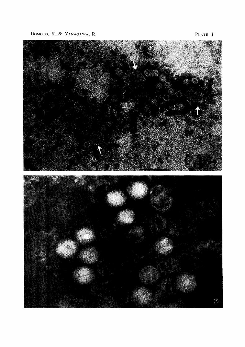
PLATE I
Fig. 1
EXPLANATION OF PLATES
ICHV, Matsuda (+); ICHV (indicated with arrows) and many small
vius particles are seen.
The small virus particles, hexagonal, include empty particles.
Fig. 2 ICHV, FD (0); only ICHV particles are seen.
Figs. 1 and 2 were the preparations negatively stained with 1 % phospho
tungstic acid.
Scales indicate 100 mfl.
DOMOTO, K. & YANAGAWA, R. PLATE I

PLATE II
Fig. 3 The small VIrus and ICHV particles in the same nucleus (18 hr
after inoculation)
The small virus and ICHV particles are located separately from
each other. ICRV particles attached to cell surface are also seen.
Scale indicates 1 p.
Fig. 4 The nucleus containing only small virus particles, which aggregate
and scatter (72 hr after inoculation)
Scale indicates 250 mp.

DOMOTO, K. & Y ANAGA W A, R. PLATE II
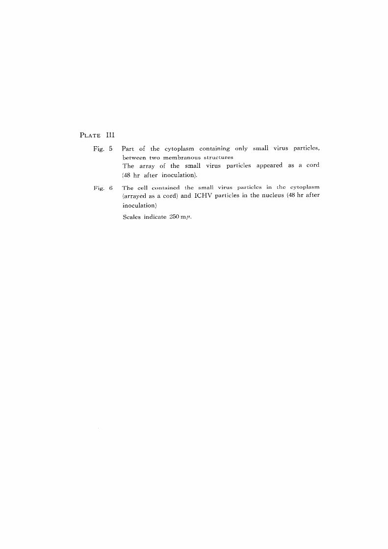
PLATE III
Fig. 5
Fig. 6
Part of the cytoplasm containing only small Virus particles,
between two membranous structures
The array of the small virus particles appeared as a cord
(48 hr after inoculation).
The cell contained the small virus particles in the cytoplasm.
(arrayed as a cord) and ICHV particles in the nucleus (48 hr after
inoculation)
Scales indicate 250 mf1.

DOMOTO, K. & YANAGAWA, R. PLATE III

PLATE IV
Fig. 7
Fig. 8
The cell contained only the small virus particles both In the
nucleus and cytoplasm
Intracytoplasmic small Vlrus particles, between two membra
nous structures, appeared as a cord.
Intranuclear small virus particles are seen as a large packed
aggregate (48 hr after inoculation).
Scale indicates 500 mp.
Crystalline aggregate of the small virus particles m the cytoplasm
(48 hr after inoculation)
Scale indicates 250 mp.

DOMOTO, K. & YANAGA W A, R. PLATE IV

PLATE V
Fig. 9 The cell contained a small number of ICHV particles in the
nucleus and numerous ICHV particles (crystalline array) in the
cytoplasm
Note ICHV multiplied in the cytoplasm (18 hr after inoculation).
Fig. 10 The cell contained a small number of ICHV and a mass of small
virus particles in the nucleus, and numerous ICHV particles in
the cytoplasm (18 hr after inoculation).
Scales indicate 1 fl.

DaMaTO, K. & YANAGAWA, R. PLATE V