Embed Size (px)
Citation preview

THEJOURNAL OFBIOLOGICALCHEMIBTBY Vol. 238, No. 7,July1933
Printed in U.S. A.
Properties of Partially Purified Carnitine Acetyltransferase*
IRVING B. FRITZ, SUZZANNE K. SCHULTZ, AND PAUL A. SRERE
From the Departments of Physiology and Biological Chemistry, The University of Michigan, Ann Arbr, Michigan
(Received for publication, February 25, 1963)
Friedman and Fraenkel (1) demonstrated that extracts of pigeon and sheep livers contain an enzyme, carnitine acetyl- transferase (EC 2.3.1.7), which catalyzes the reaction
Acetylcarnitine + Cob G acetyl-CoA + carnitine
Subsequent reports from our laboratory showed that both carnitine (/3-hydroxy-y-trimethylammonium butyrate) and acetylcarnitinei increased oxygen uptake and oxidation of long chain fatty acids by heart homogenates incubated under condi- tions optimal for fatty acid oxidation (2, 3). Acetylcarnitine action could not be attributed simply to formation of carnitine via hydrolysis because acetylcarnitine addition augmented respiration in the absence of added coenzyme A (CoA) whereas carnitine had no such effect (3). Further, acetylcarnitine en- hanced the conversion of (Y-labeled fatty acids to COZ in the absence of CoA to a greater extent than carnitine did. The possibility that carnitine acetyltransferase and other trans- ferases might be directly involved in mediating the metabolic effects of carnitine on fatty acid metabolism has been discussed in previous publications (3-5). In the present investigation we shall describe the properties of carnitine acetyltransferase and will give results demonstrating that the 0-acyl structure of acylcarnitine derivatives has a high group potential.
EXPERIMENTAL PROCEDURE
Assay Procedures
Hydroxamate Formation at pH ?&--In crude preparations, which were unsuitable for spectrophotometric assays to be de- scribed, the hydroxamate procedure suggested by Friedman and Fraenkel (1) was employed. The assay takes advantage of the quantitative conversion of acetyl-CoA to acethydroxamate at pH 7.4, whereas acetylcarnitine reacts with hydroxylamine to only a slight extent at this pH. The amount of hydroxamate formed nonenzymatically from acetylcarnitine was variable, de- pending upon pH, initial acetylcarnitine concentration, and time of incubation at 35”. When this assay was used, enzyme units were calculated on the basis of micromoles of hydroxamate
* Supported by United States Public Health Service Grants A-1465 and A-1682. A preliminary report of this work was pre- sented at the 47th annual meetingof the Federation of American Societies for Experimental Bioloav at Atlantic Citv. New Jer- sey, April 1963. -
-_ _ ,
1 Acetylcarnitine is used to designate the 0-acetyl ester of carnitine. Similar terminoloas has been emnloved for acetvl- -- norcarnitine as well as for acylcarnitine derivatives of various chain lengths.
formed in 30 minutes at 35” in tubes containing 10.0 pmoles of nn-acetylcarnitine, 400 pmoles of hydroxylamine at pH 7.4, variable amounts of enzyme preparation, and 0.34 mg of CoA (Pabst, 55% pure) in a total volume of 1.5 ml. Control tubes contained no CoA.
NADH Production in a Coupled Enzyme System-After partial purification of the enzyme preparation, it was possible to use a spectrophotometric method dependent upon the rate of genera- tion of acetyl-CoA from acetylcarnitine, in which the citrate- condensing enzyme assay system of Ochoa (6) was employed. Cuvettes contained an excess of malate, malate dehydrogenase (California Corporation for Biochemical Research, porcine heart), NAD (Pabst), crystalline pig heart citrate-condensing enzyme (7), and CoA buffered at pH 8.0 with Tris-HCl. Changes in absorbancy at 340 rnp were followed after addition of nn-acetylcarnitine and the enzyme preparation. It was necessary to add cyanide to inhibit NADH oxidase, which re- mained in enzyme preparations until final purification steps. Concentrations of all constituents are listed in legends to Table II and Fig. 1. The reaction could be initiated by addition of either CoA, acetylcarnitine, or carnitine acetyltransferase as the final component. Concentrations of acetyl-CoA synthesized from acetic anhydride and CoA (8) were estimated as equivalents of NADH formed when all other components of the coupled citrate-condensing enzyme-malate dehydrogenase assay were in excess.
Formation of CoA from Acyl-CoA Derivatives-With puri- fied enzyme preparations, the reaction was followed by determin- ing the rate of appearance of sulfhydryl groups from fatty acyl- CoA derivatives according to the procedure of Ellman (9). Constituents added to cuvettes are listed in the legend to Fig. 4. The reagent, 5,5’-dithiobis-(2dinitrobenzoic acid), did not in- hibit the initial velocity of the transfer reaction. When carnitine acetyltransferase was preincubated with the above reagent for 15 minutes under conditions cited, enzymatic activity was abolished.
Changes in Absorbancy at %X%’ rnp-With purified preparations, the cleavage of the thioester bond of acetyl-CoA or other acyl- CoA derivatives catalyzed by carnitine acetyltransferase in the presence of carnitine was assayed by recording the decrease in absorbancy at 232 rnK (10). Alternatively, the formation of thioester bonds was followed by recording the increase in ab- sorption at 232 rnp when acetylcarnitine and CoA were added initially. Spectrophotometric determinations were performed manually with the aid of a Beckman DU spectrophotometer in early experiments, but data reported were obtained primarily
by guest on August 4, 2018
http://ww
w.jbc.org/
Dow
nloaded from

2510 Carnitine Acetyltransferase Vol. 238, No. 7
with a Gilford automatic recording attachment to the spectro- photometer.
Enzyme Extraction and PuriJication Procedures
Pig hearts were obtained immediately after slaughter from Peters Sausage Company, Ann Arbor. They were either ex- tracted directly or frozen for subsequent use, since it was found that storage of frozen hearts for several months did not lower enzyme yields.
Approximately 200 g of heart ventricle were trimmed free of visible fat and were cut into thin strips. The weighed muscle was rinsed several times in an excess of ice-cold 0.25 M sucrose containing 0.005 M Versene (sodium EDTA) at pH 7.4, and 50-g portions were homogenized in an ice-jacketed semimicro Monel container. The Waring Blendor motor was run at low speeds by employing a Variac set at 60 to 70 volts. The mix- ture was kept cold by interrupting homogenization at 30-second intervals for 60-second cooling periods. Total homogenization time exclusive of cooling was 5 minut.es per 50-g portion. The pooled homogenate in approximately 400 ml of the sucrose- Versene solution was filtered through one layer of cheesecloth at O-5” and centrifuged at 10,000 X g for 10 minutes. All sub- sequent steps were performed with reagents maintained at O-2”. The residue was extracted by homogenization twice in the Monel blendor as described above, with 150 ml of 0.1 M KzHPOl con- taining 5 X lop3 M sodium EDTA for each extraction of the residue. The supernatant fractions were pooled, protein was determined spectrophotometrically (ll), and Ca3(PO& gel (12) was added to give a final ratio of 1 mg of gel per mg of protein. After the gel had been left in contact with the protein solution for 30 to 60 minutes with occasional stirring, it was collected by centrifugation, washed once with 150 ml of 0.05 M KzHP04, and extracted three times by successive addition of 50-, 30-, and 20-ml portions of 0.4 M KZHPOI buffer at pH 8.0. Pooled supernatant fractions were adjusted to pH 6.5 by slow addition of 1 N acetic acid, and then 20 g of solid (NH&SO, were gradually added per 100 ml of solution to give a 35% saturated solution. After about 15 minutes, the precipitate was collected by centrifugation at 10,000 X g for 10 minutes. The supernatant solution was brought to pH 8.0 by the addition of 1 N NaOH, and approxi- mately 40 g more of (NHa)$04 per 100 ml of solution were added over a period of hours at 0” to -5” with stirring until the solu- tion was saturated. The suspension was allowed to stand for approximately 1 hour at -5”, and was then centrifuged at 18,000 X g for 1 hour. In other preparations, the saturated solution had been allowed to stand overnight, with no precep- tible difference in results. The residue was taken up in 25 to 50 ml of 0.01 M KH2P04 buffer at pH 6.5 containing 0.005 M
sodium EDTA. To 30 ml of solution containing approximately 2.5 mg of protein per ml, 7.5 ml of absolute ethanol at -15” were added very slowly with stirring. The final temperature of the alcoholic solution was -5”. One hour later, the suspension was centrifuged at 10,000 X g for 30 minutes at -5”, and the pellet was discarded. Precipitates subsequently formed in the supernatant solutions stored at 0” to - 5” were discarded without loss of enzymatic activity. Carnitine acetyltransferase in the supernatant solution proved stable for at least 3 months when stored at this temperature.
Enzyme purification up to the ethanol step has been per- formed for five different preparations, and fairly uniform yields have been obtained with specific activities varying from 0.18 to
0.40 unit per mg of protein; a carnitine acetyltransferase unit is defined as the amount of enzyme required to catalyze the forma- tion of 1 pmole of acetyl-CoA per minute. This represents an average purification of lo- to 25-fold. Ethanol precipitation has given more variable results. In our best of three prepara- tions with this step, the supernatant of the ethanol fraction had a specific activity of 4.6 units per mg, representing a 250-fold purification. In other preparations, however, the comparable specific activity figures were only 0.58 and 0.77. If the alcoholic supernatant fraction contained acetyl-CoA hydrolase, we were able to free it from carnitine acetyltransferase by first removing alcohol by dialysis, reprecipitating the enzyme with 80% satu- rated (NH&!J’O~, and then repeating the Ca3(P04)2 gel adsorp- tion and elution procedure described above. All experiments reported with “purified preparations” were performed with en- zyme solutions having a specific activity of 0.58 to 4.6 units per milligram.
Materials and Analytical Procedures
Acetyl-, propionyl-, butyryl-, pentanyl-, hexanyl-, octanyl-, and decanyl-CoA were synthesized from their respective an- hydrides and CoA. For the synthesis of octanyl and decanyl derivatives, equal volumes of tetrahydrofuran and water were used to solubilize the anhydride. After anhydride addition and when no free sulfhydryl groups remained, excess anhydride and free acid were extracted with ether, and tetrahydrofuran was re- moved under vacuum. Acyl-CoA concentration was approxi- mated by determining absorbancy at 232 and 260 rnp (13). A more accurate analysis was performed by measuring CoA released in the coupled carnitine acetyltransferase reaction described above. Protein was estimat,ed spectrophotometrically as de- scribed by Layne (11). Carnitine was generously supplied by International Minerals and Chemical Corporation, Skokie, Illi- nois, and carnitine derivatives were obtained from sources pre- viously reported (3). Norcarnitine and its derivatives were gifts from Riker Laboratories, Inc., Northridge, California.
RESULTS
Purification of Enzyme and Properties of Assay System
Table I shows the results obtained during a typical purification procedure. It is apparent that carnitine acetyltransferase was not soluble in the sucrose fraction under conditions of homogeni- zation described in “Experimental Procedure,” but that the transferase could readily be extracted with K2HP04. The final enzyme preparation was free of acetyl-CoA hydrolase and acetyl- carnitine hydrolase. Both these substrates could be recovered quantitatively when incubated with carnitine acetyltransferase for 30 minutes at 35” in the absence of carnitine or CoA.
An outline of the reactions of the coupled enzyme system employed is presented in Table II, together with data demon- strating the dependency of NADH production on components listed. Since NADH formation did not occur in the citrate- condensing enzyme assay in the absence of CoA, it appears that acetylcarnitine was unable to condense directly with oxaloace- tate. Acetyl-CoA formation from acetylcarnitine was necessary to permit the reaction sequence to proceed. Purified prepara- tions were nearly free of NADH oxidase, but still retained citrate- condensing enzyme and malate dehydrogenase.
When all components except acetylcarnitine were present in excess, the assay could be used to determine acetylcarnitine
by guest on August 4, 2018
http://ww
w.jbc.org/
Dow
nloaded from

July 1963 I. B. Fritz, S. K. Schultx, and P. A. Srere 2511
TABLE I
Partial purification of carnitine acf3tyltransferase
from pig heart ventricle
Pig heart ventricle was extracted and purified as described in “Experimental Procedure.” Acetyl-CoA generation in all assays except in the initial homogenate was estimated by assuming that 1 rmole of NADH was formed for every micromole of acetyl-CoA presented to the malate dehydrogenase-citrate-condensing enzyme assay system (6). NADH was measured by recording changes in absorbancy at 340 rnp with the system described in Table II, with the value of 6.2 X lo3 M-I cm-1 used as the molar absorbancy index. For values shown in parentheses, obtained with whole homogenates, acetyl-Cob generation from acetyl- carnitine was determined by the method of Friedman and Fraenkel (1). For details, see the text.
Fraction and procedure
Whole homogenate (200 g/ 400 ml) . .
Sucrose supernatant.. KzHPOa supernatant. . Elution from Caa(POh)z
gel with 0.4 M KK~HPOI. 35% saturation with
(NH4) zSO4 ; precipitate discarded; 100% satura- tion with (NH&SOa; pre- cipitate taken up in 0.01 MKHzPOIatpH6.5.....
Supernatant after treat- ment with 20 volume % ethanol.
After dialysis: 80% satura- tion with (NH,) zSO~; precipitate taken up in 0.01 M KHsPOl at pH 6.5
-
1
?“,“a’t”e” yield
_-
% unitsjmg protein
4,350 (217) 100 0.014) 1 2,750 0 0 0 0 2,100 157 72 0.075 5.3
875 150 69 0.18 13
250 98 45 28
99 76 35
38 52 24
0.40
0.77
1.40
55
100
Total units
-
Specific activity
-
concentrations (Fig. I). Approximately 0.8 pmole of NADH was formed per 2.0 pmoles of nn-acetylcarnitine added, suggest- ing that only one-half of the added substrate was acceptable to the enzyme. It is likely that (+)-acetylcarnitine is an unsuit- able substrate, since (-)-carnitine is the natural isomer in muscle (14). Further, the acetyl group of ( -)-acetylcarnitine was completely oxidized by mitochondrial preparations investi- gated by Bremer (5), whereas only one-half of the m-derivative was utilized.
Initial velocities in this coupled reaction sequence have been plotted against acetylcarnitine concentrations in a Lineweaver and Burk graph (15). Under these conditions, the apparent K, is 6.2 X 1O-4 M for nn-acetylcarnitine, and the K, for (-)- acetylcarnitine may be estimated at 3.1 X 10m4 M if t,he (+)- isomer proves to be enzymatically inactive (Fig. 2).
Estimation of Equilibrium Constant
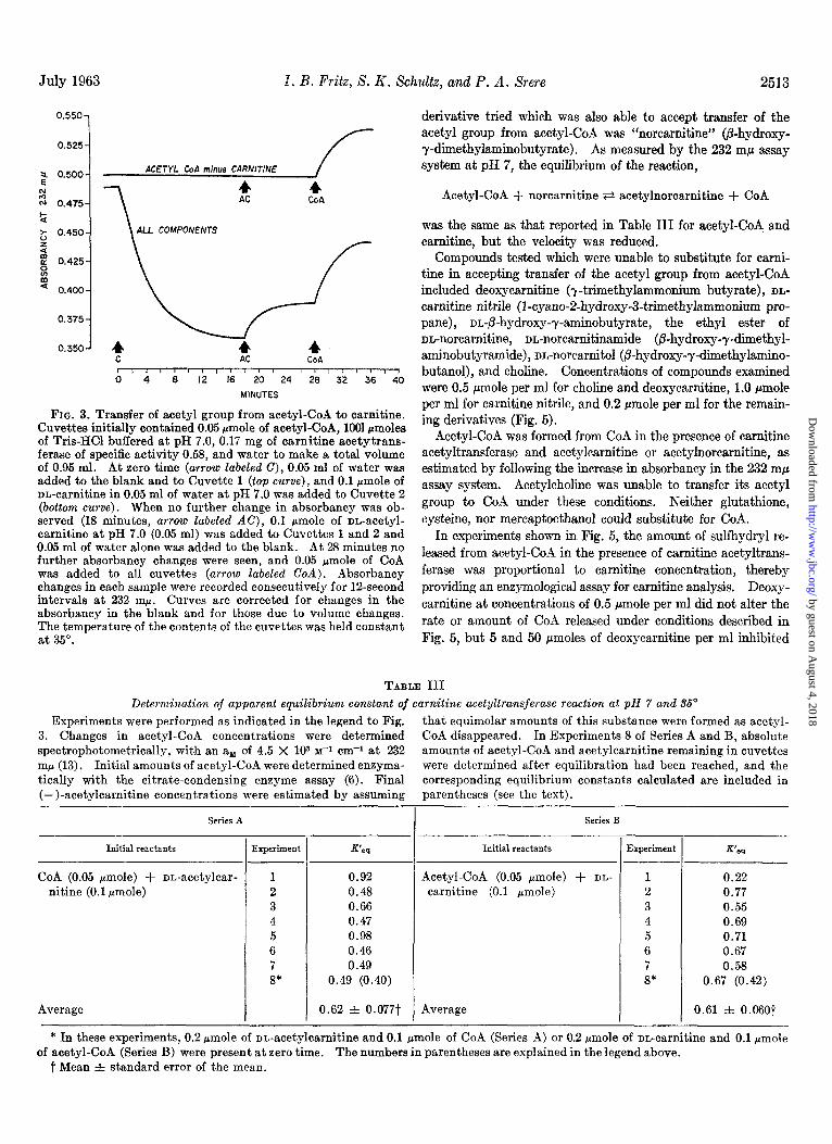
The equilibrium constant has been estimated by following the disappearance or appearance of thioester bonds spectrophoto- metrically at 232 rnp (lo), with an ay of 4.5 X lo3 M-~ cm-r for acetyl-CoA at this wave length (13). Data shown from a single experiment in Fig. 3 and results from eight experiments sum-
marized in Table III indicate that the apparent equilibrium constant at pH 7.0 was approximately 0.6 toward acetyl-CoA formation when the initial reactants were either nn-acetylcarni- tine and Coil or acetyl-CoA and nn-carnitine. For these calcu- lations, it was assumed that only the (-)-isomers were en- zymatically active and that the reactants and products were exclusively those cited in Table II for the carnitine acetyltrans- ferase reaction. In the experiment shown in Fig. 3, acetyl-CoA and carnitine were initially incubated with carnitine acetyl- transferase. After no further change in absorbancy occurred, acetylcarnintine and CoA were added at separate times. Cal- culations to determine the apparent equilibrium constant were made by assuming that acetylcarnitine formed initially was equal to acetyl-CoA which disappeared. By substitution into the equation,
K’ - (acetyl-CoA) (carnitine)
w - (CoA) (acetylcarnitine)
the values obtained in the experiment cited in Fig. 3 were 0.55 in the first phase, in which there was a decrease in absorbancy of 0.129, corresponding to a disappearance of 0.0287 pmole of acetyl-CoA; 0.50 after acetylcarnitine addition, in which there was an increased absorbancy of 0.031, corresponding to an in- crease of 0.007 pmole of acetyl-CoA; and 0.44 after COA addi- tion, in which the absorbancy increased by 0.054, corresponding
to an increase of 0.012 pmole of acetyl-CoA. This experiment is the same as Experiment 3, Series B, of TabIe III.
TABLE II
Spectrophotometric assay for carnitine acetyltransferase
Cuvettes having all components added to the system con- tained, in a final volume of 1.0 ml: 100 rmoles of Tris-HCl, pH 7.8, at 35”; 2.5 pmoles of NAD; 10 pmoles of on-potassium malate; 1.0 rmole of NaCN; 30 pmoles of on-acetylcarnitine; 0.17 mg of CoA (Pabst, 55% pure) ; 0.07 unit of malate dehydrogenase (Cali- fornia Corporation for Biochemical Research); and 0.07 unit of crystalline pig heart citrate-condensing enzyme (7). In the deletion experiments, 0.005 mg of carnitine acetyltransferase of the specific activity shown was present. NADH formation was determined by measuring absorbancy changes per minute at 340 rnp. NADH production rate was linear from 12 seconds after addition of enzyme for periods of at least 10 minutes.
The steps in the reaction sequence are outlined as follows.
Malate2- + NAD+ F? oxaloacetate+ + NADH + Hf
Acetylcarnitine + CoA ti acetyl-CoA + carnitine
Acetyl-CoA + Hz0 + oxaloacetate2- a citrates- + CoA + H+
Sum: MalateX- -I- acetylcarnitine + Hz0 + NAD+ ti carnitine + citrate3- + NADH + 2 H+
Components added NADH formed
mpmoles~min
All minus carnitine acetyltransferase. 0 All (0.005 mg of carnitine acetyltransferase) 16 All (0.010 mg of carnitine acetyltransferase) 32
Minus acetylcarnitine........................... 0 Minus CoA................ . .._....... 0 Minus malate................................... 0 Minus citrate-condensing enzyme. 9 Minus malate dehydrogenase. . . . 15
by guest on August 4, 2018
http://ww
w.jbc.org/
Dow
nloaded from

2512 Carnitine Acetyltransferase Vol. 238, No. 7
/
G ii 82 3 I /
I J
DL-ACETYL CARNITINE (p moles )
FIG. 1. Total NADH formed in the coupled carnitine acetyl- transferase-malate dehydrogenase-citrate-condensing enzyme sys- tem at various acetylcarnitine concentrations. Cuvettes con- tained all components listed in the legend to Table II at identical concentrations, except that amounts of nn-acetylcarnitine added were varied as indicated, and 0.04 mg of carnitine acetyltrans- ferase of specific activity 4.6 was present in each cuvette. The reaction was initiated by acetylcarnitine addition, and absorbancy at 340 rnp was recorded until no further changes occurred. For samples containing more than 0.25 pmole of bn-acetylcarnitine per ml, absorbancy at 340 rnp was determined in aliquots which had been diluted 1:lO after incubation for 30 minutes at 35”, by which time the reaction had gone to completion.
Final concentrations of acetylcarnitine and acetyl-CoA in these cuvettes were determined by an independent method. Acetyl-CoA and carnitine were incubated at higher concentra- tions with carnitine acetyltransferase as indicated above. Cuvettes were then heated at 70” for 1 minute to inactivate carnitine acetyltransferase, a.fter which acetyl-CoA was measured with the citrate-condensing enzyme (6). After the analysis was completed, carnitine acetyltransferase and CoA were added to the same cuvettes for determination of acetylcarnitine by methods shown in Fig. 1. Equilibrium constant values calcu- lated were in fair agreement with other determinations (see Experiments 8 of Series A and B, Table III).
Substrate SpeciJicity
Fatty Acyl-CoA Derivatives-Under conditions shown in Table IV, the acyl groups of acetyl-CoA, propionyl-Cod, and butyryl-CoA were transferred to carnitine at approximately the same rates, but the transfer of longer chain acyl groups pro- ceeded at reduced velocities. We have no evidence concerning the possibility that more than one short chain carnitine acyl- transferase exists. The rate of release of CoA from acyl-CoA derivatives when a different amount of enzyme was used is plotted in Fig. 4. The acyl group of palmityl-CoA was not transferred by carnitine acetyltransferase, nor did the prepara- tion contain a palmityl-CoA hydrolase. After reactions shown in Fig. 4 and Table IV had apparently stopped, addition of more enzyme resulted in further CoA release from hexanyl-, octanyl-, and decanyl-CoA, but in no further CoA release when
acetyl-, propionyl-, or butyryl-CoA was substrate. This prob- ably occurred because carnitine acetyltransferase was slowly inactivated by interaction with the reagent, 5,5’-dithiobis-(2- dinitrobenzoic acid), used in the sulfhydryl group analysis (9). It is possible, however, to determine concentrations of specific acyl-CoA derivatives by measurement of sulfhydryl released when sufficient amounts of enzyme are added.
Reversible transfer of acyl groups from other acyl-CoA deriva- tives to carnitine was also followed by the 232 rnp assay, and simi- lar results were obtained in that the rate of transfer was slower for longer chain acyl groups. At each enzyme concentration tried, the initial velocity for short chain length acyl transfer, i.e. acetyl, propionyl, and butyryl, was greater than that observed for transfer of longer chain length acyl groups. Acyl-CoA deriva- tives incubated in the presence of carnitine acetyltransferase did not release CoA unless carnitine was added to the system, demonstrating that the enzyme preparation did not contain acyl-CoA hydrolase.
Carnitine and Derivatives-CoA liberation from acetyl-CoA incubated with carnitine acetyltransferase was proportional to carnitine added to the system (Fig. 5). The only carnitine
0.40
0.35
7 0.30
i!
z ?- 0.25
.c E
N ‘2 0.20
-7, 0.15
0. IO
/ 0.05
/
/’
r
-2 -I 0 I 2 3 4 5
$ X lo3 DL-ACETYLCARNITINE (M-‘)
FIG. 2. Determination of the apparent K, for acetylcarnitine in the coupled carnitine acetyltransferase-malate dehydrogenase- citrate-condensing enzyme system. Cuvettes contained all com- ponents listed in the legend to Table II at identical concentra- tions, except that concentrations of nn-acetylcarnitine were varied as indicated, and 0.04 mg of carnitine acetyltransferase of specific activity 4.6 was present in each cuvette. The reaction was ini- tiated by acetylcarnitine addition, and absorbancy changes at 340 rnp were recorded. The rate of NADH formation was linear for at least 1 minute after addition of 0.2 or more pmole of DL- acetylcarnitine.
by guest on August 4, 2018
http://ww
w.jbc.org/
Dow
nloaded from
July 1963 1. B. Fritz, S. K. Schultz, and P. A. Srere 2513
0.550-
0.525-
2 0.500- ACETYL Cod minus CARNITINE
2 4 N 0.475- CoA
t z 0.450- ALL COMPONENTS
3 zl % 0.425
ii * 0.400
C AC COP, ),I I I I,,, I I I I I I I / I I,, 0 4 8 12 16 20 24 28 32 36 40
MINUTES
FIG. 3. Transfer of acetyl group from acetyl-CoA to carnitine. Cuvettes initially contained 0.05 pmole of acetyl-CoA, 1001 crnaoles of Tris-HCl buffered at pH 7.0, 0.17 mg of carnitine acetytrans- ferase of specific activity 0.58, and water to make a total volume of 0.95 ml. At zero time (arrow labeled C), 0.05 ml of water was added to the blank and to Cuvette 1 (top curve), and 0.1 pmole of nn-carnitine in 0.05 ml of water at pH 7.0 was added to Cuvette 2 (bottom curve). When no further change in absorbancy was ob- served (18 minutes, arrow labeled AC), 0.1 pmole of nn-acetyl- carnitine at pH 7.0 (0.05 ml) was added to Cuvettes 1 and 2 and 0.05 ml of water alone was added to the blank. At 28 minutes no further absorbancy changes were seen, and 0.05 rmole of CoA was added to all cuvettes (arrow labeled CoA). Absorbancy changes in each sample were recorded consecutively for la-second intervals at 232 rnp. Curves are corrected for changes in the absorbancy in the blank and for those due to volume changes. The temperature of the contents of the cuvettes was held constant at 35”.
derivative tried which was also able to accept transfer of the acetyl group from acetyl-CoA was “norcarnitine,, (p-hydroxy- y-dimethylaminobutyrate). As measured by the 232 rnp assay system at pH 7, the equilibrium of the reaction,
Acetyl-CoA + norcarnitine F? acetylnorcarnitine + CoA
was the same as that reported in Table III for acetyl-CoA and carnitine, but the velocity was reduced.
Compounds tested which were unable to substitute for carni- tine in accepting transfer of the acetyl group from acetyl-CoA included deoxycarnitine (T-trimethylammonium butyrate), DL-
carnitine nitrile (l-cyano-2-hydroxy-3-trimethylammonium pro-
pane), nr&hydroxy-y-aminobutyrate, the ethyl ester of nn-norcarnitine, nn-norcarnitinamide @-hydroxy-y-dimethyl- aminobutyramide), nn-norcarnitol (&hydroxy-y-dimethylamino- butanol), and choline. Concentrations of compounds examined were 0.5 pmole per ml for choline and deoxycarnitine, 1.0 pmole per ml for carnitine nitrile, and 0.2 pmole per ml for the remain- ing derivatives (Fig. 5).
Acetyl-CoA was formed from CoA in the presence of carnitine acetyltransferase and acetylcarnitine or acetylnorcarnitine, as estimated by following the increase in absorbancy in the 232 rnp assay system. Acetylcholine was unable to transfer its acetyl group to CoA under these conditions. Neither glutathione, cysteine, nor mereaptoethanol could substitute for CoA.
In experiments shown in, Fig. 5, the amount of sulfhydryl re- leased from acetyl-CoA in the presence of carnitine acetyltrans- ferase was proportional to carnitine concentration, thereby providing an enzymological assay for carnitine analysis. Deoxy- carnitine at concentrations of 0.5 pmole per ml did not alter the rate or amount of CoA released under conditions described in Fig. 5, but 5 and 50 pmoles of deoxycarnitine per ml inhibited
TABLE III Determination of apparent equilibriunz constant of carnitine acetyltransferase reaction al pH 7 and 3.5’
Experiments were performed as indicated in the legend to Fig. that equimolar amounts of this substance were formed as acetyl- 3. Changes in acetyl-CoA concentrations were determined CoA disappeared. In Experiments 8 of Series A and B, absolute spectrophotometrically, with an an of 4.5 X lo3 ~-1 cm+ at 232 amounts of acetyl-CoA and acetylcarnitine remaining in cuvettes rnp (13). Initial amounts of acetyl-CoA were determined eneyma- were determined after equilibration had been reached, and the tically with the citrate-condensing enzyme assay (6). Final corresponding equilibrium constants calculated are included in (-)-acetylcarnitine concentrations were estimated by assuming parentheses (see the text).
Series A
Initial reactants
CoA (0.05 pmole) + rm-acetylcar- nitine (0.1 pmole)
Average
-
1 --
k%periment K’eq
1 0.92 2 0.48 3 0.66 4 0.47 5 0.98 6 0.46 7 0.49 s* 0.49 (0.40)
0.62 f 0.077t
- I
--
Series B
Initial reactants
Acetyl-CoA (0.05 hmole) + DL-
carnitine (0.1 pmole)
Average
T
-
Experiment
1 0.22 2 0.77 3 0.55 4 0.69 5 0.71 6 0.67 7 0.58 8* 0.67 (0.42)
0.61 f 0.0607
* In these experiments, 0.2 Fmole of nn-acetylcarnitine and 0.1 pmole of CoA (Series A) or 0.2 pmole of nn-carnitine and 0.1 rmole of acetyl-CoA (Series B) were present at zero time. The numbers in parentheses are explained in the legend above.
t Mean i standard error of the mean.
by guest on August 4, 2018
http://ww
w.jbc.org/
Dow
nloaded from

2514 Carnitine Acetyltransferase Vol. 238, No. 7
TABLE IV
Cleavage of aeyl-CoA derivatives my carnitine acetyltransferase
All cuvettes contained, in a total volume of 1.0 ml: 0.05 pmole of the designated acyl-Coa derivative prepared as described in “Experimental Procedure”; 100 pmoles of Tris-HCl, pH 7.8, at 35”; 0.2 pmole of 5,5’-dithiobis-(2.nitrobenzoic acid); and 0.035 mg of carnitine acetyltransferase of specific activity 1.4. The reaction was initiated by addition of enzyme, and absorbancy changes at 412 rnp obtained 3Oseconds later were used to compute values shown, assuming an aM of 13.6 X lo3 M-’ cm-r (9). Blank cuvettes contained no carnitine. The same relative values were obtained at all enzyme concentrations tried. Results shown are the average figures obtained from at least three separate de- terminations.
Substrate CoA released in initial 30 seconds
Acetyl-CoA ............................... Propionyl-CoA. ........................... Butyryl-CoA. ............................. Pentanoy-CoA. ........................... Hexanoyl-CoA ............................ Octanoyl-CoA ....... ‘ ..... , .............. Decanoyl-CoA ............................ Palmityl-CoA*. ...........................
m)mwles
30 37 30 11
7 4 1 0
* Palmityl-CoA was synthesized by the method of Seubert (16).
the reaction by 65 and 96’%, respectively, when 0.1 pmole of nn-carnitine per ml was present.
pH Optimum Determination
The decrease in absorbancy at 232 rnp at various hydrogen ion concentrations during the first minute after addition of carnitine acetyltransferase to cuvettes containing 0.05 nmole of
both acetyl-CoA and carnitine is plotted in the top part of Fig. 6. Enzyme activity decreased at pH values below 7.1, and maximal activity was unchanged from pH 7.1 to 8.2. The rate decreased to less than half maximum at pH 8.9. Total ab- sorbancy change in a 15-minute period was constant at all hydrogen ion concentrations examined up to pH 8.6, but at higher pH values, the enzyme was inactivated (Fig. 6, bottom).
We have obtained carnitine acetyltransferase activity in ex- tracts of rat liver and brain, but levels were lower than those found in pig or rat heart preparations. A detailed study of en- zyme distribution in these and other organs has not yet been attempted, but we have observed that extracts of heart mito- chondria gave highest yields of carnitine acetyltransferase per mg of initial protein. The enzyme was also present, however, in 25,000 X g supernatant fractions of fresh rat hearts homogenized in 0.25 M sucrose containing 1 mM sodium EDTA when an all glass conical tissue homogenizer was employed.
DISCUSSION
Carnitine acetyltransferase catalyzes the reversible transfer of short chain acyl groups to carnitine from acyl-CoA derivatives. Although this enzyme lacks the ability to transfer long chain groups, a separate transferase has been found in both mitochon- dria and soluble cytoplasmic fractions which catalyzes the reac- tion (17, 18)
Palmitylcarnitine + CoA + palmityl-CoA + carnitine
The long chain carnitine acyltransferase is not yet sufficiently pure to permit characterization of its properties. It will be of interest to determine whether the substrate specificity toward carnitine and its derivatives is the same as that reported here for carnitine acetyltransferase, since an identical specificity is required to obtain stimulation of long chain fatty acid oxidation by heart muscle preparations (3). I f this is the case, it will offer additional support to recent suggestions that acylcarnitine derivatives are involved in processes by which carnitine cataly- tically enhances oxidation of fatty acids by several tissues (2-4). Evidence indicating that carnitine may serve as a shuttle for the transport of long chain acyl groups to the fatty acid oxidase site from extramitochondrial portions of the cell has been presented both by Bremer (19) and by us (17, 18), and has recently been reviewed (20).
A function similar to that formulated for long chain carnitine acyltransferase (18) may be postulated for carnitine acetyltrans- ferase, in that the enzyme whose properties have been reported here could mediate the transfer of acetyl groups from mitochon- dria to sites of fatty acid synthesis in the cytoplasm. Although this hypothesis remains to be tested, there is ample evidence
0.8
0.7
0.f
2
z 0.5
t
g 0.4 z s
5 z 0.3
0.2
0. I
0
ACETYL CoA B BUTYRYL CoA
HEXANYL CoA
0 2 4 6 8 10 12
MINUTES
FIG. 4. Specificity of carnitine acetyltransferase toward acyl- CoA derivatives of various chain lengths. Cuvettes contained equimolar amounts of acyl-CoA (0.06 rmole), 1.0 rmole of DL- carnitine, 0.02 mg of carnitine acetyltransferase of specific activity 4.6, 0.2 .umole of 5,5’-dithiobis-(2.nitrobenzoic acid); 100 rmoles of Tris-HCl buffered at pH 7.0 at 35”, and water to 1.0 ml. Blank cuvettes had all components except carnitine. The reaction was started by enzyme addition at zero time. Tracings are corrected for absorbancy changes in the blank, which amounted to an ab- sorbancy of 0.070 at 12 minutes under the conditions cited. Since results obtained with acetyl-CoA (open circles) and butyryl-CoA (triangles) were indistinguishable, a single curve was drawn for these substrates.
by guest on August 4, 2018
http://ww
w.jbc.org/
Dow
nloaded from

July 1963 I. B. Fritz, X. K. Schultz, and P. A. Xrere 2515
that acetylcarnitine can penetrate mitochondrial barriers. Ad- dition of acetylcarnitine stimulated oxygen uptake by heart muscle preparations (3) and by mitochondria from several tissues (5) in the absence of added CoA. The increase in respiration most probably resulted from oxidation of acetyl groups in the tricarboxylic acid cycle after initial conversion of acetylcarnitine to acetyl-CoA within mitochondria, catalyzed by mitochondrial carnitine acetyltransferase. As Bremer indicated, however, the possibility remained that the increased respiration that fol- lowed acetylcarnitine addition could have resulted from trans- acetylation independent of acetyl-CoA formation, possibly by direct condensation with oxaloacetate to form citrate (5). This possibility can now be ruled out on the basis of data shown in Table II. Since NADH was not formed in the coupled citrate- condensing enzyme system unless both CoA and carnitine acetyl- transferase were added to cuvettes containing acetylcarnitine, it may be concluded that the citrate-condensing enzyme cannot accept the acetyl group directly from acetylcarnitine. Available evidence is consonant with the concept that acetylcarnitine must first be converted to acetyl-CoA before being metabolized fur- ther.
The partial purification of carnitine acetyltransferase has per- mitted us to estimate the equilibrium constant of that reaction by employing procedures described in Fig. 3. When Friedman and Fraenkel (1) initially demonstrated the carnitine acetyl- transferase reaction, they postulated that the 0-acetyl ester of acetylcarnitine might have a high group potential. The im- pure nature of their enzyme preparation did not allow Fraenkel and Friedman to substantiate their hypothesis. The present data, which demonstrate an equilibrium constant of approxi- mately 0.6 toward acetyl-CoA formation under conditions de- scribed, show that the group potential of the 0-acyl structure is indeed of a higher order than would be expected of an ordinary secondary ester. The AP for hydrolysis of acetylcarnitine to carnitine and acetate may be estimated by summing the follow- ing reactions.
Acetylcarnitine + CoA * acetyl-CoA + carnitine (AFO’
= +0.3 kcal) (A) Acetyl-CoA + Hz0 + acetate + H+ + CoA (AFO’
= -8.2 kcal at pH 7.0 (21)) (B)
Sum: Acetylcarnitine + Hz0 * acetate + Hf + carnitine (APO’ = -7.9 kcal at pH 7.0) (C)
As Jaenicke and Lynen (21) have pointed out, a high group potential of the 0-acyl ester of acetylcarnitine is contrary to usual expectations. It is not found, for example, in the 0-acetyl group of acetylcholine (21). Since norcarnitine was able to substitute for carnitine in the acetyltransferase reaction at pH 7.0 without changing the equilibrium position, it appears that the energy of the 0-acetyl group was not altered by removal of a methyl group from the nitrogen of acetylcarnitine.
In an examination of substrate specificity of carnitine acetyl- transferase, we have demonstrated that removal of a methyl group to. form norcarnitine decreased the velocity of the reac- tion, whereas substitution of a cyano, a hydroxyl, an amide, or an ester grouping for the original carboxyl group abolished activity of the derivative molecule in the enzymatic assay at concentrations employed. Since a free carboxyl group appears to be required, it is possible that the sequence shown in Diagram 1 occurs during the carnitine acetyltransferase reaction.
0.450
0.400
d 1 E- 0.350 N
a i 2 0.300
4 1 2 1 g 0.250 ,
1 I , I I I I
0 0.02 0.04 0.06 0.08 0.10 0.12 p moles DL-CARNITINE OR DERIVATIVE /ml.
FIG. 5. Effects of carnitine and derivatives on the release of CoA from acetyl-CoA in the presence of carnitine acetyl- transferase. Results were obtained by following absorbancy at 412 rnp. Blank cuvettes contained, in 1.0 ml, the following com- ponents: 0.2 rmole of 5,5’-dithiobis-(2.nitrobenxoic acid), 100 @moles of Tris-HCl buffered at pH 8.0 at 35”; 0.05 rmole of acetyl- CoA, and 0.02 mg of carnitine acetyltransferase of specific activity 4.6. Other cuvettes, appropriately labeled on the graph, con- tained the same components plus indicated amounts of DL-carni-
tine or nn-norcarnitine. In experiments labeled R on the graph, cuvettes contained one of the following derivatives: choline, deoxycarnitine, m-8-hydroxy-r-aminobutyrate, nn-norcarnitol, nn-carnitine nitrile, nn-norcarnitinamide, or the ethyl ester of nn-norcarnitine. Maximal concentrations examined of these latter derivatives were varied between 0.2 and 1 .O rmole per ml, as stated in the text. CoA was measured by the procedure of Ellman (9). DL-Norcarnitine at 0.2 pmole per ml resulted in a release of CoA corresponding to an absorbancy of 0.289, giving a continuation of the straight line labeled norcarnitine. Reac- tions were started by addition of enzyme. The blank absorbancy, subtracted from all points on the above graph, was 0.114 and was contributed chiefly by interaction of the dye with protein. Solid points represent analyses performed in the presence of 0.5 rmole of deoxycarnitine per ml.
Intermediate I might be stabilized by interaction of the O- with positively charged groups on the enzyme. Similar reaction sequences have been postulated to account for unexpectedly high rates of hydrolyses of certain esters (22). Alternatively, the positively charged amino group may be involved in the transi- tion states (23). It will be of interest to examine acetyl-CoA formation from acetylnorcarnitine and CoA at pH values above the pK of the amino group to see whether a positively charged nitrogen is in fact required in the carnitine acetyltransferase reaction.
by guest on August 4, 2018
http://ww
w.jbc.org/
Dow
nloaded from

2516 Carnitine Acetyltransferase Vol. 238, No. i’
.06 1 0
if’ 05-
2 ; .04-
z G1
E‘s .03- -
@
G .02 - 0 PHOSPHATE BUFFER
0 TRIS BUFFER
5 * GLYCINE-NaOH BUFFER
5
.Ol - I I I I I / I I
8
3
.13-
.12-
.06-
.05 -. 6.0
I , I I ! 6.5 7.0 7.5 6.0 9.5 9.0 9.5 10.0
pH at 35* C
FIG. 6. Effects of pH on carnitine acetyltransferase activity. obtained with a single addition of enzyme. Buffers used were Acetyl-CoA (0.05 pmole per ml) and 0.1 rmole of nn-carnitine per 0.05 M KHzPOI (circles), 0.1 M Tris-HCl (squares), and 0.05 M ml were present in all ouvettes together with buffer at pH values glycine with NaOH added to give designated pH values (triangles). indicated. The reaction was initiated by addition of 0.035 mg of Immediately after the end of incubation at 35”, pH values of these carnitine acetyltransferase of specific activity 1.4. Absorbancy solutions were measured with glass electrodes (Beckman model change at 232 rnp during the first minute is plotted against pH G) at room temperature, and suitable correction factors were in the top graph; the bottom graph shows total absorbancy change introduced to calculate pH values at 35”.
DIAGRAM 1
i
CHI O-
‘c’
OCOCH, 0’ ‘0
(CHs)aNCHz&HCH&OO FI (CH&NCH2 HCH&=O ci + -L + Acetylcarnitine I
Since enzymatic activity was abolished when carnitine acetyl- transferase was incubated with 5,5’-dithiobis-(2-dinitrobenzoic acid), it is possible that sulfhydryl groups of the enzyme may be involved in the formation of an acylthio-enzyme intermediate in the reaction sequence.
SUMMARY
Carnitine acetyltransferase was extracted from pig heart and was partially purified. It catalyzed the reaction
Acetylcarnitine + CoA F? acetyl-CoA + carnitine
The apparent equilibrium constant was 0.6 at pH 7.0 and at 35”, on the assumption that only the (-)-isomers were enzymatically active. It was concluded that the 0-acyl ester of acylcarnitine derivatives had a high group potential, and that the AFO’ for acetylcarnitine hydrolysis was approximately -7.9 kcal.
Substrate snecificity of carnitine acetyltransferase toward
carnitine or its derivatives was investigated by examining co- enzyme A (CoA) release from acetyl-CoA, and it was found that the analogous compounds containing an amide, cyano, hydroxyl, or ester group instead of the carboxyl were inactive at concentrations examined. The removal of one methyl group from carnitine to form norcarnitine did not abolish activity in the enzymatic assay, although the velocity of the transfer was less than that observed with equimolar carnitine concentrations. fl-Hydroxy-y-aminobutyric acid was unable to accept acetyl transfer. Acetyl-CoA, propionyl-CoA, and butyryl-CoA reacted at approximately the same rates, but carnitine acetyltransferase catalyzed the transfer of acyl groups of longer chain lengths at slower velocities under the conditions described. Decanyl-CoA reacted slowly, and palmityl-CoA was inactive. The enzyme had slight or no fatty acyl-CoA hydrolase or acetylcarnitine esterase activity. Carnitine acetyltransferase did not react with choline or its derivatives. Of various assay procedures used in the purification and characterization of carnitine acetyl-
by guest on August 4, 2018
http://ww
w.jbc.org/
Dow
nloaded from

July 1963 I. B. Fritz, S. K. Schultz, and P. A. Srere 2517
transferase, the system employed to measure CoA release from acetyl-CoA seemed most appropriate as an enzymological assay for carnitine in biological material, since a linear relationship was established between carnitine concentration and sulfhydryl groups released. The possible functional significance of carni- tine acetyltransferase was discussed, and efforts were made to relate data presented to previous findings that carnitine catalyt- ically enhances fatty acid oxidation. On the basis of the ob- served substrate speciftcity, a reaction sequence was postulated.
Achmowledgment-The authors are happy to acknowledge the contributions of Dr. Robert I-I. Abeles to the discussion of pos- sible intermediates in the carnitine acetyltransferase reaction.
REFERENCES
1. FRIEDMAN, S., AND FRAENKEL, G., Arch. Biochenz. Biophys.* 69, 491 (1955).
2. FRITZ, I.B., AND KAPLAN, E.,inH. PEETERS (Editor), Protides of the biological jluids, proceedings of the seventh colloquium, Elsevier, Amsterdam, 1960, p. 252.
3. FRITZ, I. B., KAPLAN, E., AND YUE, K. T., Am. J. Physiol., 202, 117 (1962).
4. FRITZ, I. B., Physiol. Revs., 41, 52 (1961). 5. BREMER, J., J. Biol. Chem., 237, 2228 (1962). 6. OCHOA, S., in D. SHEMIN (Editor), Biochemical preparations,
VoZ. 6, John Wiley and Sons, Inc., New York, 1957, p. 19. 7. SRERE, P. A., AND KOSICKI, G. W., J. Biol. Chem., 236, 2557
(1961). 8. OCHOA, S., in S. P. COLOWICK AND N. O.KAPLAN (Editors)
Methods in enzymology, Vol. I, Academic Press, Inc., New York, 1955, p. 688.
9. ELLMAN, G. L., Arch. Biochem. Biophys., 82, 70 (1959). 10. SRERE, P. A., SEUBERT, W., AND LYNEN, F., Biochim. et Bio-
phys. Acta, 33, 313 (1959). 11. LAYNE, E., in S. P. COLOWICK AND N. 0. KAPLAN (Editors),
Methods in enzymology, Vol. III, Academic Press, Inc., New York, 1957, p. 451. --.
12. COLOWICK.S. P..in S.P. COLOWICK AND N.O.KAPLAN (Edi- tors), Mkhods’in enzymology, Vol. I, Academic Press, Inc., Newkork, 1955, p. 98. --.
13. STADTMAN. E.R..inS.P. COLOWICKANDN.O.KAPLAN (Edi- tors), Methods k enzymology, Vol. III, Academic Press, Inc., Newkork, 1957, p. 931. --’
14. FRAENKEL. G.. AND FRIEDMAN. S.. Vitamins and Hormones. 16, 73 (1957):
, I
15. DIXON, M., AND WEBB, E. C., Enzymes, Academic Press, Inc., New York, 1958, p. 21.
16. SEUBERT, W., in H. A. LARDY (Editor), Biochemical prepara- tions, Vol. 7, John Wiley and Sons, Inc., New York, 1960, p. 80.
17. FRITZ, I. B., AND YUE, K. T. N., Physiologist, 6, 144 (1962). 18. FRITZ, I. B., AND YUE, K. T. N., J. Lipid Research, in press. 19. BREMER, J., J. Biol. Chem., 237, 3628 (1962). 20. FRITZ, I. B., in R. PAOLETTI AND D. KRITCHEVSKY (Editors),
Advances in lipid research, Vol. I, Academic Press, Inc., New York, 1963, in press.
21. JAENICKE, L., AND LYNEN, F.,inP.D. BOYER, H. LARDY,AND K. MYRBHCK (Editors), The enzymes, Vol. III, Academic Press, Inc., New York, 1960, p. 30.
22. BENDER, M. L., Chem. Revs., 60, 53 (1960). 23. BRUICE, T. C., AND BENEOVIC, S. J.,J. Am. Chem.Soc., 86, 1
(1963).
by guest on August 4, 2018
http://ww
w.jbc.org/
Dow
nloaded from

Irving B. Fritz, Suzzanne K. Schultz and Paul A. SrereProperties of Partially Purified Carnitine Acetyltransferase
1963, 238:2509-2517.J. Biol. Chem.
http://www.jbc.org/content/238/7/2509.citation
Access the most updated version of this article at
Alerts:
When a correction for this article is posted•
When this article is cited•
to choose from all of JBC's e-mail alertsClick here
http://www.jbc.org/content/238/7/2509.citation.full.html#ref-list-1
This article cites 0 references, 0 of which can be accessed free at
by guest on August 4, 2018
http://ww
w.jbc.org/
Dow
nloaded from


![carnitine deficiency[1]](https://img.pdfslide.net/doc/110x75/577d20c11a28ab4e1e93ae46/carnitine-deficiency1.jpg)