Embed Size (px)
Citation preview
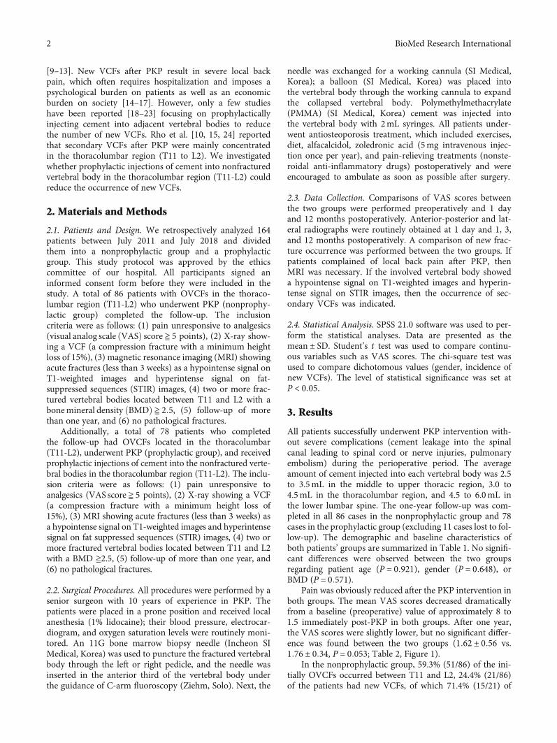
Research ArticleProphylactic Percutaneous Kyphoplasty Treatment forNonfractured Vertebral Bodies in Thoracolumbar forOsteoporotic Patients
Fei Lei ,1 Wen He ,2 Xinggui Tian ,1 Zhongyang Li,3 Lipeng Zheng,1 Jianping Kang,1
and Daxiong Feng 1
1Department of Spine Surgery, The Affiliated Hospital of Southwest Medical University, No. 25 Taiping St., Luzhou,646000 Sichuan, China2Department of Library, Southwest Medical University, No. 1 Xianglin Road of Longma District, Luzhou, 646000 Sichuan, China3Department of Orthopedics Surgery, West China Hospital, Sichuan University, No. 37 Guoxue St. of Wuhou District, Chengdu,610041 Sichuan, China
Correspondence should be addressed to Daxiong Feng; [email protected]
Received 15 September 2019; Revised 6 February 2020; Accepted 29 February 2020; Published 9 April 2020
Academic Editor: Eiichi Ishikawa
Copyright © 2020 Fei Lei et al. This is an open access article distributed under the Creative Commons Attribution License, whichpermits unrestricted use, distribution, and reproduction in any medium, provided the original work is properly cited.
Purpose. The occurrence of new vertebral compression fractures (VCFs) is a common complication after percutaneous kyphoplasty(PKP). Secondary VCFs after PKP occur predominantly in the thoracolumbar segment (T11 to L2). Prophylactic injections ofcement into vertebral bodies in order to reduce new VCFs have rarely been reported. The main purpose of this study was toinvestigate whether prophylactically injecting cement into a nonfractured vertebral body at the thoracolumbar level (T11-L2)could reduce the occurrence of new VCFs. Methods. From July 2011 to July 2018, PKP was performed in 86 consecutive patientswith osteoporotic vertebral compression fractures (OVCFs) in the thoracolumbar region (T11-L2). All patients selectedunderwent PKP because of existing OVCFs (nonprophylactic group). Additionally, 78 consecutive patients with fracturedvertebrae in the thoracolumbar region (T11-L2) with OVCFs underwent PKP and received prophylactic injections of cementinto their nonfractured vertebrae in the thoracolumbar region (T11-L2) (prophylactic group). The visual analog scale (VAS)scores and incidence of new VCFs after PKP were compared between the two groups. Results. The mean VAS scores improvedfrom 8:00 ± 0:79 preoperatively to 1:62 ± 0:56 at the last follow-up in the nonprophylactic group and improved from 8:17 ± 0:84to 1:76 ± 0:34 in the prophylactic group (P > 0:05). In the nonprophylactic group, 21 of 86 patients (24.4%) developed newVCFs within one year after PKP, of whom 15 patients (71.4%) developed VCFs within 3 months. In the prophylactic group, 8 of78 patients (10.3%) developed new VCFs within one year, and 6 of these 8 patients (75%) developed new VCFs within 3months. The incidence of new VCFs was significantly higher in the nonprophylactic group than that in the prophylactic groupat one year (P = 0:018), but there were no statistically significant differences at three months (P = 0:847). Conclusions.Prophylactic injections of cement into nonfractured (T11-L2) vertebral bodies reduced the incidence of secondary VCFs afterPKP in patients with OVCFs, but there was no significant difference in local back pain (VAS) scores between the two groups.
1. Introduction
Percutaneous kyphoplasty (PKP) is a minimally invasivevertebral augmentation technique that includes the injec-tion of polymethylmethacrylate (PMMA) into fracturedosteoporotic bodies, which can relieve local back painquickly and reduce the number of complications due to
long-term bedrest [1–4]. These techniques are widely usedto treat patients with osteoporosis vertebral compressionfractures (OVCFs), metastatic tumors, and multiple mye-loma [5–8]. However, these techniques may be associatedwith several complications, such as an elevated risk fornew vertebral compression fractures (VCFs), cement leak-age, pulmonary embolism, and spinal cord or nerve injuries
HindawiBioMed Research InternationalVolume 2020, Article ID 8593516, 7 pageshttps://doi.org/10.1155/2020/8593516
[9–13]. New VCFs after PKP result in severe local backpain, which often requires hospitalization and imposes apsychological burden on patients as well as an economicburden on society [14–17]. However, only a few studieshave been reported [18–23] focusing on prophylacticallyinjecting cement into adjacent vertebral bodies to reducethe number of new VCFs. Rho et al. [10, 15, 24] reportedthat secondary VCFs after PKP were mainly concentratedin the thoracolumbar region (T11 to L2). We investigatedwhether prophylactic injections of cement into nonfracturedvertebral body in the thoracolumbar region (T11-L2) couldreduce the occurrence of new VCFs.
2. Materials and Methods
2.1. Patients and Design. We retrospectively analyzed 164patients between July 2011 and July 2018 and dividedthem into a nonprophylactic group and a prophylacticgroup. This study protocol was approved by the ethicscommittee of our hospital. All participants signed aninformed consent form before they were included in thestudy. A total of 86 patients with OVCFs in the thoraco-lumbar region (T11-L2) who underwent PKP (nonprophy-lactic group) completed the follow-up. The inclusioncriteria were as follows: (1) pain unresponsive to analgesics(visual analog scale ðVASÞ score ≧ 5 points), (2) X-ray show-ing a VCF (a compression fracture with a minimum heightloss of 15%), (3) magnetic resonance imaging (MRI) showingacute fractures (less than 3 weeks) as a hypointense signal onT1-weighted images and hyperintense signal on fat-suppressed sequences (STIR) images, (4) two or more frac-tured vertebral bodies located between T11 and L2 with abonemineral density ðBMDÞ ≧ 2:5, (5) follow-up of morethan one year, and (6) no pathological fractures.
Additionally, a total of 78 patients who completedthe follow-up had OVCFs located in the thoracolumbar(T11-L2), underwent PKP (prophylactic group), and receivedprophylactic injections of cement into the nonfractured verte-bral bodies in the thoracolumbar region (T11-L2). The inclu-sion criteria were as follows: (1) pain unresponsive toanalgesics (VAS score ≧ 5 points), (2) X-ray showing a VCF(a compression fracture with a minimum height loss of15%), (3) MRI showing acute fractures (less than 3 weeks) asa hypointense signal on T1-weighted images and hyperintensesignal on fat suppressed sequences (STIR) images, (4) two ormore fractured vertebral bodies located between T11 and L2with a BMD ≧2.5, (5) follow-up of more than one year, and(6) no pathological fractures.
2.2. Surgical Procedures. All procedures were performed by asenior surgeon with 10 years of experience in PKP. Thepatients were placed in a prone position and received localanesthesia (1% lidocaine); their blood pressure, electrocar-diogram, and oxygen saturation levels were routinely moni-tored. An 11G bone marrow biopsy needle (Incheon SIMedical, Korea) was used to puncture the fractured vertebralbody through the left or right pedicle, and the needle wasinserted in the anterior third of the vertebral body underthe guidance of C-arm fluoroscopy (Ziehm, Solo). Next, the
needle was exchanged for a working cannula (SI Medical,Korea); a balloon (SI Medical, Korea) was placed intothe vertebral body through the working cannula to expandthe collapsed vertebral body. Polymethylmethacrylate(PMMA) (SI Medical, Korea) cement was injected intothe vertebral body with 2mL syringes. All patients under-went antiosteoporosis treatment, which included exercises,diet, alfacalcidol, zoledronic acid (5mg intravenous injec-tion once per year), and pain-relieving treatments (nonste-roidal anti-inflammatory drugs) postoperatively and wereencouraged to ambulate as soon as possible after surgery.
2.3. Data Collection. Comparisons of VAS scores betweenthe two groups were performed preoperatively and 1 dayand 12 months postoperatively. Anterior-posterior and lat-eral radiographs were routinely obtained at 1 day and 1, 3,and 12 months postoperatively. A comparison of new frac-ture occurrence was performed between the two groups. Ifpatients complained of local back pain after PKP, thenMRI was necessary. If the involved vertebral body showeda hypointense signal on T1-weighted images and hyperin-tense signal on STIR images, then the occurrence of sec-ondary VCFs was indicated.
2.4. Statistical Analysis. SPSS 21.0 software was used to per-form the statistical analyses. Data are presented as themean ± SD. Student’s t test was used to compare continu-ous variables such as VAS scores. The chi-square test wasused to compare dichotomous values (gender, incidence ofnew VCFs). The level of statistical significance was set atP < 0:05.
3. Results
All patients successfully underwent PKP intervention with-out severe complications (cement leakage into the spinalcanal leading to spinal cord or nerve injuries, pulmonaryembolism) during the perioperative period. The averageamount of cement injected into each vertebral body was 2.5to 3.5mL in the middle to upper thoracic region, 3.0 to4.5mL in the thoracolumbar region, and 4.5 to 6.0mL inthe lower lumbar spine. The one-year follow-up was com-pleted in all 86 cases in the nonprophylactic group and 78cases in the prophylactic group (excluding 11 cases lost to fol-low-up). The demographic and baseline characteristics ofboth patients’ groups are summarized in Table 1. No signifi-cant differences were observed between the two groupsregarding patient age (P = 0:921), gender (P = 0:648), orBMD (P = 0:571).
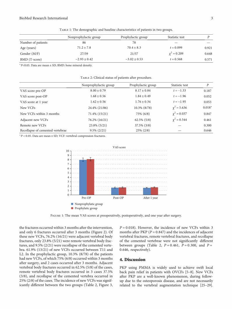
Pain was obviously reduced after the PKP intervention inboth groups. The mean VAS scores decreased dramaticallyfrom a baseline (preoperative) value of approximately 8 to1.5 immediately post-PKP in both groups. After one year,the VAS scores were slightly lower, but no significant differ-ence was found between the two groups (1:62 ± 0:56 vs.1:76 ± 0:34, P = 0:053; Table 2, Figure 1).
In the nonprophylactic group, 59.3% (51/86) of the ini-tially OVCFs occurred between T11 and L2, 24.4% (21/86)of the patients had new VCFs, of which 71.4% (15/21) of
2 BioMed Research International
the fractures occurred within 3 months after the intervention,and only 6 fractures occurred after 3 months (Figure 2). Ofthese new VCFs, 76.2% (16/21) were adjacent vertebral bodyfractures, only 23.8% (5/21) were remote vertebral body frac-tures, and 9.5% (2/21) were recollapse of the cemented verte-bra. 61.9% (13/21) of new VCFs occurred between T11 andL2. In the prophylactic group, 10.3% (8/78) of the patientshad new VCFs, of which 75% (6/8) occurred within 3 monthsafter surgery, and 2 cases occurred after 3 months. Adjacentvertebral body fractures occurred in 62.5% (5/8) of the cases,remote vertebral body fractures occurred in 3 cases 37.5%(3/8), and recollapse of the cemented vertebra occurred in25% (2/8) of the cases. The incidence of new VCFs was signif-icantly different between the two groups (Table 2, Figure 3,
P = 0:018). However, the incidence of new VCFs within 3months after PKP (P = 0:847) and the incidences of adjacentvertebral fractures, remote vertebral fractures, and recollapseof the cemented vertebrae were not significantly differentbetween groups (Table 2, P = 0:461, P = 0:300, and P =0:646, respectively).
4. Discussion
PKP using PMMA is widely used to achieve swift localback pain relief in patients with OVCFs [5–8]. New VCFsafter PKP are a well-known phenomenon, during follow-up due to the osteoporosis disease, and are not necessarilyrelated to the vertebral augmentation technique [25–29].
Table 2: Clinical status of patients after procedure.
Nonprophylactic group Prophylactic group Statistic test P
VAS score pre-OP 8:00 ± 0:79 8:17 ± 0:84 t = −1:33 0.187
VAS score post-OP 1:68 ± 0:56 1:84 ± 0:49 t = −1:96 0.052
VAS score at 1 year 1:62 ± 0:56 1:76 ± 0:34 t = −1:95 0.053
New VCFs 24.4% (21/86) 10.3% (8/78) χ2 = 5:636 0.018∗
New VCFs within 3 months 71.4% (15/21) 75% (6/8) χ2 = 0:037 0.847
Adjacent new VCFs 76.2% (16/21) 62.5% (5/8) χ2 = 0:544 0.461
Remote new VCFs 23.8% (5/21) 37.5% (3/8) — 0.300
Recollapse of cemented vertebrae 9.5% (2/21) 25% (2/8) — 0.646∗P < 0:05. Data are mean ± SD; VCF: vertebral compression fractures.
A�er 1 yearPost-OP
VAS score
Pre-OP0123456789
10
Nonprophylatic groupProphylatic group
Figure 1: The mean VAS scores at preoperatively, postoperatively, and one year after surgery.
Table 1: The demographic and baseline characteristics of patients in two groups.
Nonprophylactic group Prophylactic group Statistic test P
Number of patients 86 78 — —
Age (years) 71:2 ± 7:8 70:4 ± 8:3 t = 0:099 0.921
Gender (M/F) 27/59 21/57 χ2 = 0:209 0.648
BMD (T-score) −2:93 ± 0:42 −3:02 ± 0:53 t = 0:568 0.571∗P<0.05. Data are mean ± SD; BMD: bone mineral density.
3BioMed Research International
(a) (b) (c) (d)
(e) (f) (g) (h)
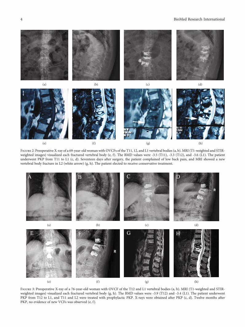
Figure 2: Preoperative X-ray of a 69-year-old woman with OVCFs of the T11, 12, and L1 vertebral bodies (a, b). MRI (T1-weighted and STIR-weighted images) visualized each fractured vertebral body (e, f). The BMD values were -3.5 (T11), -3.3 (T12), and -3.6 (L1). The patientunderwent PKP from T11 to L1 (c, d). Seventeen days after surgery, the patient complained of low back pain, and MRI showed a newvertebral body fracture in L2 (white arrow) (g, h). The patient elected to receive conservative treatment.
(a) (b) (c) (d)
(e) (f) (g) (h)
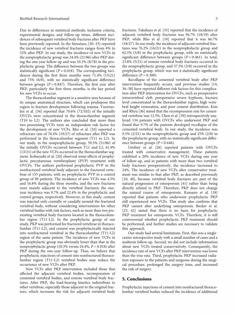
Figure 3: Preoperative X-ray of a 76-year-old woman with OVCF of the T12 and L1 vertebral bodies (a, b). MRI (T1-weighted and STIR-weighted images) visualized each fractured vertebral body (g, h). The BMD values were -3.9 (T12) and -3.4 (L1). The patient underwentPKP from T12 to L1, and T11 and L2 were treated with prophylactic PKP. X-rays were obtained after PKP (c, d). Twelve months afterPKP, no evidence of new VCFs was observed (e, f).
4 BioMed Research International
Due to differences in statistical methods, inclusion criteria,experimental designs, and follow-up times, different inci-dences of subsequent vertebral body fractures after PKP havebeen previously reported. In the literature, [30–33] reportedthe incidence of new vertebral fractures ranges from 8% to52% after PKP. In our study, the incidence of new VCFs inthe nonprophylactic group was 24.4% (21/86) after PKP dur-ing the one-year follow-up and was 10.3% (8/78) in the pro-phylactic group. The difference between the two groups wasstatistically significant (P = 0:018). The corresponding inci-dences during the first three months were 71.4% (15/21)and 75% (6/8), with no statistically significant differencebetween groups (P = 0:847). Therefore, the first year afterPKP, particularly the first three months, is the key periodfor new VCFs to occur.
The thoracolumbar segment is a sensitive area because ofits unique anatomical structure, which can predispose thisregion to fracture development following trauma. Voormo-len et al. [34] reported that 70.6% (72/102) of the initiallyOVCFs were concentrated in the thoracolumbar segment(T10 to L2). The authors also concluded that more thantwo preexisting VCFs were an independent risk factor forthe development of new VCFs. Rho et al. [10] reported arefracture rate of 70.4% (19/27) of refracture after PKP wasperformed in the thoracolumbar segment (T11 to L2). Inour study, in the nonprophylactic group, 59.3% (51/86) ofthe initially OVCFs occurred between T11 and L2, 61.9%(13/21) of the new VCFs occurred in the thoracolumbar seg-ment. Kobayashi et al. [20] observed some effects of prophy-lactic percutaneous vertebroplasty (PVP) treatment withOVCFs. The authors performed prophylactic PVP in thenonfractured vertebral body adjacent to the fractured verte-brae of 155 patients, with no prophylactic PVP in a controlgroup of 89 patients. The incidence of new VCFs was 4.5%and 16.8% during the three months, and the new fractureswere mainly adjacent to the vertebral fractures; the one-year incidence was 9.7% and 22.4% in the prophylactic andcontrol groups, respectively. However, in this study, cementwas injected only cranially or caudally around the fracturedvertebral body, without considering interventions for othervertebral bodies with risk factors, such as more than two pre-existing vertebral body fractures located in the thoracolum-bar region (T11-L2). In the prophylactic group of ourstudy, PKP was performed for fractured vertebrae in thoraco-lumbar (T11-L2), and cement was prophylactically injectedinto nonfractured vertebral in the thoracolumbar (T11-L2)region of the same patient. The incidence of new VCFs inthe prophylactic group was obviously lower than that in thenonprophylactic group (10.3% versus 24.4%, P = 0:018) afterPKP during the one-year follow-up. Thus, we believe thatprophylactic injections of cement into nonfractured thoraco-lumbar region (T11-L2) vertebral bodies may reduce theoccurrence of new VCFs after PKP.
New VCFs after PKP intervention included those thataffected the adjacent vertebral bodies, recompression ofcemented vertebral bodies, and remote vertebral body frac-tures. After PKP, the load-bearing kinetics redistribute toother vertebrae, especially those adjacent to the original frac-ture, which increases the risk for adjacent vertebral body
fractures. Takahara et al. [35] reported that the incidence ofadjacent vertebral body fractures was 94.7% (18/19) afterPKP, while Rho et al. [10] reported that it was 66.7%(18/27). In our study, the incidence of adjacent vertebral frac-tures was 76.2% (16/21) in the nonprophylactic group and62.5% (5/8) in the prophylactic group, with no statisticallysignificant difference between groups (P = 0:461). In total,23.8% (5/21) of remote vertebral body fractures occurred inthe nonprophylactic group, and 37.5% (3/8) occurred in theprophylactic group, which was not a statistically significantdifference (P = 0:300).
Recollapse of the cemented vertebral body after PKPintervention frequently occurs, and previous studies [14,36–38] have reported different risk factors for this complica-tion after PKP intervention for OVCFs, such as preoperativeintravertebral cleft, preoperative severe kyphosis, fracturelevel concentrated in the thoracolumbar region, high verte-bral height restoration, and poor cement distribution. Kimand Rhyu [36] stated that the recompression rate for cemen-ted vertebrae was 12.5%. Chen et al. [39] retrospectively ana-lyzed 134 patients with OVCFs who underwent PKP andfound that 9.7% of the patients developed recollapse of thecemented vertebral body. In our study, the incidence was9.5% (2/21) in the nonprophylactic group and 25% (2/8) inthe prophylactic group, with no statistically significant differ-ence between groups (P = 0:646).
Lindsay et al. [26] reported patients with OVCFstreated with conservative management. These patientsexhibited a 20% incidence of new VCFs during one yearof follow-up, and in patients with more than two vertebralbody fractures preoperatively, the incidence increased to24%. The incidence of new VCFs after conservative treat-ment was similar to that after PKP, as described previously[10, 40], because vertebral body fractures are part of thenatural progression of osteoporosis [41] rather than beingdirectly related to PKP. Therefore, PKP does not changethe natural course of osteoporosis. Kamano et al. [18]reported that patients who underwent prophylactic PKPstill experienced new VCFs. This study also confirms thatPKP cannot alter underlying osteoporosis. Becker et al.[23, 42] stated that there is no basis for prophylacticPKP treatment for osteoporotic VCFs. Therefore, it is stillcontroversial whether prophylactic PKP treatment shouldbe performed, and further studies are necessary to validatethis approach.
Our study had several limitations. First, this was a single-center retrospective study with a small number of cases and amidterm follow-up. Second, we did not include informationabout new VCFs treated conservatively. Consequently, theincidence rate of new VCFs after PKP intervention was lowerthan the true rate. Third, prophylactic PKP increased radia-tion exposure to the patients and surgeons during the surgi-cal procedure, prolonged the surgery time, and increasedthe risk of surgery.
5. Conclusions
Prophylactic injections of cement into nonfractured thoraco-lumbar vertebral bodies reduced the incidence of additional
5BioMed Research International
secondary VCFs after PKP in patients with OVCFs, but therewas no significant difference in pain relief between groups.
Data Availability
The datasets used and/or analyzed during the current studyare available from the corresponding author on reasonablerequest.
Conflicts of Interest
All authors declare that they have no conflict of interests.
Acknowledgments
This study was funded by the Scientific Research ProjectFunds of Health and Family Planning Commission ofSichuan Province (17PJ209) and the Office of Science &Technology and Intellectual Property of Luzhou City,Sichuan Province (2015LSKZ146). We would like to thankthe relevant staff for guidance and assistance and for theirsupport and collaboration in our hospital.
References
[1] M. J. McGirt, S. L. Parker, J.-P. Wolinsky, T. F. Witham,A. Bydon, and Z. L. Gokaslan, “Vertebroplasty and kypho-plasty for the treatment of vertebral compression fractures:an evidenced-based review of the literature,” The Spine Jour-nal, vol. 9, no. 6, pp. 501–508, 2009.
[2] A. T. Chen, D. B. Cohen, and R. L. Skolasky, “Impact of non-operative treatment, vertebroplasty, and kyphoplasty on sur-vival and morbidity after vertebral compression fracture inthe medicare population,” The Journal of Bone and Joint Sur-gery, vol. 95, no. 19, pp. 1729–1736, 2013.
[3] I. Lieberman and M. K. Reinhardt, “Vertebroplasty andkyphoplasty for osteolytic vertebral collapse,” Clinical Ortho-paedics and Related Research, vol. 415, pp. S176–S186, 2003.
[4] I. H. Lieberman, S. Dudeney, M. K. Reinhardt, and G. Bell,“Initial outcome and efficacy of “kyphoplasty” in the treatmentof painful osteoporotic vertebral compression fractures,”Spine, vol. 26, no. 14, pp. 1631–1637, 2001.
[5] S. Boonen, D. A. Wahl, L. Nauroy et al., “for the CSA FractureWorking Group of the International Osteoporosis Founda-tionBalloon kyphoplasty and vertebroplasty in the manage-ment of vertebral compression fractures,” OsteoporosisInternational, vol. 22, no. 12, article 1639, pp. 2915–2934,2011.
[6] I. A. Grafe, K. da Fonseca, J. Hillmeier et al., “Reduction ofpain and fracture incidence after kyphoplasty: 1-year out-comes of a prospective controlled trial of patients with primaryosteoporosis,” Osteoporosis International, vol. 16, no. 12,pp. 2005–2012, 2005.
[7] J. W. Bae, H.-S. Gwak, S. Kim et al., “Percutaneous vertebro-plasty for patients with metastatic compression fractures ofthe thoracolumbar spine: clinical and radiological factorsaffecting functional outcomes,” The Spine Journal, vol. 16,no. 3, pp. 355–364, 2016.
[8] A. Saracen and Z. Kotwica, “Treatment of multiple osteopo-rotic vertebral compression fractures by percutaneous cement
augmentation,” International Orthopaedics, vol. 38, no. 11,pp. 2309–2312, 2014.
[9] A. S. Mudano, J. Bian, J. U. Cope et al., “Vertebroplasty andkyphoplasty are associated with an increased risk of secondaryvertebral compression fractures: a population-based cohortstudy,” Osteoporosis International, vol. 20, no. 5, pp. 819–826, 2009.
[10] Y.-J. Rho, W. J. Choe, and Y. I. Chun, “Risk factors predictingthe new symptomatic vertebral compression fractures afterpercutaneous vertebroplasty or kyphoplasty,” European SpineJournal, vol. 21, no. 5, pp. 905–911, 2012.
[11] B. G. Lee, J.-H. Choi, D.-Y. Kim, W. R. Choi, S. G. Lee, andC. N. Kang, “Risk factors for newly developed osteoporoticvertebral compression fractures following treatment for osteo-porotic vertebral compression fractures,” The Spine Journal,vol. 19, no. 2, pp. 301–305, 2019.
[12] M. J. Nieuwenhuijse, H. Putter, A. R. van Erkel, and P. D. S.Dijkstra, “New vertebral fractures after percutaneous verteb-roplasty for painful osteoporotic vertebral compression frac-tures: a clustered analysis and the relevance of intradiskalcement leakage,” Radiology, vol. 266, no. 3, pp. 862–870,2013.
[13] J.-P. Tourtier and S. Cottez, “Pulmonary cement embolismafter vertebroplasty,” The New England Journal of Medicine,vol. 366, no. 3, pp. 258–258, 2012.
[14] X. Li, X. Lou, X. Lin, and J. Du, “Refracture of osteoporotic ver-tebral body concurrent with cement fragmentation at the pre-viously treated vertebral level after balloon kyphoplasty: a casereport,” Osteoporosis International, vol. 25, no. 5, pp. 1647–1650, 2014.
[15] X. Yi, H. Lu, F. Tian et al., “Recompression in new levels afterpercutaneous vertebroplasty and kyphoplasty compared withconservative treatment,” Archives of Orthopaedic and TraumaSurgery, vol. 134, no. 1, pp. 21–30, 2014.
[16] W. Yu,W. Xu, X. Jiang, D. Liang, andW. Jian, “Risk factors forrecollapse of the augmented vertebrae after percutaneous ver-tebral augmentation: a systematic review and meta-analysis,”World Neurosurgery, vol. 111, pp. 119–129, 2018.
[17] M. Syed, N. Patel, S. Jan, M. S. Harron, K. Morar, andA. Shaikh, “New symptomatic vertebral compression fractureswithin a year following vertebroplasty in osteoporoticwomen,” AJNR American Journal of Neuroradiology, vol. 26,no. 6, pp. 1601–1604, 2005.
[18] H. Kamano, A. Hiwatashi, N. Kobayashi et al., “New vertebralcompression fractures after prophylactic vertebroplasty inosteoporotic patients,” AJR American Journal of Roentgenol-ogy, vol. 197, no. 2, pp. 451–456, 2011.
[19] M. C. Eichler, C. Spross, A. Ewers, R. Mayer, and F. A. Külling,“Prophylactic adjacent-segment vertebroplasty followingkyphoplasty for a single osteoporotic vertebral fracture andthe risk of adjacent fractures: a retrospective study and clinicalexperience,” Journal of Neurosurgery: Spine, vol. 25, no. 4,pp. 528–534, 2016.
[20] N. Kobayashi, Y. Numaguchi, S. Fuwa et al., “Prophylactic ver-tebroplasty: cement injection into non-fractured vertebralbodies during percutaneous vertebroplasty,” Academic Radiol-ogy, vol. 16, no. 2, pp. 136–143, 2009.
[21] J. Langdon, J. Bernard, and S. Molloy, “Prophylactic stabiliza-tion of vertebral body metastasis at risk of imminent fractureusing balloon kyphoplasty,” Spine, vol. 34, no. 13, pp. E469–E472, 2009.
6 BioMed Research International
[22] M. Kurutz, P. Varga, and G. Jakab, “Prophylactic vertebro-plasty versus kyphoplasty in osteoporosis: a comprehensivebiomechanical matched-pair study by in vitro compressivetesting,” Medical Engineering & Physics, vol. 65, pp. 46–56,2019.
[23] S. Becker, M. Garoscio, J. Meissner, A. Tuschel, and M. Ogon,“Is there an indication for prophylactic balloon kyphoplasty?A pilot study,” Clinical Orthopaedics and Related Research,vol. 458, pp. 83–89, 2007.
[24] L. Ning, S. Wan, C. Liu, Z. Huang, H. Cai, and S. Fan, “Newlevels of vertebral compression fractures after percutaneouskyphoplasty: retrospective analysis of styles and risk factors,”Pain Physician, vol. 18, no. 6, pp. 565–572, 2015.
[25] D. F. Kallmes andM. E. Jensen, “Percutaneous vertebroplasty,”Radiology, vol. 229, no. 1, pp. 27–36, 2003.
[26] R. Lindsay, S. Silverman, C. Cooper et al., “Risk of new verte-bral fracture in the year following a fracture,” JAMA,vol. 285, no. 3, pp. 320–323, 2001.
[27] N. Tanigawa, A. Komemushi, S. Kariya, H. Kojima,Y. Shomura, and S. Sawada, “Radiological follow-up of newcompression fractures following percutaneous vertebroplasty,”Cardiovascular and Interventional Radiology, vol. 29, no. 1,pp. 92–96, 2006.
[28] A. A. Uppin, J. A. Hirsch, L. V. Centenera, B. A. Pfiefer, A. G.Pazianos, and I. S. Choi, “Occurrence of new vertebral bodyfracture after percutaneous vertebroplasty in patients withosteoporosis,” Radiology, vol. 226, no. 1, pp. 119–124, 2003.
[29] C. C. Lin, I. H. Chen, T. C. Yu, A. Chen, and P. S. Yen, “Newsymptomatic compression fracture after percutaneous verteb-roplasty at the thoracolumbar junction,” AJNR American Jour-nal of Neuroradiology, vol. 28, no. 6, pp. 1042–1045, 2007.
[30] F. Grados, C. Depriester, G. Cayrolle, N. Hardy, H. Deramond,and P. Fardellone, “Long-term observations of vertebral osteo-porotic fractures treated by percutaneous vertebroplasty,”Rheumatology, vol. 39, no. 12, pp. 1410–1414, 2000.
[31] C. A. H. Klazen, A. Venmans, J. de Vries et al., “Percutaneousvertebroplasty is not a risk factor for new osteoporotic com-pression fractures: results from VERTOS II,” American Jour-nal of Neuroradiology, vol. 31, no. 8, pp. 1447–1450, 2010.
[32] H.-L. Ren, J. Jiang, J. Chen, and J. Wang, “Risk factors of newsymptomatic vertebral compression fractures in osteoporoticpatients undergone percutaneous vertebroplasty,” EuropeanSpine Journal, vol. 24, no. 4, pp. 750–758, 2015.
[33] N. Tanigawa, S. Kariya, A. Komemushi et al., “Percutaneousvertebroplasty for osteoporotic compression fractures: long-term evaluation of the technical and clinical outcomes,” Amer-ican Journal of Roentgenology, vol. 196, no. 6, pp. 1415–1418,2011.
[34] M. H. J. Voormolen, P. N. M. Lohle, J. R. Juttmann, Y. van derGraaf, H. Fransen, and L. E. H. Lampmann, “The risk of newosteoporotic vertebral compression fractures in the year afterpercutaneous vertebroplasty,” Journal of Vascular and Inter-ventional Radiology, vol. 17, no. 1, pp. 71–76, 2006.
[35] K. Takahara, M. Kamimura, H. Moriya et al., “Risk factors ofadjacent vertebral collapse after percutaneous vertebroplastyfor osteoporotic vertebral fracture in postmenopausalwomen,” BMC Musculoskeletal Disorders, vol. 17, no. 1,p. 12, 2016.
[36] Y.-Y. Kim and K.-W. Rhyu, “Recompression of vertebral bodyafter balloon kyphoplasty for osteoporotic vertebral compres-
sion fracture,” European Spine Journal, vol. 19, no. 11,pp. 1907–1912, 2010.
[37] T. Leslie-Mazwi and H. G. Deen, “Repeated fracture of a verte-bral body after treatment with balloon kyphoplasty: case illus-tration,” Journal of Neurosurgery: Spine, vol. 4, no. 3, p. 270,2006.
[38] J. Niu, H. Zhou, Q. Meng, J. Shi, B. Meng, and H. Yang, “Fac-tors affecting recompression of augmented vertebrae after suc-cessful percutaneous balloon kyphoplasty: a retrospectiveanalysis,” Acta Radiologica, vol. 56, no. 11, pp. 1380–1387,2015.
[39] Y.-J. Chen, W.-H. Chen, H.-T. Chen, and H.-C. Hsu, “Repeatneedle insertion in vertebroplasty to prevent re-collapse ofthe treated vertebrae,” European Journal of Radiology,vol. 81, no. 3, pp. 558–561, 2012.
[40] P. D. Delmas, H. K. Genant, G. G. Crans et al., “Severity ofprevalent vertebral fractures and the risk of subsequent verte-bral and nonvertebral fractures: results from the MORE trial,”Bone, vol. 33, no. 4, pp. 522–532, 2003.
[41] G. Ballane, J. A. Cauley, M. M. Luckey, and G. El-Hajj Fulei-han, “Worldwide prevalence and incidence of osteoporoticvertebral fractures,” Osteoporosis International, vol. 28, no. 5,pp. 1531–1542, 2017.
[42] J. Mathis, H. Deramond, and S. Belkoff, “Standards for the per-formance of percutaneous vertebroplasty: American College ofRadiology and Society of Interventional Radiology Guide-lines,” in Percutaneous Vertebroplasty and Kyphoplasty,pp. 223–245, Springer, New York, NY, USA, 2006.
7BioMed Research International