Embed Size (px)
Citation preview
Prophylactic Venous Supercharged RadialCollateral Artery Perforator Propeller Flap:Improved Outcome in Perforator Propeller FlapsDamir Kosutic, MD, PhD, FRCS (Plast)1 Pratap Dutta, MBBS, MS, MRCSEd1
Ingrid Kieran, MD, MRCS, MB, BCh, BAO1
1Department of Plastic Surgery, The Christie NHS Foundation Trust,Manchester, United Kingdom
J Reconstr Microsurg Open 2016;1:45–47.
Address for correspondence Damir Kosutic, MD, PhD, FRCS (Plast),Department of Plastic Surgery, The Christie NHS Foundation Trust,Wilmslow Road, M20 4BX, Manchester, United Kingdom(e-mail: [email protected]).
Propeller flaps provide a safe and reliable reconstructiveoption. Following flap rotation intraoperatively, a degree ofvenous stasis occurs. Difficulty lies in predicting whetherthis is likely to be transient or not. These flaps are thereforefrequently at risk of venous congestion which can result inpartial or total flap necrosis.1 This results in delayedwoundhealing and often necessitates further surgical proceduresto achieve the final outcome. This defeats the originalintention of performing a reliable, one-stage, and aestheti-cally pleasing reconstructive technique. Subsequent man-agement of flap necrosis can include conservative andsurgical management. Long-term dressings and negativepressure therapy, with or without eventual skin grafting,will result in wound healing, but can take time and areincapacitating for patients. The final result, by these means,can potentially negatively impact upper limb function interms of scar contracture and appear less aestheticallypleasing. We present two prophylactically venous super-charged radial collateral artery perforator (RCAP) propellerflaps in the upper limb and suggest this is a more reliableapproach than the standard RCAP flap technique.
Case 1
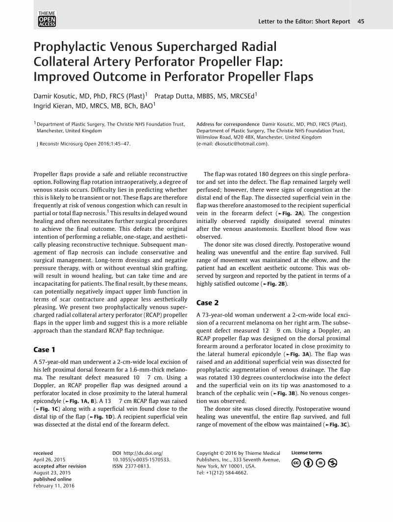
A 57-year-old man underwent a 2-cm-wide local excision ofhis left proximal dorsal forearm for a 1.6-mm-thick melano-ma. The resultant defect measured 10 � 7 cm. Using aDoppler, an RCAP propeller flap was designed around aperforator located in close proximity to the lateral humeralepicondyle (►Fig. 1A, B). A 13 � 7 cm RCAP flap was raised(►Fig. 1C) along with a superficial vein found close to thedistal tip of the flap (►Fig. 1D). A recipient superficial veinwas dissected at the distal end of the forearm defect.
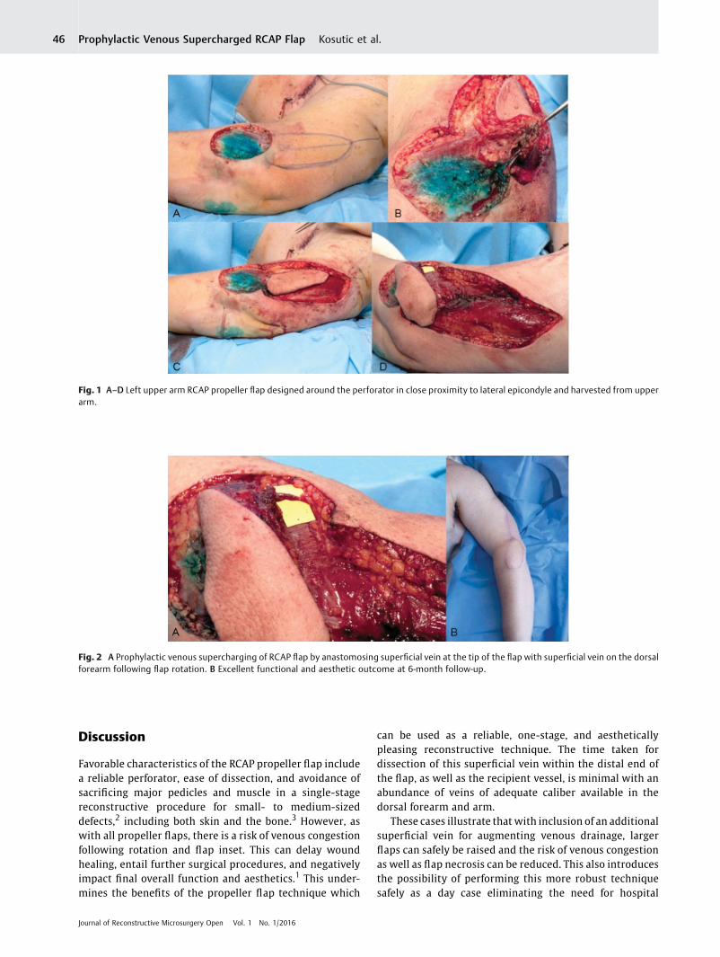
The flap was rotated 180 degrees on this single perfora-tor and set into the defect. The flap remained largely wellperfused; however, there were signs of congestion at thedistal end of the flap. The dissected superficial vein in theflap was therefore anastomosed to the recipient superficialvein in the forearm defect (►Fig. 2A). The congestioninitially observed rapidly dissipated several minutesafter the venous anastomosis. Excellent blood flow wasobserved.
The donor site was closed directly. Postoperative woundhealing was uneventful and the entire flap survived. Fullrange of movement was maintained at the elbow, and thepatient had an excellent aesthetic outcome. This was ob-served by surgeon and reported by the patient in terms of ahighly satisfied outcome (►Fig. 2B).
Case 2
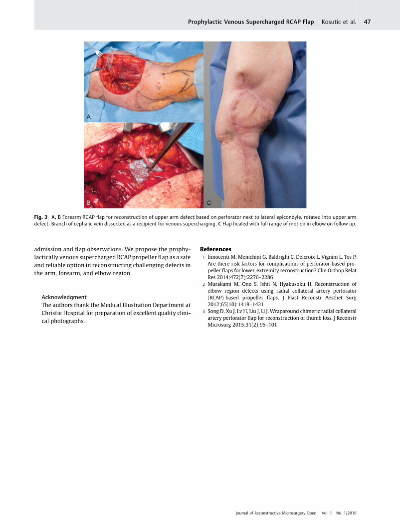
A 73-year-old woman underwent a 2-cm-wide local exci-sion of a recurrent melanoma on her right arm. The subse-quent defect measured 12 � 9 cm. Using a Doppler, anRCAP propeller flap was designed on the dorsal proximalforearm around a perforator located in close proximity tothe lateral humeral epicondyle (►Fig. 3A). The flap wasraised and an additional superficial vein was dissected forprophylactic augmentation of venous drainage. The flapwas rotated 130 degrees counterclockwise into the defectand the superficial vein on its tip was anastomosed to abranch of the cephalic vein (►Fig. 3B). No venous conges-tion was observed.
The donor site was closed directly. Postoperative woundhealing was uneventful, the entire flap survived, and fullrange of movement of the elbow was maintained (►Fig. 3C).
receivedApril 26, 2015accepted after revisionAugust 23, 2015published onlineFebruary 11, 2016
DOI http://dx.doi.org/10.1055/s-0035-1570533.ISSN 2377-0813.
Copyright © 2016 by Thieme MedicalPublishers, Inc., 333 Seventh Avenue,New York, NY 10001, USA.Tel: +1(212) 584-4662.
THIEME
Letter to the Editor: Short Report 45
Discussion
Favorable characteristics of the RCAP propeller flap includea reliable perforator, ease of dissection, and avoidance ofsacrificing major pedicles and muscle in a single-stagereconstructive procedure for small- to medium-sizeddefects,2 including both skin and the bone.3 However, aswith all propeller flaps, there is a risk of venous congestionfollowing rotation and flap inset. This can delay woundhealing, entail further surgical procedures, and negativelyimpact final overall function and aesthetics.1 This under-mines the benefits of the propeller flap technique which
can be used as a reliable, one-stage, and aestheticallypleasing reconstructive technique. The time taken fordissection of this superficial vein within the distal end ofthe flap, as well as the recipient vessel, is minimal with anabundance of veins of adequate caliber available in thedorsal forearm and arm.
These cases illustrate that with inclusion of an additionalsuperficial vein for augmenting venous drainage, largerflaps can safely be raised and the risk of venous congestionas well as flap necrosis can be reduced. This also introducesthe possibility of performing this more robust techniquesafely as a day case eliminating the need for hospital
Fig. 1 A–D Left upper arm RCAP propeller flap designed around the perforator in close proximity to lateral epicondyle and harvested from upperarm.
Fig. 2 A Prophylactic venous supercharging of RCAP flap by anastomosing superficial vein at the tip of the flap with superficial vein on the dorsalforearm following flap rotation. B Excellent functional and aesthetic outcome at 6-month follow-up.
Journal of Reconstructive Microsurgery Open Vol. 1 No. 1/2016
Prophylactic Venous Supercharged RCAP Flap Kosutic et al.46
admission and flap observations. We propose the prophy-lactically venous supercharged RCAP propeller flap as a safeand reliable option in reconstructing challenging defects inthe arm, forearm, and elbow region.
AcknowledgmentThe authors thank the Medical Illustration Department atChristie Hospital for preparation of excellent quality clini-cal photographs.
References1 Innocenti M, Menichini G, Baldrighi C, Delcroix L, Vignini L, Tos P.
Are there risk factors for complications of perforator-based pro-peller flaps for lower-extremity reconstruction? Clin Orthop RelatRes 2014;472(7):2276–2286
2 Murakami M, Ono S, Ishii N, Hyakusoku H. Reconstruction ofelbow region defects using radial collateral artery perforator(RCAP)-based propeller flaps. J Plast Reconstr Aesthet Surg2012;65(10):1418–1421
3 Song D, Xu J, Lv H, Liu J, Li J. Wraparound chimeric radial collateralartery perforator flap for reconstruction of thumb loss. J ReconstrMicrosurg 2015;31(2):95–101
Fig. 3 A, B Forearm RCAP flap for reconstruction of upper arm defect based on perforator next to lateral epicondyle, rotated into upper armdefect. Branch of cephalic vein dissected as a recipient for venous supercharging. C Flap healed with full range of motion in elbow on follow-up.
Journal of Reconstructive Microsurgery Open Vol. 1 No. 1/2016
Prophylactic Venous Supercharged RCAP Flap Kosutic et al. 47