Embed Size (px)
Citation preview
Proposal Form
STRATEGIC INDO-SWEDISH COOPERATIVE
PROGRAMME ON “HEALTH AND DISEASE PREVENTION”
A. PROJECT
1. Title of Project
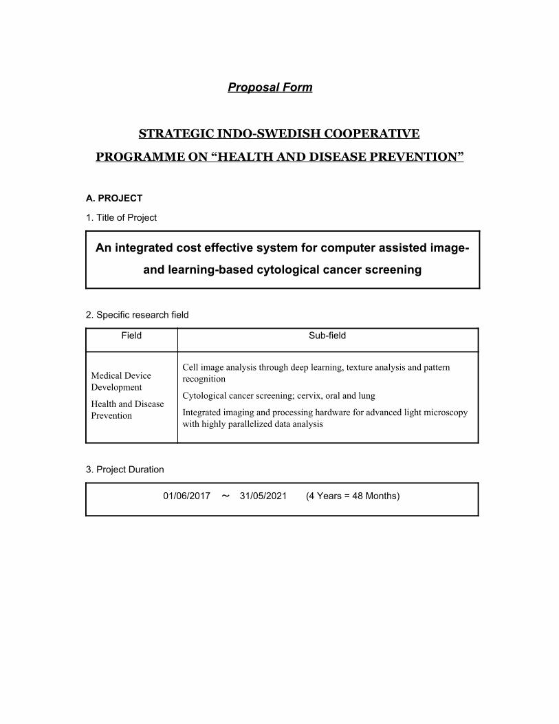
An integrated cost effective system for computer assisted image-
and learning-based cytological cancer screening
2. Specific research field
Field Sub-field
Medical Device Development
Health and Disease Prevention
Cell image analysis through deep learning, texture analysis and pattern recognition
Cytological cancer screening; cervix, oral and lung
Integrated imaging and processing hardware for advanced light microscopy with highly parallelized data analysis
3. Project Duration
01/06/2017 31/05/2021 (4 Years = 48 Months)
2
4. Summary of Project
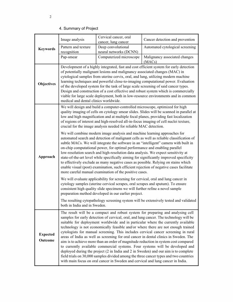
Keywords
Image analysis Cervical cancer, oral cancer, lung cancer
Cancer detection and prevention
Pattern and texture recognition
Deep convolutional neural networks (DCNN)
Automated cytological screening
Pap-smear Computerized microscope Malignancy associated changes (MACs)
Objectives
Development of a highly integrated, fast and cost efficient system for early detection of potentially malignant lesions and malignancy associated changes (MAC) in cytological samples from uterine cervix, oral, and lung, utilizing modern machine learning techniques and powerful close-to-imaging computational power. Evaluation of the developed system for the task of large scale screening of said cancer types. Design and construction of a cost effective and robust system which is commercially viable for large scale deployment, both in low-resource environments and in common medical and dental clinics worldwide.
Approach
We will design and build a computer-controlled microscope, optimized for high quality imaging of cells on cytology smear slides. Slides will be scanned in parallel at low and high magnification and at multiple focal planes, providing fast localization of regions of interest and high-resolved all-in-focus imaging of cell nuclei texture, crucial for the image analysis needed for reliable MAC detection.
We will combine modern image analysis and machine learning approaches for automated search and detection of malignant cells as well as reliable classification of subtle MACs. We will integrate the software in an “intelligent” camera with built in on-chip computational power, for optimal performance and enabling parallel low-resolution search and high-resolution data analysis. We expect sensitivity at state-of-the-art level while specifically aiming for significantly improved specificity to effectively exclude as many negative cases as possible. Relying on stains which enable visual (post) examination, such efficient rejection of negative cases facilitate more careful manual examination of the positive cases.
We will evaluate applicability for screening for cervical, oral and lung cancer in cytology samples (uterine cervical scrapes, oral scrapes and sputum). To ensure consistent high quality slide specimens we will further refine a novel sample preparation method developed in our earlier project.
The resulting cytopathology screening system will be extensively tested and validated both in India and in Sweden.
Expected
Outcome
The result will be a compact and robust system for preparing and analysing cell samples for early detection of cervical, oral, and lung cancer. The technology will be suitable for deployment worldwide and in particular where the currently available technology is not economically feasible and/or where there are not enough trained cytologists for manual screening. This includes cervical cancer screening in rural areas of India as well as screening for oral cancer in dental clinics in Sweden. The aim is to achieve more than an order of magnitude reduction in system cost compared to currently available commercial systems. Four systems will be developed and deployed during the project (2 in India and 2 in Sweden) and our aim is to complete field trials on 30,000 samples divided among the three cancer types and two countries with main focus on oral cancer in Sweden and cervical and lung cancer in India.
3
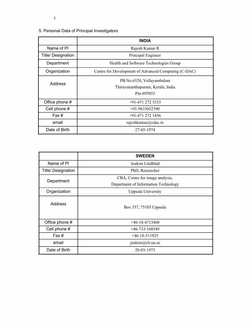
5. Personal Data of Principal Investigators
INDIA
Name of PI Rajesh Kumar R
Title/ Designation Principal Engineer
Department Health and Software Technologies Group
Organization Centre for Development of Advanced Computing (C-DAC)
Address
PB.No.6520, Vellayambalam
Thiruvananthapuram, Kerala, India
Pin-695033
Office phone # +91-471 272 3333
Cell phone # +91-9633032390
Fax # +91-471 272 3456
email [email protected]
Date of Birth 27-05-1974
SWEDEN
Name of PI Joakim Lindblad
Title/ Designation PhD, Researcher
Department CBA, Centre for image analysis,
Department of Information Technology
Organization Uppsala University
Address
Box 337, 75105 Uppsala
Office phone # +46-18-4713460
Cell phone # +46-733-168549
Fax # +46-18-511925
email [email protected]
Date of Birth 20-03-1973
4
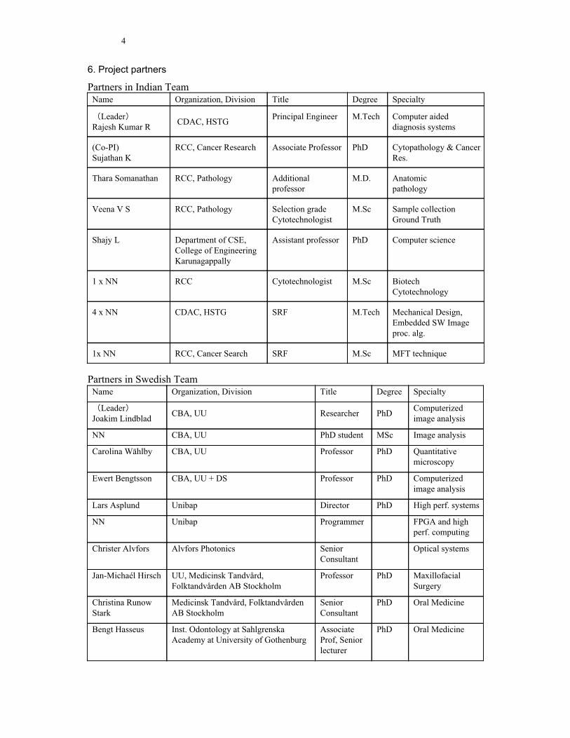
6. Project partners
Partners in Indian Team Name Organization, Division Title Degree Specialty
Leader Rajesh Kumar R
CDAC, HSTG Principal Engineer M.Tech
Computer aided diagnosis systems
(Co-PI) Sujathan K
RCC, Cancer Research Associate Professor PhD Cytopathology & Cancer Res.
Thara Somanathan RCC, Pathology Additional professor
M.D. Anatomic pathology
Veena V S RCC, Pathology Selection grade Cytotechnologist
M.Sc Sample collection Ground Truth
Shajy L Department of CSE, College of Engineering Karunagappally
Assistant professor PhD Computer science
1 x NN RCC Cytotechnologist M.Sc Biotech Cytotechnology
4 x NN CDAC, HSTG SRF M.Tech Mechanical Design, Embedded SW Image proc. alg.
1x NN RCC, Cancer Search SRF M.Sc MFT technique
Partners in Swedish Team Name Organization, Division Title Degree Specialty
Leader Joakim Lindblad
CBA, UU Researcher PhD Computerized image analysis
NN CBA, UU PhD student MSc Image analysis
Carolina Wählby CBA, UU Professor PhD Quantitative microscopy
Ewert Bengtsson CBA, UU + DS Professor PhD Computerized image analysis
Lars Asplund Unibap Director PhD High perf. systems
NN Unibap Programmer FPGA and high perf. computing
Christer Alvfors Alvfors Photonics Senior Consultant
Optical systems
Jan-Michaél Hirsch UU, Medicinsk Tandvård, Folktandvården AB Stockholm
Professor PhD Maxillofacial Surgery
Christina Runow Stark
Medicinsk Tandvård, Folktandvården AB Stockholm
Senior Consultant
PhD Oral Medicine
Bengt Hasseus Inst. Odontology at Sahlgrenska Academy at University of Gothenburg
Associate Prof, Senior lecturer
PhD Oral Medicine
5
B. TECHNICAL INFORMATION
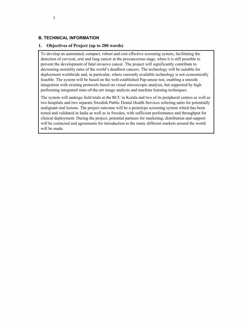
1. Objectives of Project (up to 200 words)
To develop an automated, compact, robust and cost effective screening system, facilitating the detection of cervical, oral and lung cancer at the precancerous stage, when it is still possible to prevent the development of fatal invasive cancer. The project will significantly contribute to decreasing mortality rates of the world’s deadliest cancers. The technology will be suitable for deployment worldwide and, in particular, where currently available technology is not economically feasible. The system will be based on the well-established Pap-smear test, enabling a smooth integration with existing protocols based on visual microscopic analysis, but supported by high performing integrated state-of-the-art image analysis and machine learning techniques.
The system will undergo field trials at the RCC in Kerala and two of its peripheral centres as well as two hospitals and two separate Swedish Public Dental Health Services referring units for potentially malignant oral lesions. The project outcome will be a prototype screening system which has been tested and validated in India as well as in Sweden, with sufficient performance and throughput for clinical deployment. During the project, potential partners for marketing, distribution and support will be contacted and agreements for introduction to the many different markets around the world will be made.
6
2. Justification for collaboration, including background to the proposed project, each
partner’s expertise and specific contribution to the project, and the added value of
the proposed collaboration (up to 400 words)
This is the continuation and extension of a fruitful collaboration between the Indian and Swedish teams. Through several projects since 2009, financed partly from Vinnova, VR and SRL (SIDA/VR) in Sweden and the MeitY in India, we have jointly developed a prototype system for cervical cancer screening which has been tested on more than 1100 patient samples at RCC, in Kerala, India. It has met and exceeded design specifications in terms of sensitivity and specificity but needs around 45 minutes of operator supported scanning time per slide. We now plan to create a system with sufficient throughput based on a dedicated digital microscope unit with optimized imaging and image analysis software and hardware, and to extend the function to screen also for oral and lung cancer. To do this we need to join forces between complementary expertise in Sweden and India.
The Swedish team has been active in research on cervical and oral cancer screening for 40 years and has expert knowledge of state-of-the-art in the field. The Centre for Image Analysis (CBA) is a leading research centre in medical image analysis, but has limited capacity in building integrated HW and SW systems. Thus, the collaboration with CDAC on the Indian side and Unibap in Sweden is vital.
Unibap is a company designing and manufacturing vision systems with high performance imaging capability and built in computational resources utilizing a heterogeneous architecture. Primary application areas are industrial automation and robotics. Unibap was one of the winners of 33-listan in 2016 and is also an ESA BIC (Business incubation centre).
CDAC focuses on applied research in electronics and IT and has extensive experience in designing and deploying computerized systems for all kinds of applications, including medical. CDAC also has an in-house pilot production facility. The Indian PI is currently responsible for leading medical imaging and medical electronics projects at CDAC.
RCC is a premier cancer care hospital with extensive experience in screening for cervical and oral cancer. They will provide the domain expertise and ground truth and has also develop an improved specimen preparation technique which will be used in this project.
The Swedish odontological expertise have extensive clinical experience in diagnosis and handling of malignant and premalignant oral lesions. They have jointly published approximately 200 articles and supervised a number of projects and PhD students in the relevant field. Their inclusion ensures access to oral samples and level medical expertise.
7
3. Descriptions of the Cooperative research project
Background
The need for cell based cancer screening
A most effective ways of decreasing cancer mortality is to detect the cancer early, before it becomes invasive and metastasizing. The Pap-smear (Papanicolaou test) has been very successful in reducing cervical cancer mortality in the western world, where it is used for population screening. But the visual microscopic screening requires highly trained cytotechnologists, is time consuming and expensive. Most women in the world are therefore still not offered screening, causing around 250 000 women to die annually from cervical cancer, of those around 72 000 in India [ http://www.iarc.fr/ ]. Another common and rapidly increasing cancer worldwide is oral cancer (no. 6 in overall incidence (Jemal A. Cancer J Clin 2010; 60:277–300) and no. 1 among men in India (Brocklehurst P. Cochrane Database of Systematic Reviews, July 2013) causing more than 130 000 annual deaths; the most important determinant factor in cancer survival is early
diagnosis (Onizawa K. Oral Oncol. 2003; 39(8):781-8). The most common oral cancers are very similar to cervical cancers and cell samples can be obtained in a similar way through brushing the relevant epithelial surfaces (Sankaranarayanan R. Oral Oncol 2013; 49:314–21). No large screening programs are yet in place for oral cancer, but it is very likely that a screening system for cervical cancer can be adapted for oral cancer.
Lung cancer is the most common and deadliest of all cancers worldwide. There are currently no effective ways of screening for lung cancer. But cells from the lungs can be found in sputum and through improved specimen preparation procedures (Shajy L, Int. J Appl. Eng. Res. 2015;Vol.10, No.70, 217-225) there are good possibilities that cell image analysis methods will be applicable also for screening high risk groups (e.g. smokers) against lung cancer.
Automation for cell based cancer screening
The fact that visual microscopic screening of slide specimen for early detection of cervical cancer is very laborious and time consuming has led to efforts to automate the screening through computerized image analysis. Since the start of this millennium commercial computer-assisted screening (CAS) systems exists. Those systems are, however, very expensive to purchase and maintain. (Bengtsson E, Comp. Math. Meth. Med., 2014: 12p.). Thus, most screening is still done visually, limiting the deployment to richer parts of the world.
The group in Uppsala has been involved in research in this field since 1973, producing a few hundred scientific papers and more than a dozen PhD theses. Since 2009 the group at CBA has been collaborating with C-DAC(T) and RCC, in Kerala, India, to develop an image analysis system for Pap-stained cell samples from cervical cancer screening, so that early malignancy associated changes and atypical cells can be detected and used as a basis for a new kind of computer assisted screening device; one that is suitable for wide deployment in developing countries like India.
8
A first research prototype system has been built and used to analyze over 1100 slides, yielding a sensitivity of 80% (93% for the more serious HSIL lesions) and specificity of 60% (Deepak R U, J. Cytol. Histol., 2015, 6p.). These results indicate that a clinically viable system can be built based on our approach. The current system has not been optimized for speed and at present needs around 45 minutes to scan and analyze around 10% of the specimen area. For a final product faster image acquisition and processing is a requirement.
Our work so far has been focused on cervical cancer but we see strong possibilities of extending it to oral cancer, using basically the same sample collection and preparation techniques. By a modified sample collection and preparation technique the image analysis system should also be applicable for lung cancer screening. We thus see the possibility of creating a novel cost effective screening system addressing three of the most common
and deadly cancers worldwide with particular potential for deployment in developing countries.
Impact
Our previous work on cervical cancer demonstrate that our approach can yield sufficient accuracy. In the proposed project we will develop an optimized, tightly integrated scanning and image analysis hardware/software system, designed to be cost effective both to produce, but even more importantly, to maintain in low resource environments. We further propose to extend the application area to oral and lung cancer, two common cancers of major and growing concern worldwide, not the least in developing countries like India.
We will develop an automated, compact, robust and cost effective screening system, facilitating the detection of cervical, oral and lung cancer at the precancerous stage, when it is still possible to prevent the development of fatal invasive cancer. The project will significantly contribute to decreasing mortality rates of the world’s deadliest cancers. The technology will be suitable for deployment worldwide and, in particular, where currently available technology is not economically feasible. The system will be based on the well-established Pap-smear test, enabling a smooth integration with existing protocols based on visual microscopic analysis, but with improved specimen preparation techniques and supported by high performing well integrated state-of-the-art image analysis and machine learning techniques.
The system will undergo field trials at the RCC in Kerala and two of its peripheral centers as well as two hospitals and two separate Public Dental Health Services referring units for potentially malignant oral lesions in Sweden. The project outcome will be a prototype screening system which has been tested and validated in India as well as in Sweden, with sufficient performance and throughput for clinical deployment.
In late 2016, the Govt of India has issued an operational framework making screening for cervical, breast and oral cancers mandatory for eligible persons above 30 years age, which is around 400 million people. Doing that in conventional screening method will be extremely laborious and time consuming. Our solution, which is scientific and based on a proven method of cytological examination, is applicable to cervical and oral cancer screening. It will help to augment the objective, by screening out the majority of negative cases. If we are successful in adapting the system also for sputum based lung cancer screening there are additional very large high risk groups that would benefit from such screening.
9
In Sweden the system could be used to decrease cost and increase efficiency of the cervical cancer screening program. But its strong potential is to become instrumental in introducing a new screening programs for oral cancer at Swedish dental clinics which currently have no cytological expertise available.
The potential market is of course not limited to the two participating countries. Globally there are more than 2 billon women in the age group where cervical cancer screening is relevant. Ideally they should be screened every 2-3 years. Similarly there are great needs to introduce screening for oral cancer. Even if we focus on high risk groups, such as those with excessive consumption of tobacco and those with existing potentially malignant lesions there are several hundred million cases a year that would benefit from screening. Lung cancer could offer even greater needs and potential. Given that our system could process around 20.000 samples per year at least 100,000 systems would be needed. They would in full deployment world-wide potentially prevent a million deaths per year.
In economic terms the current screening costs are in India around Rs.200–1000 (25–125 SEK) per case including consumables. The current costs in Sweden and other western countries are significantly higher but the Indian cost levels are more relevant on a global scale. Estimating a cost for the screening work exclusive of consumables to be Rs.400, the global annual labor cost for screening would then be around Rs.80.000 Crore or 100 billion SEK. In most parts of the world, screening programs are the responsibility of the government and heavily subsidized. The proposed system could relieve the governments of financial burden while still bringing a larger population under the screening programs.
Developing a functioning system does of course not automatically imply that we can reach this huge market. We will during the project make large efforts to find collaboration partners to facilitate large scale deployment. And it is clear that the potential impact is enormous.
Implementation
Our existing collaboration has resulted in a prototype system with sufficient sensitivity and specificity for a clinically acceptable cervical screening system (Deepak R U, J. Cytol. Histol., 2015, 6p.). However, the present system is too slow and difficult to use for large scale deployment. The proposed project will elevate the system throughput to a level suitable for large scale screening and deliver a highly integrated, easy to use versatile analysis unit also adapted to screening for other cancers. To achieve this we will development and implement improvements on several levels:
Specimen Preparation
The conventional way of preparing cell samples, the Pap smears, are typically large, non-uniform, cluttered specimens containing lots of blood cells and other artefacts that obscure the epithelial cells. Liquid based preparations are much cleaner and more uniform, but the preparation technique is very expensive. The RCC has devised a low cost mono-layering technique (during the past collaborative project) called the Mega Funnel Technique that is simple to use and low cost. The technique will be further developed and validated in our project.
Scanning platform
A cell sample for screening for cancer is deposited on a standard glass slide of 2.5cm × 7.5cm with cells spread over at least 10 sq.cm. With the mega funnel technique the cell sample area
10
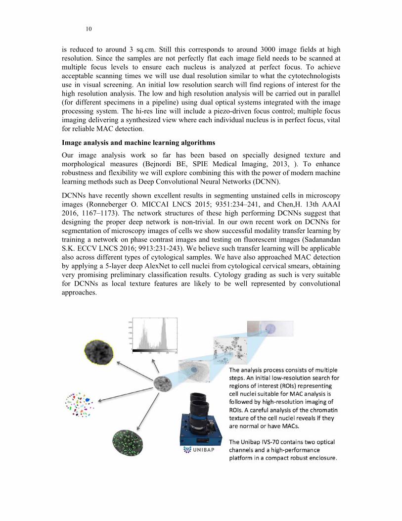
is reduced to around 3 sq.cm. Still this corresponds to around 3000 image fields at high resolution. Since the samples are not perfectly flat each image field needs to be scanned at multiple focus levels to ensure each nucleus is analyzed at perfect focus. To achieve acceptable scanning times we will use dual resolution similar to what the cytotechnologists use in visual screening. An initial low resolution search will find regions of interest for the high resolution analysis. The low and high resolution analysis will be carried out in parallel (for different specimens in a pipeline) using dual optical systems integrated with the image processing system. The hi-res line will include a piezo-driven focus control; multiple focus imaging delivering a synthesized view where each individual nucleus is in perfect focus, vital for reliable MAC detection.
Image analysis and machine learning algorithms
Our image analysis work so far has been based on specially designed texture and morphological measures (Bejnordi BE, SPIE Medical Imaging, 2013, ). To enhance robustness and flexibility we will explore combining this with the power of modern machine learning methods such as Deep Convolutional Neural Networks (DCNN).
DCNNs have recently shown excellent results in segmenting unstained cells in microscopy images (Ronneberger O. MICCAI LNCS 2015; 9351:234–241, and Chen,H. 13th AAAI 2016, 1167–1173). The network structures of these high performing DCNNs suggest that designing the proper deep network is non-trivial. In our own recent work on DCNNs for segmentation of microscopy images of cells we show successful modality transfer learning by training a network on phase contrast images and testing on fluorescent images (Sadanandan S.K. ECCV LNCS 2016; 9913:231-243). We believe such transfer learning will be applicable also across different types of cytological samples. We have also approached MAC detection by applying a 5-layer deep AlexNet to cell nuclei from cytological cervical smears, obtaining very promising preliminary classification results. Cytology grading as such is very suitable for DCNNs as local texture features are likely to be well represented by convolutional approaches.
11
Image processing hardware
We will exploit the unique power of the IVS-70 platform developed by Unibap AB, to provide a highly integrated unit which is easy to distribute and maintain, also in low-resource environments. The IVS-70 platform offers high performance computational power integrated on-chip with a dual imaging pipeline. The dual 5Mpixel sensors (5 um pixel size) with global shutter are read out at 50 fps (can be increased to 250 fps) and the first stages of calculations are performed in an FPGA, and higher up in the hierarchy both GPUs and CPUs are used. The compute system is TRL 9 according to NASA (four systems has been fully working in space for 8 months). This architecture enables high resolution processing at high speed, not impaired by otherwise limiting data transmission rates. The hardware design is well suited for distributing the different steps of the processing pipeline onto the most appropriate processing units: close to image filtering steps, such as the initial convolutional layers of a DCNN, or neighbourhood lookup for texture measures such as α-LBPs, can be processed in parallel at high speed utilizing the on-chip FPGA unit (3 tflops); following initial data reduction the high performance GPU offers flexible and general parallel processing in standardized environments such as CUDA and OpenCL; whereas high-level processing and scheduling is well supported by the N-core CPU.
Actor constellations
As mentioned in the background section the CBA has a very long background in developing cell image analysis systems and has been working together with CDAC(T) and RCC since 2009 developing a system for cervical cytology screening. We are now extending this work to other major cancer types and at the same time reimplementing and extending the software to use high performance hardware and deep convolutional neural networks. To be able to do this we are extending our project team to include experts on oral cancer: Jan-Michaél Hirsch, Christina Runow Stark and Bengt Hasseus. Dr Sujathan, who has been part of the existing work on cervical cancer, is also highly qualified in research on lung cancer. Dr. Thara Somanathan is an experienced cytopathologist and will provide samples and ground truth necessary for DCNN training. On the technical side we will work together with Unibap who are producing high performance, robust, cost effective hardware for parallel image acquisition and analysis. Optical system design will be supported by Christer Alvfors.
Rajesh Kumar from CDAC heads multiple projects on computer aided diagnosis and is well experienced in hardware-software integration and complete product development lifecycle. He also has good industry background in the development of test automation systems. On the Indian side CDAC and RCC will focus mainly on Cervix and lung cancer while the Swedish team will focus on oral cancer. The development of software algorithms on both sides will be on the common platform provided by Unibab. CDAC has inhouse capability for mechanical engineering and pilot production of the complete prototype.
12
Handling of Intellectual Property
An agreement will be entered into between the Swedish and Indian teams on the formal arrangement for sharing Intellectual Property Rights and Commercial rights. As the national policies on IPR also varies between the two countries, the general principle agreed upon is as follows:
1. the Swedish teams will share the IP and commercial rights for the whole of Europe 2. the India teams will share the IP and commercial rights for India between themselves
and DBT as per DBT guidelines 3. For the rest of the world, both side will have independent rights to market and sell the
product, the details will be worked out during the first months of the project. 4. Unibab will take the lead in commercialization of the technology in Sweden and rest of
Europe 5. C-DAC has pilot production capability and will produce the first few units inhouse.
Parallely will explore transfer of technology for production and commercialization in accordance with DBT guidelines.
13
4. Plan for the execution of the Cooperative Project
At the start of our project we have a jointly developed system for detecting early pre- malignant changes and malignancies for cervical cancer which has been tested on more than 1100 patients and found to work well, although far too slowly. We will develop that into a new-generation system ready for widespread deployment in five significant ways, which form the sub-goals of our project and thus our milestones, numbered as in the following list:
1. Improved image analysis
a. Adding the recently highly successful deep convolutional neural network approach to cell segmentation and classification to our previously developed set of feature extraction and classification routines. 2. Increased throughput
a. Re-implementing the image acquisition and image analysis in a tightly integrated hardware with FPGA and GPU support for parallel computing of the low level, heavy computational levels. b. Introduction of a low resolution mapping stage which covers the whole specimen in less than 100 image fields and finds where cell material of interest is available for high resolution analysis. c. Integrating the low and high resolution imaging paths on parallel optical channels in the same image analysis hardware system physically linked by a high performance and robust computer control of x, y and z movement of the specimen. 3. Application to new cancer types
a. Develop screening for lung cancer based on sputum cytology based on cell material collected mainly in India. b. Adapting our screening approach to oral cancer based on material collected mainly in Sweden 4. Running field studies of the final system on a total of 30 000 cases
a. cervical cancer samples from several screening units in India b. samples from oral lesions collected at three different dental care clinics in Sweden and in India c. sputum samples collected from high risk groups in India 5. Development of marketing and support strategies
a. Explore suitable collaboration partners for the different markets where our system could have great international impact
Deliverables
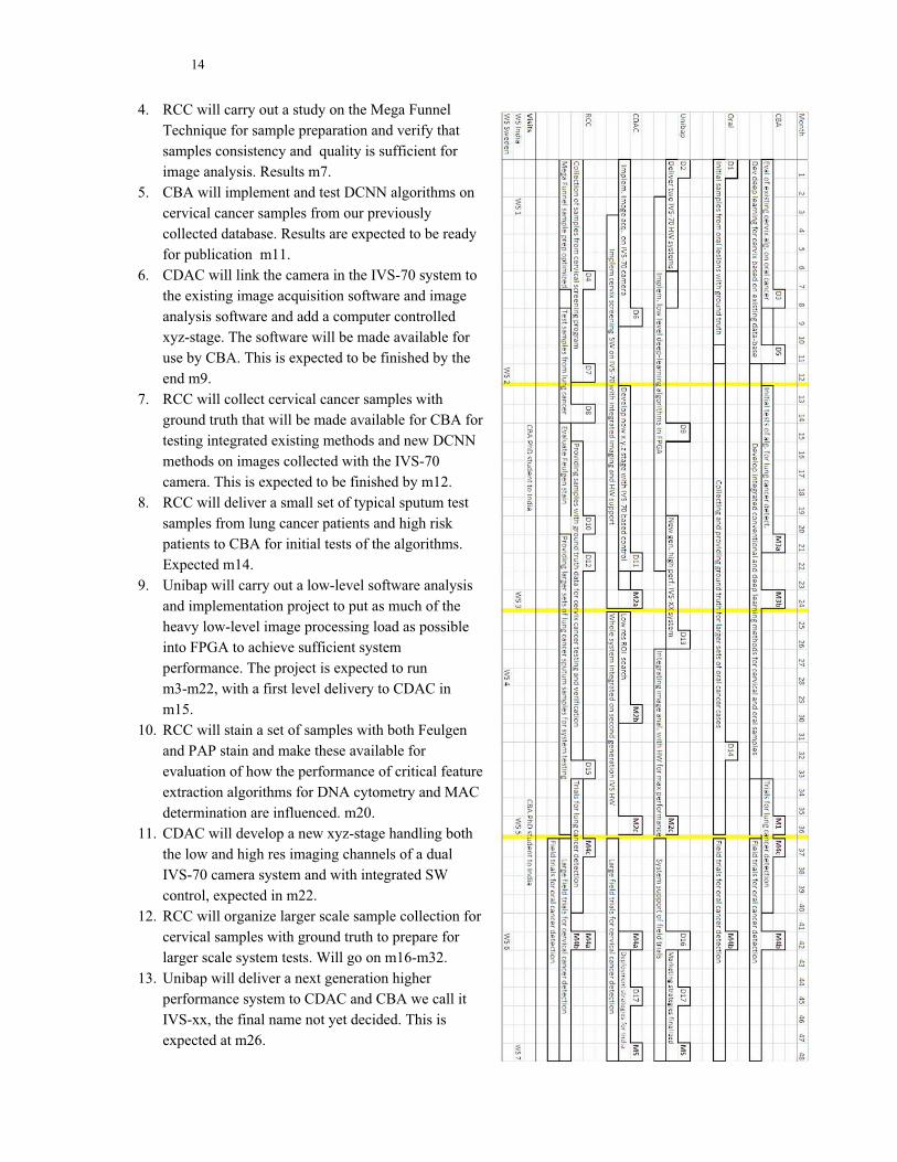
In addition to the Milestones, which mark major achievements in our project, there are a number of deliverables, or internal checkpoints, at which a component of importance for the progress of the project has been completed by one of the participating groups and made available to the other groups that need it. The chart below gives a graphical representation of the activities, milestones and deliverables in the project. The different activities are marked for the main responsible partner. Several activities will also involve work and support from other partners. The dependencies among the deliverables and activities are somewhat implicit in the chart and therefore described here in more detail, with expected delivery at month m.
1. A small set of microscope slides with cells from normal and malignant oral lesions will be made available for CBA by the participating oral/dental clinics. Expected at m1.
2. Unibap will deliver two standard IVS-70 systems, each with a single monochrome camera for mounting on existing microscopes at CBA and CDAC. Expected at m1.
3. CBA will use images of cells from D1 to evaluate if current algorithms developed for cervical cancer also work for oral cancer. Results are expected at m8.
14
4. RCC will carry out a study on the Mega Funnel Technique for sample preparation and verify that samples consistency and quality is sufficient for image analysis. Results m7.
5. CBA will implement and test DCNN algorithms on cervical cancer samples from our previously collected database. Results are expected to be ready for publication m11.
6. CDAC will link the camera in the IVS-70 system to the existing image acquisition software and image analysis software and add a computer controlled xyz-stage. The software will be made available for use by CBA. This is expected to be finished by the end m9.
7. RCC will collect cervical cancer samples with ground truth that will be made available for CBA for testing integrated existing methods and new DCNN methods on images collected with the IVS-70 camera. This is expected to be finished by m12.
8. RCC will deliver a small set of typical sputum test samples from lung cancer patients and high risk patients to CBA for initial tests of the algorithms. Expected m14.
9. Unibap will carry out a low-level software analysis and implementation project to put as much of the heavy low-level image processing load as possible into FPGA to achieve sufficient system performance. The project is expected to run m3-m22, with a first level delivery to CDAC in m15.
10. RCC will stain a set of samples with both Feulgen and PAP stain and make these available for evaluation of how the performance of critical feature extraction algorithms for DNA cytometry and MAC determination are influenced. m20.
11. CDAC will develop a new xyz-stage handling both the low and high res imaging channels of a dual IVS-70 camera system and with integrated SW control, expected in m22.
12. RCC will organize larger scale sample collection for cervical samples with ground truth to prepare for larger scale system tests. Will go on m16-m32.
13. Unibap will deliver a next generation higher performance system to CDAC and CBA we call it IVS-xx, the final name not yet decided. This is expected at m26.
15
14. The oral/dental clinics will deliver at least 100 cases of samples from oral lesions to CBA to be used for development of an optimized screening system for that application in preparation for field trials. Data collection will go on m12-m36.
15. RCC will collect a larger set of lung cancer sputum samples for delivery to CBA for a trial of a system for lung cancer screening. The data collection will go on m21-m33.
16. During the field trials Unibap will be available with support if any system errors occur in the HW and SW developed by Unibap. m37-m48.
17. Form a market consortium for commercialization, coordinated by Unibap in Sweden and C-DAC in India, m45.
After the project period we will have a fully functional and extensively tested prototype system that has identical or better performance to existing commercial systems, can be supplied at significantly lower cost with sufficient throughput to be cost effective, and designed to minimize the need for specialized technical maintenance and support and thus facilitate deployment worldwide.
During the project, advanced image analysis and machine learning algorithms will be made available through several high-class conference and journal publications. We will also create a repository of digitized image data of cervical, oral and sputum smears which will be useful for further research and trainings.
At the bottom of the graphical chart we have also marked the preliminary plan for joint workshops. At each workshop we expect on average 4 persons to travel from one country to the other. The duration is planned to be 5-7 days each time. We have experience from arranging this kind of workshops from our collaboration in the previous project and find it a useful part of the project work. The plan for the workshops are as follows: 1. In, India m3. Project kick off, for the new partners of the project to get to know each other and for
the initial project plan to be made more detailed. Will also include a training on Unibap platform SDK to C-DAC engineers
2. In Sweden, m12. When deliverables 6 and 7 have occurred, representatives from CDAC and RCC will come to Uppsala to discuss the experiences of using the new camera system.
3. In India, m24. When milestone M2a has been reached, the new integrated screening system will be demonstrated. CBA will present results relating to M3a and M3b.
4. In Sweden m28, when deliverable 13 is reached Unibap will present the new hardware generation in detail to system experts from CDAC.
5. In India, m36. When milestones M2b and M2c have been reached we have a fully integrated operational system in India and a Swedish delegation will go there to study its function in preparation for installing a copy in Sweden.
6. In Sweden, m42. When the field trials are in full progress we will have a workshop in Sweden to discuss the success of the project so far and our efforts at finding partners for marketing and distributing the system in relevant regions of the world.
7. In India, m48. At the end of the project we will have a final workshop in India to conclude and to have contacts with partners interested in distributing the system.
In addition to the workshops we are planning for two longer visits to India for the Swedish PhD student to be recruited to the project. The first one will be roughly from m16-m18 to learn how to use the developed first generation prototype and also to understand lung cytology samples as prepared at RCC. The second visit will be around m36-m38 as part of the field trial on lung cancer.
16
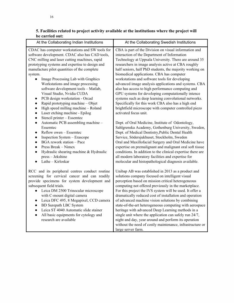
5. Facilities related to project activity available at the institutions where the project will
be carried out:
At the Collaborating Indian Institutions At the Collaborating Swedish Institutions
CDAC has computer workstations and SW tools for software development. CDAC also has CAD tools, CNC milling and laser cutting machines, rapid prototyping systems and expertise to design and manufacture pilot quantities of the complete system.
● Image Processing Lab with Graphics Workstations and image processing software development tools – Matlab, Visual Studio, Nvidia CUDA
● PCB design workstation - Orcad ● Rapid prototyping machine – Objet ● High speed milling machine – Roland ● Laser etching machine - Epilog ● Stencil printer – Essemtec ● Automatic PCB assembling machine –
Essemtec ● Reflow owen – Essemtec ● Inspection System – Erascope ● BGA rework station – Pace ● Press Break – Nimex ● Hydraulic shearing machine & Hydraulic
press – Jekshine ● Lathe – Kirloskar
RCC and its peripheral centres conduct routine screening for cervical cancer and can readily provide specimens for system development and subsequent field trials.
● Leica DM 2500 Trinocular microscope with C-mount digital camera
● Leica DFC 495, 8 Megapixel, CCD camera ● BD Surepath LBC System ● Leica ST 4040 Automatic slide stainer ● All basic equipments for cytology and
research are available
CBA is part of the Division on visual information and interaction of the Department of Information Technology at Uppsala University. There are around 35 researchers in image analysis active at CBA roughly half seniors, half PhD students, the majority working on biomedical applications. CBA has computer workstations and software tools for developing advanced image analysis applications and systems. CBA also has access to high performance computing and GPU systems for developing computationally intence systems such as deep learning convolutional networks. Specifically for this work CBA also has a high end brightfield microscope with computer controlled piezo activated focus unit. Dept. of Oral Medicine, Institute of Odontology, Sahlgrenska Academy, Gothenburg University, Sweden, Dept. of Medical Dentistry,Public Dental Health Service, Södersjukhuset, Stockholm, Sweden Oral and Maxillofacial Surgery and Oral Medicine have expertise on premalignant and malignant oral soft tissue conditions. In addition to the clinical expertise there are all modern laboratory facilities and expertise for molecular and histopathological diagnosis available. Unibap AB was established in 2013 as a product and solutions company focused on intelligent visual perception based on mission critical heterogeneous computing not offered previously in the marketplace. For this project the IVS system will be used. It offer a dramatically reduced cost of installation and operation of advanced machine vision solutions by combining state-of-the-art heterogeneous computing with aerospace heritage with advanced Deep Learning methods in a single unit where the application can safely run 24/7, night and day, year around and perform its operation without the need of costly maintenance, infrastructure or large server farm.
17
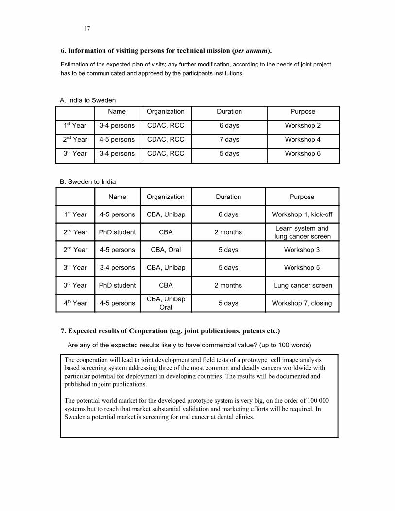
6. Information of visiting persons for technical mission (per annum ).
Estimation of the expected plan of visits; any further modification, according to the needs of joint project
has to be communicated and approved by the participants institutions.
A. India to Sweden
Name Organization Duration Purpose
1st Year 3-4 persons CDAC, RCC 6 days Workshop 2
2nd Year 4-5 persons CDAC, RCC 7 days Workshop 4
3rd Year 3-4 persons CDAC, RCC 5 days Workshop 6
B. Sweden to India
Name Organization Duration Purpose
1st Year 4-5 persons CBA, Unibap 6 days Workshop 1, kick-off
2nd Year PhD student CBA 2 months Learn system and
lung cancer screen
2nd Year 4-5 persons CBA, Oral 5 days Workshop 3
3rd Year 3-4 persons CBA, Unibap 5 days Workshop 5
3rd Year PhD student CBA 2 months Lung cancer screen
4th Year 4-5 persons CBA, Unibap
Oral 5 days Workshop 7, closing
7. Expected results of Cooperation (e.g. joint publications, patents etc.)
Are any of the expected results likely to have commercial value? (up to 100 words)
The cooperation will lead to joint development and field tests of a prototype cell image analysis based screening system addressing three of the most common and deadly cancers worldwide with particular potential for deployment in developing countries. The results will be documented and published in joint publications. The potential world market for the developed prototype system is very big, on the order of 100 000 systems but to reach that market substantial validation and marketing efforts will be required. In Sweden a potential market is screening for oral cancer at dental clinics.
18
8. Personal and Professional Data (CV) of Indian and Swedish PIs must be attached.
CV of Rajesh Kumar R (PI on Indian side)
Completed B.Tech in Electronics Engineering (1996) and M.Tech in Computer Science(1999) from Cochin University of Science and Technology. Over 17 years of experience in complete product development lifecycle and over 12 years of project management experience in internationally collaborated projects. Primary research interest is in the area of medical image processing and computer aided diagnosis. Currently leading multiple projects in these areas at the Centre for Development of Advanced Computing. Has successfully developed automated systems for early detection of cervical cancer, breast cancer and diabetic retinopathy and is an investigator in other similar collaborated projects. Generated research grants worth over 150 million rupees from various
funding agencies. In 2013, received CDAC’s 2ndBest Project Award for the work on cervical
cancer. In 2012, along with Prof. Ewert Bengtsson, Uppsala University was granted 654 kSEK by the Swedish Research Council. Has been a project guide to M.Tech students. Has over 20 publications, 2 patent applications and 13 copyrights. Between Jun 1999-Oct 2000 served in ER&DCI(now CDAC) as Research Associate. Between Oct 2000 – Mar 2009 was employed as Project Manager with Robert Bosch, the leading Tier I automotive company in the world. Was responsible for conceiving, design & development of hardware-in-loop & and software controlled vehicle simulators and fully automated test systems for engine management ECUs. Achieved more than 60% cost reduction and deployed over 200 systems with major automotive clients across the globe generating savings over 2 million dollars. Was also nominated for the biennial Bosch world wide Business Excellence Examples award (2007) as the Indian entry. Relevant Publications
[1] "Cervical Cancer Screening - New Insights", International Conference on Cancer Care and Cure, Dec 1-2 2016, Dubai [2] "Computer Aided Screening of Cervical Cancer using Methodologies of Quantitative Cytology", International Conference on Cytopathology 2015, 31 Aug - 02 Sep 2015, Toronto, Canada [3] “Computer Assisted Pap Smear Analyser for Cervical Cancer Screening using Quantitative Microscopy”, Journal of Cytology & Histology. doi 10.4172/2157-7099.S3-010 [4] “Automated Identification of Neutrophils in PAP Smear Images”, IEEE's International Conferences for Convergence of Technology(I2CT-2014 ), Pune, India [5] “Automated Cervical Cancer Screening System Using PAP Smear Images”, Annual Convention of Indian Association for Cancer Research ( IACR 2014), Kollam, India [6] “A Novel Approach for Koilocyte Identification in Cervical Smears Using Fourier Descriptors, International Conference on Biomedical Engineering and Assistive Technologies (BEATS-2014), Chandigargh, India [7] “Cluster detection and field-of-view quality rating - Applied to automated Pap-smear analysis”, ICPRAM 2013 Barcelona, Spain [8] “A fast and reliable approach to cell nuclei segmentation in PAP stained cervical smears”, CSI Transactions on ICT (December 2013) 1(4):309–315, DOI 10.1007/s40012-013-0028-y [9] “Debris removal in Pap-smear images”, Computer Methods and Programs in Biomedicine, http://dx.doi.org/10.1016/j.cmpb.2013.02.008 [10] “Automated Calibration of Microscope based on Image Processing Methods”, International Conference on Signal and Image Processing (ICSIP 2012), Coimbatore, India. [11] “Cluster Detection in Cytology Images using the Cellgraph Method”, IEEE International Symposium on IT in Medicine and Education, 2012, Hokkaido, Japan
19
[12] “Demosaicing: Study and Application in Cytology Image Analysis”, IEEE Indicon 2012, Kochi, India [13] “Detection and Removal of Artifacts from Cervical Cytology Images”, IEEE International Symposium on IT in Medicine and Education, 2011, Guangzhou, China [14] “Automated Screening in Cervical Cytology”, 41st Annual Conference of Indian Academy of Cytologists, Cytocon 2011, Dharward, Karnataka Relevant Patents and Copyrights
[1] “A Method and System for Automated Screening of Cervical Cancer”, Patent Applied 4098/CHE/2015 [2] “CellMarker – A GUI based software for Training set Creation and Automated Ground Truth Validation in Cytology Image Analysis”, copyright filed – SW-7458/2013 [3] “eSmear - A cervical smear analysis and reporting software”, copyright filed - SW-6416/2013 [4] “CerviSCAN – A Graphical User Interface Software for Automated Analysis and Classification of Cervical Smears”, copyright filed - SW-7352/2013
CV of Joakim Lindblad (PI on Swedish side)
Lindblad has a solid background in Image processing, particularly with medical applications and in microscopy. After spending some time abroad and working in industry, he is now returning to Swedish academia. He completed an M.Sc. in Engineering Physics (1997) and a Ph.D. in Computerized Image Analysis, Centre for Image Analysis (2003) at Uppsala University. PhD thesis: “Development of Algorithms for Digital Image Cytometry”. Post Doctoral fellow (2004-5) at Cancer Imaging, BC Cancer Research Centre, Vancouver, Canada: “Use of colour to improve the accuracy of image based cancer diagnostics”. Assistant professorship at SLU, Uppsala, Sweden (2006-11), FIMEK, Novi Sad, Serbia (2010-13). Associate professor Novi Sad University, Serbia (2013-16). Researcher at Mathematical Institute SANU, Belgrade, Serbia (2015-). Researcher at Centre for Image Analysis, Uppsala (2015-). He is one of the inventors (Golf Magazine's Innovator Award 2008) and main developer of the technology of Protracer AB (founded in 2007) and is since 2014 Head of Research at Protracer AB, Stockholm, Sweden. Protracer performs real time video analysis for sports TV broadcasts. It is the leading enterprise in the field and is contracted for TV coverage of the world’s major Golf tournaments since 2008.
He has supervised 7 M.Sc. students and co-supervised 4 Ph.D. students to completion, and is currently co-supervising 4 Ph.D. students. He has more than 60 high quality publications in the field of Image Analysis (945 citations), he has acquired a lot of experience with both theoretical and applied development and use of image analysis methods and software. He has excellent programming skills and a lot of practical experience of turning ideas into products. He is currently PI for the VINNOVA Innovation project 2015-01667 “Predictive modelling of real time video of outdoor scenes captured with a moving handheld camera” as well as the Swedish Research Links project 2015-05878 “Image analysis for reliable and cost effective cancer detection”. The latter project is in collaboration with the Indian co-PI, Dr. Sujathan. His experience with robust real-time image and video processing running 24/7 in outdoor environment combined with strong theoretical background in high precision image analysis will ensure that developed methods will reach a high level of usability. Being highly skilled in both medical image analysis and high performance real-time analysis using parallel architectures, he is very well suited to lead the Swedish side of this project.
20
Relevant publications:
● V. Ilić, J. Lindblad, N. Sladoje. Precise Euclidean distance transforms in 3D from voxel coverage representation. Pattern Recognition Letters, Vol. 65, pp. 184-191, 2015.
● A. Tanács, J. Lindblad, N. Sladoje, and Z. Kato. Estimation of Linear Deformations of 2D and 3D Fuzzy Objects. Pattern Recognition, Vol 48, No. 4, pp. 1387-1399, 2015.
● J. Lindblad and N. Sladoje. Linear time distances between fuzzy sets with applications to pattern matching and classification. IEEE Transactions on Image Processing, Vol. 23, No. 1, pp. 126-136, 2014.
● V. Ćurić, J. Lindblad, N. Sladoje, H. Sarve, and G. Borgefors. A new set distance and its application to shape registration. Pattern Analysis and Applications, Vol 17, No. 1, pp 141-152, 2014.
● M. Gavrilovic, J. C. Azar, J. Lindblad, C. Wählby, E. Bengtsson, C. Busch, and I. B. Carlbom. Blind Color Decomposition of Histological Images. IEEE Transactions on Medical Imaging, Vol. 32, No. 6, pp. 983-994, 2013.
● J. Lindblad and N. Sladoje. Coverage Segmentation Based on Linear Unmixing and Minimization of Perimeter and Boundary Thickness. Pattern Recognition Letters, Vol. 33, No. 6, pp. 728-738, 2012.
● T. Lukić, J. Lindblad, and N. Sladoje. Regularized image denoising based on spectral gradient optimization. Inverse Problems, Vol. 27, No. 8, pp. 085010, 2011.
● F. Malmberg, J. Lindblad, N. Sladoje, and I. Nyström. A Graph-based Framework for Sub-pixel Image Segmentation. Theoretical Computer Science, Vol. 412, No 15, pp. 1338-1349, 2011.
● N. Sladoje, J. Lindblad, and I. Nyström. Defuzzification of spatial fuzzy sets by feature distance minimization. Image and Vision Computing, Vol. 29, No 2-3, pp. 127-141, 2011.
● B. Bajić, J. Lindblad, N. Sladoje. Single image super-resolution reconstruction in presence of mixed Poisson-Gaussian noise. in Proceedings of the 6th IEEE International Conference on Image Processing Theory, Tools and Applications (IPTA), IEEE, Oulu, Finland, Dec. 2016.
● B. Bajić, J. Lindblad, N. Sladoje. Restoration of images degraded by signal-dependent noise based on energy minimization: an empirical study. Journal of Electronic Imaging (SPIE), Vol. 25, No. 4, 043020, 2016.
● S. Dražić, N. Sladoje, J. Lindblad. Estimation of Feret's Diameter from Pixel Coverage Representation of a Shape. Pattern Recognition Letters, Vol. 80, pp. 37-45, 2016.
● Bajić, J. Lindblad, N. Sladoje. Blind Restoration of Images Degraded with Mixed Poisson-Gaussian Noise with Application in Transmission Electron Microscopy. In Proceedings of the 13th IEEE International Symposium on Biomedical Imaging (ISBI), IEEE, pp. 123-127, Prague, Czech Republic, April 2016.
● A. Suveer, N. Sladoje, J. Lindblad, A. Dragomir, and I.-M. Sintorn. Automated Detection of Cilia in Low Magnification Transmission Electron Microscopy Images Using Template Matching. In Proceedings of the 13th IEEE International Symposium on Biomedical Imaging (ISBI), IEEE, pp. 386-390, Prague, Czech Republic, April 2016.
● V. Ilić, J. Lindblad, N. Sladoje. Signature of a shape based on its pixel coverage representation. In Proceedings of the 19th international conference on Discrete Geometry for Computer Imagery (DGCI), LNCS-9647, pp. 181-193, Nantes, France, April 2016.
● J. Lindblad, N. Sladoje, A. Suveer, A. Dragomir, I.-M. Sintorn. High-resolution reconstruction by feature distance minimization from multiple views of an object. In Proceedings of the 5th IEEE International Conference on Image Processing Theory, Tools and Applications (IPTA), IEEE, pp. 29-34, Orléans, France, Nov. 2015.
● K. Lidayová, J. Lindblad, N. Sladoje, H. Frimmel, C. Wang, and Ö. Smedby. Coverage Segmentation of 3D Thin Structures. In Proceedings of the 5th IEEE International Conference
21
on Image Processing Theory, Tools and Applications (IPTA), IEEE, pp. 23-28, France, Nov. 2015.
● M Delić, J. Lindblad, and N. Sladoje. αLBP - a Novel Member of the Local Binary Pattern Family Based on α-cutting. In Proceedings of the 9th IEEE International Symposium on Image and Signal Processing and Analysis (ISPA), IEEE, pp. 13-18, Croatia, Sept. 2015.
● J. Lindblad, E. Bengtsson, and N. Sladoje. Microscopy Image Enhancement for Cost-Effective Cervical Cancer Screening. In Proceedings of the 19th Scandinavian Conference on Image Analysis (SCIA), LNCS-9127, pp. 441-452,Denmark, June 2015.
● J. Lindblad and N. Sladoje. Exact Linear Time Euclidean Distance Transforms of Grid Line Sampled Shapes. In Proceedings of the 12th International Symposium on Mathematical Morphology (ISMM), LNCS-9082, pp. 645-656, Reykjavik, Iceland, May 2015.
● B. Bajić, J. Lindblad, and N. Sladoje. An Evaluation of Potential Functions for Regularized Image Deblurring. In Proceedings Part I of the 11th International Conference on Image Analysis and Recognition (ICIAR), LNCS-8814, pp. 150-158, Portugal, Oct. 2014.
● J. Lindblad, N. Sladoje, P. Malm, E. Bengtsson, R. Moshavegh, and A. Mehnert. Optimizing optics and imaging for pattern recognition based screening tasks. In Proceedings International Conference on Pattern Recognition (ICPR), IEEE, pp. 3333-3338, Sweden, Aug. 2014.
● K. Lidayova, J. Lindblad, N. Sladoje and H. Frimmel. Coverage segmentation of thin structures by linear unmixing and local centre of gravity attraction. In Proceedings of the 8th IEEE International Symposium on Image and Signal Processing and Analysis (ISPA), IEEE, pp. 83-88, Trieste, Italy, Sept. 2013.
9. Complementary resources.
The Swedish PI at CBA and the Indian co-PI at RCC, received in 2015 a joint grant for research collaboration within the project “Image analysis for reliable and cost effective cancer detection” from the Swedish Research Link program of the Swedish Research Council: SRL grant no. 2015-05878.
The grant is for the period January 1, 2016 until December 31, 2018. The total granted amount is 300.000 SEK per year for a total of 900.000 SEK. The grant finances networking and knowledge exchange between Sweden and India. This proposed project fits well with the specification of the awarded SRL grant and we will use it to co-finance our traveling costs.
22
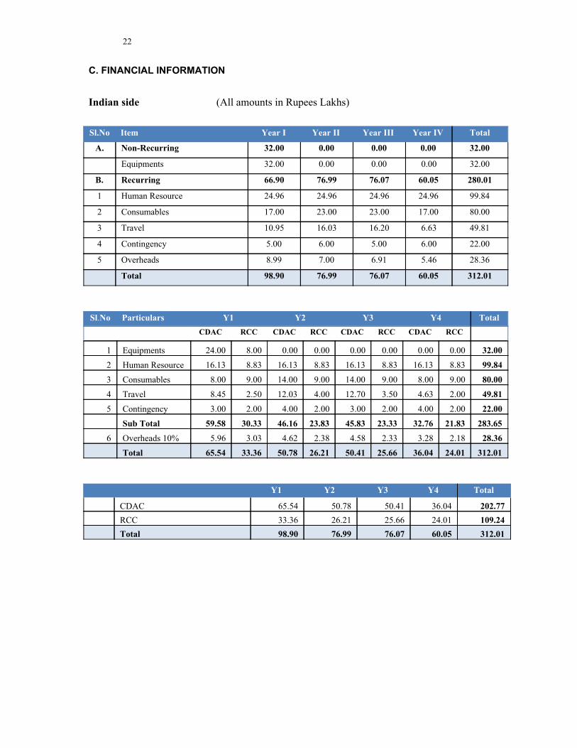
C. FINANCIAL INFORMATION
Indian side (All amounts in Rupees Lakhs)
Sl.No Item Year I Year II Year III Year IV Total
A. Non-Recurring 32.00 0.00 0.00 0.00 32.00
Equipments 32.00 0.00 0.00 0.00 32.00
B. Recurring 66.90 76.99 76.07 60.05 280.01
1 Human Resource 24.96 24.96 24.96 24.96 99.84
2 Consumables 17.00 23.00 23.00 17.00 80.00
3 Travel 10.95 16.03 16.20 6.63 49.81
4 Contingency 5.00 6.00 5.00 6.00 22.00
5 Overheads 8.99 7.00 6.91 5.46 28.36
Total 98.90 76.99 76.07 60.05 312.01
Sl.No Particulars Y1 Y2 Y3 Y4 Total
CDAC RCC CDAC RCC CDAC RCC CDAC RCC
1 Equipments 24.00 8.00 0.00 0.00 0.00 0.00 0.00 0.00 32.00
2 Human Resource 16.13 8.83 16.13 8.83 16.13 8.83 16.13 8.83 99.84
3 Consumables 8.00 9.00 14.00 9.00 14.00 9.00 8.00 9.00 80.00
4 Travel 8.45 2.50 12.03 4.00 12.70 3.50 4.63 2.00 49.81
5 Contingency 3.00 2.00 4.00 2.00 3.00 2.00 4.00 2.00 22.00
Sub Total 59.58 30.33 46.16 23.83 45.83 23.33 32.76 21.83 283.65
6 Overheads 10% 5.96 3.03 4.62 2.38 4.58 2.33 3.28 2.18 28.36
Total 65.54 33.36 50.78 26.21 50.41 25.66 36.04 24.01 312.01
Y1 Y2 Y3 Y4 Total
CDAC 65.54 50.78 50.41 36.04 202.77
RCC 33.36 26.21 25.66 24.01 109.24
Total 98.90 76.99 76.07 60.05 312.01
23
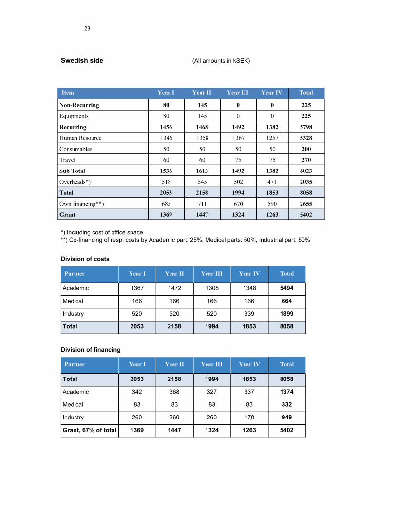
Swedish side (All amounts in kSEK)
Item Year I Year II Year III Year IV Total
Non-Recurring 80 145 0 0 225
Equipments 80 145 0 0 225
Recurring 1456 1468 1492 1382 5798
Human Resource 1346 1358 1367 1257 5328
Consumables 50 50 50 50 200
Travel 60 60 75 75 270
Sub Total 1536 1613 1492 1382 6023
Overheads*) 518 545 502 471 2035
Total 2053 2158 1994 1853 8058
Own financing**) 685 711 670 590 2655
Grant 1369 1447 1324 1263 5402
*) Including cost of office space
**) Co-financing of resp. costs by Academic part: 25%, Medical parts: 50%, Industrial part: 50%
Division of costs
Partner Year I Year II Year III Year IV Total
Academic 1367 1472 1308 1348 5494
Medical 166 166 166 166 664
Industry 520 520 520 339 1899
Total 2053 2158 1994 1853 8058
Division of financing
Partner Year I Year II Year III Year IV Total
Total 2053 2158 1994 1853 8058
Academic 342 368 327 337 1374
Medical 83 83 83 83 332
Industry 260 260 260 170 949
Grant, 67% of total 1369 1447 1324 1263 5402
Annexure
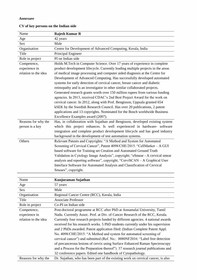
CV of key persons on the Indian side
Name Rajesh Kumar R
Age 42 years
Sex Male
Organisation Centre for Development of Advanced Computing, Kerala, India
Title Principal Engineer
Role in project PI on Indian side
Competence,
experience in
relation to the idea
Holds M.Tech in Computer Science. Over 17 years of experience in complete
product development lifecycle. Currently leading multiple projects in the areas
of medical image processing and computer aided diagnosis at the Centre for
Development of Advanced Computing. Has successfully developed automated
systems for early detection of cervical cancer, breast cancer and diabetic
retinopathy and is an investigator in other similar collaborated projects.
Generated research grants worth over 150 million rupees from various funding
agencies. In 2013, received CDAC’s 2nd Best Project Award for the work on
cervical cancer. In 2012, along with Prof. Bengtsson, Uppsala granted 654
kSEK by the Swedish Research Council. Has over 20 publications, 2 patent
applications and 13 copyrights. Nominated for the Bosch worldwide Business
Excellence Examples award (2007).
Reasons for why the
person is a key
Has, in collaboration with Sujathan and Bengtsson, developed existing system
which this project enhances. Is well experienced in hardware- software
integration and complete product development lifecycle and has good industry
background in the development of test automation systems.
Others Relevant Patents and Copyrights: “A Method and System for Automated
Screening of Cervical Cancer”, Patent 4098/CHE/2015. “CellMarker – A GUI
based software for Training set Creation and Automated Ground Truth
Validation in Cytology Image Analysis”, copyright; “eSmear - A cervical smear
analysis and reporting software”, copyright; “CerviSCAN – A Graphical User
Interface Software for Automated Analysis and Classification of Cervical
Smears”, copyright.
Name Kunjuraman Sujathan
Age 57 years
Sex Male
Organisation Regional Cancer Centre (RCC), Kerala, India
Title Associate Professor
Role in project Co-PI on Indian side
Competence,
experience in
relation to the idea
Post-doctoral programme at RCC after PhD at Annamalai University, Tamil
Nadu. Currently Assoc. Prof. at Div. of Cancer Research of the RCC, Kerala.
Currently four research projects funded by different agencies. 4 national awards
received for his research works. 5 PhD students currently under his supervision
and 2 PhDs awarded. Patent application filed: (Indian Complete Patent Appl.
No. 4098/CHE/2015: “A Method and system for automated screening of
cervical cancer”) and submitted (Ref. No.: 0080NF2016: “Label free detection
of precancerous lesions of cervix using Surface Enhanced Raman Spectroscopy
and a Process for the Preparation thereof”). 37 research journal publications and
32 conference papers. Edited one handbook of Cytopathology.
Reasons for why the Dr. Sujathan, who has been part of the existing work on cervical cancer, is also
person is a key
highly qualified in research on lung cancer. He will ensure samples and ground
truth necessary for DCNN training.
Others Developed a low cost monolayering technique (MFT) for cervical specimens, a
vital component for good image analysis
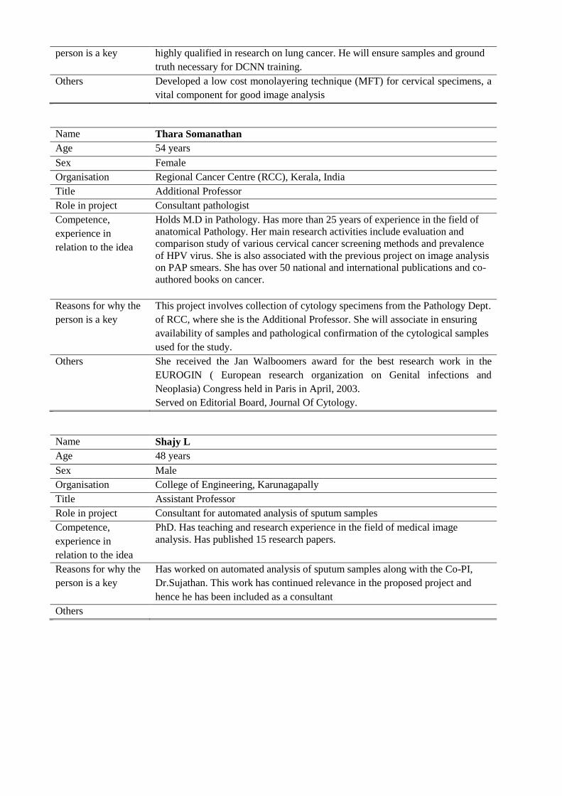
Name Thara Somanathan
Age 54 years
Sex Female
Organisation Regional Cancer Centre (RCC), Kerala, India
Title Additional Professor
Role in project Consultant pathologist
Competence,
experience in
relation to the idea
Holds M.D in Pathology. Has more than 25 years of experience in the field of
anatomical Pathology. Her main research activities include evaluation and
comparison study of various cervical cancer screening methods and prevalence
of HPV virus. She is also associated with the previous project on image analysis
on PAP smears. She has over 50 national and international publications and co-
authored books on cancer.
Reasons for why the
person is a key
This project involves collection of cytology specimens from the Pathology Dept.
of RCC, where she is the Additional Professor. She will associate in ensuring
availability of samples and pathological confirmation of the cytological samples
used for the study.
Others She received the Jan Walboomers award for the best research work in the
EUROGIN ( European research organization on Genital infections and
Neoplasia) Congress held in Paris in April, 2003.
Served on Editorial Board, Journal Of Cytology.
Name Shajy L
Age 48 years
Sex Male
Organisation College of Engineering, Karunagapally
Title Assistant Professor
Role in project Consultant for automated analysis of sputum samples
Competence,
experience in
relation to the idea
PhD. Has teaching and research experience in the field of medical image
analysis. Has published 15 research papers.
Reasons for why the
person is a key
Has worked on automated analysis of sputum samples along with the Co-PI,
Dr.Sujathan. This work has continued relevance in the proposed project and
hence he has been included as a consultant
Others
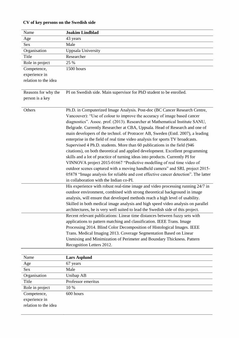
CV of key persons on the Swedish side
Name Joakim Lindblad
Age 43 years
Sex Male
Organisation Uppsala University
Title Researcher
Role in project 25 %
Competence,
experience in
relation to the idea
1500 hours
Reasons for why the
person is a key
PI on Swedish side. Main supervisor for PhD student to be enrolled.
Others Ph.D. in Computerized Image Analysis. Post-doc (BC Cancer Research Centre,
Vancouver): “Use of colour to improve the accuracy of image based cancer
diagnostics”. Assoc. prof. (2013). Researcher at Mathematical Institute SANU,
Belgrade. Currently Researcher at CBA, Uppsala. Head of Research and one of
main developers of the technol. of Protracer AB, Sweden (Estd. 2007), a leading
enterprise in the field of real time video analysis for sports TV broadcasts.
Supervised 4 Ph.D. students. More than 60 publications in the field (946
citations), on both theoretical and applied development. Excellent programming
skills and a lot of practice of turning ideas into products. Currently PI for
VINNOVA project 2015-01667 “Predictive modelling of real time video of
outdoor scenes captured with a moving handheld camera” and SRL project 2015-
05878 “Image analysis for reliable and cost effective cancer detection”. The latter
in collaboration with the Indian co-PI.
His experience with robust real-time image and video processing running 24/7 in
outdoor environment, combined with strong theoretical background in image
analysis, will ensure that developed methods reach a high level of usability.
Skilled in both medical image analysis and high speed video analysis on parallel
architectures, he is very well suited to lead the Swedish side of this project.
Recent relevant publications: Linear time distances between fuzzy sets with
applications to pattern matching and classification. IEEE Trans. Image
Processing 2014. Blind Color Decomposition of Histological Images. IEEE
Trans. Medical Imaging 2013. Coverage Segmentation Based on Linear
Unmixing and Minimization of Perimeter and Boundary Thickness. Pattern
Recognition Letters 2012.
Name Lars Asplund
Age 67 years
Sex Male
Organisation Unibap AB
Title Professor emeritus
Role in project 10 %
Competence,
experience in
relation to the idea
600 hours
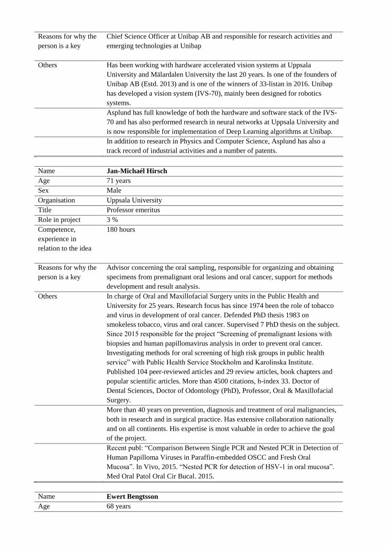
Reasons for why the
person is a key
Chief Science Officer at Unibap AB and responsible for research activities and
emerging technologies at Unibap
Others Has been working with hardware accelerated vision systems at Uppsala
University and Mälardalen University the last 20 years. Is one of the founders of
Unibap AB (Estd. 2013) and is one of the winners of 33-listan in 2016. Unibap
has developed a vision system (IVS-70), mainly been designed for robotics
systems.
Asplund has full knowledge of both the hardware and software stack of the IVS-
70 and has also performed research in neural networks at Uppsala University and
is now responsible for implementation of Deep Learning algorithms at Unibap.
In addition to research in Physics and Computer Science, Asplund has also a
track record of industrial activities and a number of patents.
Name Jan-Michaél Hirsch
Age 71 years
Sex Male
Organisation Uppsala University
Title Professor emeritus
Role in project 3 %
Competence,
experience in
relation to the idea
180 hours
Reasons for why the
person is a key
Advisor concerning the oral sampling, responsible for organizing and obtaining
specimens from premalignant oral lesions and oral cancer, support for methods
development and result analysis.
Others In charge of Oral and Maxillofacial Surgery units in the Public Health and
University for 25 years. Research focus has since 1974 been the role of tobacco
and virus in development of oral cancer. Defended PhD thesis 1983 on
smokeless tobacco, virus and oral cancer. Supervised 7 PhD thesis on the subject.
Since 2015 responsible for the project “Screening of premalignant lesions with
biopsies and human papillomavirus analysis in order to prevent oral cancer.
Investigating methods for oral screening of high risk groups in public health
service” with Public Health Service Stockholm and Karolinska Institute.
Published 104 peer-reviewed articles and 29 review articles, book chapters and
popular scientific articles. More than 4500 citations, h-index 33. Doctor of
Dental Sciences, Doctor of Odontology (PhD), Professor, Oral & Maxillofacial
Surgery.
More than 40 years on prevention, diagnosis and treatment of oral malignancies,
both in research and in surgical practice. Has extensive collaboration nationally
and on all continents. His expertise is most valuable in order to achieve the goal
of the project.
Recent publ: “Comparison Between Single PCR and Nested PCR in Detection of
Human Papilloma Viruses in Paraffin-embedded OSCC and Fresh Oral
Mucosa”. In Vivo, 2015. “Nested PCR for detection of HSV-1 in oral mucosa”.
Med Oral Patol Oral Cir Bucal. 2015.
Name Ewert Bengtsson
Age 68 years
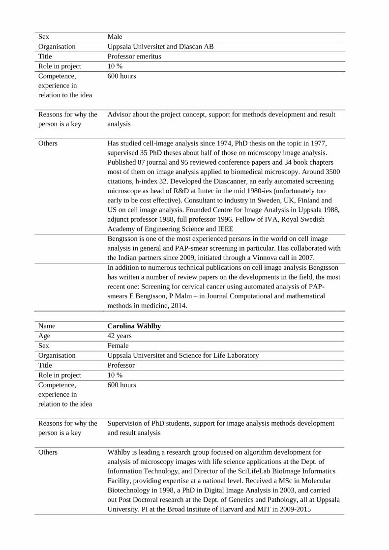
Sex Male
Organisation Uppsala Universitet and Diascan AB
Title Professor emeritus
Role in project 10 %
Competence,
experience in
relation to the idea
600 hours
Reasons for why the
person is a key
Advisor about the project concept, support for methods development and result
analysis
Others Has studied cell-image analysis since 1974, PhD thesis on the topic in 1977,
supervised 35 PhD theses about half of those on microscopy image analysis.
Published 87 journal and 95 reviewed conference papers and 34 book chapters
most of them on image analysis applied to biomedical microscopy. Around 3500
citations, h-index 32. Developed the Diascanner, an early automated screening
microscope as head of R&D at Imtec in the mid 1980-ies (unfortunately too
early to be cost effective). Consultant to industry in Sweden, UK, Finland and
US on cell image analysis. Founded Centre for Image Analysis in Uppsala 1988,
adjunct professor 1988, full professor 1996. Fellow of IVA, Royal Swedish
Academy of Engineering Science and IEEE
Bengtsson is one of the most experienced persons in the world on cell image
analysis in general and PAP-smear screening in particular. Has collaborated with
the Indian partners since 2009, initiated through a Vinnova call in 2007.
In addition to numerous technical publications on cell image analysis Bengtsson
has written a number of review papers on the developments in the field, the most
recent one: Screening for cervical cancer using automated analysis of PAP-
smears E Bengtsson, P Malm – in Journal Computational and mathematical
methods in medicine, 2014.
Name Carolina Wählby
Age 42 years
Sex Female
Organisation Uppsala Universitet and Science for Life Laboratory
Title Professor
Role in project 10 %
Competence,
experience in
relation to the idea
600 hours
Reasons for why the
person is a key
Supervision of PhD students, support for image analysis methods development
and result analysis
Others Wählby is leading a research group focused on algorithm development for
analysis of microscopy images with life science applications at the Dept. of
Information Technology, and Director of the SciLifeLab BioImage Informatics
Facility, providing expertise at a national level. Received a MSc in Molecular
Biotechnology in 1998, a PhD in Digital Image Analysis in 2003, and carried
out Post Doctoral research at the Dept. of Genetics and Pathology, all at Uppsala
University. PI at the Broad Institute of Harvard and MIT in 2009-2015
developing the CellProfiler software for image-based cell screening. Board
member of Swedish Bioimaging and the National Microscopy Infrastructure,
and active in the international cytometry community as ISAC scholar since 2014
and an ERC consolidator since 2015. Supervise/ed 15 PhD theses on microscopy
image analysis and published 52 journal- and reviewed conference papers on the
subject. Currently supervising PhD and MSc students on Convolutional Neural
Networks (CNNs) for cell screening.
With a strong background in biotechnology, genetics and pathology combined
with image processing and current activities on CNNs, Wählby will be valuable
for providing expertise, coordination and communication within the proposed
project.
Recent papers on CNNs for image-based screening:
“Deep Fish: Deep Learning-Based Classification of Zebrafish Deformation for
High-Throughput Screening”. J. of Biomolecular Screening, 2016. “Feature
Augmented Deep Neural Networks for Segmentation of Cells”. ECCV 2016.