Embed Size (px)
Citation preview
Prosomatostatin-derived Antrin Is Present in Gastric D Cells and in Portal BloodM. Ravazzola,* R. Benoit,t N. Ling,* and L. Orci**Institut d'Histologie et d'Embryologie, 1211 Geneve 4, Switzerland; tDepartment of Medicine, Montreal General Hospital, McGillUniversity, Montreal H3GIA4, Canada; and *Laboratories for Neuroendocrinology, The Salk Institute, La Jolla, California 92037
Abstract
The three widely distributed peptides derived from proso-
matostatin are the original neurohormone somatostatin-14,somatostatin-28, and somatostatin-28(1-12), which are all de-rived from the COOHterminus of the precursor. Recently a
decapeptide derived from the NH2terminus of the prohormonehas been identified in extracts of rat gastric antrum and namedantrin. Data now show that in both rats and humans this new
molecular form is concentrated in the D cell of the gastrointes-tinal mucosa together with somatostatin-28(1-12). The high-est concentration of antrin immunofluorescent cells is locatedin the mucosa of the gastric antrum. Ultrastructural studiesperformed on pyloric D cells using the protein A-gold tech-nique reveals that antrin is present in the same secretory gran-
ules as somatostatin-28(1-12) and detectable in one-third of allsecretory granules. Acid extracts of rat hepatic portal plasmacontain a peptide similar or identical to antrin, indicating thatthe antral decapeptide circulates in blood.
Introduction
The notion that peptide hormones are derived from precursor
proteins by posttranslational modification is now well recog-
nized (1). During the maturation process several peptides may
be generated together with the hormone. These other peptides,often called cryptic peptides, are important for three reasons:
(a) their presence provides information on the processingevents, (b) some are better markers for a given peptidergicsystem than the hormone itself, and (c) among these peptides,a few could serve a biological role. After a new molecular formhas been identified in a given tissue extract, several questionsare raised, such as whether it may represent an artifact ofextraction. If the peptide is not produced artifactually it isimportant to know the cell type from which it originates andwhether it can be secreted by these cells.
Somatostatin is a bioactive peptide of 14 or 28 amino acidsthat generally acts as a paninhibitor in the neuroendocrinesystem. It is derived from the COOH-terminal end of a 10-kDprecursor protein, prosomatostatin (2-5) (Fig. 1). We have
Address correspondence to Dr. M. Ravazzola, Departement de Mor-phologie, Institut d'Histologie et d'Embryologie, Centre Medical Uni-versitaire, 1, rue Michel-Servet, 1211 Geneve 4, Switzerland.
Receivedfor publication 23 May 1988 and in revisedform 2 Sep-tember 1988.
recently isolated a peptide from rat stomach that correspondsto the first 10 amino acids at the NH2terminus of mammalianprosomatostatin (6). In the rat, the highest concentration ofthe decapeptide is present in the gastric antrum, hence theproposed name antrin. It is presently assumed that antrin isformed after cleavage at a monobasic site very similar to thatleading to formation of somatostatin-28 and somatostatin-28(1-12). In the present work we provide the first immunocy-tochemical data on the cellular and ultrastructural localizationof antrin and compare it with somatostatin-28(1-12), an ex-cellent marker of the prosomatostatin system in tissue (7, 8).Evidence is also presented for the antral decapeptide circulat-ing in hepatic portal blood in rats.
Methods
AntiseraTo obtain specific antibodies that recognize only antrin, not proso-matostatin or any of the other prosomatostatin-derived peptides, onemust direct the formation of gammaglobulin toward the COOHter-minus of antrin. This was achieved by immunizing rabbits with amixture of synthetic antrin and methylated BSA or with the analogue[TYR3]antrin (3-10) coupled to BSA as described previously for sev-eral other peptides (9). The immuno plasma generated by the firstmethod (Fig. 2) was selected for immunocytochemical studies (A09).An antibody population with higher affinity (A 14) was obtained by thesecond method and it was selected for quantitation of antrin by RIA. Apreviously characterized antibody population (S320) against somato-statin-28(1-12) was used as a marker of the prosomatostatin sys-tem (7).
ImmunocytochemistryLight microscopy. Pieces of pancreas and gastrointestinal mucosa ofnormal adult Sprague-Dawley rats fixed by vascular perfusion with 1%glutaraldehyde were dehydrated and embedded in Epon. Consecutivesemithin sections (1 um) attached on glass slides were incubated by theindirect immunofluorescence method (10). Sections were treated toremove Epon (1 1) and then incubated for 2 h at room temperaturewith the primary antisera A09 (diluted 1:1,000) for antrin or S320(diluted 1:4,000) for somatostatin-28(1-12) immunoreactivity. Sec-tions were washed with PBS and then exposed to FITC-conjugatedgoat anti-rabbit IgG for 1 h. After washing and counterstaining with0.03% Evans Blue the sections were examined with a fluorescencemicroscope (Axiophot; Carl Zeiss, Inc., Oberkochen, FRG). Thepro-cedure for the fixation and embedding of the human fetal tissue isdetailed in reference 12. Sections were etched with 5% (vol/vol) H202before immunofluorescence labeling. Control of specificity was carriedout by applying anti-antrin serum previously absorbed with 10 4g/mlantrin, prosomatostatin(1-32), somatostatin-14, somatostatin-28, or
somatostatin-28(1-12).Electron microscopy. Thin sections attached on nickel grids were
immunostained by the protein A-gold (pAg)1 technique (13) with anti-
1. Abbreviations used in this paper: pAg, protein A-gold.
362 M. Ravazzola, R. Benoit, N. Ling, and L. Orci
J. Clin. Invest.© The American Society for Clinical Investigation, Inc.0021-9738/89/02/0362/05 $2.00Volume 83, February 1989, 362-366

IlI I ll1 24-25 56-57 88-89 101-102 116
-E-- PRE - PROSOMATOSTATIN -
I1I_.
- 24 1 10 32 64 76 92
-1 -__10ALA-PRO-SER-ASP-PRO-ARG-LEU-ARG-GLN-PHE
ANTRIN
1ss28(1l2 l ss4:l.......... ........SS2 S28....... .........
Figure 1. Schematic representation ofthe 1 16-amino acid precursor of mam-malian somatostatin. It contains a sig-nal peptide or "pre" region of 24amino acids preceding the 92 aminoacid prosomatostatin of 10 kD. Antrinrepresents the first 10 amino acids atthe amino terminus of prosomatostatinwhile somatostatin- 14 represents thelast 14 amino acids at the carboxyl ter-minus. Direct and indirect evidence in-dicates that cleavage occurs at Gly24-Ala25, Leu56-Leu57, Arg88-Ser89, andArg'01-Lys'02. Antrin is probably gener-ated through cleavage at Lys37-Ser38(not shown), a monobasic site alsopresent in catfish as Arg37-Ser38.
antrin diluted 1:3,000 or anti-somatostatin diluted 1:4,000. Gold par-ticles 10 nm in diameter were used. After immunolabeling sectionswere double stained with uranyl acetate and lead citrate and examinedin an electron microscope (model 301; Philips Electron Optics, Eind-hoven, The Netherlands).
The number of secretory granules expressing antrin immunoreac-tivity was quantified on photographic prints of 8-10 D cells (magnifi-cation of 34,000). 500-600 secretory granules were evaluated. Secre-tory granules showing at least four gold particles were counted asimmunoreactive for antrin.
Gel permeation chromatography of hepatic portal plasmaRat hepatic portal plasma was extracted with 2 M acetic acid, theextract was loaded onto octadecylsilylsilica cartridges and the retentateeluted with acetonitrile. The material eluted with acetonitrile wasdried, dissolved in 5.2 Macetic acid, and loaded onto a Sephadex G-75gel permeation chromotography column. Each column fraction wasmonitored for the presence of antrin using an RIA in which iodinated[TYR3]antrin(3-10) was used as the ligand.
Results
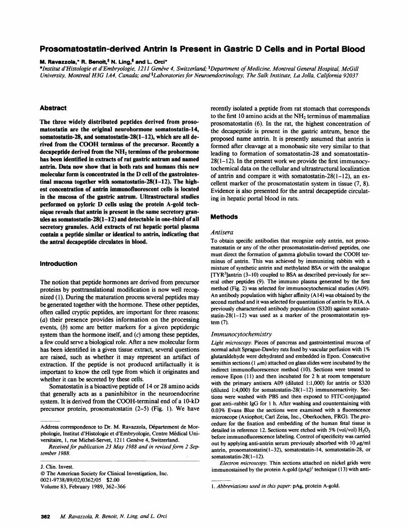
Immunofluorescence with A09 anti-antrin serum on sectionsof pancreas and gastrointestinal mucosa of rat revealed thatantrin immunofluorescence was undetectable in pancreaticislets, seldom present in epithelial cells of oxyntic and duo-denal mucosa, but clearly represented in endocrine cells of thegastric antrum (Fig. 3 A). All antrin immunofluorescent cellsexamined reacted also with S320 antibody. The staining forantrin was more sparse than that for somatostatin-28(1-12)which, when present, produced a diffuse cytoplasmic staining(compare Fig. 3, A and B). The same experiments performedin human fetal gastric tissue yielded similar results for bothantrin and somatostatin-28(1-12) immunostaining (Fig. 3, Cand D). Control of specificity showed that among the peptidesused to adsorb the anti-antrin serum before the immunolabel-ing, only antrin abolished the staining.
At the ultrastructural level antrin immunoreactivity, local-
Somatostatin.14 Figure 2. Characteristics of A09, an an-100 - .. Somatostatin-28 tibody population produced against an-
p-o matdotptin.28(1.12) trin. A NewZealand White rabbit was80 p Enr. GnRHimmunized with an emulsion contain-
Prosomatostatin (1-32) ing synthetic antrin (14), methylated
F 60 - "FN .. BSA, and complete Freund adjuvantz '. Antrin supplemented with 10 mgkilled myco-m[40- lTYR3] Antrin *. bacterium tuberculosis as previously
co '\ITYROI Antrin described (9). Booster injections weredone with the same emulsion but with-
20-\20
out bacteria. 10 d after the secondboost the immune plasma was collectedand used at a 1/100,000 dilution for
1 pg 10 Pg 100 P9 1 ng 10 ng 100 ng RIA. The ligand was 125I [TYR3]-Peptide Per RIA Tube antrin(3-10) which was obtained after
iodination of 2.5 ,g [TYR3]antrin(3-10) with 20 jig chloramine T during 38 s followed by addition of 75 ,ug sodium metabisulfite and I mghuman serum albumin. The label waspurified on carboxymethyl cellulose (CM 52; Whatman Laboratory Products, Inc., Clifton, NJ) using 0.04 Mammonium acetate, pH 4.6, asthe elution buffer. Net binding of label to antibody was 31% after 44 h incubation at 4°C in RIA buffer (9). The specificity studies representedhere show that cross-reactivity with preproSS(25-33)-TYR (i.e., [TYR'0]antrin) and prosomatostatin(1-32) is 0.03% and 0.003%, respectively,indicating that A09 recognizes the COOH-terminal region of antrin. This was confirmed by incubating '251-[TYR'0]antrin with A09 diluted at1/10,000. No binding was observed (data not shown).
Antrin in Rats and Humans 363
Figure 3. Localization of antrin and somatostatin-28(1-12) immunoreactivity by immunofluorescence on consecutive semithin sections of an-tral mucosa from adult rat (A, B) and 17-wk human fetus (C, D). The same D cells (arrowheads) show antrin and somatostatin-28(1-12) im-munoreactivity. However, the pattern of antrin immunolabeling of individual cells appears rather sparse compared with the very bright andcompact immunofluorescence of somatostatin-28(1-12). A, B: X800; C, D: X630.
ized by pAg, was observed over the secretory granules. Labeledgranules frequently coexisted with granules devoid of immu-nolabeling (Fig. 4, A and C). Quantitative evaluation of an-trin-positive granules showed that they represented 33%of thepopulation of secretory granules in rat D cells and 15% inhuman fetal D cells.
Comparing serial sections, one incubated with anti-antrinand the other with anti-somatostatin-28(1-12), it was foundthat the secretory granules containing antrin also containedsomatostatin-28(1-12) (Fig. 4, A-D). By contrast, many so-matostatin-positive granules do not contain antrin. Inasmuchas the antiserum recognizes the COOHterminus of proso-matostatin 1-10 (antrin) it appears that the processing of pro-somatostatin to antrin occurs in only a subpopulation of thegranules that process prosomatostatin to prosomatostatin-28(1-12). Antrin could be a late cleavage product, occurringonly in "old" granules, and never be secreted. Whenextract ofrat hepatic portal plasma was subjected to gel permeationchromatography, it was found that a single peak of immuno-reactivity eluted in a zone compatible with a peptide the size ofantrin (- 1,200 mol wt) (Fig. 5).
Discussion
The findings obtained with a specific antibody population di-rected toward antrin indicate (a) that the decapeptide ispresent in secretory granules that also contain somatostatin-28(1-12), and (b) that it is detectable in only a fraction ofsomatostatin-containing granules in a subpopulation of so-
matostatin-producing cells. This suggests differential precursor
processing or product stability within the granules, and antrincould thus be a marker for the presence of specific cleavageenzymes.
Antrin cannot be detected by immunohistochemistry ingastric nerves, nor can it be detected in the D cells of thepancreas. This restricted tissue distribution, namely that theendocrine cells of the stomach are the predominant site ofantrin accumulation, suggests that the measurement of antrinin blood may represent a means by which to evaluate specifi-cally the response of the gastric D cell during digestion. To-gether with the demonstration of this peptide in the portalcirculation, it also suggests that antrin acts as a hormone dur-ing digestion. The high degree of amino acid sequence conser-
364 M. Ravazzola, R. Benoit, N. Ling, and L. Orci

42*~~~~~~~~~~~~~~~~~~~
;r 'Sf#,F'st;'Si -itl~k 6 ,+,'4;S ATs;fi'
A~~~~~~~~~~~~~~~~~~~~~~~~~~~~~~~~~~~~~~A
~~~~~~~~~~~~~~~~~~~~~~~~~~~~~~-
Figure 4. Localization of antrin and somatostatin-28(1 -12) at the ultrastructural level on consecutive thin sections of antral D cells of rat (A, B)and human fetus (C, D) processed by the pAg method. In electron microscopy antrin was revealed over a population (33% in rat, 15% inhuman) of secretory granules while somatostatin-28( 1-12) was present on virtually all the secretory granules. Serially cut secretory granulescontaining antrin and somatostatin-28( 1-12) immunoreactivity are numbered. Arrowheads point to secretory granules with somatostatin-28( 1-12) but not antrin immunolabeling. A, B: X34,000; C, D: X40,000.
CL0
x:>Il
I -a: 0I a-J z
^ - ~~~~~120
~~~~- _0
44 60 76 92
Fraction
Figure 5. Sephadex G-75 gel permeation chromatography of an acid ex-tract of 78 ml rat portal plasma. Hepatic portal blood was collected incold heparinized syringes from adult Sprague-Dawley rats fed ad lib. Aftercentrifugation at 12,800 g for 2 min at 40C the plasma was added to 2 Macetic acid (1 vol. for 1 vol.), vortexed, frozen in liquid nitrogen, and keptat -20'C until purification on Sep-Paks (Waters Associates, Milford,MA). The material eluted from octadecylsilylsilica cartridges with 80%acetonitrile in 0.1 %trifluoroacetic acid was lyophilized. The dry powderwas dissolved in 5.2 Macetic acid and loaded onto a Sephadex G-75 col-umn (2.6 X 107 cm). Antrin radioimmunoactivity was measured with an-
. tibody A14 in fractions 23-92. A major peak of immunoreactive materialwas detected at 0.7 Kay [(Vx - Vo)/(Vsit - V0)], which is compatible with a
t peptide of 1,200-1,300 mol wt. The column was previously calibratedwith cytochrome c, 12,384 mol wt, human fl-endorphin, 3,466 mol wt, lu-teinizing-hormone releasing hormone (LHRH), 1,182 mol wt, and NaCl,58.5 mol wt. The peak seen in the void volume represents crystalline BSA(Pentex; Miles Laboratories, Inc., Elkhart, IN) 67,000 mol wt, used forcoating the column. Flow rate was 21 ml/h and fraction size was 7 ml.
Antrin in Rats and Humans 365
0 '5>
o0.6 -
0.4-0.2 -
28
0.0 0.2 0.4 0.6 0.8 1.0
Kav
Los tAti W_28( ..4-_.'. "' -. I- _;, .,2r. I* 0" A
44-
4
gp
0.
vation from catfish to man in the region of prosomatostatincorresponding to antrin argues in favor of a biological role forthat molecule (5).
Acknowledgments
Wethank Dr. J.-D. Vassalli for critical reading of the manuscript, Ms.Gina Gravel, A.-M. Lucini, G. Moussard, and G. Perrelet for technicalassistance, Mr. G. Negro and P.-A. Ruttimann for photographic work,and Ms. I. Bernard for typing.
This work was supported by the Swiss National Science Founda-tion (grant 3.404.86), The Medical Research Council of Canada (grantMA9145), and The National Institutes of Health (grants HD-09690and DK-188'1 1).
References
1. Douglass, J., 0. Civelli, and E. Herbert. 1984. Polyprotein geneexpression: generation of diversity of neuroendocrine peptides. Annu.Rev. Biochem. 53:665-715.
2. Brazeau, P., W. Vale, R. Burgus, N. Ling, M. Butcher, J. Rivier,and R. Guillemin. 1973. Hypothalamic peptide that inhibits the secre-tion of immunoreactive pituitary growth hormone. Science (Wash.DC). 179:77-79.
3. Pradayrol, L., H. Jornvall, V. Mutt, and A. Ribet. 1980. N-termi-nally extended somatostatin: the primary structure of somatostatin-28.FEBS(Fed. Eur. Biochem. Soc.) Lett. 109:55-58.
4. Benoit, R., P. B6hlen, N. Ling, A. Briskin, F. Esch, P. Brazeau,S.-Y. Ying, and R. Guillemin. 1982. Presence of somatostatin-28-(l-12)' in hypothalamus and pancreas. Proc. Nati. Acad. Sci. USA.79:917-921.
5. Argos, P., W. L. Taylor, C. D. Minth, and J. E. Dixon. 1983.Nucleotide and aminoacid sequence comparison of preprosomato-statins. J. Biol. Chem. 258:8788-8793.
6. Benoit, R., N. Ling, and F. Esch. 1987. A new prosomatostatin-derived peptide reveals a pattern for prohormone cleavage at monoba-sic sites. Science (Wash. DC). 238:1126-1129.
7. Ravazzola,'M., R. Benoit, R. Guillemin, and L. Orci. 1983.Immunocytochemical localization of prosomatostatin fragments inmaturing and mature secretory granules of pancreatic and gastrointes-tinal D-cells. Proc. Natl. Acad. Sci. USA. 80:215-218.
8. Morrison, J. H., R. Benoit, P. J. Magistretti, and F. E. Bloom.1983. Immunohistochemical distribution of prosomatostatin relatedpeptides in cerebral cortex. Brain Res. 262:344-351.
9. Benoit, R., N. Ling, P. Brazeau, S. Lavielle, and R. Guillemin.1987. Strategies for antibody production and radioimmunoassays. InNeuromethods: Peptides. Vol. 6. A. A. Boulton, G. B. Baker, and Q. J.Pittman, editors. Humana Press, Clifton, NJ. 43-72.
10. Coons, A. H., E. H. Leduc, and J. M. Connolly. 1955. Studieson antibody production. I. A method for the histochemical demonstra-tion of specific antibody and its application to a study of the hyperim-mune rabbit. J. Exp. Med. 102:49-63.
11. Maxwell, M. H. 1978. Two rapid and simple methods used forthe removal of resin from 1.0 um thick epoxy sections. J. Microsc.(Lond.). 112:253-255.
12. Like, A. A., and L. Orci. 1972. Embryogenesis of the humanpancreatic islets: a light and ele4tron microscopic study. Diabetes.21(Suppl. 2):511-534.
13. Roth, J., M. Bendayan, and L. Orci. 1978. Ultrastructurallocalization of intracellular antigens by the use of protein A-gold com-plex. J. Histochem. Cytochern. 26:1074-1081.
14. Ling, N., F. Esch, D. Davis, M. Mercado, M. Regno, P. Bohlen,P. Brazeau, and R. Guillemin. 1980. Solid phase synthesis of somato-statin-28. Biochem. Biophys. Res. Commun. 95:945-951.
366 M. Ravazzola, R. Benoit, N. Ling, and L. Orci