Embed Size (px)
Citation preview
Prostaglandin E2-Bisphosphonate Conjugates: Potential Agents forTreatment of Osteoporosis
Laurent Gil, a,y Yongxin Han, a Evan E. Opas, b Gideon A. Rodan, b Re jean Ruel, a
J. Gregory Seedor, b Peter C. Tyler a,{ and Robert N. Younga,*aDepartment of Medicinal Chemistry, Merck Frosst Centre for Therapeutic Research, P.O. Box 1005, Pointe Claire-Dorval, QueÂbec,
Canada H9R 4P8bDepartment of Bone Biology and Osteoporosis, Merck Research Laboratories, West Point, PA 19486, USA
Received 3 September 1998; accepted 3 December 1998
Dedicated to the Memory of Professor Sir Derek H. R. Barton
AbstractÐConjugates of bisphosphonates (potential bone resorption inhibitors) and prostaglandin E2 (a bone formation enhancer)were prepared and evaluated for their ability to bind to bone and to liberate, enzymatically, free PGE2. The conjugate 3, an amide atC-1 of PGE2 proved to be too stable in vivo while conjugate 6, a thioester, was too labile. Several PGE2, C-15 ester-linked con-jugates (18, 23, 24 and 31) were prepared and conjugate 23 was found to bind e�ectively to bone in vitro and in vivo and to liberatePGE2 at an acceptable rate. A 4-week study in a rat model of osteoporosis showed that 23 was better tolerated and more e�ective asa bone growth stimulant than daily maximum tolerated doses of free PGE2. # 1999 Elsevier Science Ltd. All rights reserved.
Introduction
Osteoporosis is the most common metabolic bonedisease which a�ects 40±50% of the elderly female and10±15% of the elderly male population.1 The diseaseinvolves the gradual loss of bone mass as a result of animbalance between the bone resorption activity ofosteoclasts and the bone formation activity of osteo-blasts.2 A number of pharmaceutical agents have beendeveloped to treat this disease. These compounds can bedivided into two groups: bone resorption inhibitors suchas bisphosphonates3 (e.g. Fosamax1) and bone formationstimulants.4±8 Bisphosphonates are analogues of pyr-ophosphates which are absorbed tightly onto hydroxy-apitite surfaces and, due to this process, bisphosphonatesare targetted to bone.9 While bisphosphonates representan important class of drugs for the treatment of osteo-porosis, their value is generally manifested by preventionof bone loss and thus, for treatment of more advanceddisease, there has been great interest in the discovery of
safe and e�ective bone formation stimulants. Examplesof bone-activating agents may include parathyroid hor-mone,4 growth hormone,5 ¯uoride,6 possibly certainvitamin D metabolites7 and prostaglandin E2 (PGE2).
8
Indeed, a number of studies have demonstrated thatbone formation can be stimulated in vivo by systemicinjection of PGE2.
10 Furthermore, substantial new boneformation has been observed on the controlled release ofPGE2 from implanted PGE2-containing polymers11 indi-cating that PGE2 acts locally in bone. Unfortunately,such implants are impractical in a normal therapeuticsetting and the pharmaceutical utility of systemic PGE2
is greatly reduced due to side e�ects and metabolicinstability.
Conjugates of PGE2 and bisphosphonates described inthis report represent a new class of compounds whichcould circumvent the problems associated with PGE2.PGE2 (or an analogue) chemically coupled to a bisphos-phonate could be e�ectively delivered to bone due tothe property of the bisphosphonate to bind to bone.Gradual hydrolysis of the conjugate could then liberatea bone resorption inhibitor (the bisphosphonate moiety)and a bone formation enhancer (the PGE2 moiety). Totest this hypothesis, it was necessary to devise methodsto couple bisphosphonates and PGE2 in a way compat-ible with the chemical and biochemical instability ofPGE2 through a linkage which was suitably stable forthe conjugate to survive intact during the time necessary
0968-0896/99/$ - see front matter # 1999 Elsevier Science Ltd. All rights reserved.
PII: S0968-0896(99)00045-0
Bioorganic & Medicinal Chemistry 7 (1999) 901±919
Key words: Osteoporosis; prostaglandin E2; bisphosphonate; alen-dronate; conjugate.*Corresponding author.y Present address: Departamento de Quimica, ICEx-UFMG, Av.Antonio Carlos, 6627-Pampulha, CEP 31270-190 Belo Horizonte,Minas Gerais, Brazil.{ Present address: Industrial Research Limited, Grace®eld ResearchCentre, Grace®eld Road, P.O. Box 31-310, Lower Hutt, New Zealand.
for uptake into bone in vivo, and suitably labile to besubsequently released by metabolic hydrolysis. Ideally,a hydrolytic half-life of 1 to 4 days was sought. In orderto monitor e�ectively both the uptake and release of theprostaglandin moiety in the bone, it was decided to uti-lize radioactive-labelled PGE2. In early experiments, wealso utilized radioactive-labelled bisphosphonate andthus double label analysis of plasma and bone samplesover time could be used to follow the uptake and clea-vage of the conjugate. Finally, when conjugates withappropriate properties were discovered, more complexin vivo experiments to measure their e�ect on bone for-mation were undertaken.
Results and Discussion
Synthesis and studies of conjugates linked via the C-1carboxyl group of PGE2
12
Synthesis of PGE2-alendronate conjugate 3. With thecommercial availability of tritium-labelled PGE2 (1) andthe in-house availability of 14C-labelled alendronate (2),we ®rst explored the direct coupling of alendrolate withPGE2 to provide the corresponding amide (3). The N-hydroxysuccinimide ester of PGE2 was readily preparedusing DCC as coupling agent and the active ester reac-ted e�ciently with alendronate in dioxane±water withcareful control of the pH at 8±9 in order to ensure theintegrity of the PGE2 (higher pH led to competing elim-ination of the 11-hydroxyl group to the correspondingenone) (Scheme 1). Puri®cation of the conjugate wasdi�cult but could be achieved ®rst by evaporation todryness and then dissolution of the mixture in water andabsorption onto a C18 cartridge followed by elution®rst with water and then with acetonitrile±water toprovide fractions that were essentially pure conjugate3. In this manner, quantities both of unlabelled anddouble-labelled conjugate 3 could be prepared.
In vitro and in vivo evaluation of conjugate 3. The con-jugate 3 was studied ®rst in vitro to determine its bind-ing to bone powder. These experiments showed thatirreversible in vitro binding of 3 in fetal bovine serum(FBS) to bone powder occurred to the extent of 77% ofthe 14C moiety (alendronate) and 53% of the 3H moiety(PGE2) within 1 h. Thereafter, disassociation of thePGE2 moiety could be followed during incubation withFBS by measuring the residual 14C/3H ratio. Theseexperiments indicated that disassociation of the 3H
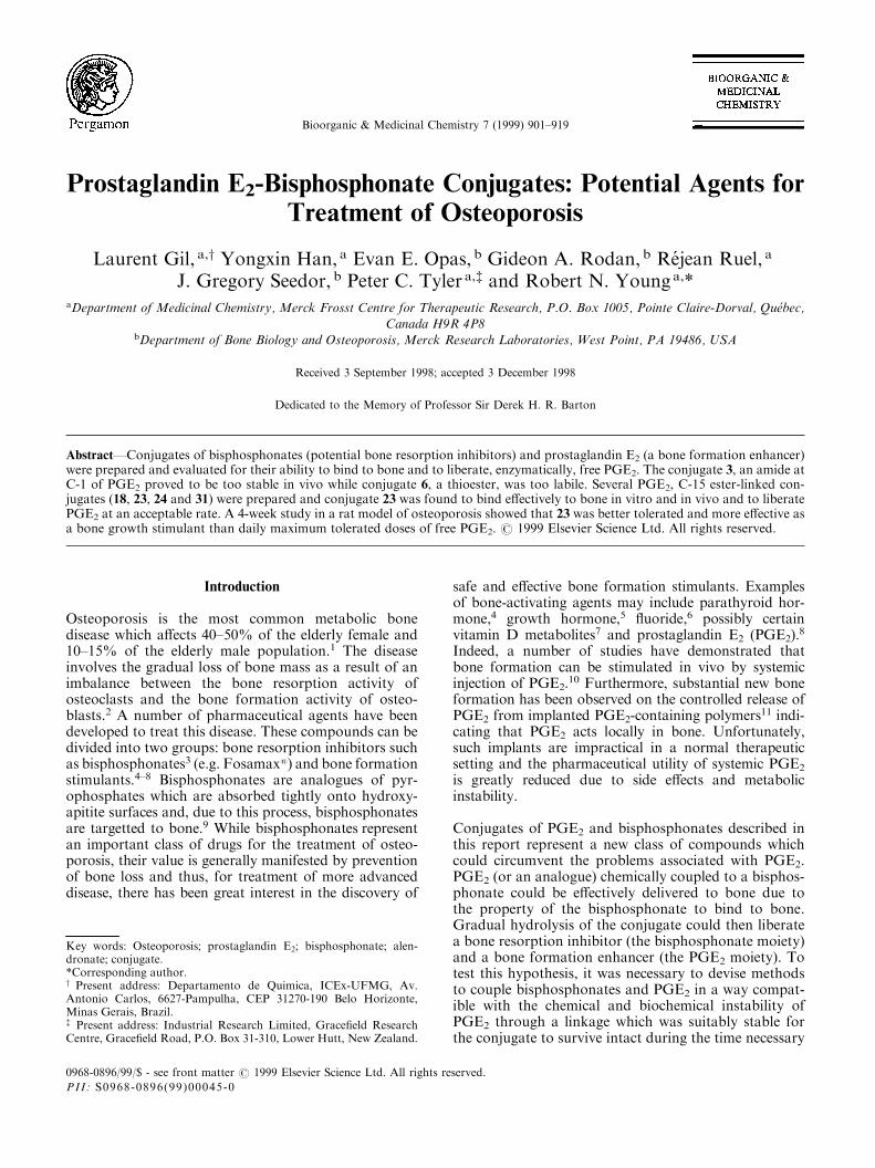
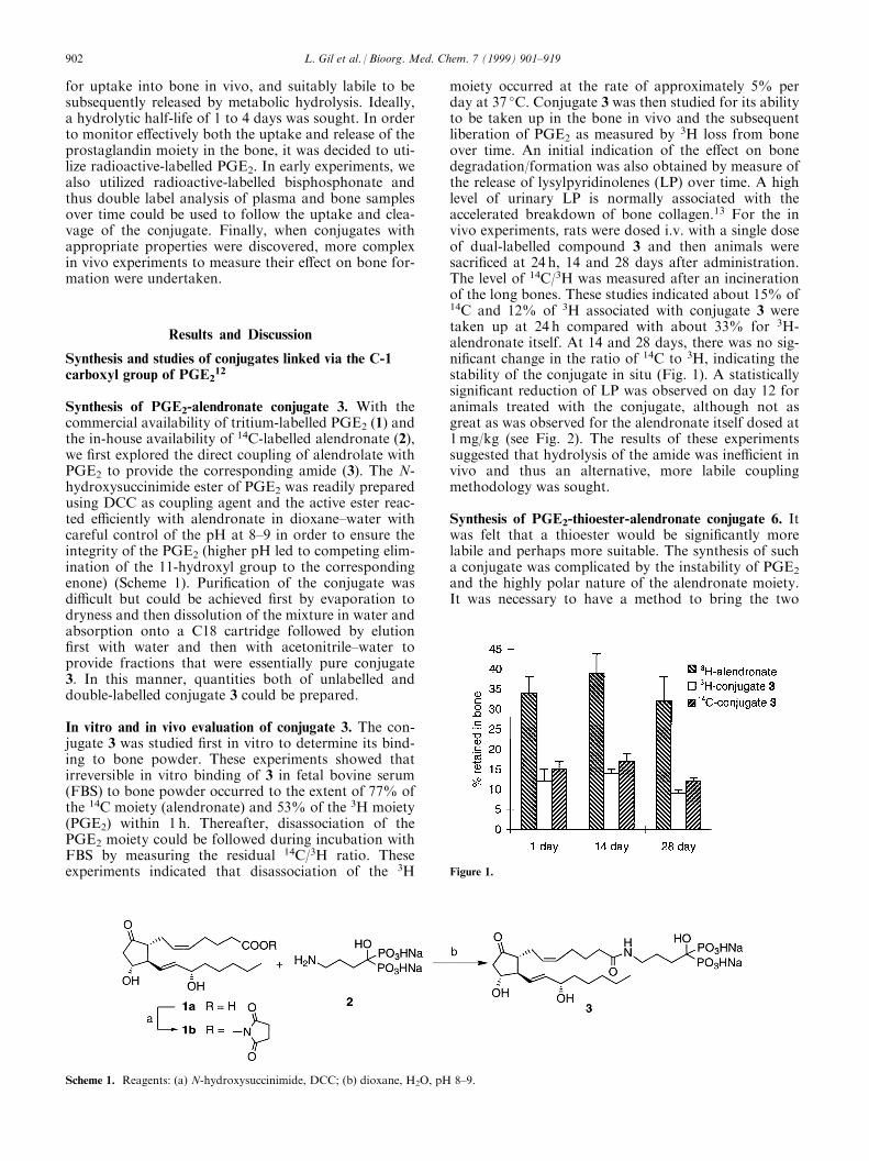
moiety occurred at the rate of approximately 5% perday at 37 �C. Conjugate 3 was then studied for its abilityto be taken up in the bone in vivo and the subsequentliberation of PGE2 as measured by 3H loss from boneover time. An initial indication of the e�ect on bonedegradation/formation was also obtained by measure ofthe release of lysylpyridinolenes (LP) over time. A highlevel of urinary LP is normally associated with theaccelerated breakdown of bone collagen.13 For the invivo experiments, rats were dosed i.v. with a single doseof dual-labelled compound 3 and then animals weresacri®ced at 24 h, 14 and 28 days after administration.The level of 14C/3H was measured after an incinerationof the long bones. These studies indicated about 15% of14C and 12% of 3H associated with conjugate 3 weretaken up at 24 h compared with about 33% for 3H-alendronate itself. At 14 and 28 days, there was no sig-ni®cant change in the ratio of 14C to 3H, indicating thestability of the conjugate in situ (Fig. 1). A statisticallysigni®cant reduction of LP was observed on day 12 foranimals treated with the conjugate, although not asgreat as was observed for the alendronate itself dosed at1mg/kg (see Fig. 2). The results of these experimentssuggested that hydrolysis of the amide was ine�cient invivo and thus an alternative, more labile couplingmethodology was sought.
Synthesis of PGE2-thioester-alendronate conjugate 6. Itwas felt that a thioester would be signi®cantly morelabile and perhaps more suitable. The synthesis of sucha conjugate was complicated by the instability of PGE2
and the highly polar nature of the alendronate moiety.It was necessary to have a method to bring the two
Scheme 1. Reagents: (a) N-hydroxysuccinimide, DCC; (b) dioxane, H2O, pH 8±9.
Figure 1.
902 L. Gil et al. / Bioorg. Med. Chem. 7 (1999) 901±919
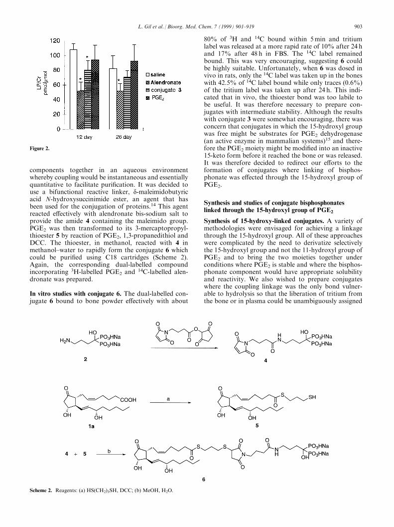
components together in an aqueous environmentwhereby coupling would be instantaneous and essentiallyquantitative to facilitate puri®cation. It was decided touse a bifunctional reactive linker, d-maleimidobutyricacid N-hydroxysuccinimide ester, an agent that hasbeen used for the conjugation of proteins.14 This agentreacted e�ectively with alendronate bis-sodium salt toprovide the amide 4 containing the maleimido group.PGE2 was then transformed to its 3-mercaptopropyl-thioester 5 by reaction of PGE2, 1,3-propanedithiol andDCC. The thioester, in methanol, reacted with 4 inmethanol±water to rapidly form the conjugate 6 whichcould be puri®ed using C18 cartridges (Scheme 2).Again, the corresponding dual-labelled compoundincorporating 3H-labelled PGE2 and 14C-labelled alen-dronate was prepared.
In vitro studies with conjugate 6. The dual-labelled con-jugate 6 bound to bone powder e�ectively with about
80% of 3H and 14C bound within 5min and tritiumlabel was released at a more rapid rate of 10% after 24 hand 17% after 48 h in FBS. The 14C label remainedbound. This was very encouraging, suggesting 6 couldbe highly suitable. Unfortunately, when 6 was dosed invivo in rats, only the 14C label was taken up in the boneswith 42.5% of 14C label bound while only traces (0.6%)of the tritium label was taken up after 24 h. This indi-cated that in vivo, the thioester bond was too labile tobe useful. It was therefore necessary to prepare con-jugates with intermediate stability. Although the resultswith conjugate 3 were somewhat encouraging, there wasconcern that conjugates in which the 15-hydroxyl groupwas free might be substrates for PGE2 dehydrogenase(an active enzyme in mammalian systems)15 and there-fore the PGE2 moiety might be modi®ed into an inactive15-keto form before it reached the bone or was released.It was therefore decided to redirect our e�orts to theformation of conjugates where linking of bisphos-phonate was e�ected through the 15-hydroxyl group ofPGE2.
Synthesis and studies of conjugate bisphosphonateslinked through the 15-hydroxyl group of PGE2
Synthesis of 15-hydroxy-linked conjugates. A variety ofmethodologies were envisaged for achieving a linkagethrough the 15-hydroxyl group. All of these approacheswere complicated by the need to derivatize selectivelythe 15-hydroxyl group and not the 11-hydroxyl group ofPGE2 and to bring the two moieties together underconditions where PGE2 is stable and where the bisphos-phonate component would have appropriate solubilityand reactivity. We also wished to prepare conjugateswhere the coupling linkage was the only bond vulner-able to hydrolysis so that the liberation of tritium fromthe bone or in plasma could be unambiguously assigned
Figure 2.
Scheme 2. Reagents: (a) HS(CH2)3SH, DCC; (b) MeOH, H2O.
L. Gil et al. / Bioorg. Med. Chem. 7 (1999) 901±919 903
to liberation of PGE2 itself. The formation of estersappeared to be the best option but initially it wasapparent that if PGE2 were to be directly acylated by abisphosphonate containing an acylating functionality,then the bisphosphonate moiety would have to be sui-tably protected and then the conjugate subsequentlydeprotected to yield the free bisphosphonate. Previousstudies on the preparation of steroid and methotrexatebisphosphonate conjugates16,17 have utilized bisphos-phonates protected as linear or branched alkyl (methyl,ethyl, isopropyl) esters. The deprotection of such estersfollowing conjugation was carried out in both cases bytreatment with bromotrimethylsilane at room tempera-ture for 2 to 3 days. We con®rmed that PGE2 wasunstable under these conditions. We chose instead toprepare benzyl-protected bisphosphonates and con-sidered that the alkylation of tetrabenzyl methylenedi-phosphonate (7a) was an appropriate route for synthesisof the required reagents.
We were unable to ®nd reports on the alkylation of tetra-benzylmethylene bisphosphonate and thus we undertook
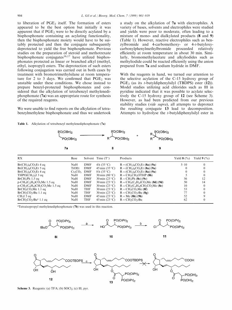
a study on the alkylation of 7a with electrophiles. Avariety of bases, solvents and electrophiles were studiedand yields were poor to moderate, often leading to amixture of mono- and dialkylated products (8 and 9)(Table 1). However, reactive electrophiles such as ben-zylbromide and 4-carbomethoxy- or 4-t-butyloxy-carbonylphenylmethylbromide proceeded relativelye�ciently at room temperature in about 30 min. Simi-larly, bromomethylacetate and alkyliodides such asmethyliodide could be reacted e�ciently using the anionprepared from 7a and sodium hydride in DMF.
With the reagents in hand, we turned our attention tothe selective acylation of the C-15 hydroxy group ofPGE2 (as its t-butyldiphenylsilyl (TBDPS) ester (12)).Model studies utilizing acid chlorides such as 11 inpyridine indicated that it was possible to acylate selec-tively the C-15 hydroxy group of 12 (see Scheme 3).However, as had been predicted from our previousstability studies (vide supra), all attempts to deprotectthe resulting conjugate 13 lead to decomposition.Attempts to hydrolyse the t-butyldiphenylsilyl ester in
Table 1. Alkylation of tetrabenzyl methylenediphosphonate (7a)
RX Base Solvent Time (T�) Products Yield 8 (%) Yield 9 (%)
Br(CH2)4CO2Et 4 eq NaH DMF 4h (35 �C) R� (CH2)4CO2Et (8a)/(9a) 5±10 0Br(CH2)4CO2Et 5 eq TiOEt DMF 45min (35 �C) R� (CH2)4CO2Et (8a)/(9a) 0 0Br(CH2)4CO2Et 4 eq Cs2CO3 DMF 8h (35 �C) R� (CH2)4CO2Et (8a)/(9a) 0 0THPO(CH2)2I 3 eq NaH DMF 30min (80 �C) R�CH2CH2OTHP (8b) 5 0BrCH2Ph 1.5 eq NaH DMF 30min (23 �C) R�CH2Ph (8c)/(9c) 56 12p-CH2(C6H4)CO2Me 1.5 eq NaH DMF 30min (23 �C) R�CH2(C6H4)CO2Me (8d)/(9d) 50 14p-CH2(C6H4)CH2CO2Me 1.5 eq NaH DMF 30min (23 �C) R�CH2(C6H4)CH2CO2Me (8e) 10 0BrCH2CO2Me 1.1 eq NaH THF 30min (23 �C) R�CH2CO2Me (8f) 53 0BrCH2CO2tBu 1.1 eq NaH THF 30min (23 �C) R�CH2CO2tBu (8g) 77 0CH3I 5 eq NaH DMF 45min (23 �C) R�Me (8h)/(9h) 52 9BrCH2CO2tBu
a 1.1 eq NaH THF 45min (23 �C) R�CH2CO2tBu 62 0
aTetraisopropyl methylenediphosphonate (7b) was used in this reaction.
Scheme 3. Reagents: (a) TFA; (b) SOCl2; (c) 11, pyr.
904 L. Gil et al. / Bioorg. Med. Chem. 7 (1999) 901±919
the corresponding tetrabenzyl bisphosphonate (8g) bytreatment with tri¯uoracetic acid (TFA) lead to partialdebenzylation, suggesting that this reagent was toounstable to be useful.
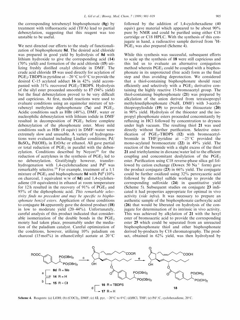
We next directed our e�orts to the study of functionali-zation of bisphosphonate 8d. The desired acid chloridewas prepared in good yield by hydrolysis of 8d withlithium hydroxide to give the corresponding acid (14)(70% yield) and formation of the acid chloride (15) uti-lizing freshly distilled oxalyl chloride in DMF. Thecrude acid chloride 15 was used directly for acylation ofPGE2-TBDPS in pyridine atÿ20 �C to 0 �C to provide thedesired C-15 acylated adduct 16 in 42% yield accom-panied with 31% recovered PGE2-TBDPS. Hydrolysisof the silyl ester proceeded smoothly to 17 (94% yield)but the ®nal debenzylation proved to be very di�cultand capricious. At ®rst, model reactions were used toevaluate conditions using an equimolar mixture of tet-rabenzyl methylene diphosphonate (7a) and PGE2.Acidic conditions such as HBr (48%), DMF, water ornucleophilic debenzylation with lithium iodide in DMFresulted in decomposition of PGE2 before completedebenzylation of the phosphonate ester. More mildconditions such as HBr (4 equiv) in DMF±water wereextremely slow and unusable. A variety of hydrogena-tions were evaluated including Lindlar conditions; Pd/BaSO4, Pd(OH)2 in EtOAc or ethanol. All gave partialor total reduction of PGE2 in parallel with the deben-zylation. Conditions described by Noyori18 for thereduction of acetylenes in the synthesis of PGE2 led tono debenzylation. Gratifyingly however, transfer-hydrogenation with 1,4-cyclohexadiene and Pd0 wasremarkably selective.19 For example, treatment of a 1/1mixture of PGE2 and bisphosphonate 8d with Pd0 (10%on charcoal, 1 equivalent w/w of 8d) and 1,4-cyclohex-adiene (10 equivalents) in ethanol at room temperaturefor 12 h resulted in the recovery of 91% of PGE2 and95% of the diphosphonic acid. This remarkable selec-tivity ®nds no precedent and may be speci®c to bispho-sphonate benzyl esters. Application of these conditionsto conjugate 16 apparently gave the desired product (18)in low to moderate yield (20±44%). Unfortunately,careful analysis of this product indicated that consider-able isomerization of the double bonds in the PGE2
moiety had taken place, presumably under the media-tion of the paladium catalyst. Careful optimization ofthe conditions, however, utilizing 10% paladium oncharcoal (15mol%) in ethanol/ethyl acetate at 20 �C
followed by the addition of 1,4-cyclohexadiene (40equiv) gave material which appeared to be about 90%pure by NMR and could be puri®ed using either C18cartridge or C18 HPLC. With the synthesis of this con-jugate in hand, a radioactive sample derived from 3H-PGE2 was also prepared (Scheme 4).
While this synthesis was successful, subsequent e�ortsto scale up the synthesis of 18 were still capricious andthis led us to evaluate an alternative conjugationmethod wherein PGE2 could be coupled with a bisphos-phonate in its unprotected (free acid) form as the ®nalstep and thus avoiding deprotection. We consideredthat a thiol-containing bisphosphonate should reacte�ciently and selectively with a PGE2 derivative con-taining the highly reactive 15-bromoacetyl group. Thethiol-containing bisphosphonate (21) was prepared byalkylation of the anion derived from tetraisopropylmethylenediphosphonate (NaH, DMF) with 3-acetyl-thiopropyliodide (19) to provide the thioacetate (20)in 90% yield. Hydrolysis of the thioester and the iso-propyl phosphonate esters proceeded concomitantly byre¯uxing in HCl followed by concentration to drynessunder high vacuum. The resulting thiol 21 was useddirectly without further puri®cation. Selective ester-i®cation of PGE2-TBDPS (12) with bromoacetyl-bromide in THF/pyridine at ÿ25 �C provided themono-acylated bromoacetate (22) in 49% yield. Thereaction of the bromide with a slight excess of the thiol21 and triethylamine in dioxane/water led to the e�cientcoupling and concomitant desilylation of the PGE2
ester. Puri®cation using C18 reverse-phase silica gel fol-lowed by cation exchange (Dowex 50 Na+ form) gavethe product conjugate (23) in 66% yield. The conjugatecould be further oxidized using 32% peroxyacetic acidfollowed by dimethyl sul®de workup to provide thecorresponding sulfoxide (24) in quantitative yield(Scheme 5). Subsequent studies on conjugate 23 indi-cated it had properties appropriate for optimal in vivoactivity (vide infra). It was necessary to prepare anauthentic sample of the bisphosphonate carboxylic acid(26) that would be liberated on hydrolysis of the con-jugate for determination of its intrinsic in vivo activity.This was achieved by alkylation of 21 with the hexylester of bromoacetic acid to provide the correspondingester 25 which could be separated from an unreactedbisphosphosphonate thiol and other bisphosphonatederived by-products by C18 chromatography. The prod-uct, obtained in 62% yield, was then hydrolyzed by
Scheme 4. Reagents: (a) LiOH; (b) (COCl)2, DMF; (c) 12, pyr, ÿ20�C to 0�C; (d)HCl, THF; (e) Pd�/C, cyclohexadiene, 20�C.
L. Gil et al. / Bioorg. Med. Chem. 7 (1999) 901±919 905
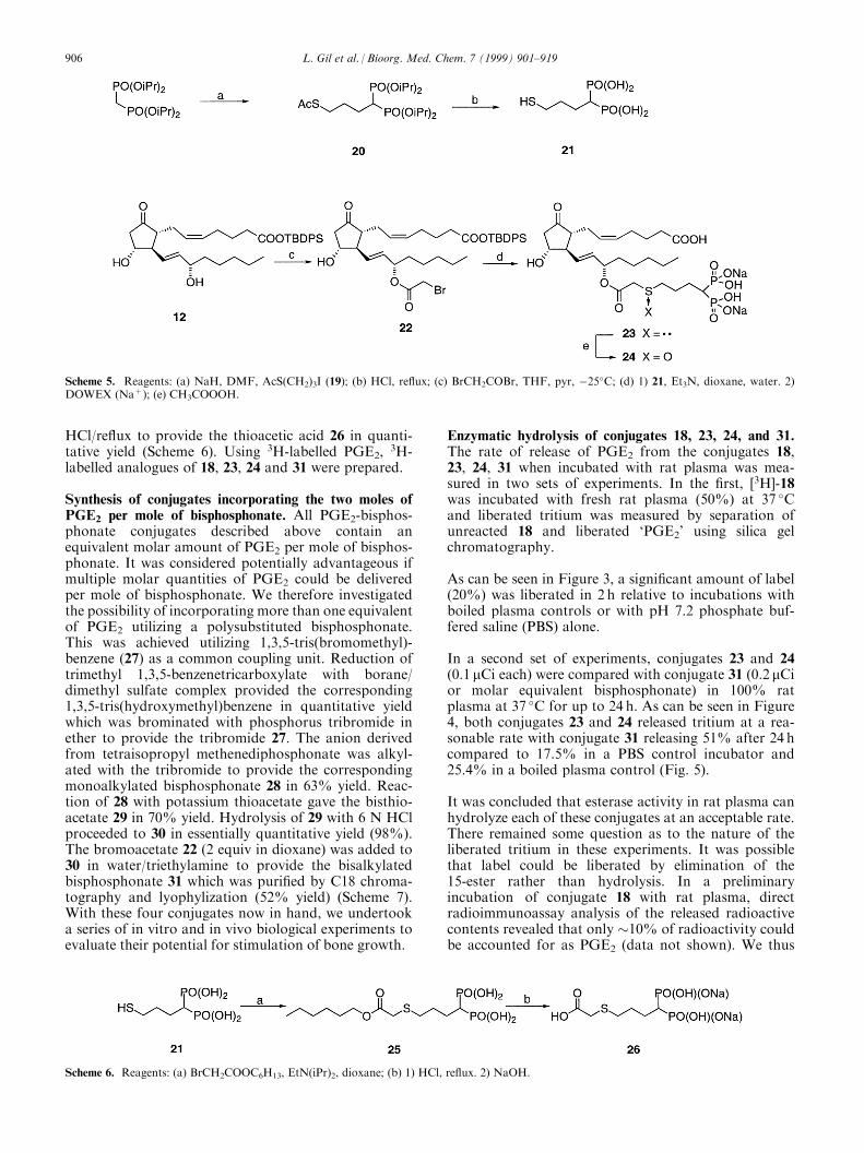
HCl/re¯ux to provide the thioacetic acid 26 in quanti-tative yield (Scheme 6). Using 3H-labelled PGE2,
3H-labelled analogues of 18, 23, 24 and 31 were prepared.
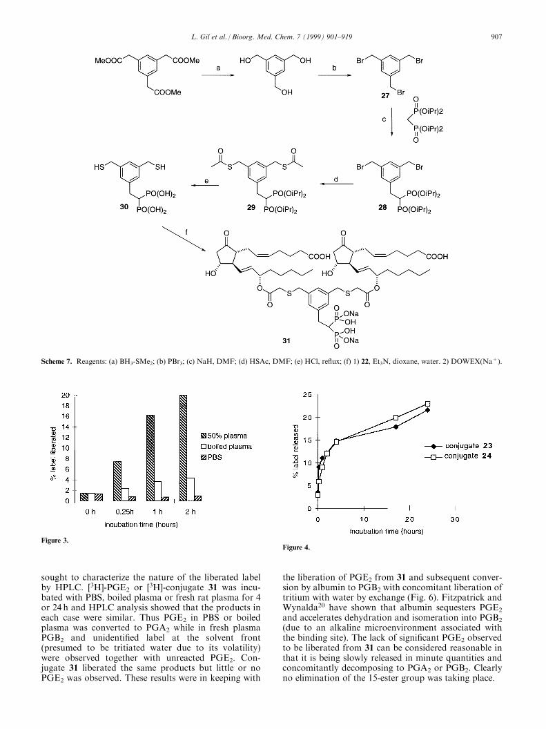
Synthesis of conjugates incorporating the two moles ofPGE2 per mole of bisphosphonate. All PGE2-bisphos-phonate conjugates described above contain anequivalent molar amount of PGE2 per mole of bisphos-phonate. It was considered potentially advantageous ifmultiple molar quantities of PGE2 could be deliveredper mole of bisphosphonate. We therefore investigatedthe possibility of incorporating more than one equivalentof PGE2 utilizing a polysubstituted bisphosphonate.This was achieved utilizing 1,3,5-tris(bromomethyl)-benzene (27) as a common coupling unit. Reduction oftrimethyl 1,3,5-benzenetricarboxylate with borane/dimethyl sulfate complex provided the corresponding1,3,5-tris(hydroxymethyl)benzene in quantitative yieldwhich was brominated with phosphorus tribromide inether to provide the tribromide 27. The anion derivedfrom tetraisopropyl methenediphosphonate was alkyl-ated with the tribromide to provide the correspondingmonoalkylated bisphosphonate 28 in 63% yield. Reac-tion of 28 with potassium thioacetate gave the bisthio-acetate 29 in 70% yield. Hydrolysis of 29 with 6 N HClproceeded to 30 in essentially quantitative yield (98%).The bromoacetate 22 (2 equiv in dioxane) was added to30 in water/triethylamine to provide the bisalkylatedbisphosphonate 31 which was puri®ed by C18 chroma-tography and lyophylization (52% yield) (Scheme 7).With these four conjugates now in hand, we undertooka series of in vitro and in vivo biological experiments toevaluate their potential for stimulation of bone growth.
Enzymatic hydrolysis of conjugates 18, 23, 24, and 31.The rate of release of PGE2 from the conjugates 18,23, 24, 31 when incubated with rat plasma was mea-sured in two sets of experiments. In the ®rst, [3H]-18was incubated with fresh rat plasma (50%) at 37 �Cand liberated tritium was measured by separation ofunreacted 18 and liberated `PGE2' using silica gelchromatography.
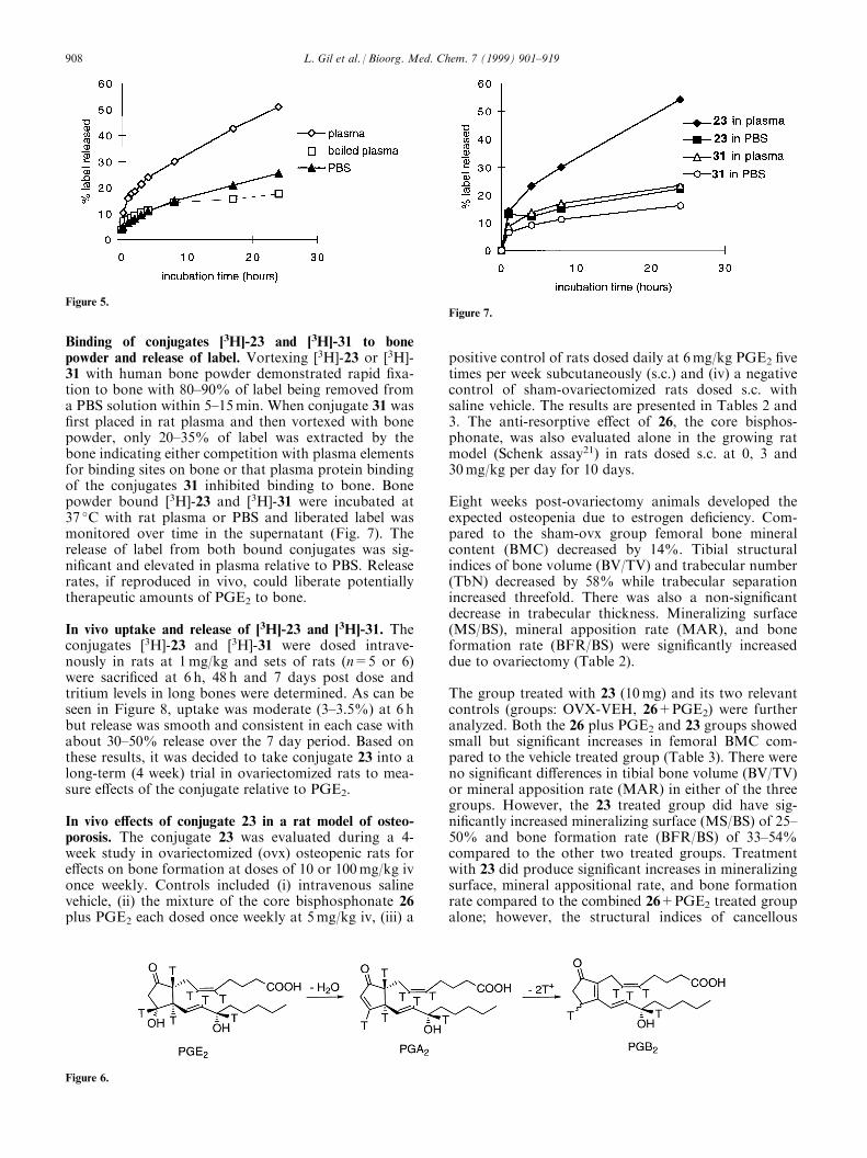
As can be seen in Figure 3, a signi®cant amount of label(20%) was liberated in 2 h relative to incubations withboiled plasma controls or with pH 7.2 phosphate buf-fered saline (PBS) alone.
In a second set of experiments, conjugates 23 and 24(0.1 mCi each) were compared with conjugate 31 (0.2 mCior molar equivalent bisphosphonate) in 100% ratplasma at 37 �C for up to 24 h. As can be seen in Figure4, both conjugates 23 and 24 released tritium at a rea-sonable rate with conjugate 31 releasing 51% after 24 hcompared to 17.5% in a PBS control incubator and25.4% in a boiled plasma control (Fig. 5).
It was concluded that esterase activity in rat plasma canhydrolyze each of these conjugates at an acceptable rate.There remained some question as to the nature of theliberated tritium in these experiments. It was possiblethat label could be liberated by elimination of the15-ester rather than hydrolysis. In a preliminaryincubation of conjugate 18 with rat plasma, directradioimmunoassay analysis of the released radioactivecontents revealed that only �10% of radioactivity couldbe accounted for as PGE2 (data not shown). We thus
Scheme 5. Reagents: (a) NaH, DMF, AcS(CH2)3I (19); (b) HCl, re¯ux; (c) BrCH2COBr, THF, pyr, ÿ25�C; (d) 1) 21, Et3N, dioxane, water. 2)DOWEX (Na+); (e) CH3COOOH.
Scheme 6. Reagents: (a) BrCH2COOC6H13, EtN(iPr)2, dioxane; (b) 1) HCl, re¯ux. 2) NaOH.
906 L. Gil et al. / Bioorg. Med. Chem. 7 (1999) 901±919
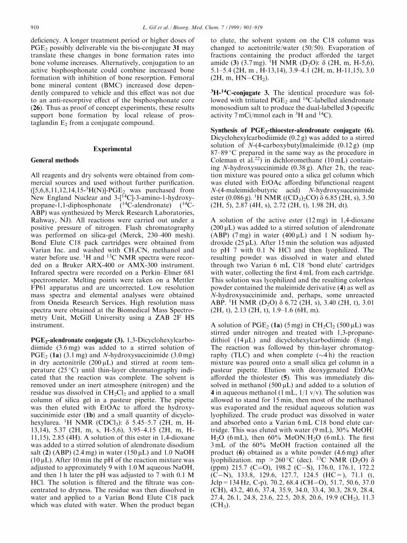
sought to characterize the nature of the liberated labelby HPLC. [3H]-PGE2 or [3H]-conjugate 31 was incu-bated with PBS, boiled plasma or fresh rat plasma for 4or 24 h and HPLC analysis showed that the products ineach case were similar. Thus PGE2 in PBS or boiledplasma was converted to PGA2 while in fresh plasmaPGB2 and unidenti®ed label at the solvent front(presumed to be tritiated water due to its volatility)were observed together with unreacted PGE2. Con-jugate 31 liberated the same products but little or noPGE2 was observed. These results were in keeping with
the liberation of PGE2 from 31 and subsequent conver-sion by albumin to PGB2 with concomitant liberation oftritium with water by exchange (Fig. 6). Fitzpatrick andWynalda20 have shown that albumin sequesters PGE2
and accelerates dehydration and isomeration into PGB2
(due to an alkaline microenvironment associated withthe binding site). The lack of signi®cant PGE2 observedto be liberated from 31 can be considered reasonable inthat it is being slowly released in minute quantities andconcomitantly decomposing to PGA2 or PGB2. Clearlyno elimination of the 15-ester group was taking place.
Scheme 7. Reagents: (a) BH3-SMe2; (b) PBr3; (c) NaH, DMF; (d) HSAc, DMF; (e) HCl, re¯ux; (f) 1) 22, Et3N, dioxane, water. 2) DOWEX(Na+).
Figure 3.Figure 4.
L. Gil et al. / Bioorg. Med. Chem. 7 (1999) 901±919 907
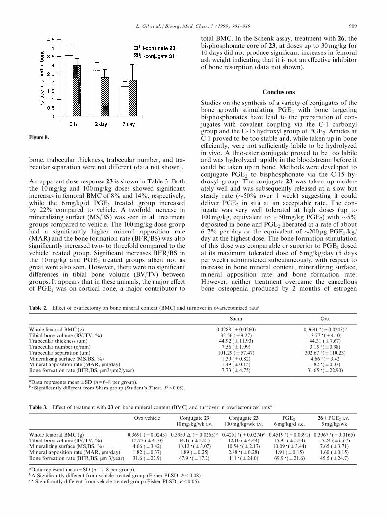
Binding of conjugates [3H]-23 and [3H]-31 to bonepowder and release of label. Vortexing [3H]-23 or [3H]-31 with human bone powder demonstrated rapid ®xa-tion to bone with 80±90% of label being removed froma PBS solution within 5±15min. When conjugate 31 was®rst placed in rat plasma and then vortexed with bonepowder, only 20±35% of label was extracted by thebone indicating either competition with plasma elementsfor binding sites on bone or that plasma protein bindingof the conjugates 31 inhibited binding to bone. Bonepowder bound [3H]-23 and [3H]-31 were incubated at37 �C with rat plasma or PBS and liberated label wasmonitored over time in the supernatant (Fig. 7). Therelease of label from both bound conjugates was sig-ni®cant and elevated in plasma relative to PBS. Releaserates, if reproduced in vivo, could liberate potentiallytherapeutic amounts of PGE2 to bone.
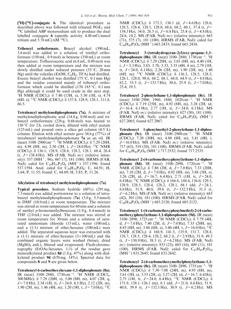
In vivo uptake and release of [3H]-23 and [3H]-31. Theconjugates [3H]-23 and [3H]-31 were dosed intrave-nously in rats at 1mg/kg and sets of rats (n=5 or 6)were sacri®ced at 6 h, 48 h and 7 days post dose andtritium levels in long bones were determined. As can beseen in Figure 8, uptake was moderate (3±3.5%) at 6 hbut release was smooth and consistent in each case withabout 30±50% release over the 7 day period. Based onthese results, it was decided to take conjugate 23 into along-term (4 week) trial in ovariectomized rats to mea-sure e�ects of the conjugate relative to PGE2.
In vivo e�ects of conjugate 23 in a rat model of osteo-porosis. The conjugate 23 was evaluated during a 4-week study in ovariectomized (ovx) osteopenic rats fore�ects on bone formation at doses of 10 or 100mg/kg ivonce weekly. Controls included (i) intravenous salinevehicle, (ii) the mixture of the core bisphosphonate 26plus PGE2 each dosed once weekly at 5mg/kg iv, (iii) a
positive control of rats dosed daily at 6mg/kg PGE2 ®vetimes per week subcutaneously (s.c.) and (iv) a negativecontrol of sham-ovariectomized rats dosed s.c. withsaline vehicle. The results are presented in Tables 2 and3. The anti-resorptive e�ect of 26, the core bisphos-phonate, was also evaluated alone in the growing ratmodel (Schenk assay21) in rats dosed s.c. at 0, 3 and30mg/kg per day for 10 days.
Eight weeks post-ovariectomy animals developed theexpected osteopenia due to estrogen de®ciency. Com-pared to the sham-ovx group femoral bone mineralcontent (BMC) decreased by 14%. Tibial structuralindices of bone volume (BV/TV) and trabecular number(TbN) decreased by 58% while trabecular separationincreased threefold. There was also a non-signi®cantdecrease in trabecular thickness. Mineralizing surface(MS/BS), mineral apposition rate (MAR), and boneformation rate (BFR/BS) were signi®cantly increaseddue to ovariectomy (Table 2).
The group treated with 23 (10mg) and its two relevantcontrols (groups: OVX-VEH, 26+PGE2) were furtheranalyzed. Both the 26 plus PGE2 and 23 groups showedsmall but signi®cant increases in femoral BMC com-pared to the vehicle treated group (Table 3). There wereno signi®cant di�erences in tibial bone volume (BV/TV)or mineral apposition rate (MAR) in either of the threegroups. However, the 23 treated group did have sig-ni®cantly increased mineralizing surface (MS/BS) of 25±50% and bone formation rate (BFR/BS) of 33±54%compared to the other two treated groups. Treatmentwith 23 did produce signi®cant increases in mineralizingsurface, mineral appositional rate, and bone formationrate compared to the combined 26+PGE2 treated groupalone; however, the structural indices of cancellous
Figure 5.
Figure 6.
Figure 7.
908 L. Gil et al. / Bioorg. Med. Chem. 7 (1999) 901±919
bone, trabecular thickness, trabecular number, and tra-becular separation were not di�erent (data not shown).
An apparent dose response 23 is shown in Table 3. Boththe 10mg/kg and 100mg/kg doses showed signi®cantincreases in femoral BMC of 8% and 14%, respectively,while the 6mg/kg/d PGE2 treated group increasedby 22% compared to vehicle. A twofold increase inmineralizing surfact (MS/BS) was seen in all treatmentgroups compared to vehicle. The 100mg/kg dose grouphad a signi®cantly higher mineral apposition rate(MAR) and the bone formation rate (BFR/BS) was alsosigni®cantly increased two- to threefold compared to thevehicle treated group. Signi®cant increases BFR/BS inthe 10mg/kg and PGE2 treated groups albeit not asgreat were also seen. However, there were no signi®cantdi�erences in tibial bone volume (BV/TV) betweengroups. It appears that in these animals, the major e�ectof PGE2 was on cortical bone, a major contributor to
total BMC. In the Schenk assay, treatment with 26, thebisphosphonate core of 23, at doses up to 30mg/kg for10 days did not produce signi®cant increases in femoralash weight indicating that it is not an e�ective inhibitorof bone resorption (data not shown).
Conclusions
Studies on the synthesis of a variety of conjugates of thebone growth stimulating PGE2 with bone targetingbisphosphonates have lead to the preparation of con-jugates with covalent coupling via the C-1 carbonylgroup and the C-15 hydroxyl group of PGE2. Amides atC-1 proved to be too stable and, while taken up in bonee�ciently, were not su�ciently labile to be hydrolyzedin vivo. A thio-ester conjugate proved to be too labileand was hydrolyzed rapidly in the bloodstream before itcould be taken up in bone. Methods were developed toconjugate PGE2 to bisphosphonate via the C-15 hy-droxyl group. The conjugate 23 was taken up moder-ately well and was subsequently released at a slow butsteady rate (�50% over 1 week) suggesting it coulddeliver PGE2 in situ at an acceptable rate. The con-jugate was very well tolerated at high doses (up to100mg/kg, equivalent to �50mg/kg PGE2) with �5%deposited in bone and PGE2 liberated at a rate of about6±7% per day or the equivalent of �200 mg PGE2/kg/day at the highest dose. The bone formation stimulationof this dose was comparable or superior to PGE2 dosedat its maximum tolerated dose of 6mg/kg/day (5 daysper week) administered subcutaneously, with respect toincrease in bone mineral content, mineralizing surface,mineral apposition rate and bone formation rate.However, neither treatment overcame the cancellousbone osteopenia produced by 2 months of estrogen
Figure 8.
Table 2. E�ect of ovariectomy on bone mineral content (BMC) and turnover in ovariectomized ratsa
Sham Ovx
Whole femoral BMC (g) 0.4288 (�0.0260) 0.3691 � (�0.0243)b
Tibial bone volume (BV/TV, %) 32.56 (�9.27) 13.77 � (�4.10)Trabecular thickness (mm) 44.92 (�11.93) 44.31 (�7.67)Trabecular number (#/mm) 7.56 (�1.99) 3.15 � (�0.98)Trabecular separation (mm) 101.29 (�57.47) 302.67 � (�110.23)Mineralizing surface (MS/BS, %) 1.39 (�0.82) 4.66 � (�3.42Mineral apposition rate (MAR, mm/day) 1.49 (�0.15) 1.82 � (�0.37)Bone formation rate (BFR/BS, mm3/mm2/year) 7.73 (�4.75) 31.65 � (�22.90)
aData represents mean�SD (n=6±8 per group).b �Signi®cantly di�erent from Sham group (Student's T test, P<0.05).
Table 3. E�ect of treatment with 23 on bone mineral content (BMC) and turnover in ovariectomized ratsa
Ovx vehicle Conjugate 2310mg/kg/wk i.v.
Conjugate 23100mg/kg/wk i.v.
PGE2
6mg/kg/d s.c.26+PGE2 i.v.5mg/kg/wk
Whole femoral BMC (g) 0.3691 (�0.0243) 0.3969 � (�0.0265)b 0.4201 � (�0.0274)c 0.4519 � (�0.0391) 0.3967 � (�0.0165)Tibial bone volume (BV/TV, %) 13.77 (�4.10) 14.16 (�3.21) 12.10 (�4.44) 15.93 (�5.34) 15.24 (�6.67)Mineralizing surface (MS/BS, %) 4.66 (�3.42) 10.13 � (�3.07) 10.54 � (�2.17) 10.09 � (�3.44) 7.65 (�3.71)Mineral apposition rate (MAR, mm/day) 1.82 (�0.37) 1.89 (�0.25) 2.88 � (�0.28) 1.91 (�0.15) 1.60 (�0.15)Bone formation rate (BFR/BS, mm 3/year) 31.6 (�22.9) 67.9 � (�17.2) 111 � (�24.0) 69.9 � (�21.6) 45.5 (�24.7)
aData represent mean�SD (n=7±8 per group).b� Signi®cantly di�erent from vehicle treated group (Fisher PLSD, P<0.08).c � Signi®cantly di�erent from vehicle treated group (Fisher PLSD, P<0.05).
L. Gil et al. / Bioorg. Med. Chem. 7 (1999) 901±919 909
de®ciency. A longer treatment period or higher doses ofPGE2 possibly deliverable via the bis-conjugate 31 maytranslate these changes in bone formation rates intobone volume increases. Alternatively, conjugation to anactive bisphosphonate could combine increased boneformation with inhibition of bone resorption. Femoralbone mineral content (BMC) increased dose depen-dently compared to vehicle and this e�ect was not dueto an anti-resorptive e�ect of the bisphosphonate core(26). Thus as proof of concept experiments, these resultssupport bone formation by local release of pros-taglandin E2 from a conjugate compound.
Experimental
General methods
All reagents and dry solvents were obtained from com-mercial sources and used without further puri®cation.([5,6,8,11,12,14,15-3H(N)]-PGE2 was purchased fromNew England Nuclear and 3-[14C]-3-amino-1-hydroxy-propane-1,1-diphosphonate (14C-alendronate) (14C-ABP) was synthesized by Merck Research Laboratories,Rahway, NJ). All reactions were carried out under apositive pressure of nitrogen. Flash chromatographywas performed on silica-gel (Merck, 230±400 mesh).Bond Elute C18 pack cartridges were obtained fromVarian Inc. and washed with CH3CN, methanol andwater before use. 1H and 13C NMR spectra were recor-ded on a Bruker ARX-400 or AMX-300 instrument.Infrared spectra were recorded on a Perkin±Elmer 681spectrometer. Melting points were taken on a MettlerFP61 apparatus and are uncorrected. Low resolutionmass spectra and elemental analyses were obtainedfrom Oneida Research Services. High resolution massspectra were obtained at the Biomedical Mass Spectro-metry Unit, McGill University using a ZAB 2F HSinstrument.
PGE2-alendronate conjugate (3). 1,3-Dicyclohexylcarbo-diimide (3.6mg) was added to a stirred solution ofPGE2 (1a) (3.1mg) and N-hydroxysuccinimide (3.0mg)in dry acetonitrile (200 mL) and stirred at room tem-perature (25 �C) until thin-layer chromatography indi-cated that the reaction was complete. The solvent isremoved under an inert atmosphere (nitrogen) and theresidue was dissolved in CH2Cl2 and applied to a smallcolumn of silica gel in a pasteur pipette. The pipettewas then eluted with EtOAc to a�ord the hydroxy-succinimide ester (1b) and a small quantity of dicyclo-hexylurea. 1H NMR (CDCl3): d 5.45±5.7 (2H, m, H-13,14), 5.37 (2H, m, s, H-5,6), 3.95±4.15 (2H, m, H-11,15), 2.85 (4H). A solution of this ester in 1,4-dioxanewas added to a stirred solution of alendronate disodiumsalt (2) (ABP) (2.4mg) in water (150 mL) and 1.0 NaOH(10 mL). After 10min the pH of the reaction mixture wasadjusted to approximately 9 with 1.0M aqueous NaOH,and then 1 h later the pH was adjusted to 7 with 0.1 MHCl. The solution is ®ltered and the ®ltrate was con-centrated to dryness. The residue was then dissolved inwater and applied to a Varian Bond Elute C18 packwhich was eluted with water. When the product began
to elute, the solvent system on the C18 column waschanged to acetonitrile/water (50/50). Evaporation offractions containing the product a�orded the targetamide (3) (3.7mg). 1H NMR (D2O): d (2H, m, H-5,6),5.1±5.4 (2H, m , H-13,14), 3.9±4.1 (2H, m, H-11,15), 3.0(2H, m, HNÿCH2).
3H-14C-conjugate 3. The identical procedure was fol-lowed with tritiated PGE2 and
14C-labelled alendronatemonosodium salt to produce the dual-labelled 3 (speci®cactivity 7mCi/mmol each in 3H and 14C).
Synthesis of PGE2-thioester-alendronate conjugate (6).Dicyclohexylcarbodiimide (0.2 g) was added to a stirredsolution of N-(4-carboxybutyl)maleimide (0.12 g) (mp87±89 �C prepared in the same way as the procedure inColeman et al.22) in dichloromethane (10mL) contain-ing N-hydroxysuccinimide (0.38 g). After 2 h, the reac-tion mixture was poured onto a silica gel column whichwas eluted with EtOAc a�ording bifunctional reagentN-(4-maleimidobutyric acid) N-hydroxysuccinimideester (0.086 g). 1H NMR ((CD3)2CO) d 6.85 (2H, s), 3.50(2H, 5), 2.87 (4H, s), 2.72 (2H, t), 1.98 2H, dt).
A solution of the active ester (12mg) in 1,4-dioxane(200 mL) was added to a stirred solution of alendronate(ABP) (7mg) in water (400 mL) and 1 N sodium hy-droxide (25 mL). After 15min the solution was adjustedto pH 7 with 0.1 N HCl and then lyophilized. Theresulting powder was dissolved in water and elutedthrough two Varian 6 mL C18 `bond elute' cartridgeswith water, collecting the ®rst 4mL from each cartridge.This solution was lyophilized and the resulting colorlesspowder contained the maleimide derivative (4) as well asN-hydroxysuccinimide and, perhaps, some unreactedABP. 1H NMR (D2O) d 6.72 (2H, s), 3.40 (2H, t), 3.01(2H, t), 2.13 (2H, t), 1.9±1.6 (6H, m).
A solution of PGE2 (1a) (5mg) in CH2Cl2 (500 mL) wasstirred under nitrogen and treated with 1,3-propane-dithiol (14 mL) and dicyclohexylcarbodiimide (8mg).The reaction was followed by thin-layer chromatog-raphy (TLC) and when complete (�4 h) the reactionmixture was poured onto a small silica gel column in apasteur pipette. Elution with deoxygenated EtOAca�orded the thiolester (5). This was immediately dis-solved in methanol (500 mL) and added to a solution of4 in aqueous methanol (1mL, 1/1 v/v). The solution wasallowed to stand for 15min, then most of the methanolwas evaporated and the residual aqueous solution waslyophilized. The crude product was dissolved in waterand absorbed onto a Varian 6mL C18 bond elute car-tridge. This was eluted with water (9mL), 30% MeOH/H2O (6mL), then 60% MeON/H2O (6mL). The ®rst3mL of the 60% MeOH fraction contained all theproduct (6) obtained as a white powder (4.6mg) afterlyophilization. mp >260 �C (dec). 13C NMR (D2O) d(ppm) 215.7 (C�O), 198.2 (CÿS), 176.0, 176.1, 172.2(CÿN), 133.8, 129.6, 127.7, 124.5 (HC=), 71.1 (t,Jclp=134Hz, C-p), 70.2, 68.4 (CHÿO), 51.7, 50.6, 37.0(CH), 43.2, 40.6, 37.4, 35.9, 34.0, 33.4, 30.3, 28.9, 28.4,27.4, 26.1, 24.8, 23.6, 22.5, 20.8, 20.6, 19.9 (CH2), 11.3(CH3).
910 L. Gil et al. / Bioorg. Med. Chem. 7 (1999) 901±919
[3H]-[14C]-conjugate 6. The identical procedure asdescribed above was followed with tritiated PGE2 and14C labelled ABP monosodium salt to produce the duallabelled conjugate 6 (speci®c activity 4.48mCi/mmoltritium and 5.76mCi/nM 14C).
Tribenzyl orthoformate. Benzyl alcohol (390mL,3.6mol) was added to a solution of triethyl ortho-formate (150mL, 0.9mol) in benzene (350mL) at roomtemperature. Tri¯uoroacetic acid (6.8mL, 0.09mol) wasthen added at room temperature and the mixture wasslowly distilled under reduced pressure (35 �C, 20mmHg) until the volatiles (EtOH, C6H6, TFA) had distilled.Excess benzyl alcohol was distilled (75 �C, 0.1mm Hg)and the residue consisted mainly of tribenzyl ortho-formate which could be distilled (170±185 �C, 0.1mmHg) although it could be used crude in the next step.1H NMR (CDCl3): d 7.40 (15H, s), 5.50 (1H, s), 4.74(6H, s); 13C NMR (CDCl3): d 137.8, 128.9, 128.1, 111.8,66.5.
Tetrabenzyl methylenediphosphonate (7a). A mixture ofmethylenediphosphonic acid (14.8 g, 0.08mol) and tri-benzyl orthoformate (226 g, 0.68mol) was heated to150 �C for 2 h, cooled down, diluted with ethyl acetate(125mL) and poured onto a silica gel column (4.5 L)column. Elution with ethyl acetate gave 34.6 g (77%) oftetrabenzyl methylenediphosphonate 7a as an oil. IR(neat) 3100±2900 cmÿ1. 1H NMR (CDCl3): d 7.29 (20H,m), 4.98 (8H, m), 2.50 (2H, t, J=24.0Hz); 13C NMR(CDCl3): d 136.1, 128.9, 128.8, 128.2, 128.1, 68.4, 26.4(t, J=138.4Hz). MS (FAB, NaI) m/z (relative inten-sity): 537 (MH+, 96), 447 (7), 181 (100). HRMS (FAB,NaI): calcd for C29H31P2O6 (MH+) 537.1596; found537.1594. Anal. calcd for C29H31P2O6: C, 64.91; H,5.64; P, 11.55; found: C, 64.69; H, 5.85; P, 11.26.
Alkylation of tetrabenzyl methylenediphosphonate (7a)
Typical procedure. Sodium hydride (60%) (291mg,7.3mmol) was added portionwise to a solution of tetra-benzyl methylenediphosphonate (7a) (3.0 g, 5.5mmol)in DMF (10.0mL) at room temperature. The mixturewas stirred at room temperature for 60min and a solutionof methyl p-bromomethylbenzoate (1.9 g, 8.4mmol) inTHF (2.0mL) was added. The mixture was stirred atroom temperature for 30min and a solution of satu-rated ammonium chloride (15mL), water (100mL),and a (1/1) mixture of ether/hexanes (100mL) wereadded. The separated aqueous layer was extracted witha (1/1) mixture of ether/hexanes (3�100mL) and thecombined organic layers were washed (brine), dried(MgSO4 anh.), ®ltered and evaporated. Flash-chroma-tography (EtOAc/hexanes, 1/1) of the residue gavemonoalkylated product 8d (1.8 g, 47%) along with dial-kylated product 9d (670mg, 14%). Spectral data forcompounds 8 and 9 are given below.
Tetrabenzyl 6-carboethoxyhexane-1,1-diphosphonate (8a).IR (neat) 3100±2880, 1730 cmÿ1. 1H NMR (CDCl3,300MHz): d 7.29 (20H, s), 5.00 (8H, m), 4.07 (2H, q,J=7.0Hz), 2.34 (1H, tt, J=24.0, 6.1Hz), 2.12 (2H, m),1.90 (2H, m), 1.46 (4H, m), 1.20 (3H, t, J=7.0Hz); 13C
NMR (CDCl3): d 173.3, 136.1 (d, J=4.6Hz) 128.6,128.5, 128.4, 128.1, 128.0, 68.6, 68.2, 60.1, 37.4 (t, J=134.3Hz), 34.0, 28.5 (t, J=6.8Hz), 25.4 (t, J=4.8Hz),24.6, 14.2. MS (FAB, NaI) m/z (relative intensity): 665(71), 575 (7), 181 (100). HRMS (FAB, NaI): calcd forC36H43P2O8 (MH+) 665.2433; found 665.2434.
Tetrabenzyl 3-(tetrahydropyran-2yl)oxy-propane-1,1-diphosphonate (8b). IR (neat) 3100±2880, 1730 cmÿ1. 1HNMR (CDCl3): d 7.29 (20H, s), 5.01 (8H, m), 4.40 (1H,t, J=3.3Hz), 3.83, 3.70, 3.53, 3.35 (4H, 4 m), 2.79 (1H,tt, J=24.0, 6.1Hz), 2.26 (2H, m), 1.90 (2H, m), 1.40(6H, m); 13C NMR (CDCl3): d 136.3, 128.5, 128.3,128.1, 128.0, 98.6, 68.2, 68.1, 68.0, 64.9 (t, J=4.8Hz),62.2, 33.5 (t, J=133.7Hz), 30.6, 25.9 (t, J=7.0Hz),25.4, 19.5.
Tetrabenzyl 2-phenylethane-1,1-diphosphonate (8c). IR(neat) 3100±2900, 1960, 1880, 1820 cmÿ1. 1H NMR(CDCl3): d 7.19 (25H, m), 4.92 (8H, m), 3.28 (2H, td,J=16.4, 6.1Hz), 2.77 (1H, tt, J=24.0, 6.1Hz). MS(FAB, NaI) m/z (relative intensity): 627 (50), 181 (100).HRMS (FAB, NaI): calcd for C36H37P2O6 (MH+)627.2065; found 627.2067.
Tetrabenzyl 1-phenylmethyl-2-phenylethane-1,1-diphos-phonate (9c). IR (neat) 3100±2900 cmÿ1. 1H NMR(CDCl3): 7.20 (30H, m), 4.80 (8H, m), 3.41 (4H, t,J=16.0Hz). MS (FAB, NaI) m/z (relative intensity):717 (65), 519 (20), 181 (100). HRMS (FAB, NaI): calcdfor C43H43P2O6 (MH+) 717.2532; found 717.2535.
Tetrabenzyl 2-(4-carbomethoxyphenyl)ethane-1,1-diphos-phonate (8d). IR (neat) 3100±2890, 1720 cmÿ1. 1HNMR (CDCl3): d 7.80 (2H, d, J=7.0Hz), 7.33 (20H,m), 7.10 (2H, d, J=7.0Hz), 4.92 (8H, m), 3.88 (3H, s),3.26 (2H, td, J=16.7, 6.4Hz), 2.71 (1H, tt, J=24.0,6.4Hz); 13C NMR (CDCl3): d 166.8, 144.4, 136.0, 129.5,128.9, 128.5, 128.4, 128.2, 128.1, 68.1 (dd, J=24.1,6.6Hz), 51.9, 40.8, 39.6 (t, J=132.5Hz), 31.3 (t,J=6.2Hz). MS (FAB, NaI) m/z (relative intensity): 685(42), 301 (10), 181 (100). HRMS (FAB, NaI): calcd forC38H39P2O8 (MH+) 685.2120; found 685.2122.
Tetrabenzyl 1-(4-carbomethoxyphenylmethyl)-2-(4-carbo-methoxy)phenyl)ethane-1,1-diphosphonate (9d). IR (neat)3100±2890, 1725 cmÿ1. 1H NMR (CDCl3): d 7.79 (4H,d, J=7.0Hz), 7.40 (2H, d, J=7.0Hz), 7.33 (20H, m),4.85 (8H, m), 3.86 (6H, s), 3.40 (4H, t, J=16.0Hz); 13CNMR (CDCl3): d 168.9, 141.5, 135.9, 131.7, 128.8,128.7, 128.5, 128.4, 128.2, 68.2 (t, J=2.9Hz), 51.9, 49.3(t, J=130.9Hz), 38.3 (t, J=6.2Hz). MS (FAB, NaI)m/z (relative intensity): 833 (23), 603 (16), 449 (11), 181(100). HRMS (FAB, NaI): calcd for C47H47P2O10
(MH+) 833.2645; found 833.2642.
Tetrabenzyl 2-(4-carbomethoxymethylphenyl)ethane-1,1-diphosphonate (8e). IR (neat) 3100±2890, 1735 cmÿ1. 1HNMR (CDCl3): d 7.30±7.08 (24H, m), 4.95 (8H, m),3.61 (3H, s), 3.53 (2H, s), 3.27 (2H, td, J=16.7, 6.4Hz),2.75 (1H, tt, J=24.0, 6.4Hz); 13C NMR (CDCl3): d171.9, 138.1±126.1 (m), 8.1 (dd, J=21.0, 6.6Hz), 51.9,40.8, 39.9 (t, J=132.1Hz), 30.9 (t, J=6.2Hz). MS
L. Gil et al. / Bioorg. Med. Chem. 7 (1999) 901±919 911
(FAB, NaI) m/z (relative intensity): 699 (21), 537 (15),271 (15), 205 (16), 197 (16), 193 (24), 181 (100). HRMS(FAB, NaI): calcd for C39H41P2O8 (MH+) 699.2277;found 699.2276.
Tetrabenzyl 2-carbomethoxyethane-1,1-diphosphonate (8f).IR (neat) 3100±2860, 1740 cmÿ1. 1H NMR (CDCl3): d7.27 (20H, m), 5.02 (8H, m), 3.43 (3H, s), 3.25 (1H, tt,J=24.0, 6.1Hz), 2.83 (1H, td, J=16.4, 6.1Hz. MS(FAB, NaI) m/z (relative intensity): 609 (42), 181 (100).HRMS (FAB, NaI): calcd for C32H35P2O8 (MH+)609.1807; found 609.1807.
Tetrabenzyl 2-t-butoxycarbonylethane-1,1-diphosphonate(8g). IR (neat) 3100±2890, 1740 cmÿ1. 1H NMR(CDCl3): d 7.28 (20H, m), 5.02 (8H, m), 3.30 (1H, tt,J=24.0, 6.1Hz), 2.82 (2H, td, J=16.4, 6.1Hz); 13CNMR (CDCl3): d 169.6 (t, J=9.1Hz), 136.1 (d, J=4.6Hz), 128.5, 128.4, 128.1, 128.0, 81.5, 68.2 (dd,J=14.8, 6.4Hz), 33.2 (t, J=135.7Hz), 31.4, 27.9.
Tetrabenzyl ethane-1,1-diphosphonate (8h). IR (neat)3080±2900 cmÿ1. 1H NMR (CDCl3): d 7.26 (20H, s),5.03 (8H, m), 2.52 (1H, tq, J=24.0, 6.8Hz), 1.48 (3H,td, J=16.4, 6.8Hz); 13C NMR (CDCl3): d 136.9, 128.5,128.3, 128.1, 98.6, 68.1, 32.0 (t, J=134.4Hz), 20.0 (t,J=6.8Hz).
Tetraisopropyl 2-t-butoxycarboxylethane-1,1-diphos-phonate (8i). Compound 8i was obtained in 62% yieldfollowing the typical procedure described above withtetraisopropyl methylenediphosphonate 7b as startingmaterial. IR (neat) 3060±2880, 1730 cmÿ1. 1H NMR(CDCl3): d 4.75 (4H, m), 2.94 (1H, tt, J=24.0, 6.1Hz),2.69 (2H, td, J=16.4, 6.1Hz), 1.44 (9H, s), 1.33 (24H,m); 13C NMR (CDCl3): d 169.9, 80.9, 71.1 (dd, J=24.7,6.7Hz), 33.9 (t, J=137.7Hz), 31.7 (t, J=4.2Hz), 27.9,23.9. MS (FAB, NaI) m/z (relative intensity): 459 (29),403 (41), 361 (25), 319 (27), 277 (40), 235 (100), 217 (97).HRMS (FAB, NaI): calcd for C19H41P2O8 (MH+)459.2276; found 459.2276.
Tetraisopropyl 2-carboxyethane-1,1-diphosphonate (10).Tri¯uoroacetic acid (5.5mL) was added to t-butyl ester8i (1.1 g, 2.4mmol) at room temperature and the mix-ture was stirred for 60 s at room temperature and evap-orated under reduced pressure. Flash-chromatography(EtOH/EtOAc, 1/9) of the residue gave carboxylic acid10 (802mg, 83%). IR (neat) 3700±2300, 1735 cmÿ1. 1HNMR (CDCl3): d 4.69 (4H, m), 2.94 (1H, tt, J=24.3,6.1Hz), 2.71 (2H, td, J=16.4, 6.1Hz), 1.26 (24H, m);13C NMR (CDCl3): d 169.9 (t, J=8.6Hz), 71.9 (dd,J=21.6, 7.0Hz), 34.0 (t, J=138.2Hz), 30.6, 23.9 (m).MS (FAB, NaI) m/z (relative intensity): 827(2M+Na+, 8), 425 (M+Na+, 44), 403 (56), 361 (15),319 (22), 277 (36), 235 (100), 217 (73). HRMS (FAB,NaI): calcd for C15H33P2O8 (MH+) 403.1650; found403.1652.
Acid chloride (11). Thionyl chloride (182 mL, 2.5mmol)was added to a solution of carboxylic acid 10 (201mg,0.5mmol) in dichloromethane (2.5mL) at room tem-perature. The mixture was heated to re¯ux for 3 h,
cooled and evaporated under reduced pressure to give200mg (95%) of acid chloride 11 used directly in thenext step. IR (neat) 1800 cmÿ1.
PGE2-t-butyl-diphenylsilyl ester (PGE2TBDPS) (12). Toa solution of PGE2 (352.5mg, 1mmol) in CH2Cl2(5mL) at 0 �C was added t-butyldiphenylsilylchloride(275 mL, 1.1mmol) and triethylamine (278 mL, 2.0mmol)consecutively via microsyringe. The mixture was stirredat 0 �C for 2 h, then the solvent was evaporated and theresidue puri®ed by ¯ash chromatography on silica geleluting with ethyl acetate to provide PGE2-TBDPS ester(12) (592mg, 100%). 1H NMR (CDCl3): d 7.65, 7.37(10H, m), 5.59 (1H, dd, J=15, 7Hz), 5.48 (1H, dd,J=15, 8Hz), 5.38 (1H, m), 5.29 (1H, m), 4.05±3.97 (2H,m), 2.68 (1H, dd, J=15, 7Hz), 2.44 (2H, dd, J=7,7Hz), 2.38±2.28 (6H, m), 2.14 (1H, dd, J=17, 9Hz),2.05 (3H, m), 1.70 (2H, m), 1.56±1.40 (2H, m), 1.35±1.24(5H, m), 1.08 (9H, s), 0.86 (3H, t, J=7Hz).
A sample of [5,6,8,11,12,14,15-3H(N)]-PGE2-TBDPSester was prepared by diluting [5,6,8,11,12,14,15-3H(N)]-PGE2 (1mCi, 100±200Ci/mmol) into 100mgPGE2 to provide a ®nal speci®c activity of 3.53mCi/mmol. The PGE2 was converted to [3H]-PGE2-TBDPSester (12) as above in 89% yield.
PGE2-TBDPS-bisphosphonate ester conjugate (13). Asolution of PGE2-TBDPS ester (61mg, 0.1mmol) inpyridine (150 mL) was added to a mixture of acid chlor-ide 9 (110mg, 0.25mmol) and pyridine (150 mL) atroom temperature. The mixture was stirred at roomtemperature for 3 h and a solution of saturated ammo-nium chloride (2mL), water (10mL), and ethyl acetate(20mL) were added. The separated aqueous layer wasextracted with ethyl acetate (3�20mL) and the com-bined organic layers were washed (brine), dried (MgSO4
anh.), ®ltered and evaporated. Flash-chromatography(EtOAc (100%) then EtOH/EtOAc, 1/9) of the residuegave PGE2-TBDPS bisphosphonate ester conjugate 13(22mg, 22%). IR (neat) 3380, 3080±2860, 1750±1720 cmÿ1. 1H NMR (CDCl3): d 7.63, 7.31 (10H, 2m),5.78±5.16 (5H, m), 4.70 (4H, m), 3.94 (1H, m), 2.89 (1H,tt, J=24.0, 6.1Hz), 2.85±1.45 (23H, m), 1.31 (24H, m),1.09 (9H, s), 0.85 (3H, m). MS (FAB, NaI) m/z (relativeintensity): 997 (M+Na+, 8), 559 (7), 515 (15), 499 (38),497 (36), 477 (60), 459 (40), 423 (46), 404 (100). HRMS(FAB, NaI): calcd for C51H80P2SiNaO12 (M+Na+)997.4792; found 997.4788.
Tetrabenzyl 2-(4-carboxyphenyl)ethane-1,1-diphosphon-ate (14). A solution of lithium hydroxide (84mg,2.0mmol) in water (1.0mL) was added to a solution ofmethyl ester 8d (455mg, 0.6mmol) in 1,4-dioxane(1.0mL) at room temperature. The mixture was stirredat room temperature for 5 h and a 1 N solution of HCl(10mL) was added. Dioxane was evaporated underreduced pressure and the mixture was diluted withEtOAc (20mL). The separated aqueous layer wasextracted with EtOAc (3�50mL) and the combinedorganic layers were washed (brine), dried (MgSO4 anh.),®ltered and evaporated. Flash-chromatography (HOAc/EtOH/EtOAc, 0.1/1/9) of the residue gave carboxylic
912 L. Gil et al. / Bioorg. Med. Chem. 7 (1999) 901±919
acid 14 (210mg, 48%). 1H NMR (CDCl3): d 9.65 (1H,br. s), 7.98 (2H, d, J=8.1Hz), 7.26 (20H, s), 7.13 (2H,d, J=8.1Hz), 4.95 (8H, m), 3.31 (2H, td, J=16.7,6.4Hz), 2.82 (1H, tt, J=24.0, 6.4Hz). 13C NMR(CDCl3): d 170.0, 144.6 (t, J=7.6Hz), 135.9, 135.8,130.1, 129.0, 128.6, 128.5, 128.4, 128.3, 128.2, 128.1, 68.3(dd, J=19.6, 6.5Hz), 39.4 (t, J=133.2Hz), 31.2 (br. s).
PGE2-TBDPS ester bisphosphonate conjugate 17.Freshly distilled oxalyl chloride (1.5 equiv) was addedto a solution of the acid 14 (177 ng, 0.264mmol) andDMF (10 mL, 0.132mmol) in dichloromethane (1mL) at0 �C. After stirring 10min the volatiles were evaporatedunder high vacuum and the residue acid chloride 15used directly. IR (neat): 1770, 1740 cmÿ1. Compound 15was dissolved in dichloromethane (100 mL), cooled toÿ20 �C and pyridine 50 mL was added followed byPGE2-TBDPS in pyridine (350 mL) and dichloro-methane (100 mL). After stirring 10min at ÿ10 �C and0.5 h at 0 �C. A solution of saturated ammonium chlo-ride was added and the mixture was extracted withEtOAc acetate (3�5mL). The organic extracts werewashed with brine, dried over magnesium sulfate andevaporated to dryness. The residue was puri®ed byHPLC (ZORBAX, 21±5�25 cm, 20mL/min EtOAc:hexane, (80/20) as eluant). The ®rst fraction corre-sponded to the C-11 regioisomer (16b) (66.4mg; 18%).The second fraction was the desired C-15 regioisomer16a (156.4mg, 42%). Ethyl acetate elution provided thebis-acylated product (20mg) and recovered PGE2-TBDPS (55.3mg, 31.4%). C-15 isomer 16b: IR (neat)3400, 3060±2860, 1740±1720 cmÿ1. 1H NMR (CDCl3): d7.78 (2H, d, J=8.2Hz), 7.66, 7.64 (4H, 2d, J=7.9Hz),7.43 7.18 (26H, m), 7.11 (2H, d, J=8.2Hz), 5.67, 5.39(4H, 2m), 5.28 (1H, m), 4.93 (8H, m), 4.05 (1H, m), 3.28(2H, td, J=16.6, 6.4Hz), 2.72 (1H, tt, J=24.0, 6.4Hz),2.71 (1H, m), 2.44 (2H, t, J=7.5Hz), 2.41±1.22 (19H,m), 1.09 (9H, s), 0.86 (3H, m). 1H NMR (CD3COCD3):d 7.85 (2H, d, J=8.3Hz), 7.73, 7.72 (4H, 2d, J=7.7Hz),7.48±7.26 (28H, m), 5.87 and 5.78 (2H, 2dd, J=15.5,7.8Hz and J=15.5, 6.5Hz, respectively), 5.52, 5.40 (3H,m), 5.03 (8H, m), 4.31 (1H, d, J=5.1Hz), 4.16 (1H, m),3.33 (2H, td, J=16.4, 6.6Hz), 3.05 (1H, tt, J=23.7,6.6Hz), 2.67 1.20 (19H, m), 1.10 (9H, s), 0.87 (3H, m);13C NMR (CD3COD3): d 214.3, 173.0, 165.9, 145.7 (t,J=7.8Hz), 137.5 (dd, J=9.0, 6.9Hz), 136.0, 130.1,129.2, 75.5, 72.4, 68.5 (m), 54.8, 53.9, 39.8 (t, J=131.1Hz), 47.5, 35.9, 35.4, 33.9, 32.3, 27.3, 25.8, 25.7,23.2, 19.6, 14.3. MS (FAB, NaI) m/z (relative intensity):1265 (M+Na+, 24), 1243 (13), 1192 (17), 819 (13), 761(16), 671 (100), 581 (62). HRMS (FAB, NaI): calcd forC73H85P2SiO12 (MH+) 1243.5286; found 1243.5287. C-11isomer 16b: IR (neat) 3400, 3060±2860, 1740±1720 cmÿ1.1H NMR (CDCl3): d 7.75 (2H, d, J=8.3Hz), 7.64 (4H,m), 7.46±7.18 (26H, m), 7.11 (2H, d, J=8.3Hz), 5.62±5.21 (5H, 4m), 5.28 (1H, m), 4.93 (8H, m), 4.06 (1H, d,J=6.6Hz), 4.00 (1H, m), 3.27 (2H, td, J=16.7, 6.5Hz),3.00 (1H, dd, J=18.3, 6.8Hz), 2.79±1.11 (19H, m), 1.09(9H, s), 0.81 (3H, br. t, J=6.7Hz).
PGE2 bisphosphonate ester conjugate 17. A solutionof PGE2-TBDPS ester conjugate 16 (145mg,0.117mmol) in THF (4mL) and 0.2 N HCl (1mL) was
stirred at room temperature for 4 h and diluted withbrine, extracted with EtOAc (4�10mL). The extractswere combined, concentrated in vacuo and the residuewas puri®ed by circular chromatography (EtOAc/hex-ane=80/20) to furnish the corresponding acid (110mg,94%). 17: IR (neat) 3680±3200, 3000±2840, 1740 cmÿ1.1H NMR (CDCl3): d 7.79 (2H, d, J=8.0Hz), 7.25 (20H,m), 7.13 (2H, d, J=8.0Hz), 5.68 (2H, 2m), 5.37 (3H,m), 4.92 (8H, m), 4.07 (1H, m), 3.26 (2H, td, J=16.6,6.2Hz), 2.84 (1H, tt, J=24.0, 6.2Hz), 2.68 (1H, br. dd,J=18.4, 7.4Hz), 2.41±1.22 (19H, m), 1.09 (9H, s), 0.86(3H, m); 13C NMR (CDCl3): d 214.8, 176.7, 165.2, 144.4(t, J=7.6Hz), 135.8 (dd, J=9.0, 6.9Hz), 131.5, 129.3,128.6, 128.5, 128.2, 74.9, 72.0, 68.3, 54.5, 53.2, 39.2 (t,J=132.9Hz), 46.2, 34.5, 33.4, 31.6, 31.1, 26.6, 25.1,24.9, 24.6, 22.5, 14.0. MS (FAB, NaI) m/z (relativeintensity): 1027 (M+Na+, 31), 671 (100). HRMS(FAB, NaI): calcd for C57H67P2O12 (MH+): 1005.4108;found: 1005.4106.
PGE2 bisphosphonate conjugate 18. In a 3mL boron-silicate test tube a solution of the acid 17 (36mg,0.036mmol) in EtOH (420mL) and EtOAc (80mL)under nitrogen was immersed in a 20 �C water bath. Tothe solution was added Pd/C (5% Pd content, 5.7 mg,0.036 mmol) followed by 1,4-cyclohexadiene (136mL,1.44mmoL) and the resultant mixture was stirred atroom temperature for 4.5 h and transferred to a 1.5mLplastic Eppendorf vial and centrifuged. The supernatantwas separated and the residue rinsed twice with ethanol(1mL). The supernatants were combined, neutralizedwith 0.5 N ammonium acetate (144mL, 0.072mmol)and concentrated. The crude product (�90% pure by1H NMR) could be puri®ed in two ways: (1) by C18mini-columns (6mL Varian Bond Elute) using water(5mL), 30% MeOH/water (5mL), 60% MeOH/water(5mL) and MeOH (5mL). The desired product elutedwith the 30% MeOH/water fraction. The fraction waslyophilized to a�ord the compound 18 (21mg, 76%); asa light-yellow ¯u�y powder. (2) by HPLC usingWaters PrepPak mbondapak1 C18 column (25�100mm,10mL/min, gradient composition: 0.5 N NH4OAc/CH3CN=90/10 to 70/30 in 10min and 70/30 for 10min,UV detection: 254 nM). The fractions thus obtainedwere lyophilized to give the desired product, 18. 1HNMR (D2O): d 7.82 (2H, d, J=7.9Hz), 7.37 (2H, d,J=7.9Hz), 5.65 (2H, m), 5.37 (2H, m), 5.19 (1H, m),4.05 (1H, m), 3.03 (2H, m), 2.65 (1H, dd, J=18.8,7.5Hz), 2.42±1.16 (21H, m), 0.71 (3H, m). 13C NMR(D2O): d 222.1, 180.0, 169.5, 133.0, 130.4, 130.3, 77.2,72.1, 53.2, 42.1, 40.4, 35.1, 34.6, 34.5, 32.4, 32.1, 31.8,25.2, 25.1, 22.9, 14.3. MS (FAB, NaI) m/z (relativeintensity): 667 (M+Na+, 4), 645 (2), 399 (4), 311 (11),293 (14), 177 (60), 136 (100). HRMS (FAB, NaI): calcdfor C29H43P2O12 (MH+): 645.2231; found: 645.2230.
Preparation of 4-mercaptobutane-1,1-diphosphonic acid(21)
3-acetylthiopropyliodide (19). To a solution of 1,3-diio-dopropane (10 g, 33.8mmol) in 10mL of anhydrousDMF at 0 �C under nitrogen was added, via a cannulaover 15min, a solution of potassium thioacetate (1.3 g,
L. Gil et al. / Bioorg. Med. Chem. 7 (1999) 901±919 913
11.3mmol) in 5mL of DMF and the mixture was stirredat 0 �C for 0.5 h, quenched with water (20mL) andextracted with ether (3�20mL). The extracts werecombined, washed with brine and dried over MgSO4,®ltered and concentrated. The residue was puri®ed by¯ash chromatography (silica gel, EtOAc/hexane, 5/95±10/90) to yield iodide 19 (2.5 g, 90%) as a light-yellowoil. IR (neat) 2960, 2920, 1689, 1418, 1350, 1210,1130 cmÿ1; 1H NMR (CDCl3) d 2.00 (2H, m), 2.26 (3H,s), 2.87 (2H, t, J=7Hz), 3.13 (2H, t, J=6.9Hz); 13CNMR (CDCl3) d 4.35, 29.70, 30.63, 32.97, 195.09.
Tetraisopropyl 4-acetylthiobutane-1,1-diphosphonate (20).To a solution of tetraisopropyl methylenediphos-phonate (9.35 g, 27mmol) in anhydrous DMF (30mL)was added NaH (0.96 g, 32mmol) portionwise and theresulting suspension was stirred at room temperaturefor 1 h. To the above solution was then introduceddropwise a solution of iodide 4 in DMF (7mL) and themixture was stirred at room temperature for 2 h, quen-ched with saturated aqueous ammonium chloride andextracted with EtOAc (3�60mL). The extracts werecombined, washed with brine and dried over Na2SO4,®ltered and concentrated in vacuo. The residue wassubjected to KughroÈ r distillation to remove the unreac-ted starting material. The residue of the distillation waspuri®ed by ¯ash chromatography (silica gel, EtOH/CH2Cl2, 0/100±3/97) to furnish bisphosphonate 20(4.5 g, 36%) as a colorless oil. IR (neat) 2980, 2930,2875, 1692, 1381, 1370, 1248 cmÿ1; 1H NMR (CDCl3): d1.27 (24H, m), 1.73±1.92 (4H, m), 2.06 (1H, tt, J=24.1,5.8Hz), 2.23 (3H, s), 2.80 (2H, t, J=7Hz), 4.70 (4H, m);13C NMR (CDCl3): d 23.06, 23.11, 23.16, 23.42, 24.30(t, J=5Hz), 27.85, 28.06 (t, J=6.6Hz), 29.71, 37.18 (t,J=135Hz), 70.10 (d, J=6.9Hz), 70.25 (d, J=7Hz),194.11; MS (FAB) m/z (relative intensity) 461 (MH+,46), 251 (100); HRMS calcd for C18H39O7P2S (MH+)461.1891, found 461.1892.
4-Mercaptobutane-1,1-diphosphonic acid (21). A solu-tion of bisphosphonate 20 (2.07 g, 4.5mmol) in 40mLof 6 N HCl was heated to re¯ux under nitrogen for 6 hand cooled to room temperature. The solution wasconcentrated under high vacuum to a�ord 21 (1.1 g,98%) as a yellowish oil. 1H NMR (D2O, 400MHz) d1.54±1.78 (4H, m), 2.08 (1H, tt, J=23.6, 5.9Hz), 2.31(2H, t, J=6.7Hz); 13C NMR (D2O, 100MHz) d 24.13,24.63 (t, J=4.5Hz), 33.46 (t, J=6.6Hz), 37.75 (t,J=128Hz); MS (FAB) m/z (relative intensity) 251(MH+, 47), 217 (39), 136 (100); HRMS calcd forC4H13O6P2S (MH+) 250.9908, found 250.9908.
15-Bromoacetyl PGE2-TBDPS ester (22). To a solutionof PGE2-TBDPS (3.3 g, 5.58mmol) in anhydrous THF(9mL) at ÿ25 �C was added pyridine (0.54mL,6.7mmol) and bromoacetylbromide (0.54mL, 6.14mmol)and the suspension was stirred 10min at ÿ25 to ÿ20 �C.The mixture was quenched with saturated aqueous am-monium chloride, warmed to room temperature andextracted with EtOAc. The organic layer was washedwith brine, dried over Na2SO4 and concentrated invacuo. The crude product was puri®ed by ¯ash chro-matography (silica gel, EtOAc/hexane, 10/90±40/60) to
yield the desired compound 22 (1.93 g, 49%) as a color-less oil. IR (neat) 3460, 2950, 2928, 2855, 1725, 1460,1424, 1270 cmÿ1; 1H NMR (CDCl3): d 0.86 (3H, t,J=6.7Hz), 1.08 (9H, s), 1.22±1.35 (6H, m), 1.52±1.77(4H, m), 1.88 (1H, b), 2.04±2.12 (3H, m), 2.17 (1H, dd,J=18.5, 9.4Hz), 2.31 (1H, m), 2.35±2.50 (2H, m), 2.44(2H, dd, J=7.7, 7.3Hz), 2.71 (1H, ddd, J=18.4, 7.3,1Hz), 3.77 (2H, s), 4.07 (1H, ddd, J=9.3, 9.3, 8.5Hz),5.21 (1H, d, J=6.9Hz), 5.25±5.44 (2H, m), 5.55 (1H,dd, J=15.4, 7.1Hz), 5.66 (1H, dd, J=15.4, 8.3Hz),7.34±7.47 (6H, m), 7.65 (4H, m); 13C NMR (CDCl3): d13.99, 19.16, 22.50, 24.75, 24.87, 25.18, 26.30, 26.68,26.96, 31.42, 34.25, 35.50, 46.18, 53.25, 54.35, 71.90,76.90, 126.45, 127.73, 130.07, 131.08, 131.33, 131.95,133.87, 135.32, 166.82, 172.76, 213.92; MS (APCI) m/z(relative intensity) 730 (81Br) ([M+NH4]
+, 15), 477(63), 149 (100); MS (FAB) m/z (relative intensity) 711(MH+, 1), 135 (100); HRMS calcd for C38H52O6SiBr(MH+) 711.2716, found 711.2715.
PGE2 bisphosphonate conjugate 23. To a solution ofbromide 22 (4.39 g, 6.16mmol) in dioxane (50mL) atroom temperature and under nitrogen was added drop-wise via a cannula a solution of thiol 21 (2.23 g,8.92mmol) and triethylamine (4.95mL, 35.68mmol) inwater (20mL) and the clear solution was stirred at roomtemperature for 2 h and concentrated. The residue waspartitioned between EtOAc and water. The aqueouslayer was washed twice with EtOAc and concentrated.The residue was puri®ed by ¯ash chromatography(silica gel C-18, MeOH/water, 0/100±60/40). The desiredproduct came out in the 30% MeOH/water fractionswhich were ®ltered on a cation exchange (DOWEX 50Na+ form, 35 g). The ®ltrate was lyophilized to givethe desired conjugate 23 (2.8 g, 66%) as a white stickysolid. 1H NMR (D2O): d 0.70 (3H, m), 1.16 (6H, m),1.40±1.60 (4H, m), 1.67±1.80 (5H, m), 1.88 (2H, m), 2.01(2H, dd, J=8, 7.4Hz), 2.08 (1H, dd, J=18.7, 9.6Hz),2.22 (3H, m), 2.35 (1H, m), 2.51 (2H, m), 2.66 (1H, dd,J=18, 7.3Hz), 3.26 (2H, s), 4.03 (1H, m), 5.10±5.20(2H, m), 5.37 (1H, m), 5.52 (1H, dd, J=15.4, 7Hz), 5.61(1H, dd, J=15.4, 8.3Hz); 13C NMR (D2O): d 14.20,22.81, 24.96, 25.25, 25.63 (t, J=5Hz), 26.59, 27.56,29.47 (t, J=7.5Hz), 31.55, 32.75, 34.29, 34.35, 37.78,39.60 (t, J=116Hz), 46.93, 53.33, 55.11, 71.87, 77.92,126.84, 132.25, 132.92, 134.54, 173.33, 183.81, 221.32;MS (FAB) m/z (relative intensity) 709 ([M+Na]+, 1.5),687 ([M+H]+, 2.7), 665 ([M+2HÿNa]+, 1.5), 115(100); HRMS calcd for C26H42O12P2SNa3 ([M+Na]+)709.1565, found 709.1564.
PGE2 bisphosphonate sulfoxide conjugate 24. To a solu-tion of conjugate 23 (10mg, 0.0145mmol) in 1mL ofMeOH was added at room temperature a 32% peraceticacid (3.37 mL, 0.016mmol) solution and the mixture wasstirred for 10min. Dimethyl sul®de was then added andafter 5min the solvents of the reaction were removed togive sulfoxide 24 (10.2mg, 100%). 1H NMR (D2O): d0.69 (3H, m), 1.08±1.23 (6H, m), 1.43±1.65 (4H, m),1.75±2.00 (7H, m), 2.07 (1H, dd, J=18.3, 9.7Hz), 2.14±2.25 (5H, m), 2.34 (1H, m), 2.65 (1H, dd, J=19, 7.4Hz),2.89 (2H, m), 3.71 (1H, d, J=14.6Hz), 3.90 (1H, d,J=14.6Hz), 4.04 (1H, m), 5.13±5.27 (2H, m), 5.32 (1H,
914 L. Gil et al. / Bioorg. Med. Chem. 7 (1999) 901±919
m), 5.51 (1H, dd, J=15.4, 6.8Hz), 5.61 (1H, dd,J=15.4, 8.3Hz); 13C NMR (D2O): d 14.45, 23.05, 25.18,25.35, 25.47, 25.67, 27.22, 31.87, 34.49, 34.63, 39.47 (t,J=117Hz), 39.59, 46.73, 51.92, 53.73, 55.12, 55.97,71.77, 78.68, 127.47, 131.92, 132.00, 135.83, 167.19,179.20, 179.38; MS (FAB) m/z (relative intensity) 747([MÿH+2Na]+, 3.5), 725 ([M+Na]+, 4), 703([M+H]+, 3), 115 (100); HRMS calcd for C26H43
O13P2SNa2 (MH+) 703.1695, found 703.1696.
Preparation of 4-carboxymethylthiobutane-1,1-diphosphonic acid (26)
Bromomethylcarbonyloxyhexane. To a solution of n-hexyl alcohol (4mL, 31.8mmol) in dichloromethane(20mL) at 0 �C was added pyridine (2.83mL, 35mmol)and dropwise bromoacetylbromide (3.05mL, 35mL).The mixture was stirred at room temperature for 2 h,quenched with water and extracted with dichloro-methane. The organic layer was dried over Na2SO4,concentrated to give bromomethyl carbonyloxyhexane(7 g, 99%) as a yellow liquid. IR (neat) 2955, 2925, 2855,1733, 1280 cmÿ1; 1H NMR (CDCl3): d 0.87 (3H, t,J=6.9Hz), 1.25±1.36 (6H, m), 1.64 (2H, m), 3.80 (2H,s), 4.14 (2H, t, J=6.8Hz); 13C NMR (CDCl3): d 13.89,22.44, 25.36, 25.88, 28.33, 31.30, 66.33, 167.23.
4-(Hexyloxycarbonylmethylthio)butane-1,1-diphosphonicacid (25). To a solution of bromomethylcarbonyloxy-hexane (0.98 g, 4.4mmol) in dioxane (16.5mL) at roomtemperature and under nitrogen was added via a can-nula a solution of thiol 21 (1.1 g, 4.4mmol) and HuÈ nig'sbase (3.06mL, 17.6mmol) in water (8.5mL). The mixturewas stirred at room temperature for 1.5 h, concentrated,washed three times with EtOAc and concentrated invacuo. The residue was ®ltered on a cation exchangeresin (DOWEX 50 Na+ form, 20 g) and the ®ltrate wasevaporated. The crude was puri®ed by ¯ash chromato-graphy (silica gel C-18, methanol/water, 0/100±60/40).The desired product eluted with 20% MeOH/waterfractions. Lyophilization of the fractions a�orded thebisphosphonate 25 (1.2 g, 62%) as a white sticky solid.1H NMR (D2O): d 0.70 (3H, m), 1.12±1.25 (6H, m),1.50 (2H, m), 1.60±1.75 (5H, m), 2.51 (2H, m), 3.26 (2H,s), 4.01 (2H, t, J=6.6Hz) ); 13C NMR (D2O): d 14.31,22.86, 25.70, 25.76, 28.69, 29.57 (t, J=7.3Hz), 31.67,32.80, 34.08, 39.60 (t, J=117Hz), 67.17, 173.91; MS(FAB) m/z (relative intensity) 459 ([M+Na]+, 17), 437([M+H]+, 12), 115 (100); HRMS calcd for C12H25O8
P2SNa2 (MH+) 437.0540, found 437.0540.
4-Carboxymethylthiobutane-1,1-diphosphonic aciddisodium salt (26)
A solution of bisphosphonate 25 (0.98 g, 2.24mmol) in30mL of 6 N HCl was heated to re¯ux for 2 h andcooled to room temperature. The solvent was evapor-ated (wash twice with water) and the residue was solu-bilized in water and neutralized with an aqueoussolution of NaOH. Lyophilization of the aqueous solu-tion gave the acid 26 (0.79 g, 100%) as a white stickysolid. 1H NMR (D2O): d 1.68±1.90 (5H, m), 2.47 (2H, t,J=7Hz), 3.09 (2H, s); 13C NMR (D2O): d 25.77 (t,
J=4.4Hz), 29.71 (t, J=7.3Hz), 32.66, 37.49, 39.53 (t,J=116.9Hz), 179.26.
Preparation of 2-(3,5-bis(mercaptomethyl)phenyl)ethane-1,1-diphosphonic acid (30)
1,3,5-Tris(hydroxymethyl)benzene. To a stirring solutionof trimethyl 1,3,5-benzenetricarboxylate (10.45 g,41.4mmol) in 70mL of anhydrous THF was added atroom temperature a 10 M solution of borane±methylsul®de complex (25mL, 248mmol) and the solution washeated to re¯ux for 3 h. The mixture was then addedslowly to 50mL of MeOH and the resulting mixturewas heated at 70 �C for 10min to remove the methylsul®de. Evaporation of solvent, washing twice with50mL of MeOH and evaporation of MeOH gave1,3,5-tris(hydroxymethyl)benzene (6.96 g, 100%). 1HNMR (D2O): d 4.52 (6H, s), 7.15 (3H, s).
1,3,5-Tris(bromomethyl)benzene (27). To a suspension of1,3,5-tris(hydroxymethyl)benzene (3.19 g, 18.98mmol)in 75mL of anhydrous ether at 0 �C was added drop-wise a solution of phosphorus tribromide (7mL,74.4mmol) in 7mL of ether and the mixture was stirredfor 1.5 h at 0 �C and 4 h at room temperature. The mix-ture was poured onto ice and extracted with ether. Thecombined ether extracts were dried over Na2SO4 andevaporated to give 1,3,5-tris(bromomethyl)benzene 27(6.35 g, 94%) as a white solid. 1H NMR (CDCl3): d 4.42(6H, s), 7.33 (3H, s).
Tetraisopropyl 2-(3,5-bis(bromomethyl)phenyl)ethane-1,1-diphosphonate (28). NaH (0.216 g, 5.4mmol) wasadded at room temperature to a solution of tetra-isopropyl methylenediphosphonate (1.77 g, 5.14mmol)in 7mL of anhydrous DMF and the suspension wasstirred for 30min under nitrogen. The resulting solutionwas transferred via a cannula to a solution of 1,3,5-tris(bromomethyl)benzene 27 (3.658, 10.2 nmol) in 8mLof anhydrous DMF. The mixture was stirred for 1.25 h,quenched with a saturated solution of ammonium chlor-ide and extracted with EtOAc (twice). The extractswere combined, washed with brine and dried overNa2SO4, ®ltered and concentrated in vacuo. The residuewas puri®ed by ¯ash chromatography (silica gel,MeOH/Ch2Cl2, 0/100±2/98) to furnish bisphosphonate28 (2 g, 63%) as a colorless oil. IR (neat) 2975, 2930,2870, 1721, 1673, 1602, 1450, 1380, 1370 cmÿ1; 1HNMR (CDCl3): d 1.11 (6H, d, J=6.3Hz), 1.14 (6H, d,J=6.2Hz), 1.19 (12H, d, J=6.2Hz), 2.37 (1H, tt,J=24, 6.2Hz), 3.07 (2H, td, J=16.4, 6.2Hz), 4.31 (4H,s), 4.62 (4H, m), 7.12 (3H, s); 13C NMR (CDCl3): d23.63, 23.66, 23.69, 23.73, 23.79, 23.93, 24.00, 31.17 (t,J=4.9Hz), 32.66, 40.42 (t, J=135Hz) 70.94 (d,J=4Hz), 70.98 (d, J=4Hz), 71.11 (d, J=4Hz), 71.28(d, J=4Hz), 127.57, 129.76, 138.00, 141.24 (t,J=7.5Hz); MS (APCI) m/z (relative intensity) 623(81Br81Br), 621 (81Br79Br), 619 (79Br79Br) (MH+, 58,100, 59), 579 (53), 537 (42), 495 (35), 453 (43); MS(FAB) m/z (relative intensity) 623 (81Br81Br), 621(81Br79Br), 619 (79Br79Br) (MH+, 36, 71, 36), 453 (82),371 (100); (MH+, 46); HRMS calcd for C22H39O6P2Br2(MH+) 619.0588, found 619.0589.
L. Gil et al. / Bioorg. Med. Chem. 7 (1999) 901±919 915
Tetraisopropyl 2-(3,5-bis(acetylthiomethyl)phenyl)eth-ane-1,1-diphosphonate (29). To a solution of bisphos-phonate 28 (2 g, 3.2mmol) in 12mL of anhydrous DMFunder nitrogen was added at 0 �C via a cannula a solu-tion of potassium thioacetate (1.1 g, 9.6mmol) in 15mLof anhydrous DMF. The mixture was stirred for 1.5 h at0 �C, quenched with water and extracted with EtOAc.The organic layer was dried over Na2SO4, ®ltered andevaporated. The residue was puri®ed by ¯ash chroma-tography (silica gel, MeOH/CH2Cl2, 0/100±2/98) togive bisthioacetate 29 (1.38 g, 70%) as a light-yellowoil. IR (neat) 2980, 2932, 2230, 1692, 1600 cmÿ1; 1HNMR (CDCl3): d 1.16 (6H, d, J=6.2Hz), 1.18 (6H, d,J=6.2Hz), 1.23 (12H, d, J=6.2Hz), 2.26 (6H, s), 2.40(1H, tt, J=24, 6.3Hz), 3.07 (2H, td, J=16.5, 6.3Hz),4.00 (4H, s), 4.67 (4H, m), 6.95 (1H, s), 6.99 (2H, s); 13CNMR (CDCl3): d 23.72, 23.75, 23.78, 23.84, 23.87,23.90, 24.14, 30.25, 31.35 (t, J=4.8Hz), 33.17, 40.59 (t,J=134Hz), 70.98 (d, J=3Hz), 71.01 (d, J=3Hz),71.15 (d, J=3Hz), 71.28 (d, J=3Hz), 127.24, 128.55,137.62, 140.99 (t, J=7.6Hz), 194.77; MS (APCI) m/z(relative intensity) 611 (MH+, 100), 569 (63), 527 (58),485 (37), 453 (31) ); MS (FAB) m/z (relative intensity)611 (MH+, 100); HRMS calcd for C26H45O8P2S2(MH+) 611.2031, found 611.2029.
2-(3,5-Bis(thiomethyl)phenyl)ethane-1,1-diphosphonic acid(30). A solution of bisthioacetate 29 (0.647 g,1.06mmol) in 20mL of 6 N HCl was heated to re¯uxunder nitrogen for 6 h and cooled to room temperature.The solution was directly concentrated under high vac-cum to a�ord the bisthiol diphosphonic acid 30 (0.373 g,98%) as an amorphous solid. 1H NMR (D2O): d 2.46(1H, tt, J=23, 6.4Hz), 3.00 (2H, td, J=16.6, 6.4Hz),3.55 (4H, s), 7.04 (3H, s); 13C NMR (D2O): d 28.75,31.41, 40.47 (t, J=126Hz), 126.73, 127.99, 141.46,142.74; MS (APCI) m/z (relative intensity) 359 (MH+,85), 325 (100); HRMS calcd for C10H17O6P2S2 (MH+)358.9941, found 358.9942.
Bis-(PGE2)-bisphosphonate conjugate (31). To a solutionof bromide 22 (103mg, 0.144mmol) in dioxane (1mL)at room temperature and under nitrogen was addeddropwise via a cannula a solution of thiol 30 (25.8mg,0.072mmol) and triethylamine (50 mL, 0.36mmol) inwater (0.5mL) and the solution was stirred at roomtemperature for 2 h and concentrated. The residue waspartitioned between EtOAc and water. The aqueouslayer was washed twice with EtOAc and ®ltered on acation exchange (DOWEX 50 Na+ form). The ®ltratewas concentrated and the residue was puri®ed by ¯ashchromatography (silica gel C18, MeOH/water, 0/100±60/40). The desired product came eluted in the 30% and60% methanol/water fractions which after lyophiliza-tion gave the desired conjugate 31 (44mg, 52%) as alight-yellow sticky solid. 1H NMR (D2O): d 0.73 (6H,m), 1.17 (12H, m), 1.43 (4H, m), 1.54 (4H, m), 1.83 (4H,m), 1.98±2.20 (13H, m), 2.36 (2H, m), 2.63 (2H, dd,J=18.5, 7.6Hz), 3.00 (2H, m), 3.05 (4H, s), 3.67 (4H, s),3.98 (2H, m), 5.07±5.20 (4H, m), 5.32 (2H, m), 5.50 (2H,dd, J=15.4, 7.3Hz), 5.61 (2H, dd, J=15.4, 8.4Hz), 6.87(1H, s), 7.17 (2H, s); 13C NMR (D2O): d 14.57, 23.13,25.33, 25.60, 26.80, 27.68, 32.03, 32.20, 33.57, 34.80,
36.57, 38.01, 42.15 (t, J=113Hz), 53.41, 53.49, 55.11,71.82, 77.50, 126.77, 128.04, 129.62, 131.99, 132.86,134.97, 137.92, 144.66, 172.53, 183.67, 220.66; MS(FAB) m/z (relative intensity) 1230 ([M+2Na]+, 0.8),1208 ([M+Na]+, 0.5), 379 (8), 114 (100).
In vitro binding of conjugate 3 to human bone powder.Dual-labelled conjugate 3 ([3H]-PGE2/[
14C]-alendron-ate) (21.64 mCi of 14C and 19.05 mCi of 3H) was placed in1mL 100% fetal bovine serum to yield a ®nal con-centration of 3.5 mM. 200 mM of this solution was incu-bated with 10mg bone powder for 1, 2, 3 and 5min withvigorous shaking. The mixture was centrifuged (20 s),125 mL aliquot was taken from each sample and countedin 10mL Atomlight in an LKB liquid scintillationcounter, 125 mL of the radioactive sample was alsocounted at 0 time. The uptake of radioactivity into thebone powder was calculated by subtracting the dpms inthe medium counted at the times indicated above fromdpms at 0 time and this number was divided by thedpms at 0 time. The data demonstrated that about 76%of the 14C-moiety and 53% of the 3H-moiety were takenup by bone particles within 1min. In a separate experi-ment, 77% 3H-ABP was taken up by bone in 1min.
In vitro dissociation of conjugate 3 from human bonepowder. Dissociation of [3H]-PGE2/[
14C]-alendronatefrom human bone powder in fetal bovine serum at 37 �Cwas measured by incubating 10mg of human bonepowder with 1mL [3H]-PGE2/[
14C]-ABP in 1mL FBSfor 5min. The mixture was centrifuged (20 s), 100 mLaliquot was taken and counted in Atomlight in an LKBliquid scintillation counter. The rest of the 900 mL solu-tion was withdrawn, the bone powder was washed oncewith 1mL phosphate bu�ered saline, 1mL fresh fetalbovine serum was added and incubated with the bonepowder for 15, 24, 39, 48, 59, 79 and 103 h in a shakingbath at 37 �C. 100 mL aliquots were withdrawn at thesetimes and counted in 10mL Atomlight in an LKB liquidscintillation counter. The release of radioactivity fromthe human bone powder into the medium was calcu-lated as follows: dpms from 100 mL of the [3H]-PGE2/[14C]-ABP at 5min were subtracted from dpms at 0time. The resulting dpms re¯ect radioactivity taken upby bone powder. The dpms obtained by counting100 mL aliquots at each time point were then divided bythe dpms taken up by bone. 13% of the 3H-moiety wasreleased into the medium at 15 h and by 103 h 32.9% ofthe radioactivity was released into the medium. About5% of the 3H moiety was released per day whereas thedpms of 14C-moiety in the medium were not sig-ni®cantly changed during this time frame.
In vivo uptake and release of dual-labelled conjugate 3and [3H]-alendronate in rat tibiae and femora
Both compounds were administered i.v. via the tail veinto Sprague±Dawley female rats as a single dose of28 nmoles of radio-labelled compound, equivalent to0.2 mCi/animal. [3H]-alendronate which was adminis-tered to nine rats corresponds to 0.1mg/kg and [3H]-PGE2/[
14C]-ABP (dual-labelled conjugate 3), which wasadministered to seven rats, corresponds to 0.24mg/kg.
916 L. Gil et al. / Bioorg. Med. Chem. 7 (1999) 901±919
After 1, 14 or 28 days, animals were sacri®ced by CO2
and the tibiae and femora were dissected, weighed andthen stored at 20 �C. The amount of radioactivityincorporated into the bone was determined by incinera-tion in a Packard combuster after ®rst air drying thebone for 3 days at ambient temperature. The percent ofthe compound retained in the skeleton at each timepoint was calculated on the basis of the radioactivity,converted to nmoles/gm bone on the assumption thatthe skeleton represents 8% of the body weight. Theskeletal retention was expressed as percent administereddose.
E�ect of conjugate 3 on bone resorption estimated byurinary excretion of lysylpyridinoline in the rat
Four-week old Sprague±Dawley female rats were injec-ted i.v. via the tail vein with equimolar weekly doses ofalendronate (1mg/kg, n=5), conjugate 3 (2.4mg/kg,n=5), PGE2 (1.4mg/kg, n=5), or saline (n=4) each.Filtered urine was collected after 12 and 26 days byhousing individual rats in metabolic cages and provid-ing them with food and water ad libitum. The overnightcollections of urine were centrifuged at 1000�g for10min to remove any particles and the supernatant ¯uidwas stored at ÿ80 �C until analysis. Lysylpyridinoline(LP) was extracted from duplicate 1mL aliquots by acidhydrolysis and subsequent low pressure CF-1 chroma-tography according to the method of Beardsworth.23 LPwas further resolved by high pressure liquid chromato-graphy according to the method of Uebelhart24 andquantitated by comparison with an external standard.Urinary creatinine was measured using the picric acidcolorimetric assay (Pharmacia Diagnostics Inc., Fair-®eld, NJ). Final results were expressed as pmoles LP permmole creatinine.
In vitro binding and dissociation of conjugate 6 to humanbone powder
Incubation of dual-labelled conjugate 6 (speci®c activity3H 4.48mCi/nM) and 14C 5.76mCi/nM in a mannersimilar to studies described for conjugate 3 led to 79%uptake of tritium and 81% uptake of 14C label. Afteruptake, incubation with fetal bovine serum, as pre-viously described for conjugate 3, led to 10% release of3H at 24 h and 17% release of 3H at 48 h compared with2% and 3% of 13C label at the same time points.
In vivo uptake and release of dual-labelled conjugate 6 inrat tibiae and femora
Dual-labelled conjugate 6 (30.8 nmoles/rat; 0.14 mCi 3H,0.18 mCi 14C) was administered in a manner as describedfor conjugate 3. Analysis of tibiae and femora indicated42.4% uptake of 13C after 1 day and 40.2% after 14days. Levels of 3H observed were 0.6% after 1 day and0.3% after 14 days.
Hydrolysis of conjugate 18 in rat plasma
In a typical experiment, a stock solution of conjugate 18(50 mL, 18 mg, 0.02 mCi) was added to 1mL solution of
rat plasma (diluted to 50% with PBS) at 37 �C and themixture was vortexed and incubated at 37 �C for 15min,1, 2 and 4 h. At each time interval, 200 mL of theincubate was pipetted into a 1mL Eppendorf vial anddiluted with 200 mL acetonitrile. The suspension wascentrifuged at 14K rpm for 3min and 200mL of thesupernatant was pipetted into the silica gel column(preconditioned with either toluene or isopropyl alco-hol). The column was then eluted with 2mL methanoland the collect solution was counted on a Beckmann2000 b-sintillation counter. The radioactivity obtaineddivided by the original loading represented the percen-tage of hydrolysis. The same experiments were carriedout using 50% boiled plasma (diluted with PBS) ascontrol and PBS as control.
Hydrolysis of conjugates 23, 24 and 31 in rat plasma
In a set of experiments essentially as described abovebut utilizing 100% rat plasma 3H-labelled conjugates 23(36 mg, 0.1 mCi), 24 (38 mg, 0.1 mCi) and 31 (62 mg,0.2 mCi) were incubated in fresh heperinized rat plasmaat 37 �C. Aliquots (100 mL) were worked up as beforeand the eluted 3H-label counted.
Characterization of 3H liberated on hydrolysis ofconjugate 31
A stock solution of 0.4 mCi conjugate 31 or [3H]-PGE2
(0.4 mCi) was incubated in either fresh rat plasma, boiledplasma or PBS (mL) at 37 �C. After 4 h or 24 h, 100 mLaliquots were removed, diluted with acetonitrile(100 mL), vortexed and centrifuged. 100 mL of super-natant was separated by HPLC (C-18, 0.5% HOAc inwater, 66%: acetonitrile 33%, 1mL/min) with e�uentmonitored by an on-line scintillation detector and UVdetector. No radioactivity was eluted under these con-ditions when 0.2 mCi, conjugate 31 (62 mg) was applied.(It was necessary to mix 0.4mg of unlabelled 31 with0.1 mCi labelled 31 to recover 0.05 mCi from the HPLC.)Radioactive peaks were identi®ed by coelution withauthentic [3H]-PGE2 and cold PGA2. An authenticsample of PGB2 was prepared by incubating PGE2
(2.4mg) with 1mL rat plasma at 37 �C for 24 h. Thesample, puri®ed by HPLC, had appropriate 1H NMR,UV and MS. To identify radioactive peaks eluting at thesolvent front with incubation of conjugate 31 for 24 h infresh rat plasma, the fraction was collected and distilled.The collected distillate had 60% of the initial counts.
Binding of conjugates [3H]-23 and [3H]-31 to humanbone powder and release of label
Studies were carried out essentially as described pre-viously for conjugate 3 using 0.1 mCi conjugate per10mg bone powder.
In vivo uptake and release of [3H]-23 and [3H]-31 in rattibiae and femora
In vivo studies were carried out essentially as pre-viously described for conjugate 3 with Sprague±Dawleyrats dosed at 1mg/kg, �0.3 mCi/rat. In vivo assay of
L. Gil et al. / Bioorg. Med. Chem. 7 (1999) 901±919 917
conjugate 23 in a rat model of osteoporosis. Brie¯y,three month old Sprague±Dawley rats were ovari-ectomized and were kept for 8 weeks prior to the startof treatment to allow the development of osteopenia.Treatment groups received 10 or 100mg/kg 23, iv (seeTable below). Control groups included: an ovar-iectomized vehicle treated group, a sham operated non-ovariectomized group, group 4 receiving equimolardoses of non-conjugated bisphosphonate (26) plusPGE2, and group 5 PGE2 alone. All animals were trea-ted for 4 weeks.
1. Ovx vehicle tx saline i.v. 1�per week2. Ovx 23 100mg/kg i.v. 1�per week3. Ovx 23 10mg/kg i.v. 1�per week4. Ovx 26+PGE2 �5mg/kg 1�per week
each i.v.
5. Ovx PGE2 6mg/kg s.c. 5�per week6. Sham±Ovx vehicle tx saline s.c. 5�per week
Animals received the ¯uorescent bone label calcein(20mg/kg ip) 14 and 4 days prior to sacri®ce. Femora,tibiae and vertebrae were removed and ®xed in 70%EtOH. The femoral bone mineral content (BMC) wasmeasured using a HOLOGIC QDR 4500A X-ray den-sitometer. Femoral length was also measured. Tibiaewere processed without decalci®cation through increas-ing concentrations of EtOH and embedded in methyl-methacrylate using a Hypercenter XP tissue processor.Five micron thick Masson's Trichrome stained sectionswere used to measure the following static histomorpho-metric variables of cancellous bone structure. Bonevolume/tissue volume (BV/TV, %), trabecular number(Tb.N, #/mm), trabecular thickness (TbTh., mm), tra-becular separation (TbSp., mm) were measured or cal-culated directly from primary measurements of tissuearea, trabecular bone area, and trabecular bone peri-meter. Ten-micron thick sections were coverslippedunstained for dynamic ¯uorochrome label measure-ments. Viewed under epi¯uorescence the length of cal-cein labeled bone surfaces and the interlabel distanceswere measured. The mineralizing surface (MS/BS, %)was calculated as one-half the length of single labeledsurface plus the length of the double labeled surfaceexpressed as a percentage of total bone surface. Thismeasures the relative amount of bone surface under-going formation. The mineral apposition rate (MAR,mm/day) was calculated as the mean of equidistantpoints between the ®rst and second label divided by thelabeling interval (14 days) and estimates the cell basedformation rate. Bone formation rate surface referent(BFR, BS, mm3/mm2/year) or the estimated 3D volumeof bone formed per measured 2-D bone area was calcu-lated as the product of mineral apposition rate (MAR)and the mineralizing surface (MS) expressed per year.
The anti-resorptive e�ect of 26, the bisphosphonate coreof 23 was also evaluated using the growing rat model(Schenk Assay). Using this model rats were treated scfor ten days at 0, 3, or 30mg/kg. After necropsy, femoraare measured for length and incinerated at 700 �C for24 h. Inhibition of bone resorption in long bones(femur) of growing rats results in increased bone
mineral content measured as femoral ash weight cor-rected for length (mg/mm).
Statistical analysis was done using the Statview(Macintosh) package. Di�erences between two groupswere tested using Students-t test. With three or moregroups, di�erences were tested using one-way analysisof variance (ANOVA). If signi®cance was found, thedi�erences in group means were tested using the FisherPLSD with a P<0.05 considered signi®cant.
References
1. Cummings, S. R.; Kelsey, J. L.; Nevitt, M. C.; O'Dowd, K.J. Epidem. Rev. 1985, 7, 178.2. Heaney, R. P. Am. Jour. Med. 1965, 39, 877.3. (a) Papapoulos, S. E.; Landman, J. O.; Bijvoet, O. L. M.;Lowik, W. G. M.; Valkema, R.; Pauwels, E. K. H. Bone 1992,13, S41. (b) Fleisch, H. J. Clin. Endo. Metab. 1993, 76, 1397±1398. (c) Rodan, G. A.; Balena, R. Ann Med. 1993, 25, 373.4. (a) Dempster, D. W.; Cosman, F.; Parisien, M.; Shen, V.;Lindsay, R. Endo. Rev. 1993, 14, 690. (b) Kimmel, D. B.; Slovik,D. M.; Lane, N. E. Rheum. Dis. Clin. No. Am. 1994, 20, 735.5. (a) Aloia, J. F.; Vaswani, A.; Meunier, P. J.; Edouard, C.M.;Arlot, M. E.; Yeh, J. K.; Cohn, S. H. Calc. Tiss Int. 1987, 40,253. (b) Holloway, L.; Butter®eld, G.; Hintz, R. L.; Gesundheit,N.; Marcus, R. J. Clin. Endo. Metab. 1994, 79, 470.6. (a) Riggs, B. L.; Hodgson, S. F.; O'Fallon, W. M.; Chao,E. Y.; Wahner, H. W.; Muhs, J. M.; Cedel, S. L.; Melton, L. J.New Engl. J. Med. 1990, 322, 802. (b) Dure-Smith, B. A.;Kraenzlin, M. E.; Farley, S. M.; Libanati, C. R.; Schulz, E. E.;Baylink, D. J. Calc. Tiss. Int. 1991, 49, S64. (c) Kleerekoper,M.; Mendlovic, D. B. Endo. Rev. 1993, 14, 312.7. (a) Bikle, D. D. Endo. Rev. 1992, 13, 765. (b) Erben, R. G.;Scutt, A. M.; Miao, D.; Kollenkirchen, U.; Haberey, M. Endo.1997, 138, 4629.8. Jee, W. S. S.; Mori, S.; Li, X. J.; Chan S. Bone 1990, 11,253.9. Sahni, M.; Guenther, H. L.; Fleisch, H.; Collin, P.; Martin,T. J. J. Clin. Invest. 1993, 6, 79.10. (a) Rodan, G. A. J. Cell. Biochem. 1991, Suppl. 15, 160.(b) Ueno, K.; Haba, T.; Woodbury, D.; Price, P.; Anderson,R.; Jee, W. S. S. Bone 1985, 6, 79. (c) Jee, W. S. S.; Ke, H. Z.;Li, X. J. Bone Mineral 1991, 15, 33±55. (d) Norrdin, R. W.; JeeW. S. S.; High W. B. 1990, 41, 139.11. Appel, L. E.; Balena, R.; Cortese, M.; Opas, E.; Rodan,G.; Seedor, G.; Zentner, G. M. J. Controlled Release 1993, 26,77.12. Tyler, P. C.; Young, R. N.; Rodan G. A. U.S. Patent 5409 911; 1995.13. Gertz, B. J.; Shao, P.; Hanson, D. A.; Quan, H.; Harris, S.T.; Genant, H. K.; Chesnut, C. H.; Eyre, D. R. Bone Min. Res.1994, 9, 135.14. Fujiwara, K.; Yasuno, M.; Kitagawa, T. J. Immunol.Meth. 1981, 45, 195.15. (a) Hansen, H. S. Prostaglandins 1976, 12, 647. (b) Pace-Asciak, C. R.; Smith, W. L. In The Enzymes; Serra, G. B., Ed.;Academic, New York, 1990; Vol. 16, pp 543±603.16. (a) Saari, W. S.; Rodan, G. A.; Fisher, T. E.; Anderson, P.S. U. S. Patent 5 183 815, 1993. (b) Bauss, F.; Esswein, A.;Rei�, K.; Sponer, G.; MuÈ ller-Beckmann, B. Calcif. Tissue Int.1996, 59, 168.17. Sturtz, G.; Appe re , G.; Breistol, K.; Fodstad, O.;Schwartsmann, G.; Hendriks, H. R. Eur. J. Med. Chem. 1992,27, 825. (b) Sturtz, G.; Couthon, H.; Fabulet, O.; Mian, M.;Rosini, S. Eur. J. Med. Chem. 1993, 28, 899.18. Suzuki, M.; Yanagisawa, A.; Noyori, R. J. Am. Chem.Soc. 1988, 110, 4718. See also Takahashi, T.; Nakazawa, M.;
918 L. Gil et al. / Bioorg. Med. Chem. 7 (1999) 901±919

Karoh, M.; Yamamoto, K. Tetrahedron Lett. 1990, 31, 7349and references therein.19. (a) Felix, A. M.; Heimer, E. P.; Lambros, T. J.; Tzou-graki, C.; Meienhofer, J. J. Org. Chem. 1978, 43, 4194. (b)Johnstone, R. A. W.; Wilby, A. H.; Entwistle, I. D. Chem.Rev. 1985, 85, 129.20. Fitzpatrick, F. A.; Wynalda, M. A. Biochemistry 1981, 20,6129.
21. Schenk, R.; Merz, W. A.; Fleisch, H. A.; Muhlbauer, R.C.; Russell, R. G. G. Calc. Tiss. Res. 1973, 11, 196.22. Coleman, Jr., L. E.; Bork, J. F.; Dunn, Jr., H. J. Org.Chem. 1959, 24, 135.23. Beardsworth, L. J.; Eyre, D. R.; Dickson, I. R. Bone Min.Res. 1990, 5, 671.24. Uebelhart, D.; Gineyts, E.; Chapuy, M. C.; Delmas, P. D.Bone Mineral 1990, 8, 87.
L. Gil et al. / Bioorg. Med. Chem. 7 (1999) 901±919 919


![OBE022, an Oral and Selective Prostaglandin F Receptor Antagonist · specific prostaglandin synthases], and metabolism via pros-taglandin dehydrogenase enzymes. Prostaglandin E 2](https://img.pdfslide.net/doc/110x75/612431e6b1d2d8488c3d852e/obe022-an-oral-and-selective-prostaglandin-f-receptor-antagonist-specific-prostaglandin.jpg)