Embed Size (px)
Citation preview
Acta Pharmacologica Sinica (2009) 30: 1092–1098 © 2009 CPS and SIMM All rights reserved 1671-4083/09 $32.00
www.nature.com/aps
npg
IntroductionBeta-glucans are glucose polymers consisting of a backbone of β-1,3-linked β-D-glucopyranosyl units with β-1,6-linked side chains of varying distribution and length. Recent stud-ies have shown that β-glucan has favorable biological effects in mammals. It is a powerful immune stimulant that can be used to treat benign and malignant tumors[1–3] and infectious diseases[4–6]. Furthermore, it lowers cholesterol and triglyc-eride levels, normalizes blood sugar levels, heals and rejuve-nates the skin and has various other beneficial effects on the immune system[7]. More recently, it was reported that glucan phosphate reduced the area of myocardial infarction (MI) in rats[8].
Some studies have suggested that β-glucans with different molecular weights might have different functions[9, 10]. In gen-eral, β-glucans with a large molecular weight (such as lentinan
and schizophyllan) can modulate immune functions and have been used for the treatment of tumors[9, 10]. Beta-glucans with intermediate or low molecular weights (such as glucan phos-phate) can be used for the treatment of ischemic diseases[8], and their effect on the immune system is reduced. Finally, β-glucans with very low molecular weights (<5000–10 000, such as laminarin) are generally considered inactive[11,12].
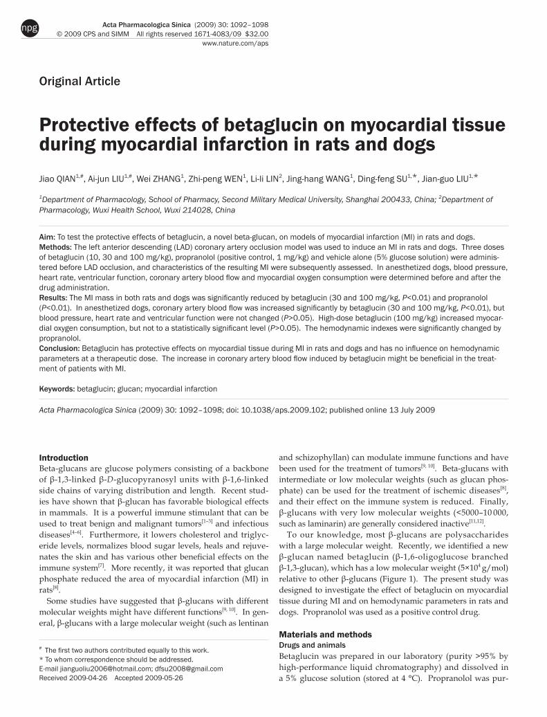
To our knowledge, most β-glucans are polysaccharides with a large molecular weight. Recently, we identified a new β-glucan named betaglucin (β-1,6-oligoglucose branched β-1,3-glucan), which has a low molecular weight (5×10⁴ g/mol) relative to other β-glucans (Figure 1). The present study was designed to investigate the effect of betaglucin on myocardial tissue during MI and on hemodynamic parameters in rats and dogs. Propranolol was used as a positive control drug.
Materials and methodsDrugs and animals Betaglucin was prepared in our laboratory (purity >95% by high-performance liquid chromatography) and dissolved in a 5% glucose solution (stored at 4 °C). Propranolol was pur-
Protective effects of betaglucin on myocardial tissue during myocardial infarction in rats and dogs Jiao QIAN1,#, Ai-jun LIU1,#, Wei ZHANG1, Zhi-peng WEN1, Li-li LIN2, Jing-hang WANG1, Ding-feng SU1,*, Jian-guo LIU1,*
1Department of Pharmacology, School of Pharmacy, Second Military Medical University, Shanghai 200433, China; 2Department of Pharmacology, Wuxi Health School, Wuxi 214028, China
Aim: To test the protective effects of betaglucin, a novel beta-glucan, on models of myocardial infarction (MI) in rats and dogs. Methods: The left anterior descending (LAD) coronary artery occlusion model was used to induce an MI in rats and dogs. Three doses of betaglucin (10, 30 and 100 mg/kg), propranolol (positive control, 1 mg/kg) and vehicle alone (5% glucose solution) were adminis-tered before LAD occlusion, and characteristics of the resulting MI were subsequently assessed. In anesthetized dogs, blood pressure, heart rate, ventricular function, coronary artery blood flow and myocardial oxygen consumption were determined before and after the drug administration. Results: The MI mass in both rats and dogs was significantly reduced by betaglucin (30 and 100 mg/kg, P<0.01) and propranolol (P<0.01). In anesthetized dogs, coronary artery blood flow was increased significantly by betaglucin (30 and 100 mg/kg, P<0.01), but blood pressure, heart rate and ventricular function were not changed (P>0.05). High-dose betaglucin (100 mg/kg) increased myocar-dial oxygen consumption, but not to a statistically significant level (P>0.05). The hemodynamic indexes were significantly changed by propranolol. Conclusion: Betaglucin has protective effects on myocardial tissue during MI in rats and dogs and has no influence on hemodynamic parameters at a therapeutic dose. The increase in coronary artery blood flow induced by betaglucin might be beneficial in the treat-ment of patients with MI.
Keywords: betaglucin; glucan; myocardial infarction Acta Pharmacologica Sinica (2009) 30: 1092–1098; doi: 10.1038/aps.2009.102; published online 13 July 2009
Original Article
# The first two authors contributed equally to this work.* To whom correspondence should be addressed. E-mail [email protected]; [email protected] Received 2009-04-26 Accepted 2009-05-26
1093
www.chinaphar.comQian J et al
Acta Pharmacologica Sinica
npg
chased from Beijing Zizhu Pharmaceutical Co, Ltd (Beijing, China). All drugs were dissolved in the vehicle (5% glucose solution) before administration, and the administration vol-ume was 5 mL/kg in both rats and dogs). Male Sprague-Daw-ley (SD) rats aged 12 weeks (300−400 g) were provided by the Sino-British SIPPR/BK Laboratory (Shanghai, China). Hybrid dogs of both sexes (n=6 in each group, body weight: 12−14 kg) were provided by the animal center of the Second Military Medical University. The rats and dogs were housed under controlled temperature (23−25 °C) and lighting (8:00–20:00 light, 20:00–8:00 dark) conditions with free access to food and drinking water. All animals used in the experiments received humane care. All surgical and experimental procedures were performed in accordance with the institutional animal care guidelines of the Second Military Medical University.
Determination of MI in rats The surgical procedure to produce an MI was performed according to a well-accepted technique[8, 13, 14]. Briefly, rats were anesthetized with pentobarbital sodium (45 mg/kg) and underwent a tracheal intubation for subsequent mechanical ventilation. The heart was exteriorized via a left thoracotomy and subjected to left anterior descending (LAD) coronary artery occlusion with a 6–0 polypropylene suture between the pulmonary outflow tract and the left atrium. The beat-ing heart was then quickly returned to its original location, the thorax was immediately closed, and the air was removed. Rats were subsequently returned to their previously described cages. Four hours after the LAD coronary artery occlusion, the MI mass was measured according to previously reported methods[15, 16]. Briefly, the rats were killed by an overdose of pentobarbital (200 mg/kg ip). The left ventricle (LV) was iso-lated and cut into four or five slices perpendicular to the car-diac long axis. The slices were stained for 30 min at 37 ºC in a 0.1% solution of nitro blue tetrazolium (NBT) and a 0.2 mol/L phosphate buffer (sodium dihydrogen phosphate 87%+diso-dium hydrogenorthophosphate 13%). NBT stained the normal tissue deep blue and the necrotic tissue white. Stained and unstained tissues were isolated and weighed separately. MI mass was expressed as a fraction of the total LV weight[17].
Determination of MI in dogs Anesthesia was induced by intravenous injection of pentobar-
bital sodium (30 mg/kg), and the dogs underwent a tracheal intubation for subsequent mechanical ventilation (SC-3 electri-cal ventilator, Shanghai Medical Equipment Factory, Shang-hai, China). A left lateral thoracotomy was performed in the fourth intercostal space, the pericardium was incised, and the heart was suspended in a pericardial cradle. Before opening and fixing the pericardium, 2 mL of 2% lidocaine was injected intravenously. Infarction was induced by occlusion of the LAD coronary artery between the first and second diagonal branches. Surface electrocardiogram (ECG, V3 lead) and epi-cardial ECG tracings were recorded by the ALC-ECG16-32 point sync-epicardial electrogram system (Shanghai Alcott Biotech Co Ltd Shanghai, China) on anesthetized dogs at baseline before LAD occlusion, immediately after LAD occlusion, and again at 30 min, 60 min, 120 min, and 180 min after drug administration to assess for changes in the ST seg-ment (the changes of surface ECG expressed as ∆ST; and the total changes of epicardial ECG expressed as Σ-ST). Three hours after LAD occlusion, the heart was stopped by injection of 2 mol/L KCl and removed from the chest. The infarction mass was determined using a method similar to the one used for rats.
Measurement of blood pressure, heart rate and ventricular function in anesthetized dogs Dogs were anesthetized with sodium pentobarbital (30 mg/kg, iv) and placed on a ventilator. A catheter connected to a pressure transducer was introduced into the femoral artery to record the blood pressure and heart rate. After making an incision in the neck, a catheter connected to a pressure trans-ducer was introduced into the LV through the right carotid artery. LV systolic pressure (LVSP), end-diastolic pressure (LVEDP), and the maximal rates of pressure rise (+dp/dt) and fall (-dp/dt) were measured or calculated for each group. The stable ventricular pressure was recorded for 10 min. The means of LVSP, LVEDP, and ±dp/dt during the last 5 min were analyzed.
Measurement of coronary artery blood flow and myocardial oxygen consumption in dogs Anesthesia was induced by intravenous injection of pentobar-bital sodium (30 mg/kg), and dogs underwent a tracheal intu-bation for subsequent mechanical ventilation. A left lateral
Figure 1. The chemical structure of betaglucin.
1094
www.nature.com/apsQian J et al
Acta Pharmacologica Sinica
npg
thoracotomy was performed in the fourth intercostal space, the pericardium was incised, and the heart was suspended in a pericardial cradle. A probe was connected to the left cir-cumflex coronary artery, and the blood flow was recorded by an electromagnetic blood flow meter system (Nihon Konden, Japan). The value was expressed as mL/min per 100 g of myocardium supplied by a coronary artery. A catheter con-nected to a pressure transducer was introduced into the femo-ral artery. Blood was collected from the femoral artery and the coronary sinus for blood gas analysis (DH-1300 blood gas ana-lyzer, Nanjing Analytical Instrument Factory Co, Ltd, Nanjing, China). The value of the myocardial oxygen consumption was expressed as the amount of blood flow in the coronary artery× (arterial oxygen saturation – coronary sinus oxygen satura-tion).
ProtocolExperiment 1: Effect of betaglucin on MIs in rats The experiment was performed in 12-week-old male SD rats. Fifty animals were randomly divided into five groups: a vehicle control group (5% glucose solution), three betaglucin groups (10, 30, and 100 mg/kg) and a propranolol group (1 mg/kg), with n=10 in each group. The drugs were injected into the abdominal cavity 30 min before LAD occlusion using the above-mentioned methods. The heart rate was recorded by the electrocardiograph. Four hours later, the rats were killed, and the heart was harvested to measure the infarct mass.
Experiment 2: Effect of betaglucin on MIs in dogs The experiment was performed in anesthetized dogs weighing from 12 to 14 kg. Thirty animals were randomly divided into five groups: a vehicle control group, three betaglucin groups (10, 30, and 100 mg/kg) and a propranolol group (1 mg/kg), with n=6 in each group. Drugs were injected via the femoral vein 30 min before LAD occlusion. Surface and epicardial ECGs were recorded using the above-mentioned methods before and after drug administration. Three hours after LAD occlusion, the dogs were killed, and the heart was harvested to measure the infarct mass.
Experiment 3: The effect of betaglucin on blood pressure, heart rate and ventricular function in anesthetized dogsThe experiment was performed in anesthetized dogs. Thirty animals were randomly divided into five groups: a vehicle control group, three betaglucin groups (10, 30, and 100 mg/kg) and a propranolol group (1 mg/kg), with n=6 in each group. Drugs were injected via the femoral vein after stabilization of vital signs for 30 min. Blood pressure, heart rate and ventricu-lar function were recorded using the above-mentioned meth-ods before and after drug administration.
Experiment 4: The effect of betaglucin on coronary artery blood flow and myocardial oxygen consumption in anesthetized dogs The experiment was performed in anesthetized dogs. Thirty animals were randomly divided into five groups: a vehicle
control group, three betaglucin groups (10, 30, and 100 mg/kg) and a propranolol group (1 mg/kg), with n=6 in each group. Drugs were injected via the femoral vein after stabilization for 30 min. Coronary artery blood flow and myocardial oxy-gen consumption were measured using the above-mentioned methods before and after administration.
Statistical analysisData are expressed as the means±standard errors. Compari-sons among the five groups or between different time points within a group were made using one-way analysis of variance (ANOVA). P<0.05 was considered statistically significant.
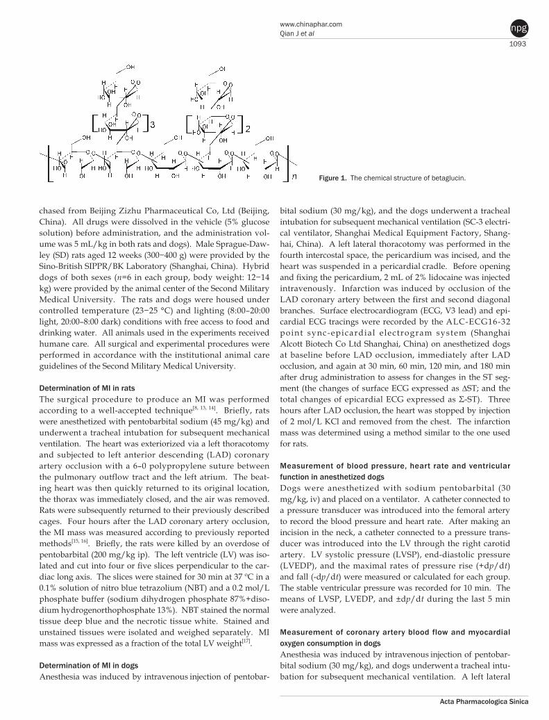
ResultsExperiment 1: Effect of betaglucin on MIs in rats Data are shown in Figure 2. None of the three doses of beta-glucin had a significant impact on the heart rate (P>0.05), whereas 1 mg/kg of propranolol markedly reduced the heart rate (P<0.05 at all time points from 5 to 180 min after admin-istration; Figure 2A). Compared with the vehicle control group, betaglucin (30 and 100 mg/kg) significantly reduced the infarct mass (absolute infarct mass: 78±5.69, 99±5.37 (respectively) vs 182±22.4 mg; ratio of infarct mass to LV: 10.6%±0.96%, 11.4%±0.76% (respectively) vs 21.3%±2.24%, P<0.05). Moreover, propranolol also significantly reduced the infarct mass in rats with MIs (absolute infarct mass: 72±5.69 vs 182±22.4 mg; ratio of infarct mass to LV: 8.81%±0.62% vs 21.3%±2.24%, P<0.05).
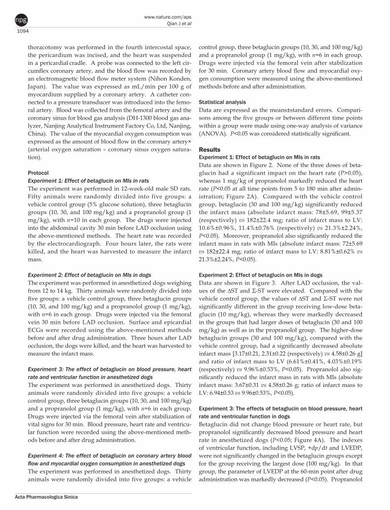
Experiment 2: Effect of betaglucin on MIs in dogs Data are shown in Figure 3. After LAD occlusion, the val-ues of the ∆ST and Σ-ST were elevated. Compared with the vehicle control group, the values of ∆ST and Σ-ST were not significantly different in the group receiving low-dose beta-glucin (10 mg/kg), whereas they were markedly decreased in the groups that had larger doses of betaglucin (30 and 100 mg/kg) as well as in the propranolol group. The higher-dose betaglucin groups (30 and 100 mg/kg), compared with the vehicle control group, had a significantly decreased absolute infarct mass [3.17±0.21, 2.31±0.22 (respectively) vs 4.58±0.26 g] and ratio of infarct mass to LV (6.61%±0.41%, 4.03%±0.19% (respectively) vs 9.96%±0.53%, P<0.05). Propranolol also sig-nificantly reduced the infarct mass in rats with MIs (absolute infarct mass: 3.67±0.31 vs 4.58±0.26 g; ratio of infarct mass to LV: 6.94±0.53 vs 9.96±0.53%, P<0.05).
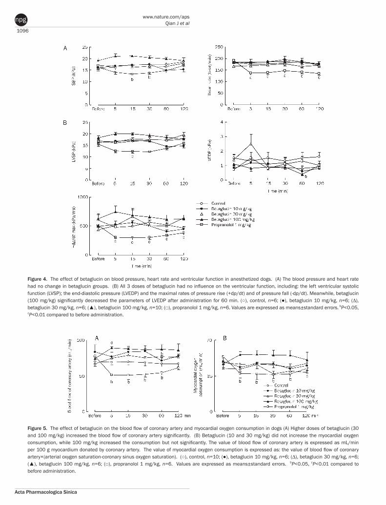
Experiment 3: The effects of betaglucin on blood pressure, heart rate and ventricular function in dogs Betaglucin did not change blood pressure or heart rate, but propranolol significantly decreased blood pressure and heart rate in anesthetized dogs (P<0.05; Figure 4A). The indexes of ventricular function, including LVSP, +dp/dt and LVEDP, were not significantly changed in the betaglucin groups except for the group receiving the largest dose (100 mg/kg). In that group, the parameter of LVEDP at the 60-min point after drug administration was markedly decreased (P<0.05). Propranolol
1095
www.chinaphar.comQian J et al
Acta Pharmacologica Sinica
npg
significantly decreased LVSP and +dp/dt in anesthetized dogs (P<0.01; Figure 4B).
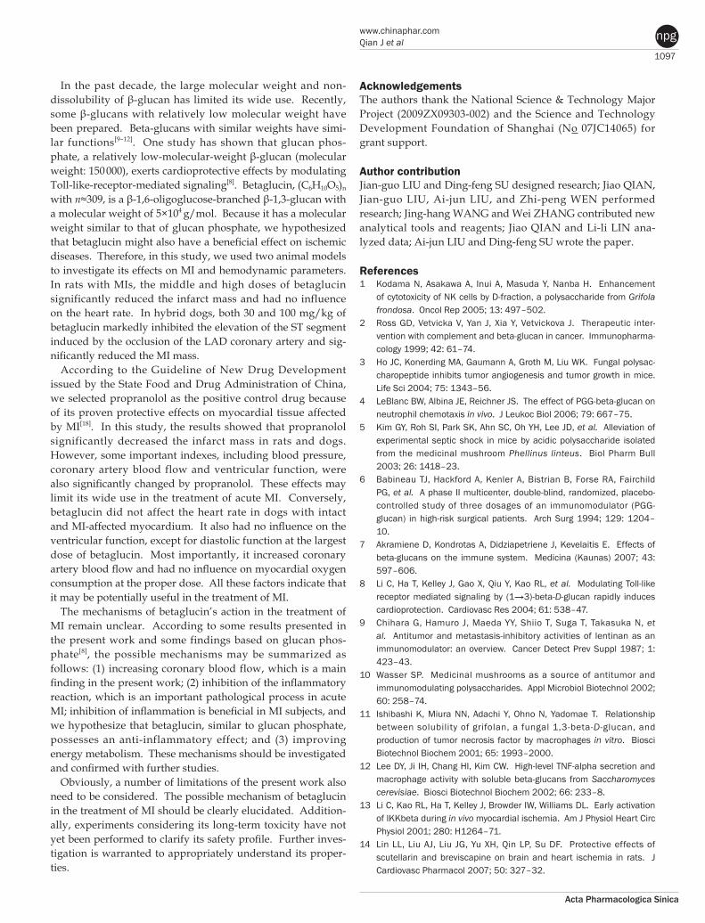
Experiment 4: The effect of betaglucin on coronary artery blood flow and myocardial oxygen consumption in dogs Data are summarized in Figure 5. Betaglucin (30 and 100 mg/kg) significantly increased coronary artery blood flow by 11.5 and 14.7 mL/min, respectively (P<0.05). Betaglucin (100 mg/kg) increased the index of oxygen consumption by about 9 mL/min, but this was not found to be statistically significant (P>0.05). However, in the propranolol-treated group, both the indexes of blood flow and oxygen consumption were signifi-cantly decreased (P<0.05).
DiscussionIn this study, our results show that betaglucin has protective effects on myocardial tissue during MI in rats and dogs and
has no side effects on hemodynamic parameters at therapeutic doses.
Figure 2. Betaglucin reduced myocardial infarction (MI) in SD rats subjected to LAD occlusion. (A) The effect of betaglucin on the heart rate of rats subjected to LAD occlusion. (B) Betaglucin reduced the MI mass after 4 h of occlusion. The heart was removed and the infarct mass was determined by NBT staining. LAD coronary artery, left anterior descending coronary artery; Infarct mass ratio was expressed as a fraction of the total left ventricle (LV). (○), control, n=10; (●), betaglucin 10 mg/kg, n=10; (∆), betaglucin 30 mg/kg, n=10; (▲), betaglucin 100 mg/kg, n=10; (□), propranolol 1 mg/kg, n=10. Values are expressed as means±standard errors. aP>0.05, bP<0.05, cP<0.01 compared to before administration.
Figure 3. Betaglucin reduced MI in dogs subjected to LAD occlusion. (A) The values of the ST segment of surface electrocardiogram (ECG) (∆ST), (B) epicardial ECG (Σ-ST) were significantly elevated by LAD occlusion. The elevation can be inhibited by higher doses of betaglucin (30 and 100 mg/kg) and propranolol (1 mg/kg) (A, B, C). (C) Compared to control group, higher doses (30 and 100 mg/kg) of betaglucin significantly reduced the infarct mass of dogs. Infarct mass ratio was expressed as a fraction of the total left ventricle (LV). Propranolol also got the similar results. (○), control, n=6; (●), betaglucin 10 mg/kg, n=6; (∆), betaglucin 30 mg/kg, n=6; (▲), betaglucin 100 mg/kg, n=6; (□), propranolol 1 mg/kg, n=6. Values are expressed as means±standard errors. aP>0.05, bP<0.05, cP<0.01 compared to vehicle control group.
1096
www.nature.com/apsQian J et al
Acta Pharmacologica Sinica
npg
Figure 4. The effect of betaglucin on blood pressure, heart rate and ventricular function in anesthetized dogs. (A) The blood pressure and heart rate had no change in betaglucin groups. (B) All 3 doses of betaglucin had no influence on the ventricular function, including: the left ventricular systolic function (LVSP); the end-diastolic pressure (LVEDP) and the maximal rates of pressure rise (+dp/dt) and of pressure fall (-dp/dt). Meanwhile, betaglucin (100 mg/kg) significantly decreased the parameters of LVEDP after administration for 60 min. (○), control, n=6; (●), betaglucin 10 mg/kg, n=6; (∆), betaglucin 30 mg/kg, n=6; (▲), betaglucin 100 mg/kg, n=10; (□), propranolol 1 mg/kg, n=6. Values are expressed as means±standard errors. bP<0.05, cP<0.01 compared to before administration.
Figure 5. The effect of betaglucin on the blood flow of coronary artery and myocardial oxygen consumption in dogs (A) Higher doses of betaglucin (30 and 100 mg/kg) increased the blood flow of coronary artery significantly. (B) Betaglucin (10 and 30 mg/kg) did not increase the myocardial oxygen consumption, while 100 mg/kg increased the consumption but not significantly. The value of blood flow of coronary artery is expressed as mL/min per 100 g myocardium donated by coronary artery. The value of myocardial oxygen consumption is expressed as: the value of blood flow of coronary artery×(arterial oxygen saturation-coronary sinus oxygen saturation). (○), control, n=10; (●), betaglucin 10 mg/kg, n=6; (∆), betaglucin 30 mg/kg, n=6; (▲), betaglucin 100 mg/kg, n=6; (□), propranolol 1 mg/kg, n=6. Values are expressed as means±standard errors. bP<0.05, cP<0.01 compared to before administration.
1097
www.chinaphar.comQian J et al
Acta Pharmacologica Sinica
npg
In the past decade, the large molecular weight and non-dissolubility of β-glucan has limited its wide use. Recently, some β-glucans with relatively low molecular weight have been prepared. Beta-glucans with similar weights have simi-lar functions[9–12]. One study has shown that glucan phos-phate, a relatively low-molecular-weight β-glucan (molecular weight: 150 000), exerts cardioprotective effects by modulating Toll-like-receptor-mediated signaling[8]. Betaglucin, (C6H10O5)n with n≈309, is a β-1,6-oligoglucose-branched β-1,3-glucan with a molecular weight of 5×10⁴ g/mol. Because it has a molecular weight similar to that of glucan phosphate, we hypothesized that betaglucin might also have a beneficial effect on ischemic diseases. Therefore, in this study, we used two animal models to investigate its effects on MI and hemodynamic parameters. In rats with MIs, the middle and high doses of betaglucin significantly reduced the infarct mass and had no influence on the heart rate. In hybrid dogs, both 30 and 100 mg/kg of betaglucin markedly inhibited the elevation of the ST segment induced by the occlusion of the LAD coronary artery and sig-nificantly reduced the MI mass.
According to the Guideline of New Drug Development issued by the State Food and Drug Administration of China, we selected propranolol as the positive control drug because of its proven protective effects on myocardial tissue affected by MI[18]. In this study, the results showed that propranolol significantly decreased the infarct mass in rats and dogs. However, some important indexes, including blood pressure, coronary artery blood flow and ventricular function, were also significantly changed by propranolol. These effects may limit its wide use in the treatment of acute MI. Conversely, betaglucin did not affect the heart rate in dogs with intact and MI-affected myocardium. It also had no influence on the ventricular function, except for diastolic function at the largest dose of betaglucin. Most importantly, it increased coronary artery blood flow and had no influence on myocardial oxygen consumption at the proper dose. All these factors indicate that it may be potentially useful in the treatment of MI.
The mechanisms of betaglucin’s action in the treatment of MI remain unclear. According to some results presented in the present work and some findings based on glucan phos-phate[8], the possible mechanisms may be summarized as follows: (1) increasing coronary blood flow, which is a main finding in the present work; (2) inhibition of the inflammatory reaction, which is an important pathological process in acute MI; inhibition of inflammation is beneficial in MI subjects, and we hypothesize that betaglucin, similar to glucan phosphate, possesses an anti-inflammatory effect; and (3) improving energy metabolism. These mechanisms should be investigated and confirmed with further studies.
Obviously, a number of limitations of the present work also need to be considered. The possible mechanism of betaglucin in the treatment of MI should be clearly elucidated. Addition-ally, experiments considering its long-term toxicity have not yet been performed to clarify its safety profile. Further inves-tigation is warranted to appropriately understand its proper-ties.
AcknowledgementsThe authors thank the National Science & Technology Major Project (2009ZX09303-002) and the Science and Technology Development Foundation of Shanghai (No 07JC14065) for grant support.
Author contributionJian-guo LIU and Ding-feng SU designed research; Jiao QIAN, Jian-guo LIU, Ai-jun LIU, and Zhi-peng WEN performed research; Jing-hang WANG and Wei ZHANG contributed new analytical tools and reagents; Jiao QIAN and Li-li LIN ana-lyzed data; Ai-jun LIU and Ding-feng SU wrote the paper.
References1 Kodama N, Asakawa A, Inui A, Masuda Y, Nanba H. Enhancement
of cytotoxicity of NK cells by D-fraction, a polysaccharide from Grifola frondosa. Oncol Rep 2005; 13: 497–502.
2 Ross GD, Vetvicka V, Yan J, Xia Y, Vetvickova J. Therapeutic inter-vention with complement and beta-glucan in cancer. Immuno pharma-cology 1999; 42: 61–74.
3 Ho JC, Konerding MA, Gaumann A, Groth M, Liu WK. Fungal polysac-charopeptide inhibits tumor angiogenesis and tumor growth in mice. Life Sci 2004; 75: 1343–56.
4 LeBlanc BW, Albina JE, Reichner JS. The effect of PGG-beta-glucan on neutrophil chemotaxis in vivo. J Leukoc Biol 2006; 79: 667–75.
5 Kim GY, Roh SI, Park SK, Ahn SC, Oh YH, Lee JD, et al. Alleviation of experimental septic shock in mice by acidic polysaccharide isolated from the medicinal mushroom Phellinus linteus. Biol Pharm Bull 2003; 26: 1418–23.
6 Babineau TJ, Hackford A, Kenler A, Bistrian B, Forse RA, Fairchild PG, et al. A phase II multicenter, double-blind, randomized, placebo-controlled study of three dosages of an immunomodulator (PGG-glucan) in high-risk surgical patients. Arch Surg 1994; 129: 1204–10.
7 Akramiene D, Kondrotas A, Didziapetriene J, Kevelaitis E. Effects of beta-glucans on the immune system. Medicina (Kaunas) 2007; 43: 597–606.
8 Li C, Ha T, Kelley J, Gao X, Qiu Y, Kao RL, et al. Modulating Toll-like receptor mediated signaling by (1→3)-beta-D-glucan rapidly induces cardioprotection. Cardiovasc Res 2004; 61: 538–47.
9 Chihara G, Hamuro J, Maeda YY, Shiio T, Suga T, Takasuka N, et al. Antitumor and metastasis-inhibitory activities of lentinan as an immunomodulator: an overview. Cancer Detect Prev Suppl 1987; 1: 423–43.
10 Wasser SP. Medicinal mushrooms as a source of antitumor and immunomodulating polysaccharides. Appl Microbiol Biotechnol 2002; 60: 258–74.
11 Ishibashi K, Miura NN, Adachi Y, Ohno N, Yadomae T. Relationship between solubility of grifolan, a fungal 1,3-beta-D-glucan, and production of tumor necrosis factor by macrophages in vitro. Biosci Biotechnol Biochem 2001; 65: 1993–2000.
12 Lee DY, Ji IH, Chang HI, Kim CW. High-level TNF-alpha secretion and macrophage activity with soluble beta-glucans from Saccharomyces cerevisiae. Biosci Biotechnol Biochem 2002; 66: 233–8.
13 Li C, Kao RL, Ha T, Kelley J, Browder IW, Williams DL. Early activation of IKKbeta during in vivo myocardial ischemia. Am J Physiol Heart Circ Physiol 2001; 280: H1264–71.
14 Lin LL, Liu AJ, Liu JG, Yu XH, Qin LP, Su DF. Protective effects of scutellarin and breviscapine on brain and heart ischemia in rats. J Cardiovasc Pharmacol 2007; 50: 327–32.
1098
www.nature.com/apsQian J et al
Acta Pharmacologica Sinica
npg
15 Nishizawa M, Kumagai H, Ichikawa M, Oshima N, Suzuki H, Saruta T. Improvement in baroreflex function by an oral angiotensin receptor antagonist in rats with myocardial infarction. Hypertension 1997; 29: 458–63.
16 Leenen FH, Yuan B. Mortality after coronary artery occlusion in different models of cardiac hypertrophy in rats. Hypertension 2001; 37: 209–15.
17 Brown GD, Gordon S. Fungal beta-glucans and mammalian immunity. Immunity 2003; 19: 311–5.
18 Lee SH, Yoon SB, Cho JR, Choi S, Jung JH, Lee N. The effects of different beta-blockers on left-ventricular volume and function after primary coronary stenting in acute myocardial infarction. Angiology 2008; 59: 676–81.
~~~~~~~~~~~~~~~~~~~~~~~~~~~~~~~~~~~~~~~~~~~~~~~~~~~~~~~~~~~~~~~~~~~~~~~~~~~~~~~~~~~~~~~~~~~~~~
SBS 16th Annual Conference & ExhibitionAdvancing the Science of Drug Discovery
2010, April 11–15Phoenix Convention Center
Phoenix, Arizona, USAwww.sbsonline.org
Session Topics♦ Lead Discovery in Immunoinflammation♦ Lead Discovery in Oncology Research♦ Lead Discovery in Neuroscience Research♦ Lead Discovery in Epigenetics: An Emerging Target Class♦ Critical Reagent: Design of Synthetic & Natural Product Libraries♦ Critical Reagents: Sample Storage, Quality Control & Distribution♦ Critical Reagents: Bioreagent Preparation for Lead Discovery♦ Assays & Automation: Best Practices in Automation & Miniaturization♦ Assays & Automation: Novel Screening Technologies for Hit Discovery♦ Assays & Automation: Novel Screening Technologies for Lead Optimization