Embed Size (px)
Citation preview

INTRODUCTION
Post-traumatic stress disorder (PTSD) is characterized byhyperarousal and recurrent stressful memories related toemotional trauma (1). PTSD has been associated with anxiety,cognitive and memory deficits (2-4).
Amygdala, hippocampus, and prefrontal cortex (PFC) arethe most affected brain regions in PTSD (5). Subjects with PTSDexert, hypo-active PFC and hippocampus, while hyper-activebasolateral amygdala (BLA) (6). The presence of GABAergicinterneurons in the entire amygdala is well known and theseneurons are important to provide inhibitory control over theamygdala (5).
Both physical stresses for example, diabetic neuropathy (7)and psychological traumatic experiences change neuronalmorphology, function, and neurochemistry (1). The presence ofabnormal apoptosis including the hippocampus, amygdala, andprefrontal cortex has been reported in various PTSD rat models(8-10). Neuronal apoptosis is related to mitochondria-mediatedapoptosis factors, such as caspase-3 (casp-3), caspase-9, and bcl-
2 associated X protein (bax) (9, 10). Stressful experiences suchas exposure to predator scent, cause increasing of levels ofcirculating adrenal steroids. Increased adrenal-glucocorticoids,inhibit cell production in the hippocampal dentate gyrus in rats(11) and reduces bcl-2 levels, and this effect causes increasedsensitivity to excitotoxic damage (12). Predator scent stress(PSS) causes neuronal damage and increased apoptosis in thehippocampus (13, 14).
Due to stress, a decrease in brain-derived neurotrophic factor(BDNF) level, an increase in glutamate and cortisol levels, andinhibition of neurogenesis may occur in the hippocampus (15).Experimental PTSD models showed that BDNF levelsdecreased; in the hippocampus (16-18) prefrontal cortex (19),and amygdala (17-20). In contrast, some studies have indicatedthat increased BDNF levels (16) and the steady-state BDNFlevels in BLA, PFC, and hippocampus (21).
Exposure to a PSS model is a commonly used model forstudying PTSD (22, 23). In the acute PSS model, rats areexposed to a reminder one week after odour stress procedure.After the reminder, anxiety, and activity evaluate by elevated
jOURNAL OF PHYSIOLOGY AND PHARMACOLOGY 2019, 70, 4, 557-571www.jpp.krakow.pl | DOI: 10.26402/jpp.2019.4.07
D. OZBEYLI1, A. AYKAC2, N. ALACA3, A.N. HAZAR-YAVUZ4, N. OZKAN1, G. SENER4
PROTECTIVE EFFECTS OF VORTIOXETINE IN PREDATOR SCENT STRESS MODELOF POST-TRAUMATIC STRESS DISORDER IN RATS:
ROLE ON NEUROPLASTICITY AND APOPTOSIS
1Department of Medical Pathology Techniques, Vocational School of Health Services, Marmara University, Istanbul, Turkey;2Department of Biophysics, Faculty of Medicine, Near East University, Nicosia, Cyprus;
4Department of Physiotherapy and Rehabilitation, Faculty of Health Sciences, Acibadem University, Istanbul, Turkey; 3Department of Pharmacology, Faculty of Pharmacy, Marmara University, Istanbul, Turkey
Post-traumatic stress disorder (PTSD) can be observed after a traumatic event. The effect of an antidepressantvortioxetine (Vrx) against PTSD is unknown. The aim of this study was to investigate the possible protective effect ofVrx in the predator scent-induced PTSD rat model. The rats were exposed to dirty cat litter for 10 min and the protocolwas repeated 1 week later with clean cat litter as a trauma reminder. The rats received Vrx (10 mg/kg/p.o.) or saline (1ml/kg/p.o.) during 7 days between two exposure sessions. Novel object recognition test, hole board test, and elevatedplus maze were performed. The b-cell lymphoma (bcl-2)/bcl-2-associated X protein (bax) ratio, brain-derivedneurotrophic factor (BDNF), caspase-3 and -9 expressions were detected using Western blotting in the amygdaloidcomplex, hippocampus, and frontal cortex. Our results indicate that increased freezing time and anxiety index in thestress-induced group is decreased with Vrx application. Vrx treatment improved deteriorated recognition memory in thestress-induced group. Decreased bcl-2/bax ratio and BDNF level and increased caspase-3 and -9 expressions in the stressgroup, improved with Vrx in the amygdala, and hippocampus. Decreased bcl-2/bax ratio and increased casp-3 and -9expressions in the stress group are ameliorated with Vrx in frontal cortex. The level of BDNF was increased with Vrxin the frontal cortex. Increased damage scores in the amygdaloid complex, hippocampal CA3, and frontal cortex in thestress group ameliorated with Vrx treatment. Our results show that if vortioxetine is administered immediately aftertrauma, it reduces anxiety, cognitive and neuronal impairment and may be protective against the development of PTSD.
K e y w o r d s : post-traumatic stress disorder, vortioxetine, brain-derived neurotrophic factor, apoptosis, recognition memory

plus maze (EPM) and hole board test (HBT) while workingmemory evaluates with a novel object recognition test (NORT)(22-24). Inescapable exposure to predator odor inducespersistent alterations in behavioral and physiological responsesin rats that imitate the symptom profile of fear and anxiety inPTSD (25). This model produces significant physiological andbehavioural alterations including increased anxiety (26),psychological stress (22), cognitive impairment (26), and meetsthe expectations of many of the diagnostic criteria of DMS-V forPTSD (27).
Numerous therapy approaches have been proposed to curethe PTSD patients (28). Selective serotonin reuptake (SSRI) hasbeen approved for the treatment although remission of PTSD isaround 30% with them (29). Animal studies have reported thattreatment before stress with antidepressants reduce behavioraldeficits related to stress. Antidepressants, including bothnorepinephrine and SSRIs, promote nerve growth(neurogenesis) in the hippocampus (30, 31) and cortex (31)while stress inhibits neurogenesis. On the other hand, the drugscurrently do not fully improve the symptoms of PTSD and theyhave side effects (3, 29). Therefore, it is really essential toexamine the underlying biological mechanisms in PTSD and itis worth investigating to develop new strategies in order toameliorate the PTSD that could be employed in this regard.
Vortioxetine (Vrx), as a novel serotonergic antidepressantwith multimodal activity, shows both antidepressant andanxiolytic effects (32, 33). Vrx is an antagonist of 5-HT1D, 5-HT3, 5-HT7 receptors, an agonist of 5-HT1A receptors, a partialagonist of 5-HT1B receptors, and an SSRI inhibiting theserotonin transporter (SERT) (32, 34). The preclinical and theclinical studies suggest that 5-HT3 and 5-HT7 receptorantagonism and 5-HT1A receptor agonism may have a positiveimpact on the cognitive functions (33). Furthermore, it has beenreported that Vrx can improve cognitive function through themodulation of different neurotransmitters (35) and potentiallyenhance memory (36). Vrx could modulate several preclinicalparameters recognized to be involved in cognitive functions. Vrxprevented the effect of stress on hippocampal LTP, increasedrapidly hippocampal cell proliferation and enhanced short-termrecognition memory in rats via its 5-HT3 receptor antagonism(37). Vrx increased hippocampal BDNF levels in rats subject tochronic unpredictable mild stress (38).
There is extensive literature showing the efficacy of Vrx ongeneralized and social anxiety disorder (39-41), however, it isdifficult to find studies that examine Vrx and PTSD. Vrx hasbeen approved for the treatment of depression however, noapproved by the Food and Drug Administration for thetreatment of PTSD. A phase IV clinical trial is currently beingconducted for the treatment of PTSD with Vrx(https://clinicaltrials.gov/ct2/show/NCT02637895).
This study aimed to investigate the possible protective effectsof Vrx on PTSD model in terms of cognitive and behaviouralfunctions, neuroplasticity and apoptotic mechanisms. In thisstudy, it has been hypothesized that Vrx has beneficial effects onPTSD may be specifically related to the level of BDNF,mitochondria-mediated apoptosis factors; pro- (bax, caspase-3,and caspase-9) and anti-apoptotic (bcl-2) proteins in theamygdaloid complex, the hippocampus, and the frontal cortex.
MATERIAL AND METHODS
Animals and conditions
All experimental procedures involving animals wereperformed in accordance with the NIH guide for the care and useof laboratory animals (NIH Publications No. 8023, revised
1978). An approval from the institutional ethical committee wasobtained from Marmara University Animal Care and UseCommittee (MUHDEK approval no: 64.2018.mar). Rats weresupplied by the Marmara University Experimental AnimalsResearch and Implementation Centre (DEHAMER).
In total 32 Wistar albino male rats, weighing 250 – 300 g,aged 12 – 14 weeks were used in this study. During theexperimental study, the rats were kept in cages (60 × 40 × 40 cm)with polyacrylic material and 4 rats were housed in each cage.Before the behavioural study, the rats were habituated with thereverse of the circadian cycle for 1 week (12-h:12-h light-darkcycles) where they were housed in rooms with a temperature of22 ± 2°C, and humidity of 50 ± 5% with food and water adlibitum.
Drugs and chemicals
Vrx was purchased from (Lundbeck, Turkey). Vrx wasfreshly dissolved in distilled water before application (10mg/kg). The dose and duration of Vrx treatment were based onprevious studies found to be effective in stress and depression inrats (36, 37, 42). All drugs were administered by gavage for 7days at the same time. All chemicals were supplied by Sigma(Sigma-Aldrich, St. Louis, MO, USA). All antibodies forimmunoblotting were purchased from Santa CruzBiotechnology, Inc (Santa Cruz, CA, USA).
Experimental design and stress protocol
After the habituation period, the rats were divided randomlyinto four experimental groups each containing eight rats (n = 32,totally). These were (i) control: physiological saline solution(SS) (1 ml/kg/p.o.) (ii) Vrx (10 mg/kg/p.o.), (iii) Stress, and (i.v.)Stress + Vrx (10 mg/kg/p.o.). All treatments were given daily atthe same time of the day (at 09.00 a.m.) for 7 days (Fig. 1).
The dirty cat litter, used by the same male cat for two days,was used as a stressor (24, 25). On the first day of theexperiment, 1 hour after Vrx or saline treatment, the stressgroups were exposed to dirty cat litter, while control and Vrxgroups were exposed to clean (fresh and unused) cat litter. In thestress protocol, each rat was kept in a cage (30 cm × 30 cm × 40cm) including 125 ml of dirty cat litter for 10 min unlike theunstressed group, which used the same amount of clean cat litterduring the same period. Clean cat litter, as a situational reminder,was used in all groups before the behavioural experiments on thelast day of the experiment (13, 22, 42).
Behavioural and cognition tests
Three different tests; NORT, HBT, and EPM tests were usedfor evaluation of cognitive and anxiety-like behaviour in rats.The EPM and HBT, extensively used to evaluate the fear oranxiety, have been validated for use in rats (43, 44). At the 7 dayof experiments, 30 minutes after the last drug administration, therats were exposed to clean cat litter for 10 min, and thenimmediately the rats were evaluated at NORT (43, 44), HBT(45), and EPM (13, 22, 42, 46); respectively (Fig. 1).
Novel object recognition test
Immediately after 10 min of clean cat litter application,NORT was performed to assess the object recognition memoryfunctions of rats. Hippocampus is important for objectrecognition memory and hippocampal lesions producedmoderate and reliable memory impairment (47). NORT is basedon the principle that rodents will spend more time exploring anew object than the familiar one. In the present study, the object
558

recognition tasks were applied to examine the short-termrecognition memory using a 1-hour delay time. A blackplexiglass box (50 cm × 50 cm × 30 cm) was used for the test.NORT has three steps as habituation, familiarizing and a test(44). Each rat has remained in the box without any objects for 10minutes for habituation. Afterward, through the familiarizationphase, a single animal is located for 5 min in the box comprisingtwo similar sample objects on opposite sides of the box. After 1-hour delay time, during the test step, the rat is put back to the boxwith two objects, which one is similar to the sample and theother is novel and was allowed to explore objects for 3 min. Theexposure time to the sample and to the novel object was recordedby a camera (43, 44). Object recognition was distinguished bymore time spent interacting with the novel object and this wasgiven a positive difference score. The difference score (A (s))was calculated by the formula below (time spent with the novelobject (B) and sample object (C)) (44, 45).
A= B – C
Hole board test
After 10 min of the NORT, the HBT was used to evaluateanxiety, exploration, and activity of the rats in an unfamiliarenvironment. The HBT performed in an apparatus with regularlyarranged equally holes each of 3.8 cm in diameter on the floor (40cm × 40 cm). The HBT was achieved by placing the rat in thecentre of the board and recorded by a camera for 5 min. The hole-board test was performed by placing the rat in the centre of theboard, and the test was recorded by a video camera for 5 min.When no movement of the head, trunk or extremities wasobserved, it was evaluated as cumulative freezing time. Increasedfreezing time indicates an elevation in anxiety (45, 48). Rearingbehaviour is defined as the rat is stationary on its back paws andraises it is forepaws off the ground, extending its body vertically.Increased rearing frequency indicates that increased activity (49).Head dipping behaviour is defined as the rat places it’s head intoone of the holes, to a minimum depth such that the ears were levelwith the floor of the apparatus. Increase head dipping indicatesthat increased exploratory tendency (49).
Elevated plus maze
Ten min after HBT, EPM was performed to evaluate anxiety-like behaviour. The wooden maze had four opposing arms, two
open (50 cm × 10 cm) and two closed (50 cm × 10 cm) and waselevated 50 cm from the ground. The circumference of the twoclosed arms was surrounded by side and end walls 50 cm high. Theopen arms had no walls. Each rat was placed in the centre of themaze facing toward the open arms. On the last day of behaviouralexperiments, behaviours of the rats on EPM were recorded using avideo camera for 5 min (= 300 s) (50). The cut-off criteria were that5 minutes spent in closed arms and no entries into the open-armson the EPM. Before placing each rat, all arms were cleaned with70% ethanol. For each rat, it was recorded the number of entries inopen arms (i), total number of entries into open and closed arms(ii), and time spent in open arms (iii). The anxiety index wascalculated (13, 42) for each rat by the following formula:
Tissue collections
The rats in all the groups were decapitated underanesthetized with thiopental sodium (50 mg/kg) immediatelyafter EPM. The brain tissues were collected from all animals andstored at –80°C for immunoblotting analysis. After decapitation,brain tissues were dissected as previously described (12, 50).The slides in the anteroposterior planes located between 7.20 –5.70 mm, 6.70 – 4.70 mm, and 13.20 – 11.20 mm anterior to theinteraural line were taken for the separation of the amygdaloidcomplex, the hippocampus, and the frontal cortex, respectively.The distance between lambda and bregma was accepted as 9.00mm, posterior to the tip of the brain (51).
Immunoblotting assay
Frozen tissues were homogenized with a 20 mM Tris-HClbuffer co ntaining the protein inhibitor cocktail and thencentrifuged at 2000 × g for 10 min. The obtained pellets, followingcentrifuge, were incubated with 0.5 mM DTT, 1% glycerol, 0.1mM EDTA, 10 mM Tris-HCl, protease inhibitors and 0.05% TritonX-100 for 60 min. Different brain tissues were used according tothe Lowry method (52) in the determination of the protein amount.
For the amygdala, the frontal cortex and the hippocampus, theprepared samples containing different concentrations of protein(50 µg, 100 µg, and 100 µg; respectively) were loaded on gelelectrophoresis and transferred onto nitrocellulose membranes at90 V for 60 min. Following the incubation overnight with primary
B
559
Fig. 1. Schematic representation of the experimental protocol. All treatments were administered at the same time of day (at 09:00 a.m).Abbreviations: EPM, elevated plus maze; HBT, hole board test; NORT, novel object recognition test; SS, saline solution; Vrx,vortioxetine.

antibodies [casp-3 (1:200) (cat. no: sc-56053), casp-9 (1:200) (cat.no: sc-56076), bcl-2 (1:200) (cat. no: sc-7382), bax (1:100) (cat.no: sc-20067), or precursor and mature BDNF (1:200) (cat. no: sc-546)] at +4°C, all the membranes were incubated with alkalinephosphatase-conjugated rabbit monoclonal anti-goat IgGsecondary antibodies (1:1000) for 1 hour. Bio-Rad MolecularAnalyst software (free edition, www.totallab.com) was used fordensitometric analysis of the membranes. The molecular weightsfor casp-3, casp-9, bcl-2, bax, BDNF and b-actin (cat. no: sc-130657) (were used for standardization in all membranes) are 20,46, 27, 23, 14/32, and 43 kDa; respectively.
Histologic analysis
The brain tissues were fixed by immersion in 10% neutralbuffered formalin, embedded in paraffin as a standard procedurefor light microscopy studies. Coronal brain sections -5 µm thick-were cut by a microtome, mounted on glass slides. Ten successivesections were selected by random systemic sampling from eachanimal and stained by Cresyl Violet for histopathologicalexaminations. Slides were examined under a light microscope(Olympus CX41, Tokyo, japan) linked to a digital camera(Kameram Dijital Mikroskopi, Turkey). Digital photographswere taken using a 10 × objective lens. The severity of thedamaged neurons in the amygdaloid complex, frontal cortex, anddentate gyrus (DG) and Cornu ammonis regions CA1, CA2, CA3of the hippocampus was scored semi-quantitatively by using agraded scale (0 to 3 score: 0 = no damage, 1 = mild damage, 2 =moderate damage, and 3 = severe damage). Degeneration ofneurons was assessed as intensely stained nuclei, deep basophiliain cytoplasm, and shrinkage of the cell (14, 45, 53, 54). Theaverage of the damage scores was calculated for each group (n =8) and statistical analysis was performed.
Statistical analysis
For statistical analysis was used the GraphPad software(Prism 6.0; GraphPad Software, San Diego, CA, USA). All datawere presented as mean ± SD. The statistical significance wasdetermined by two-way analysis of variance (ANOVA),followed by Bonferroni multiple comparison post hoc tests. Thevalue of P < 0.05 was considered to be statistically significant.
RESULTS
Novel object recognition test
According to NORT test results that were used to evaluatethe impaired short-term memory function of the rats, thedifference score was found to be significantly decreased in thestress group as compared to the control group (t = 4.031, P =0.0039; dF = 28). The difference score was increased in theStress + Vrx group compared to the stress group (t = 3.566, P =0.0116; dF = 28; Fig. 2) and was not significantly changed inVrx group as compared to the control group (P = n.s). It wasincreased by Vrx treatment respect to the stress group (t = 4.496,P = 0.0011; dF = 28; Fig. 2).
Hole board test
HBT was used to assess anxiety-like behaviour of the rats.The frequency of rearing showing vertical activity wassignificantly diminished in the stress group compared to thecontrol group (t = 4.295, P = 0.0021; dF = 28). Decreasing therearing frequency was not significantly recovered by Vrxtreatment in stress condition (t = 4.063, P = 0.0036; dF = 28).According to the test results, no effects were detected concerningthis drug in Vrx group compared to the control group (p = n.s),unlike stress group (t = 6.269, P < 0.0001; dF = 28; Fig. 3A).
There was a significant increase in the cumulative freezingtime showing anxiety in the stress group compared to the controland Vrx groups (t = 4.987, P = 0.0008; t = 4.302, P = 0.0033; dF= 28 in both groups), which was prevented by Vrx treatment (t =3.544, P = 0.0162; dF = 28). Additionally, no effects weredetected concerning this drug in Vrx group compared to thecontrol group (Fig. 3B).
There was no difference in the number of head-dips scoresshowing exploratory behaviour between all groups (Fig. 3C).
Elevated plus maze
When anxiety-like behaviours were evaluated by EPM,anxiety index (Nanxiety) of stress group was found to besignificantly higher than the control group (t = 4.401, P =0.0017; dF = 28; Fig. 4A). Vrx treatment significantly altered the
560
Fig. 2. According to NORT, difference score(s) the results of the control, stress, Vrx orStress + Vrx groups. SS (1 ml/kg/p.o.) orVrx (10 mg/kg/p.o.) treatments wereadministered for 7 days. On day 7 oftreatment, all groups were exposed to cleancat litter (for 5 min), 30 minutes aftertreatment. NORT was applied to all groupsafter 10 min of clean litter application.**P < 0.01, compared to control group; +P < 0.05 and ++P < 0.01, compared to stressgroup. Each group consists of 8 rats.Abbreviations: NORT, novel objectrecognition test; SS, saline solution; Vrx,vortioxetine.

anxiety index in the Stress + Vrx group (t = 5.948, P < 0.0001).The anxiety index was not detected differently in the Vrx groupwhen compared with the control group (Fig. 4A).
It was determined that the time spent in open arms decreasedin the stress group compared to the control group (t = 3.237, P =0.0248; dF = 28; Fig. 4B). In additionally, the decrease in the
stress group was almost returned to control values with Vrxtreatment (t = 3.900, P = 0.0053; dF = 28).
In the stress group, the number of entries in open armsdecreased compared to the control (t = 3.985, P = 0.0044; dF =28; Fig. 4C), but this reduction in the stress group wasdetermined to ameliorate with the Vrx treatment (t = 3.542, P =
561
Fig. 3. Control, Stress, Vrx or Stress + Vrxgroups rearing frequency (number/300 s)(A), cumulative freezing time (s) (B),head-dip (count/5 min) results obtainedfrom HBT. SS (1 ml/kg/p.o.) or Vrx (10mg/kg/p.o.) were administrated for 7 days.The HBT was applied to all groups 10 minafter the NORT.**P < 0.01, ***P < 0.001, compared tocontrol group; +P < 0.05, ++P < 0.01, ++++P < 0.0001, compared to stress group.&&P < 0.01 compared to Vrx group. Eachgroup consists of 8 rats. Abbreviations:HBT, Hole board test; NORT, novel objectrecognition test; SS, saline solution; Vrx, vortioxetine.

0.0123; Fig. 4C). The effect of Vrx treatment alone on the timespent in open arms and the number of entries in open arms wasnot determined (P: n.s; Fig. 4B and 4C).
The densitometric analysis of protein immunoblots
The representative immunoblotting membranes of thedifferent brain tissues were illustrated in Fig. 5. The alterationsof the bcl-2/bax expression ratio and caspase-3 and caspase-9levels were used to determine the apoptosis evaluation in the
mitochondrial apoptotic pathway. The ratio of anti-apoptotic(bcl-2) to pro-apoptotic proteins (bax) was calculated for eachrat and each group (n = 4). On the other hand, BDNF contributesto the proliferation and the survival of central nervous systemneurons, therefore BDNF levels were used to evaluate the brainneuroplasticity.
The expression of the bcl-2/bax ratio and BDNF weredecreased in the stress group compared to the control group inthe amygdaloid complex (t = 5.045, P = 0.0017; t = 4.087, P =0.0090; dF = 12, respectively; Table 1). Caspase-3 and -9 levels
562
Fig. 4. The results of EPM: N anxiety index (A),time spent (s) (B), the number of entries inopen arms (C). The EPM was applied toall groups 10 min after the HBT.*P < 0.05, **P < 0.01, compared to controlgroup; +P < 0.05, ++P < 0.01, ++++P < 0.0001,compared to stress group. Each groupconsists of 8 rats.Abbreviations: EPM, elevated plus maze;HBT, Hole board test; Vrx, vortioxetine(10 mg/kg/p.o.).

were also significantly increased in the stress group compared tothe control group in the amygdaloid complex (t = 4.561, P =0.0039; t = 7.649, P < 0.0001; dF = 12, respectively). Vrxtreatment exerted an increasing effect on bcl-2/bax ratio (t =4.036, P = 0.0099, dF = 12), BDNF expression levels (t = 5.804,P = 0.0005, dF = 12) and decreasing effect on casp-3 (t = 3.333,P = 0.0358; dF = 12) and caspase-9 (t = 0.0002, P = 6.276; dF =12) levels in the stress condition (Table 1).
When the stress and control groups were compared for thehippocampal region, it was determined that bcl-2/bax ratio (t =8.492, P < 0.0001; dF = 12) and BDNF levels (t = 6.098, P =0.0003; dF = 12) decreased and casp-3 (t = 3.715, P = 0.0177; dF= 12) and casp-9 levels (t = 4.962, P = 0.002; dF = 12) increased(Table 1). The changes in the current protein expression levels inthe stress condition were restored with Vrx treatment and it wasfound that almost returned to the control levels (t = 6.042, P =
563
Fig. 5. The photographs of membranescollected from Western blottingexperiments of Control (A), Stress (B),Vrx (C), or Stress + Vrx (D) in differentbrain regions. The immunoblots indicatethe bcl-2, bax, casp-3, casp-9, and BDNFrespectively (n = 4). b-actin was used tonormalize the amount of protein loaded ineach lane.Abbreviations: bax, bcl-2-associated Xprotein; bcl-2, b-cell lymphoma; BDNF,brain derived neurotrophic factor; casp-3,caspase-3; casp-9, caspase-9; Vrx:vortioxetine (10 mg/kg/p.o.).
Expression levels Brain region Groups
Control Stress Vrx Stress + Vrx
bcl-2/ bax ratio amygdala 0.94 ± 0.02 0.66 ± 0.06** 0.84 ± 0.02+ 0.88 ± 0.03++
hippocampus 0.91 ± 0.03 0.651 ± 0.03**** 0.8 ± 0.02*, ++ 0.84 ± 0.02+++ frontal cortex 0.87 ± 0.03 0.67 ± 0.04* 0.83 ± 0.02 0.83 ± 0.06
casp-3
amygdala 0.62 ± 0.01 0.81 ± 0.01** 0.71 ± 0.01 0.67 ± 0.05+ hippocampus 0.70 ± 0.07 0.89 ± 0.02* 0.75 ± 0.01 0.66 ± 0.05++ frontal cortex 0.69 ± 0.02 0.85 ± 0.01* 0.77 ± 0.01 0.67 ± 0.06+
amygdala 0.83 ± 0.01 1.03 ± 0.02**** 0.94 ± 0.03**, + 0.87 ± 0.01+++ casp-9 hippocampus 0.87 ± 0.03 1.07 ± 0.03** 0.94 ± 0.02+ 0.88 ± 0.04++
frontal cortex 0.91 ± 0.03 1.1 ± 0.04* 0.95 ± 0.01 0.86 ± 0.06++
BDNF
amygdala 0.33 ± 0.03 0.2 ± 0.01** 0.29 ± 0.03 0.38 ± 0.01+++ hippocampus 0.34 ± 0.02 0.17 ± 0.01*** 0.38 ± 0.02 0.36 ± 0.02+++ frontal cortex 0.25 ± 0.03 0.22 ± 0.01 0.26 ± 0.04 0.36 ± 0.01+
****P < 0.0001, ***P < 0.001, **P < 0.01, * P < 0.05, compared to control; +++P < 0.001, ++P < 0.01, +P < 0.05 compared to stress.Abbreviations: bax, Bcl-2-associated X protein; bcl-2, b-cell lymphoma; BDNF, brain derived neurotrophic factor; casp-3, caspase-3;casp-9, caspase-9; PSS, predator scent stress; PTSD, post-traumatic stress disorder; SS, saline solution (1 ml/kg/p.o.); Vrx: vortioxetine(10 mg/kg/p.o.).
Table 1. The expression levels of b-cell lymphoma (bcl-2)/bcl-2-associated X protein (bax) ratio, caspase-3 (casp-3), caspase-9 (casp-9), and brain-derived neurotrophic factor (BDNF) after saline solution or vortioxetine (Vrx) treatments in the predator scent-stress(PSS)- post-traumatic stress disorder (PTSD) rat model in all brain tissues. All membrane (for each group, n = 4) were normalized byusing b-actin antibody.

0.0003 for bcl-2/bax ratio; t = 6.695, P = 0.0001 for BDNF level;t = 4.448, P = 0.0048 for casp-3; and t = 4.663, P = 0.0033, forcasp-9).
When the stress group was compared to the control group, itwas found that the expressions of bcl-2/bax ratio have reduced in
the frontal cortex (t = 3.538, P = 0.0245; dF = 12), whereas thelevels of casp-3 (t = 3.351, P = 0.0346; dF = 12) and casp-9 (t =3.532, P = 0.0248; dF = 12) have increased (Table 1). In additionto all these findings, there was no change in BDNF expressionlevel when the results of the study were compared between the
564
Fig. 6. Histopathological scores in theamygdala (A), hippocampus (B), frontalcortex (C) regions of rats.*P < 0.05, ***P < 0.001, ****P < 0.0001;compared to control and +P < 0.05, ++P < 0.01, compared to stress. Each groupconsists of 8 rats.

control and the stress groups (P = n.s.). The increased casp-3 andcasp-9 expression levels of stressed rats were improved with Vrxtreatment (t = 3.666, P = 0.0194, dF = 12; t = 4.337, P = 0.0058,dF = 12). Also, BDNF expression level was increased with Vrxtreatment in stress condition (t = 3.967, P = 0.0112, dF = 12). Onthe other hand, it was determined that the bcl-2/bax ratio has notchanged with Vrx treatment compared to the stress group (P =n.s; Table 1). The effect of Vrx in the non-stressed rats was notdetected in the frontal cortex.
Histopathologic analysis
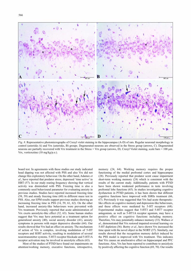
Histopathological scoring was performed to the effect of theVrx treatment on the stress-related cellular damage in theamygdaloid complex, hippocampus, and frontal cortex regions(Fig. 6A-6C). As shown in Cresyl violet, the staining of thesections displayed cells with normal histomorphology in thecontrol (Figs. 7A, 8A and 9A) and Vrx (Figs. 7B, 8B and 9B)groups. There were observed elevated damage scores in thestress group compared to the control group in the amygdaloidcomplex (t = 6.414, P < 0.0001; dF:28, Figs. 6A and 7C),hippocampal CA3 (t = 5.692, P = 0.0003; dF:28, Figs. 6B and8C), and frontal cortex (t = 7.655, P < 0.0001; dF:28, Figs. 6Cand 9C). The increased damage scores with PSS were partially
recovered with Vrx treatment in the amygdaloid complex (t =3.499, P = 0.0165; dF:28, Figs. 6A and 7D), the hippocampalCA3 (t = 3.111, P = 0.046; dF:28, Figs. 6B and 8D), and thefrontal cortex (t = 3.661, P = 0.0107; dF:28, Figs. 6C and 9D).In the other hippocampal region there were no significantdamage scores.
DISCUSSION
The major findings of the present study demonstrated thatVrx treatment improves behavioural and memory impairment inPSS rats. Stress-induced increased apoptotic parameters; bcl-2/bax ratio, casp-3 and -9 were decreased by Vrx treatments inthe amygdaloid complex, the hippocampus, and the frontalcortex except for bcl-2/bax ratio. BDNF levels of all examinedbrain regions were increased with Vrx treatments in stressconditions. Also, histological damage is reduced by Vrxtreatment in stress-induced rats.
We have found that PSS rats showed increased anxiety inboth the EPM and HBT, while these increased anxiety-likebehaviours were reversed with Vrx treatment. Previous studieshave shown PSS, had no effect on head dipping (55), decreasedhead dipping (56), that showing exploratory activity in the hole
565
Fig. 7. Representative photomicrographs of Cresyl violet staining in the amygdaloid complex of rats (A-D). Regular neuronalmorphology in Control (asterisks, A) and Vrx (asterisks, B) groups. Degenerated neurons are observed in the Stress group (arrows, C).Degenerated neurons were partially recovered with Vrx treatment in the Stress + Vrx group (arrow, D). Cresyl Violet staining, scalebars = 100 µm.Vrx, vortioxetine (10 mg/kg/p.o.).
board test. In agreements with these studies our study indicatedhead dipping was not affected with PSS and also Vrx did notchange this exploratory behaviour. On the other hand, Adamec etal., have reported that predator stress, depressed ‘time active’ inHBT (57). In our study rearing frequency showing that verticalactivity was diminished with PSS. Freezing time is also acommonly used behavioural parameter for evaluating anxiety inprevious studies. Studies have reported increased freezing time(58, 59) and steady freezing time (60) in different maze test inPSS. Also, our EPM results support previous studies showing anincreasing freezing time in PSS (14, 59, 61, 62). On the otherhand, increased anxiety-like behaviours were prevented withVrx treatment. Previously reported that acute administration ofVrx exerts anxiolytic-like effect (32, 63). Some human studiessuggest that Vrx may have potential as a treatment option forgeneralized anxiety (40), social anxiety disorder (41), anxietysymptoms in persons with major depressive disorder (64). Ourresults showed that Vrx had an effect on anxiety. The mechanismof action of Vrx is complex, involving modulation of 5-HTreceptors and SERT activity, resulting in modulation of severalneurotransmitter systems. 5-HT3 receptor blockade and 5-HT1Areceptor activation may be involved in anxiolytic effects (63).
Most of the studies of PTSD have found out impairments onattention/working memory, executive functions, retrospective,
memory (24, 64). Working memory requires the properfunctioning of the medial prefrontal cortex and hippocampus(28). Previously reported that predator scent cause impairmentshort-term working memory (24) which is consistent with theresults of the current study. Additionally, patients with PTSDhave been shown weakened performance in tests involvingprefrontal lobe functions (65). In studies investigating cognitivedysfunction in PTSD patients, it has been shown that differentcognitive functions have improved with SSRIs treatment (66,67). Previously it was suggested that Vrx had acute therapeutic-like effects on cognitive memory and depression-like behaviours,and these effects were mediated by 5-HT receptors (68).Experimental studies suggest that 5-HT3 and 5-HT7 receptorantagonism, as well as 5-HT1A receptor agonism, may have apositive effect on cognitive functions including memory.Therefore, Vrx may potentially enhance memory (33). Wallace etal., demonstrated that Vrx restored impaired reversal learning via5-HT depletion (36). Bertry et al., have shown Vrx increased thetime spent with the novel object in the NORT (37). Similarly, ourresults showed that the recognition memory deficit induced bythe PSS model is improved with Vrx treatment. This indicatesthat Vrx causes an ameliorative effect on hippocampal and PFCfunctions. Also, Vrx has been reported to contribute to anxiolysisby positively affecting the cognitive function (69, 70). Our results
566
Fig. 8. Representative photomicrographs of Cresyl violet staining in the hippocampus (A-D) of rats. Regular neuronal morphology incontrol (asterisks A) and Vrx (asterisks, B) groups. Degenerated neurons are observed in the Stress group (arrows, C). Degeneratedneurons are partially recovered with Vrx treatment in the Stress + Vrx group (arrows, D). Cresyl Violet staining, scale bars = 100 µm.Vrx, vortioxetine (10 mg/kg/p.o.).

have shown that the cognitive function deficit in PSS model hasbeen recovered by Vrx treatment. In accordance with theliterature, the improvement of the deteriorated cognitive functionmay have an anxiolytic effect as shown in our study. Previousfindings indicated that networks related to synaptic transmission,signal transduction, and neurodevelopment are modulated withVrx and may underlie Vrx’s cognitive-enhancing properties (71).Additionally, blocking 5HT3 receptors by Vrx enhances therelease of serotonin, norepinephrine, and acetylcholine whichmay be linked to pro-cognitive properties (70).
BDNF plays a critical role in synaptic plasticity (72). Thedysfunction of synaptic plasticity was reported in PTSD (66).According to the neurotrophic hypothesis of mental disordersincluding stress, there is a down-regulation of BDNF in thedifferent brain regions (72, 73). Similarly, physical stress alsocauses a decrease in systemic BDNF levels as central stressmediators in human subjects (74). On the other hand, it has beensuggested that increasing BDNF levels led to decreased anxiety-like behaviours in rats (75, 76). In the present study, thedecreased levels of the BDNF hippocampus due to PSS wereincreased by Vrx treatment, which may indicate increasedneuroplasticity with Vrx treatment. Several studies have reportedthat BDNF levels significantly increased after 1 week with acuteVrx treatment in CA1 and DG (77). In light of previous studies,
acute treatment was applied for a week to see the protectiveeffect of the drug in a process that began with stress in thecurrent study (77). Therefore, modulation of theneuroinflammation process along with neuroprotective agentsmay be useful for the treatment of mental disorders by increasingBDNF levels in PTSD patients (73). There are few studiesshowing the relationship between BDNF and stress-induceddisorders including PSS rat models after antidepressanttreatment (62,78,79). Lu et al., demonstrated that Vrx increasedthe hippocampal BDNF level in chronic unpredictable mildstressed animals (72). Furthermore, it has been demonstratedthat Vrx regulated the expression of genes associated with theplasticity in the amygdala, the hippocampus, and the frontalcortex in rodents (80). In our study, increasing expression ofBDNF in three different brain regions suggested that Vrx maystimulate neuroplasticity in stress conditions. In the presentstudy, if there is no stress exposure, Vrx did not change theBDNF levels. However, stress-induced rats have increasedBDNF levels. This suggests that Vrx may be effective in stressedconditions.
On the other hand, different PTSD models extensivelysuggest that an increase of BDNF in the amygdala (20, 62) whichplays a key role in synaptic plasticity and the formation of fearmemories. However, other studies reported that decreased BDNF
567
Fig. 9. Representative photomicrographs of Cresyl violet staining in the frontal cortex (A-D) of rats. Regular neuronal morphology incontrol (asterisks, A) and Vrx (asterisks, B) groups. Degenerated neurons are observed in the Stress group (arrows, C). Degeneratedneurons are partially recovered with Vrx treatment in the Stress + Vrx group (arrows, D). Cresyl Violet staining, scale bars = 100 µm.Vrx, vortioxetine (10 mg/kg/p.o.).

levels in the amygdala (17, 18-20). Stress-induced epigeneticchanges cause resulting in either transcriptional gene activationor repression of various genes, including BDNF (17). Our resultsshow that a decreased in BDNF levels in the amygdala in thestress group. These discrepancies in literature may depend on thetype of stress applied or the strain of the animal used.
Recent studies have shown that PTSD cause to increase inapoptosis in different brain regions, including the medialprefrontal cortex, the hippocampus, and the amygdala, which isassociated with cognition and emotion (8-12). Apoptosis isthought to alter the volume and function in certain brain regions.The networks of g-aminobutyric acid-ergic (GABAergic)interneurons in the amygdala are key factors of the brain’sinhibitory paths (6). GABA is essential for balancing betweenneuronal excitation and inhibition. Amygdala contains bothglutamatergic and GABAergic interneurons (6, 81). Impairmentof GABAergic inhibition in the amygdala can lead to behaviouralhyperexcitability, such as increased anxiety and emotionaldysregulation (82, 83). Stress may cause a reduction of theGABAergic inter-neuronal network and the development ofneuropsychological diseases (83, 84). Our findings are consistentwith previous data showing the crucial role of GABA innervationin the amygdala in fear condition. Skorzewska et al., have shownthat fear conditioning reduces the GABA levels in the BLA bydecreasing the mRNA level of the GABA synthesizing enzymeGAD67 in the amygdala and this condition leads to augmentedfear reaction (83). Our immunoblotting results indicated thatincreased apoptosis in the amygdala in the stress group. Thisneuronal apoptosis is may related to GABAergic neurons and thisleads to diminishing inhibition effect in the amygdala.Additionally, Zhao et al., have reported that predatory stressinduces hippocampal cell death by apoptosis in rats (85). Inagreement with this study our results indicated that PSS causeincreased apoptosis in the hippocampus. On the other hand,previously it has been reported that in contrast to the significantatrophy in the hippocampal regions, amygdala displays increasedendritic arborization and spine density with PSS (16). In thepresent study we did not investigate dendritic hypertrophy orvolume in amygdaloid complex.
Although there is no data about the anti-apoptotic effects ofVrx in PSS rat model, it has been shown that SSRIs were foundto have beneficial effects in animal stress models (31, 86-88).SSRIs inhibit apoptosis on hippocampus by increasing BDNFlevel in the depression model (88). According to ourimmunoblotting results in the stressed group, the increase in pro-apoptotic proteins casp-3, casp-9, and bax levels and the decreasein anti-apoptotic bcl-2 level in three different regions emphasizethe importance of apoptosis in cognitive disorders. Moreover, thesuppression of apoptosis by Vrx treatment suggests that Vrx hasa protective effect against stress disorders. In our study, theresults of the histological damage assessments have shown thatthe increase in the neuronal damage caused by stress was partiallyreversed by Vrx treatment.
This study has focused on the acute effects of Vrx. Furtherstudies are necessary in order to the long-term effects of Vrx inchronic PTSD models.
In conclusion, our results indicate that PSS causes anxietyand short-term recognition memory impairment and leads toapoptosis, and decreased BDNF levels in mainly brain regions.Neuronal deterioration is also accompanied by these findings. Ifadministered right after trauma, Vrx treatment may significantlyreduce behavioural, cognitive, and neuronal impairmentincreases neuroplasticity and may be protective against PTSDdevelopment.
Abbreviations: bax, bcl-2-associated X protein; BCIP, 5-bromo-4-chloro-3-indolyl phosphate; bcl-2, b-cell lymphoma;BLA, basolateral amygdala; BDNF, brain-derived neurotrophic
factor; BSA, bovine serum albumin; CA, cornu ammonis; casp-3, caspase-3; casp-9, caspase-9; DG, dentate gyrus; EDTA,ethylenediaminetetraacetic acid; EPM, elevated plus maze;GABA, gama amino butiric acid; HBT, hole board test; i.v.,intravenously kDa: kilodaltons; MeA, medial amygdala; NBT,nitro blue tetrazolium; NORT, novel object recognition test; PFC,prefrontal cortex; PMSF, phenylmethanesulfonyl fluoride; p.o.,perorally; PSS, predator scent-stress; SS, saline solution; PTSD,post-traumatic stress disorder; SD, standard deviation; SSRI,selective serotonin reuptake inhibitor; TBS, Tris buffer saline;TBS-T, Tris buffer saline 0.05% Tween-20; Vrx, vortioxetine.
This research did not receive any specific grant from fundingagencies in the public, the commercial, or the not-for-profitsectors.
Conflict of interests: None declared.
REFERENCES
1. Girgenti Mj, Hare BD, Ghosal S, Duman RS. Molecular andcellular effects of traumatic stress: implications for PTSD.Curr Psychiatry Rep 2017; 19: 85. doi: 10.1007/s11920-017-0841-3
2. Schoenfeld Tj, Rhee D, Martin L, Smith S, Sonti A,Padmanaban VS. New neurons restore structural andbehavioural abnormalities in a rat model of PTSD.Hippocampus 2019; 29: 848-861.
3. Hoskins M, Pearce j, Bethell A, et al. Pharmacotherapy forpost-traumatic stress disorder: systematic review and meta-analysis. Br J Psychiatry 2015; 206: 93-100.
4. Elzinga BM, Bremner jD. Are the neural substrates ofmemory the final common pathway in posttraumatic stressdisorder (PTSD) J Affect Disord 2002; 70:1-17. doi:10.1016/s0165-0327(01)00351-2
5. Zhang LM, Zhang YZ, Li YF. The progress ofneurobiological mechanisms on PTSD. Chin PharmacolBull 2010; 26: 704-707.
6. Etkin A, Wager TD. Functional neuroimaging of anxiety: ameta-analysis of emotional processing in PTSD, socialanxiety disorder, and specific phobia. Am J Psychiatry 2007;164: 1476-1488.
7. Chis IC, Clichici A, Nagy AL, Oros A, Catoi C, Clichici S.Quercetin in association with moderate exercise trainingattenuates injuries induced by experimental diabetes insciatic nerves. J Physiol Pharmacol 2017; 68: 877-886.
8. jia Y, Han Y, Wang X, Han F. Role of apoptosis in the post-traumatic stress disorder model-single prolonged stressedrats. Psychoneuroendocrinology 2018; 95: 97-105.
9. Xiao B, Yu B, Wang HT, Han F, Shi YX. Single-prolongedstress induces apoptosis by activating cytochromeC/caspase-9 pathway in a rat model of post-traumatic stressdisorder. Cell Mol Neurobiol 2011; 31: 37-43.
10. Ding j, Han F, Shi Y. Single-prolonged stress inducesapoptosis in the amygdala in a rat model of post-traumaticstress disorder. J Psychiatr Res 2010; 44: 48-55.
11. Kuhn HG, Palmer TD, Fuchs E. Adult neurogenesis: acompensatory mechanism for neuronal damage. Eur ArchPsychiatry Clin Neurosci 2001; 251: 152-158.
12. Du j, Wang Y, Hunter R, et al. Dynamic regulation ofmitochondrial function by glucocorticoids. Proc Natl Acad SciUSA 2009; 106: 3543-3548.
13. Aykac A, Aydyn B, Cabadak H, Goren MZ. The change inmuscarinic receptor subtypes in different brain regions ofrats treated with fluoxetine or propranolol in a post-traumatic stress disorder model. Behav Brain Res 2012; 232:124-129.
568

14. Ozbeyli D, Gokalp AG, Koral T, et al. Protective effect ofexercise and sildenafil on acute stress and cognitivefunction. Physiol Behav 2015; 151: 230-237.
15. Bremmer Dj. Traumatic stress: effects on the brain.Dialogues Clin Neurosci 2006; 8: 445-461.
16. Cohen H, Kozlovsky N, Matar MA, Zohar j, Kaplan Z.Distinctive hippocampal and amygdalar cytoarchitecturalchanges underlie specific patterns of behavioral disruptionfollowing stress exposure in an animal model of PTSD. EurNeuropsychopharmacol 2014; 24: 1925-1944.
17. Solanki N, Alkadhi I, Atrooz F, Patki G, Salim S. Grapepowder prevents cognitive, behavioral, and biochemicalimpairments in a rat model of posttraumatic stress disorder.Nutr Res 2015; 35: 65-75.
18. Li G, Wang G, Shi j, et al. Trans-resveratrol amelioratesanxiety-like behaviors and fear memory deficits in a ratmodel of post-traumatic stress disorder. Neuropharmacology2018; 133: 181-188.
19. Asim M, Hao B, Yang YH, et al. Ketamine alleviates feargeneralization through GluN2B-BDNF signaling in mice.Neurosci Bull 2019; Aug 23: doi: 10.1007/s12264-019-00422-47
20. ji LL, Ye Y, Nie PY, et al. Dysregulation of miR-142 resultsin anxiety-like behaviors following single prolonged stress.Behav Brain Res 2019; 365: 157-163.
21. Zoladz PR, Park, CR, Halonen jD, et al. Differentialexpression of molecular markers of synaptic plasticity in thehippocampus, prefrontal cortex, and amygdala in response tospatial learning, predator exposure, and stress-inducedamnesia. Hippocampus 2011; 22; 577-589.
22. Cohen H, Matar MA, joseph Z. Animal models of post-traumatic stress. Preclinical models of neurologic andpsychiatric disorders. Curr Protoc Neurosci 2013; 9.45: doi:10.1002/0471142301.ns0945s64.
23. Adamec R, Bartoszyk GD, Burton P. Effects of systemicinjections of vilazodone, a selective serotonin reuptakeinhibitor and serotonin 1A receptor agonist, on anxietyinduced by predator stress in rats. Eur J Pharmacol 2004;504: 65-77.
24. Morrow BA, Roth RH, Elsworth jD. TMT, a predator odor,elevates mesoprefrontal dopamine metabolic activity anddisrupts short-term working memory in the rat. Brain ResBull 2000; 52: 519-523.
25. Shallcross j, Hamor P, Bechard AR, Romano M, KnackstedtL, Schwendt M. The divergent effects of CDPPB andcannabidiol on fear extinction and anxiety in a predator scentstress model of PTSD in rats. Front Behav Neurosci 2019;13: 91. doi: 10.3389/fnbeh.2019.00091
26. Zoladz PR, Conrad CD, Fleshner M, Diamond DM. Acuteepisodes of predator exposure in conjunction with chronicsocial instability as an animal model of post-traumatic stressdisorder. Stress 2008; 11: 259-281.
27. American Psychiatric Association. Diagnostic and StatisticalManual of Mental Disorders. Arlington VA, AmericanPsychiatric Publishing, 2013.
28. Torrisi S, Leggio GM, Drago F, Salomone S. Therapeuticchallenges of post-traumatic stress disorder: focus on thedopaminergic system. Front Pharmacol 2019; 10: 404. doi:10.3389/fphar.2019.00404
29. Davidson jR. Pharmacologic treatment of acute and chronicstress following trauma: J Clin Psychiatry 2006; 67: 34-39.
30. Czeh B, Michaelis T, Watanabe T, et al. Stress-inducedchanges in cerebral metabolites, hippocampal volume, and cellproliferation are prevented by antidepressant treatment withtianeptine. Proc Natl Acad Sci USA 2001; 98: 12796-12801.
31. Lucassen Pj, Fuchs E, Czeh B. Antidepressant treatmentwith tianeptine reduces apoptosis in the hippocampal
dentate gyrus and temporal cortex. Biol Psychiatry 2004;55: 789-796.
32. Sowa-Kucma M, Panczyszyn-Trzewik P, Misztak P, et al.Vortioxetine: a review of the pharmacology and clinicalprofile of the novel antidepressant. Pharmacol Rep 2017; 69:595-601.
33. Mork A, Montezinho LP, Miller S, et al. Vortioxetine (LuAA21004), a novel multimodal antidepressant, enhancesmemory in rats. Pharmacol Biochem Behav 2013; 105:41-50.
34. Sanchez C, Asin KE, Artigas F. Vortioxetine, a novelantidepressant with multimodal activity: review of preclinicaland clinical data. Pharmacol Ther 2015; 145: 43-57.
35. Pehrson AL, Hillhouse TM, Haddjeri N, et al. Task- andtreatment length-dependent effects of vortioxetineonscopolamine-induced cognitive dysfunction and hippocampalextracellular acetylcholine in rats. J Pharmacol Exp Ther2016; 358: 472-482.
36. Wallace A, Pehrson AL, Sanchez C, Morilak DA.Vortioxetine restores reversal learning impaired by 5-HTdepletion or chronic intermittent cold stress in rats. Int JNeuropsychopharmacol 2014; 17: 1695-1706.
37. Betry C, Etievant A, Pehrson A, Sanchez C, Haddjeri N.Effect of the multimodal acting antidepressant vortioxetineon rat hippocampal plasticity and recognition memory. ProgNeuropsychopharmacol Biol Psychiatry 2015; 58: 38-46.
38. Lu Y, Ho CS, McIntyre RS, Wang W, Ho RC. Effects ofvortioxetine and fluoxetine on the level of brain derivedneurotrophic factors (BDNF) in the hippocampus of chronicunpredictable mild stress-induced depressive rats. Brain ResBull 2018; 142: 1-7. doi: 10.1016/j.brainresbull.2018.06.007
39. Orsolini L, Tomasetti C, Valchera A, et al. New advances inthe treatment of generalized anxiety disorder: the multimodalantidepressant vortioxetine. Expert Rev Neurother 2016; 16:483-495.
40. Pae CU, Wang SM, Han C, Lee Sj, Patkar AA, Masand PS,Serretti A. Vortioxetine, a multimodal antidepressant forgeneralized anxiety disorder: a systematic review and meta-analysis. J Psychiatr Res 2015; 64: 88-98.
41. Liebowitz MR, Careri j, Blatt K, et al. Vortioxetine versusplacebo in major depressive disorder comorbid with socialanxiety disorder. Depress Anxiety 2017; 34: 1164-1172.
42. Pellow S, Chopin P, File SE, Briley M. Validation of open-closed arm entries in an elevated plus maze as measure ofanxiety in the rat. J Neurosci Methods 1985; 14: 149-167.
43. Ozbeyli D, Sari G, Ozkan N, et al. Protective effects ofdifferent exercise modalities in an Alzheimer’s disease-likemodel. Behav Brain Res 2017; 328: 159-177.
44. Bevins R, Besheer j. Object recognition in rats and mice: aone-trial non-matching to sample learning task to studyrecognition memory. Nat Protoc 2006; 1: 1306-1311.
45. Kasimay-Cakir O, Ellek N, Salehin N, et al. Protective effectof low dose caffeine on psychological stress and cognitivefunction. Physiol Behav 2017; 168: 1-10.
46. Aykac A, Karanlik B. The expression level of muscarinic M1receptor subtypes in different regions of rat brain. MarmaraMed J 2017; 30: 162-168.
47. Broadbent Nj, Gaskin S, Squire LR, Clark RE. Objectrecognition memory and the rodent hippocampus. LearnMem 2010; 17: 5-11.
48. Kasimay O, Guzel E, Gemici A, et al. Colitis-inducedoxidative damage of the colon and skeletal muscle isameliorated by regular exercise in rats: the anxiolytic role ofexercise. Exp Physiol 2006; 91: 897-906.
49. Adamec R, Berton O, Razek WA. Viral vector induction ofCREB expression in the periaqueductal gray induces apredator stress-like pattern of changes in pCREB expression,
569
neuroplasticity, and anxiety in rodents. Neural Plast 2009;2009: 904568. doi: 10.1155/2009/904568
50. Cohen H, Kaplan Z, Matar MA, Loewenthal U, Kozlovsky N,Zohar j. Anisomycin, a protein synthesis inhibitor, disruptstraumatic memory consolidation and attenuates posttraumaticstress response in rats. Biol Psychiatry 2006; 60: 767-776.
51. Paxinos G, Watson C. The Rat Brain in StereotaxicCoordinates. London, Academic Press, 1986.
52. Lowry OH, Rosebrough Nj, Farr AL, Randall Rj. Proteinmeasurement with the folin-phenol reagents. J Biol Ther1951; 191: 33-43.
53. Koo E, Sheldon RA, Lee BS, Vexler ZS, Ferriero DM.Effects of therapeutic hypothermia on white matter injuryfrom murine neonatal hypoxia-ischemia. Pediatr Res 2017;82: 518-526.
54. Koyuncuoglu T, Vyzdyklar C, Uren D, et al. Obestatinimproves oxidative brain damage and memory dysfunctionin rats induced with an epileptic seizure. Peptides 2017; 90:37-47.
55. Adamec R, Muir C, Grimes M, Pearcey K. Involvement ofnoradrenergic and corticoid receptors in the consolidation ofthe lasting anxiogenic effects of predator stress. Behav BrainRes 2007; 179: 192-207.
56. Adamec R, Fougere D, Risbrough V. CRF receptor blockadeprevents initiation and consolidation of stress effects onaffect in the predator stress model of PTSD. Int JNeuropsychopharmacol 2010; 13: 747-757.
57. Adamec R, Head D, Soreq H, Blundell j. The role of the readthrough variant of acetylcholinesterase in anxiogenic effects ofpredator stress in mice. Behav Brain Res 2008; 189: 180-190.
58. Dopfel D, Perez PD, Verbitsky A, et al. Individual variabilityin behavior and functional networks predicts vulnerabilityusing an animal model of PTSD. Nat Commun 2019; 10:2372. doi: 10.1038/s41467-019-09926-z
59. Hoffman jR, Ostfeld I, Kaplan Z, Zohar j, Cohen H.Exercise enhances the behavioral responses to acute stress inan animal model of PTSD. Med Sci Sports Exerc 2015; 47:2043-2052.
60. Saridogan GE, Aykac A, Cabadak H, Cerit C, Caliskan M,Goren MZ. d-Cycloserine acts via increasing the GluN1protein expressions in the frontal cortex and decreases theavoidance and risk assessment behaviors in a rat traumaticstress model. Behav Brain Res 2015; 293: 227-233.
61. Hoffman jR, Ostfeld I, Stout jR, Harris RC, Kaplan Z,Cohen H. b-alanine supplemented diets enhance behavioralresilience to stress exposure in an animal model of PTSD.Amino Acids 2015; 47: 1247-1257.
62. Cohen H, Zohar j, Carmi L. Effects of agomelatine onbehaviour, circadian expression of period 1 and period 2clock genes and neuroplastic markers in the predator scentstress rat model of PTSD. World J Biol Psychiatry 2018;Nov 1: 1-19. doi: 10.1080/15622975.2018.1523560
63. Guilloux jP, Mendez-David I, Pehrson A, et al.Antidepressant and anxiolytic potential of the multimodalantidepressant vortioxetine (Lu AA21004) assessed bybehavioural and neurogenesis outcomes in mice.Neuropharmacology 2013; 73: 147-159.
64. Scott jC, Woods SP, Wrocklage KM, Schweinsburg BC,Southwick SM, Krystal jH. Prospective memory inposttraumatic stress disorder. J Int Neuropsychol Soc 2016;22: 724-734.
65. Kocak EE, Kilic C. Cognitive dysfunctions in posttraumaticstress disorder. Turk Psikiyatri Derg 2017; 28: 1-7.
66. Fani N, Kitayama N, Ashraf A, et al. Neuropsychologicalfunctioning in patients with posttraumatic stress disorderfollowing short-term paroxetine treatment. PsychopharmacolBull 2009; 42: 53-68.
67. Vermetten E, Vythilingam M, Southwick SM, Charney DS,Bremner jD. Long-term treatment with paroxetine increasesverbal declarative memory and hippocampal volume inposttraumatic stress disorder. Biol Psychiatry 2003; 54:693-702.
68. du jardin KG, Liebenberg N, Mueller HK, Elfving B,Sanchez C, Wegener G. Differential interaction with theserotonin system by S-ketamine, vortioxetine, and fluoxetinein a genetic rat model of depression. Psychopharmacology(Berl) 2016; 233: 2813-2825.
69. McIntyre RS, Xiao HX, Syeda K, et al. The prevalence,measurement, and treatment of the cognitivedimension/domain in major depressive disorder. CNS Drugs2015; 29: 577-589.
70. Stahl SM. Modes and nodes explain the mechanism of actionof vortioxetine, a multimodal agent (MMA): blocking 5HT3receptors enhances release of serotonin, norepinephrine, andacetylcholine. CNS Spectrums 2015; 20: 455-459.
71. Waller jA, Nygaard SH, Li Y, et al. Neuroplasticitypathways and protein-interaction networks are modulated byvortioxetine in rodents. BMC Neurosci 2017; 18: 56. doi:10.1186/s12868-017-0376-x
72. Lu B, Nagappan G, Lu Y. BDNF and synaptic plasticity,cognitive function, and dysfunction. Handb Exp Pharmacol2014; 220: 223-250.
73. Lee B, Hong R, Lim P, et al. The ethanolic extract of Araliacontinentalis ameliorates cognitive deficits via modificationsof BDNF expression and anti-inflammatory effects in a ratmodel of post-traumatic stress disorder. BMC ComplementAltern Med 2019; 19: 9-11.
74. Verbickas V, Baranauskiene N, Eimantas N, et al. Effect ofsprint cycling and stretch-shortening cycle exercises on theneuromuscular, immune and stress indicators in young men.J Physiol Pharmacol 2017; 68: 125-132.
75. Calabrese F, Molteni, R, Racagni G, Riva MA. Neuronalplasticity: a link between stress and mood disorders.Psychoneuroendocrinology 2009; 34: 208-216.
76. Song X, Liu B, Cui L, et al. Silibinin amelioratesanxiety/depression-like behaviors in amyloid b-treated rats byupregulating BDNF/TrkB pathway and attenuating autophagyin hippocampus. Physiol Behav 2017; 179: 487-493.
77. Chen F, Danladi j, Ardalan M, et al. A critical role ofmitochondria in BDNF-associated synaptic plasticity afterone-week vortioxetine treatment. Int J Neuropsychopharmacol2018: 21: 603-615.
78. Aykac A, Oncul S, Onur R. Social isolation and predatorscent tests alter brain BDNF levels differentially accordingto gender, in rats and effects of fluoxetine. J Res Pharm2018; 22: 190-198.
79. Yu H, Chen jj, Zeng BQ, Zhong QP, Xu jP, Liu YG. Role ofcAMP/CREB/BDNF signaling pathway in anti-depressiveeffect of vortioxetine in mice. Nan Fang Yi Ke Da Xue XueBao 2017; 37: 107-112.
80. Waller jA, Tamm jA, Abdourahman A, Pehrson AL, Li Y,Cajina M, Sanchez C. Chronic vortioxetine treatment inrodents modulates gene expression of neurodevelopmentaland plasticity markers. Eur Neuropsychopharmacol 2017;27: 192-203.
81. Bhatnagar S, Vining C, Denski K. Regulation of chronicstress-induced changes in hypothalamic-pituitary-adrenalactivity by the basolateral amygdala. Ann NY Acad Sci 2004;1032: 315-319.
82. Prager EM, Bergstrom HC, Wynn GH, Braga MF. Thebasolateral amygdala g-aminobutyric acidergic system inhealth and disease. J Neurosci Res 2016; 94: 548-567.
83. Skorzewska A, Wislowska-Stanek A, Lehner M, et al.Corticotropin releasing factor receptor 1 antagonist
570

differentially inhibits freezing behavior and changes gamma-aminobutyric acidergic activity in the amygdala in low- andhigh-anxiety rats. J Physiol Pharmacol 2017; 68: 35-46.
84. jie F, Yin G, Yang W, et al. Stress in regulation of GABAamygdala system and relevance to neuropsychiatricdiseases. Front Neurosci 2018; 12: 562. doi:10.3389/fnins.2018.00562
85. Zhao H, Xu H, Xu X, Young D. Predatory stress induceshippocampal cell death by apoptosis in rats. Neurosci Lett2007; 421: 115-120.
86. Aykac A, Cabadak H, Goren MZ. Altered ratio ofproapoptotic and antiapoptotic proteins in different brainregions of female rats in model of post-traumatic stressdisorder. Turk J Biochem 2015; 40: 1-7. doi:10.5505/tjb.2015.50479
87. Djordjevic A, Djordjevic j, Elakovic I, Adzic M, Matic G,Radojcic MB. Effects of fluoxetine on plasticity and
apoptosis evoked by chronic stress in rat prefrontal cortex.Eur J Pharmacol 2000; 693: 37-44.
88. Huang X, Mao YS, Li C, Wang H, ji jL. Venlafaxine inhibitsapoptosis of hippocampal neurons by up-regulating brain-derived neurotrophic factor in a rat depression model,Pharmazie 2014; 69: 909-916.
R e c e i v e d : May 27, 2019A c c e p t e d : August 28, 2019
Author’s address: Assoc. Prof. Dr. Asli Aykac, Departmentof Biophysics, Faculty of Medicine, Near East University, NearEast Boulevard, Nicosia, 99138, Cyprus.E-mail: [email protected]; [email protected]
571