Embed Size (px)
Citation preview
The Egyptian Journal of Hospital Medicine Vol., 23: 231 – 244 June 2006 I.S.S.N: 12084
1687 – 2002
Protective Role Of Proanthocyanidin Against Carbon Tetrachloride
Induced Toxicity In Irradiated Rats
Mona A. El-Gawish, Amira T. Ebrahim*, Fatma A. Eid*
National Center for Radiation Research and Technology-Atomic Energy
Authority. Zoology Department, Faculty of Science,
Al-Azhar University*.
Abstract
The hepatic injury induced by carbon tetrachloride (CCl4 ) as well as gamma-irradiation
has taken most attention. Prevention is one of the essential ways of controlling this toxicity and the use of natural plant compounds as grape seed extract can be considered as one of the most
significant elements in this prevention. The present study is also designed to evaluate the
hepatoprotective effects of grape seed extract (100mg/kg B.wt daily) against the toxic effect of CCl4 ( 0.3 ml/kg B.wt twice weekly for 8 weeks) and/ or fractionated doses of 2 Gy γ-radiation
day after day up to 10 Gy in male rats.
Results obtained could be summarized as follows: Combined treatments of CCl4 and γ-
radiation induced a significant increase in malondialdehyde (MDA) level, and decreased level of reduced glutathione (GSH). It also increased serum enzymes aspartate transferase (AST),
alanine transferase (ALT), a lkaline phosphatase (ALP) and gamma - glutamyl transpeptidase
(γ-GT) activities and decreased Hb%, RBCs, WBCs and platelets count. Proanthocyanidin administration improved the significant increase in MDA level and ameliorated serum enzymes
as well as improved the decreased level of GSH content of irradiated rats or those treated with
CCl4. It also could normalize the blood constituents Hb%, RBCs, WBCs and platelets count.
The data of the present study declared that proanthocyanidin is bioavailable, potent free radical scavenger and exhibits antioxidant properties against CCl4 and gamma-irradiation induced
hepatic injury.
Introduction
Virtually occupational exposures to toxic concentrations of carbon-tetrachloride
vapours are possible during industrial use
of the chemicals (NCI, 1985). It has been reported that oral exposure produced
marked central nervous system depression
and remarkable liver injury (Piyachaturawt
et al., 1995). Injury produced by CCl4 seems to be
mediated by reactive metabolite-trichloro-
methyl free radical (.CCl3) formed by
hemolytic cleavage of trichloromethyl
peroxy free radical (Cl3COO.) formed by
the reaction of CCl3 with O2 (Slater, 1982), which attach enoic fatty acids in the
membranes of endoplasmic reticulum,
leading to secondary free radicals, which
are subjected to attack by oxygen, and subsequent process,termed lipid peroxid-
ation, which produces damage to memb-
ranes and enzymes (Poli et al., 1989). The beneficial properties of flavo-
noids, including proanthocyanidins, have
been extensively researched (Murray and
Pizzorno, 1999). In addition to their antio-xidant and free radical scavenging activity
(Faria et al., 2006), proanthocyanidins
found in grape seed extract have been reported to have antibacterial, anticarcino-
genic, antiviral, anti-allergic, anti-inflamm-
atory effects (Blazso et al., 1994) vaso-dilatory actions (Bagchi et al., 1998),
antimutagenic agents, provide cardioprote-
ction by maintaining vascular permeability
231
Protective Role Of Proanthocyanidin Against Carbon ……….
232
(Bagchi et al., 2003) and the maintainance
of DNA integrity (Bagchi et al., 1998).
Material and Methods
Animals:
A total of 120 male Albino rats weighing
130-140 gm were used as experimental
animals. The rats were housed 6 animals/ cage with controlled air, temperature and
relative humidity and maintained on
standard cube pellets with free access to water. The diet consisted of not less than
20% protein, 5% fibers, 3% fats, 6.5% ash
and supplied with vitamins and minerals
mixture.
Gamma –irradiation procedure:
Irradiation process was performed
using Gamma Cell-40 achieved by Egypt's National Center for Radiation Research and
Technology (NCRRT), Cairo. The dose rate
was 0.66 Gy/min. at the time of experime-ntation.
Chemicals:
Grape Seed extract (Proanthoc-yanidin) was purchased from Natur’s Purest
(West Los Angeles, CA 90025). it was
dissolved in DMSO and diluted to the appropriate concentration by sterilized
saline solution. Carbon-tetrach-lorid was
diluted to the appropriate concentration by
olive oil.
Animal groups:
The experimental animals were divided into 8 groups (each group contains
15 rats) as follows:
Group1: Untreated control rats. Group2: Animals were exposed to
fractionated doses of γ-irradiation (2Gy
,day after day, up to 10 Gy). Group3:Animals were injected
intraperitoneally (i.p.) with CCl4 alone
(0.3ml/kg B.wt) twice weekly for 8 weeks
(Yasuda et al ., 2000). Group4: Animals were first treated with
CCl4 as described in group 3 (0.3 ml/kg
B.wt.) and then exposed to fractionated doses of γ-irradiation (2Gy, day after day,
up to 10 Gy).
Group5: Animals were given orally a
natural antioxidant (Proanthocyanidine)
daily for one week at a dose of 100 mg/kg B.wt (Bagchi et al., 2001).
Group6: Animals were first given
proanthocyanidin orally (daily for one week
at a dose of 100 mg/kg B.wt), then exposed to fractionated doses of γ-irradiation (2Gy
day after day up to 10 Gy).
Group7: Animals were given proantho-cyanidin orally (daily for one week at a
dose of 100 mg/kg B.wt), before the i.p.
injection with CCl4 and continuing for
another 8 weeks parallel with the injection of CCl4
( 0.3 ml/ kg B.wt.)
Group8: Animals were given proantho-cyanidin orally (daily for one week at a
dose of 100 mg/kg B.wt), before the
injection with CCl4 and continuing for another 8 weeks parallel with the injection
of CCl4 (0.3mg/kg B.wt) followed by
exposure to fractionated doses of γ-
irradiation (2Gy, day after day, up to 10 Gy). All experimental groups were
sacrificed after one day, 7 days, and 14
days post treatment or irradiation.
Biochemical studies:
Animals were sacrificed by slight ether anesthesia. A portion of blood sam-
ples were immediately collected and used
for the determination of reduced glutathione
(GSH) level as described by Beutler, (1963) as well as WBCs, RBCs, Hb %, and
thrombocyte count were estimated
according to Dacie and Lewis (1991). Another portion of blood was taken for the
separation of serum for the determination of
malonedialdehyde (MDA) according to the
method of Yoshioka et al., (1979). AST, ALT, ALP and γ-GT were measured as
described by Reitman and Frankel (1957),
John and Bauer (1982) and Szasz (1969) respectively. The data were statistically
analyzed following the student’s test (t)
(Snedecor and Cochran 1980).
Results
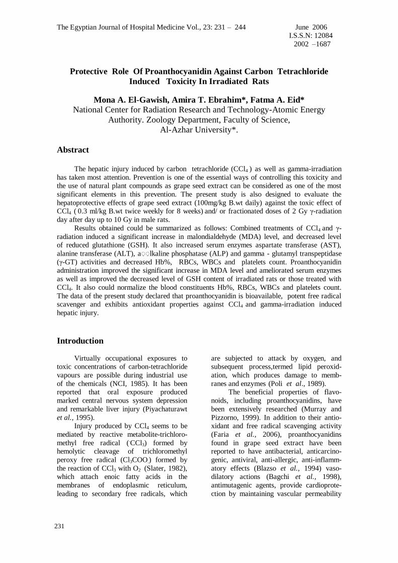
Exposure of rats to whole body γ-
irradiation or those received CCl4 induced
Mona A. El-Gawish et al
233
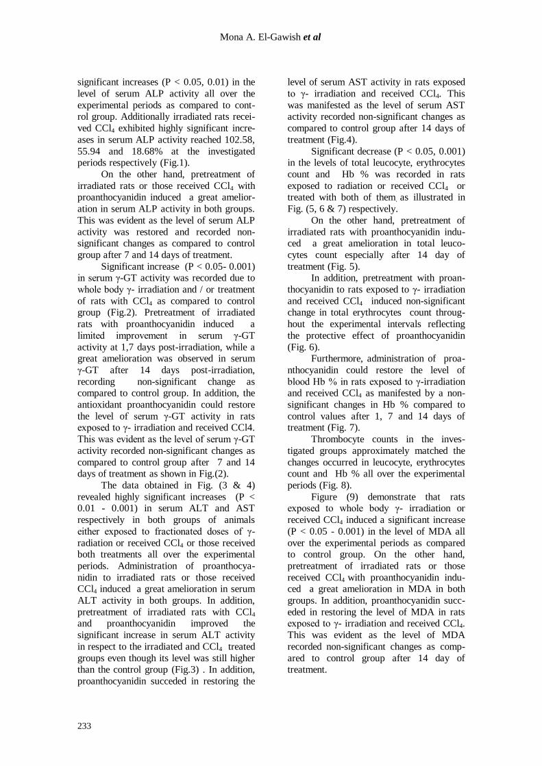
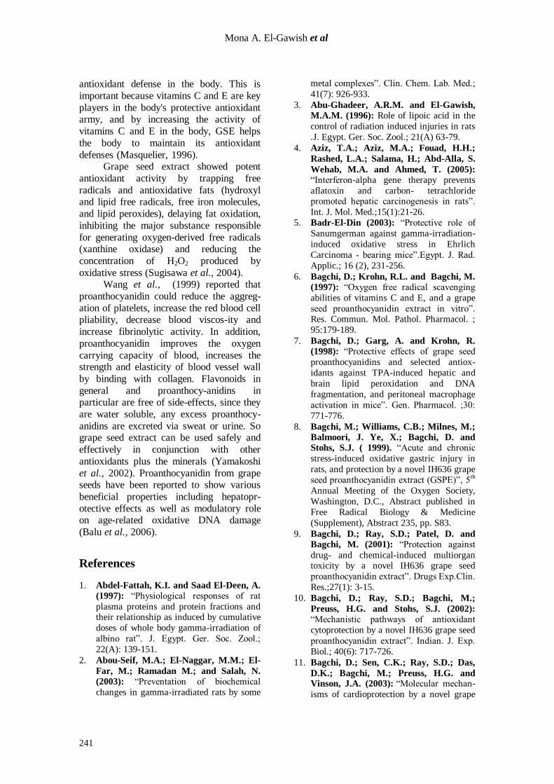
significant increases (P < 0.05, 0.01) in the
level of serum ALP activity all over the
experimental periods as compared to cont-rol group. Additionally irradiated rats recei-
ved CCl4 exhibited highly significant incre-
ases in serum ALP activity reached 102.58,
55.94 and 18.68% at the investigated periods respectively (Fig.1).
On the other hand, pretreatment of
irradiated rats or those received CCl4 with proanthocyanidin induced a great amelior-
ation in serum ALP activity in both groups.
This was evident as the level of serum ALP
activity was restored and recorded non-significant changes as compared to control
group after 7 and 14 days of treatment.
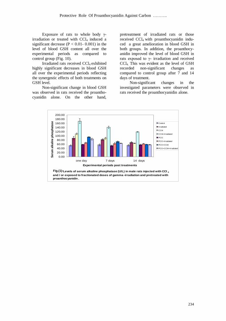
Significant increase (P < 0.05- 0.001) in serum γ-GT activity was recorded due to
whole body γ- irradiation and / or treatment
of rats with CCl4 as compared to control group (Fig.2). Pretreatment of irradiated
rats with proanthocyanidin induced a
limited improvement in serum γ-GT
activity at 1,7 days post-irradiation, while a great amelioration was observed in serum
γ-GT after 14 days post-irradiation,
recording non-significant change as compared to control group. In addition, the
antioxidant proanthocyanidin could restore
the level of serum γ-GT activity in rats exposed to γ- irradiation and received CCl4.
This was evident as the level of serum γ-GT
activity recorded non-significant changes as
compared to control group after 7 and 14 days of treatment as shown in Fig.(2).
The data obtained in Fig. (3 & 4)
revealed highly significant increases (P < 0.01 - 0.001) in serum ALT and AST
respectively in both groups of animals
either exposed to fractionated doses of γ-
radiation or received CCl4 or those received both treatments all over the experimental
periods. Administration of proanthocya-
nidin to irradiated rats or those received CCl4 induced a great amelioration in serum
ALT activity in both groups. In addition,
pretreatment of irradiated rats with CCl4 and proanthocyanidin improved the
significant increase in serum ALT activity
in respect to the irradiated and CCl4 treated
groups even though its level was still higher than the control group (Fig.3) . In addition,
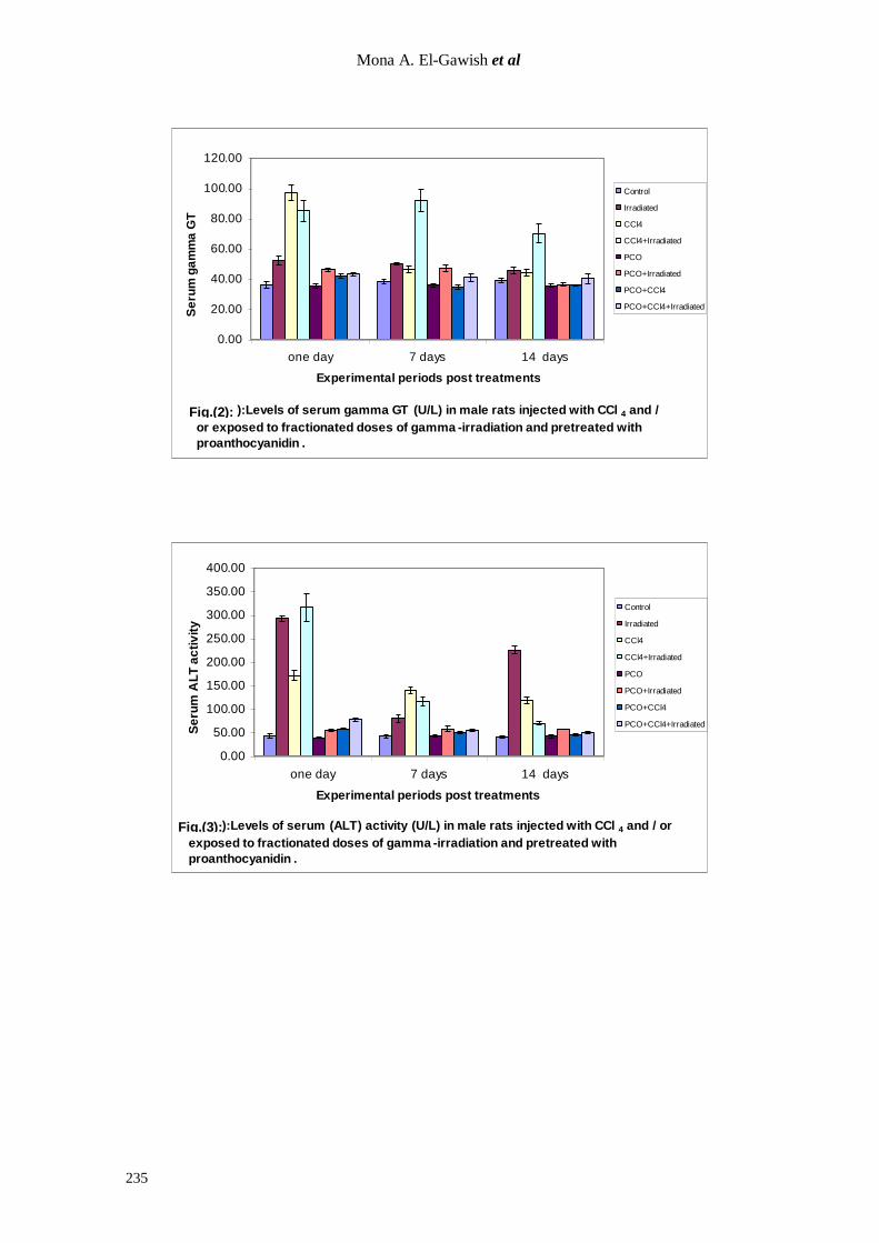
proanthocyanidin succeded in restoring the
level of serum AST activity in rats exposed
to γ- irradiation and received CCl4. This
was manifested as the level of serum AST activity recorded non-significant changes as
compared to control group after 14 days of
treatment (Fig.4).
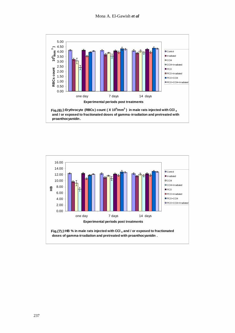
Significant decrease (P < 0.05, 0.001) in the levels of total leucocyte, erythrocytes
count and Hb % was recorded in rats
exposed to radiation or received CCl4 or treated with both of them. as illustrated in
Fig. (5, 6 & 7) respectively.
On the other hand, pretreatment of
irradiated rats with proanthocyanidin indu-ced a great amelioration in total leuco-
cytes count especially after 14 day of
treatment (Fig. 5). In addition, pretreatment with proan-
thocyanidin to rats exposed to γ- irradiation
and received CCl4 induced non-significant change in total erythrocytes count throug-
hout the experimental intervals reflecting
the protective effect of proanthocyanidin
(Fig. 6). Furthermore, administration of proa-
nthocyanidin could restore the level of
blood Hb % in rats exposed to γ-irradiation and received CCl4 as manifested by a non-
significant changes in Hb % compared to
control values after 1, 7 and 14 days of treatment (Fig. 7).
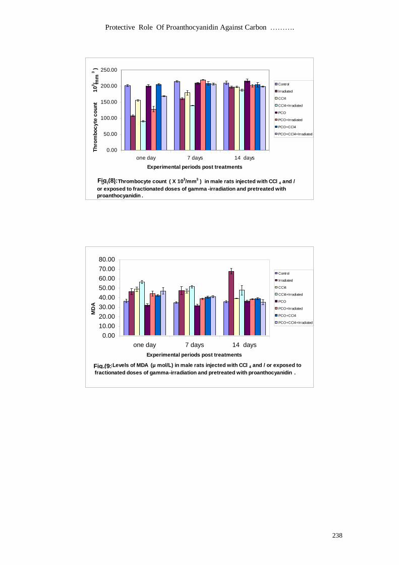
Thrombocyte counts in the inves-
tigated groups approximately matched the
changes occurred in leucocyte, erythrocytes count and Hb % all over the experimental
periods (Fig. 8).
Figure (9) demonstrate that rats exposed to whole body γ- irradiation or
received CCl4 induced a significant increase
(P < 0.05 - 0.001) in the level of MDA all
over the experimental periods as compared to control group. On the other hand,
pretreatment of irradiated rats or those
received CCl4 with proanthocyanidin indu-ced a great amelioration in MDA in both
groups. In addition, proanthocyanidin succ-
eded in restoring the level of MDA in rats exposed to γ- irradiation and received CCl4.
This was evident as the level of MDA
recorded non-significant changes as comp-
ared to control group after 14 day of treatment.
Protective Role Of Proanthocyanidin Against Carbon ……….
234
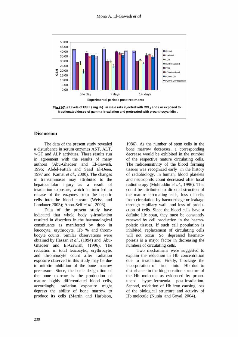
Exposure of rats to whole body γ-
irradiation or treated with CCl4 induced a
significant decrease (P < 0.01- 0.001) in the level of blood GSH content all over the
experimental periods as compared to
control group (Fig. 10).
Irradiated rats received CCl4 exhibited highly significant decreases in blood GSH
all over the experimental periods reflecting
the synergestic effects of both treatments on GSH level.
Non-significant change in blood GSH
was observed in rats received the proantho-
cyanidin alone. On the other hand,
pretreatment of irradiated rats or those
received CCl4 with proanthocyanidin indu-
ced a great amelioration in blood GSH in both groups. In addition, the proanthocy-
anidin improved the level of blood GSH in
rats exposed to γ- irradiation and received
CCl4. This was evident as the level of GSH recorded non-significant changes as
compared to control group after 7 and 14
days of treatment. Non-significant changes in the
investigated parameters were observed in
rats received the proanthocyanidin alone.
0.00
20.00
40.00
60.00
80.00
100.00
120.00
140.00
160.00
180.00
200.00
one day 7 days 14 days
Experimental periods post treatments
Se
rum
alk
alin
e p
ho
sp
ha
tas
e
( U / L )
Control
Irradiated
CCl4
CCl4+Irradiated
PCO
PCO+Irradiated
PCO+CCl4
PCO+CCl4+Irradiated
Fig .(9):Levels of serum alkaline phosphatase (U/L) in male rats injected with CCl 4
and / or exposed to fractionated doses of gamma -irradiation and pretreated with
proanthocyanidin .
Fig.(1):
Mona A. El-Gawish et al
235
0.00
20.00
40.00
60.00
80.00
100.00
120.00
one day 7 days 14 days
Experimental periods post treatments
Se
rum
ga
mm
a G
T
( U / L )
Control
Irradiated
CCl4
CCl4+Irradiated
PCO
PCO+Irradiated
PCO+CCl4
PCO+CCl4+Irradiated
Fig .(10):Levels of serum gamma GT (U/L) in male rats injected with CCl 4 and /
or exposed to fractionated doses of gamma -irradiation and pretreated with
proanthocyanidin .
0.00
50.00
100.00
150.00
200.00
250.00
300.00
350.00
400.00
one day 7 days 14 days
Experimental periods post treatments
Se
rum
AL
T a
cti
vit
y
( U / L )
Control
Irradiated
CCl4
CCl4+Irradiated
PCO
PCO+Irradiated
PCO+CCl4
PCO+CCl4+Irradiated
Fig .(8):Levels of serum (ALT) activity (U/L) in male rats injected with CCl 4 and / or
exposed to fractionated doses of gamma -irradiation and pretreated with
proanthocyanidin .
Fig.(2):
Fig.(3):
Protective Role Of Proanthocyanidin Against Carbon ……….
236
0.00
50.00
100.00
150.00
200.00
250.00
300.00
350.00
400.00
450.00
500.00
one day 7 days 14 days
Experimental periods post treatments
Se
rum
AS
T a
cti
vit
y
( U / L )
Control
Irradiated
CCl4
CCl4+Irradiated
PCO
PCO+Irradiated
PCO+CCl4
PCO+CCl4+Irradiated
Fig .(7):Levels of serum (AST) activity (U/L) in male rats injected with CCl 4 and / or
exposed to fractionated doses of gamma -irradiation and pretreated with
proanthocyanidin .
0.00
1.00
2.00
3.00
4.00
5.00
6.00
7.00
one day 7 days 14 days
Experimental periods post treatments
WB
Cs
co
un
t
( X
10
3/ m
m3 )
Control
Irradiated
CCl4
CCl4+Irradiated
PCO
PCO+Irradiated
PCO+CCl4
PCO+CCl4+Irradiated
Fig .(16): Total leucocytic (WBCs) count ( X 103/mm
3 ) in male rats injected with CCl 4
and / or exposed to fractionated doses of gamma -irradiation and pretreated with
proanthocyanidin .
Fig.(4):
Fig.(5):
Mona A. El-Gawish et al
237
0.00
0.50
1.00
1.50
2.00
2.50
3.00
3.50
4.00
4.50
5.00
one day 7 days 14 days
Experimental periods post treatments
RB
Cs
co
un
t
( X
10
6/ m
m3 )
Control
Irradiated
CCl4
CCl4+Irradiated
PCO
PCO+Irradiated
PCO+CCl4
PCO+CCl4+Irradiated
Fig .(17):Erythrocyte (RBCs ) count ( X 106/mm
3 ) in male rats injected with CCl 4
and / or exposed to fractionated doses of gamma -irradiation and pretreated with
proanthocyanidin .
0.00
2.00
4.00
6.00
8.00
10.00
12.00
14.00
16.00
one day 7 days 14 days
Experimental periods post treatments
HB
%
Control
Irradiated
CCl4
CCl4+Irradiated
PCO
PCO+Irradiated
PCO+CCl4
PCO+CCl4+Irradiated
Fig .(18):HB % in male rats injected with CCl 4 and / or exposed to fractionated
doses of gamma-irradiation and pretreated with proanthocyanidin .
Fig.(6):
Fig.(7):
Protective Role Of Proanthocyanidin Against Carbon ……….
238
0.00
50.00
100.00
150.00
200.00
250.00
one day 7 days 14 days
Experimental periods post treatments
Th
rom
bo
cy
te c
ou
nt
( X
10
3/ m
m3 )
Control
Irradiated
CCl4
CCl4+Irradiated
PCO
PCO+Irradiated
PCO+CCl4
PCO+CCl4+Irradiated
Fig .(20):Thrombocyte count ( X 103/mm
3 ) in male rats injected with CCl 4 and /
or exposed to fractionated doses of gamma -irradiation and pretreated with
proanthocyanidin .
0.00
10.00
20.00
30.00
40.00
50.00
60.00
70.00
80.00
one day 7 days 14 days
Experimental periods post treatments
MD
A
( μ
mo
l
/ L )
Control
Irradiated
CCl4
CCl4+Irradiated
PCO
PCO+Irradiated
PCO+CCl4
PCO+CCl4+Irradiated
Fig .(5):Levels of MDA (μ mol/L) in male rats injected with CCl 4 and / or exposed to
fractionated doses of gamma-irradiation and pretreated with proanthocyanidin .
Fig.(8):
Fig.(9:
Mona A. El-Gawish et al
239
0.00
5.00
10.00
15.00
20.00
25.00
30.00
35.00
40.00
45.00
50.00
one day 7 days 14 days
Experimental periods post treatments
GS
H
( mg
% )
Control
Irradiated
CCl4
CCl4+Irradiated
PCO
PCO+Irradiated
PCO+CCl4
PCO+CCl4+Irradiated
Fig .(6):Levels of GSH ( mg % ) in male rats injected with CCl 4 and / or exposed to
fractionated doses of gamma-irradiation and pretreated with proanthocyanidin .
Discussion
The data of the present study revealed
a disturbance in serum enzymes AST, ALT, γ-GT and ALP activities. These results run
in agreement with the results of many
authors (Abu-Ghadeer and El-Gawish, 1996; Abdel-Fattah and Saad El-Deen,
1997 and Kamat et al., 2000). The changes
in transaminases may attributed to the
hepatocellular injury as a result of irradiation exposure, which in turn led to
release of the enzymes from the hepatic
cells into the blood stream (Weiss and Landauer 2003); Abou-Seif et al., 2003).
Data of the present study have
indicated that whole body γ-irradiation resulted in disorders in the haematological
constituents as manifested by drop in
leucocyte, erythrocyte, Hb % and throm-
bocyte counts. Similar observations were obtained by Hassan et al., (1994) and Abu-
Ghadeer and El-Gawish, (1996). The
reduction in total leucocytic, erythrocyte, and thrombocyte count after radiation
exposure observed in this study may be due
to mitotic inhibition of the bone marrow precursors. Since, the basic designation of
the bone marrow is the production of
mature highly differentiated blood cells,
accordingly, radiation exposure might depress the ability of bone marrow to
produce its cells (Martin and Harbison,
1986). As the number of stem cells in the
bone marrow decreases, a corresponding decrease would be exhibited in the number
of the respective mature circulating cells.
The radiosensitivity of the blood forming tissues was recognized early in the history
of radiobiology. In human, blood platelets
and neutrophils count decreased after local
radiotherapy (Mohiuddin et al., 1996). This could be attributed to direct destruction of
the mature circulating cells, loss of cells
from circulation by haemorrhage or leakage through capillary wall, and loss of produ-
ction of cells. Since the blood cells have a
definite life span, they must be constantly renewed by cell production in the haemo-
poietic tissues. If such cell population is
inhibited, replacement of circulating cells
will not occur. So, depressed haemato-poiesis is a major factor in decreasing the
numbers of circulating cells.
Two mechanisms were suggested to explain the reduction in Hb concentration
due to irradiation. Firstly, blockage the
incorporation of iron into Hb due to disturbance in the biogeneration structure of
the Hb molecule as evidenced by prono-
unced hyper-ferraemia post-irradiation.
Second, oxidation of Hb iron causing loss of the biological structure and activity of
Hb molecule (Nunia and Goyal, 2004).
Fig.(10):
Protective Role Of Proanthocyanidin Against Carbon ……….
240
The results of the present study
showed that whole body γ-irradiation serio-
usly stimulated lipid peroxidation produ-ction. This was evident from the highly
significant increase in MDA as one of the
main end products of lipid peroxidation.
The recorded increase in MDA could be explained on the basis of that ionizing
radiation induced lipid peroxidation
through the production of active oxygen species, which attack the polyunsaturated
fatty acids of the phospholipids of cell
membrane (Gatsko et al., 1990).
In the view of the data obtained in the present work, there was a significant decr-
ease in blood glutathione. Such decrease
was in accordance with the data reported by EL-Gawish et al. (1997 & 2000) and
Abou-seif et al. (2003), Badr-El-Din
(2003). The recorded decrease in the reduced GSH could be attributed to its
conversion to the oxidized form with
minimal chance to be recycled to the
reduced form again due to inhibition of GSH reductase (Kergonou et al., 1986).
Jiangui and Sun (1993), found a correlation
between the increase of lipoperoxide in irradiated mice and the decrease of the
antioxidant defense system. This decrease
in the scavenging system located extracellularly and inracellulary gives rise
to excessive production of free radicals.
The data of the present study revealed
that administration of CCl4 induced similar changes in the investigated parame-ters to
those exposed to γ-radiation. Injury
produced by CCl4 seems to be mediated by reactive metabolite-trichloromethyl free
radical (.CCl3) formed by the hemolytic
cleavage of trichloromethylperoxy free
radical (Cl3COO.) formed by the reaction of
.CCl3 with O2 (Slater, 1982). This biotr-
ansformation is catalyzed by a cytochrome
P450-dependent monooxygenase to yield trichloromethyl and chlorine free radicals.
The trichloromethyl free radical is then
thought to attach enoic fatty acids in the membranes of the endoplasmic reticulum,
leading to secondary free radicals within
the fatty acids, which is subjected to attack
by oxygen, and the subsequent process, which is termed lipid peroxidation,
produces damage to membranes and
enzymes (Poli , 1989). Thus the toxicity of
CCl4 depends on the cleavage of carbon-
chloride bond to generate trichloromethyle free radical (
.CCl3) ( Lee et al., 2004 ).
Consequently, the CCl4–derived free radi-
cals can initiate a process of autocatalytic
lipid peroxidation by attacking the methyl bridges of unsaturated fatty side-chains of
microsomal lipids (Yan et al., 2003).
The decline in glutathione content together with increased lipid peroxidation
following CCl4 administration were repo-
rted by Kus et al., (2004); Shukla et al.,
(2004) and Valcheva et al., (2004). CCl4 treatment generates free radicals
that trigger a cascade of events resulting in
liver dysfunction (Luo et al., 2004 and Aziz et al., 2005) as reflected by significant
increase in serum AST, ALT, ALP and γ-
GT activities (Gao et al., 2004; Ha et al., 2005),
The data of the present study
proved that oral administration of proa-
nthocyanidin ameliorated all the invest-igated parameters in animals exposed to
fractionated doses of gamma-irradiation or
treated with CCl4. Our results are in agreement with Bagchi et al., (1998), Ray
et al.(2000), Abou seif et al., (2003) and
Weiss and Landauer (2003). Proanthocyanidins, a group of
polyphenolic bioflavonoids, have been
reported to exhibit a wide range of biolog-
ical, pharmacological and chemoprotective properties against oxygen free radicals
(Bagchi et al., 1997, 2002 and Ray et al.,
2000). It has been reported by Bagchi et al.,
(1999), that proanthocyanidin provided
significant protection against acute and
chronic stress-induced gastrointestinal and hepatic oxidative injury as demonstrated by
reduced lipid peroxidation, DNA damage
and membrane microviscosity. Grape seed extracts (GSE) contain a
total of 92% to 95% the gallic esters of
proanthocyanidins (PCO) (Veluri et al., 2006). These compounds are the most
active free radical scavenging PCOs
(Bagchi et al., 1998). GSE recycles and
significantly extends the lifetime of vitamin C and potentiates vitamin E, and thereby
creates a more powerful, synergistic
Mona A. El-Gawish et al
241
antioxidant defense in the body. This is
important because vitamins C and E are key
players in the body's protective antioxidant army, and by increasing the activity of
vitamins C and E in the body, GSE helps
the body to maintain its antioxidant
defenses (Masquelier, 1996). Grape seed extract showed potent
antioxidant activity by trapping free
radicals and antioxidative fats (hydroxyl and lipid free radicals, free iron molecules,
and lipid peroxides), delaying fat oxidation,
inhibiting the major substance responsible
for generating oxygen-derived free radicals (xanthine oxidase) and reducing the
concentration of H2O2 produced by
oxidative stress (Sugisawa et al., 2004). Wang et al., (1999) reported that
proanthocyanidin could reduce the aggreg-
ation of platelets, increase the red blood cell pliability, decrease blood viscos-ity and
increase fibrinolytic activity. In addition,
proanthocyanidin improves the oxygen
carrying capacity of blood, increases the strength and elasticity of blood vessel wall
by binding with collagen. Flavonoids in
general and proanthocy-anidins in particular are free of side-effects, since they
are water soluble, any excess proanthocy-
anidins are excreted via sweat or urine. So grape seed extract can be used safely and
effectively in conjunction with other
antioxidants plus the minerals (Yamakoshi
et al., 2002). Proanthocyanidin from grape seeds have been reported to show various
beneficial properties including hepatopr-
otective effects as well as modulatory role on age-related oxidative DNA damage
(Balu et al., 2006).
References
1. Abdel-Fattah, K.I. and Saad El-Deen, A.
(1997): “Physiological responses of rat
plasma proteins and protein fractions and their relationship as induced by cumulative
doses of whole body gamma-irradiation of
albino rat”. J. Egypt. Ger. Soc. Zool.;
22(A): 139-151.
2. Abou-Seif, M.A.; El-Naggar, M.M.; El-
Far, M.; Ramadan M.; and Salah, N.
(2003): “Preventation of biochemical
changes in gamma-irradiated rats by some
metal complexes”. Clin. Chem. Lab. Med.;
41(7): 926-933.
3. Abu-Ghadeer, A.R.M. and El-Gawish,
M.A.M. (1996): Role of lipoic acid in the
control of radiation induced injuries in rats
.J. Egypt. Ger. Soc. Zool.; 21(A) 63-79.
4. Aziz, T.A.; Aziz, M.A.; Fouad, H.H.;
Rashed, L.A.; Salama, H.; Abd-Alla, S.
Wehab, M.A. and Ahmed, T. (2005): “Interferon-alpha gene therapy prevents
aflatoxin and carbon- tetrachloride promoted hepatic carcinogenesis in rats”.
Int. J. Mol. Med.;15(1):21-26.
5. Badr-El-Din (2003): “Protective role of
Sanumgerman against gamma-irradiation-
induced oxidative stress in Ehrlich
Carcinoma - bearing mice”.Egypt. J. Rad.
Applic.; 16 (2), 231-256.
6. Bagchi, D.; Krohn, R.L. and Bagchi, M.
(1997): “Oxygen free radical scavenging
abilities of vitamins C and E, and a grape
seed proanthocyanidin extract in vitro”. Res. Commun. Mol. Pathol. Pharmacol. ;
95:179-189.
7. Bagchi, D.; Garg, A. and Krohn, R.
(1998): “Protective effects of grape seed
proanthocyanidins and selected antiox-
idants against TPA-induced hepatic and
brain lipid peroxidation and DNA
fragmentation, and peritoneal macrophage
activation in mice”. Gen. Pharmacol. ;30:
771-776.
8. Bagchi, M.; Williams, C.B.; Milnes, M.;
Balmoori, J. Ye, X.; Bagchi, D. and
Stohs, S.J. ( 1999). “Acute and chronic
stress-induced oxidative gastric injury in
rats, and protection by a novel IH636 grape
seed proanthocyanidin extract (GSPE)”, 5th
Annual Meeting of the Oxygen Society,
Washington, D.C., Abstract published in
Free Radical Biology & Medicine
(Supplement), Abstract 235, pp. S83.
9. Bagchi, D.; Ray, S.D.; Patel, D. and
Bagchi, M. (2001): “Protection against
drug- and chemical-induced multiorgan toxicity by a novel IH636 grape seed
proanthocyanidin extract”. Drugs Exp.Clin.
Res.;27(1): 3-15.
10. Bagchi, D.; Ray, S.D.; Bagchi, M.;
Preuss, H.G. and Stohs, S.J. (2002): “Mechanistic pathways of antioxidant
cytoprotection by a novel IH636 grape seed
proanthocyanidin extract”. Indian. J. Exp.
Biol.; 40(6): 717-726.
11. Bagchi, D.; Sen, C.K.; Ray, S.D.; Das,
D.K.; Bagchi, M.; Preuss, H.G. and Vinson, J.A. (2003): “Molecular mechan-
isms of cardioprotection by a novel grape
Protective Role Of Proanthocyanidin Against Carbon ……….
242
seed proanthocyanidin extract”. Mutat.
Res.; 523: 87- 97.
12. Balu M, Sangeetha P, Murali G, and
Panneerselvam C. (2006): Modulatory
role of grape seed extract on age-related
oxidative DNA damage in central nervous
system of rats. Brain Res Bull.
15;68(6):469-73.
13. Beutler. E.; Duron, O. and Kelly, B.M.
(1963): “Improved method of the determi-
nation of blood glutathione”. J. Lab. & Clin. Med.; 61(5): 882-888.
14. Blazsó, G.; Gábor, M.; Sibbel, R. and
Rohdewald, P. (1994): “Anti-inflamm-
atory and super-oxide radical scavenging
activities of a procyanidins containing
extract from the bark of Pinus pinaster Sol.
and its fractions”. Pharm. Pharmacol. Lett.
(3): 217-220.
15. Dacie, J.V. and Lewis, S.M. (1991): “Basic practical haematology. In: practical
haematology”: 7th ed., the English Languages Book Society and Churchill
Livingstonne., 37 – 58.
16. El-Gawish, M.A.M.; Abady, M.M. and
Said, O.Z. (1997): “Potential prophylactic
role of ginseng against biochemical hazards
of whole body gamma-irradiation in albino
rats.” Egypt. J. Med. Sci.; 18(2): 583-598.
17. El-Gawish, M.A.; Mahdy, E.M.E.; Hafez,
M.N. and El-Hadidy, M. E.A. (2000): “Protective effects of natural isoflavone On
1,2-dimethylhydrazine carcino-genesis in irradiated rats”. Cancer Molecular Biology,
7(4):1527-1543.
18. Faria A, Calhau C, de Freitas V, and
Mateus N. (2006): Procyanidins as antiox-
idants and tumor cell growth modulators. J
Agric Food Chem. 54(6):2392-7.
19. Gao, J; Tang, X.; Dou, H.; Fan, Y.;
Zhao, X. and Xu, Q. (2004): “Hepatopr-
otective activity of Terminalia catappa L.
leaves and its two triterpenoids. J. Pharm.
Pharmacol.”.; 56(11):1449-1455.
20. Gatsko, G.G.; Mazhul, L.M.; Shabli-
nskaya, O.V. and Volykhina, V.E.
(1990): “The influence of ionizing radiation
on lipid peroxidation in rat blood”.
Radiobiology; 30 (3): 413 – 415.
21. Ha, K.T.; Yoon, S.J.; Choi, D.Y.; Kim,
D.W.; Kim, J.K. and Kim, C.H. (2005):
“Protective effect of Lycium chinense fruit
on CCl4 induced-hepatotoxicity”.
J. Ethnopharmacol.; 15; 96(3): 529-535.
22. Hassan, S.H.M.; Abu-Ghadeer A.R.M.;
Osman, S.A.A. and Roushdy, H.M. (1994): “Possible curative role of the
antipsychotic drug : Fluphenazine: against
Post-irradiation injury in rats”. Egypt. J.
Rad. Sci. Applic.; 7(2): 181- 200.
23. Jiangui, J. and Sun, G. (1993): “Effect of
fractionated gamma-irradiation on SOD
activity and lipoperoxide count in liver and
kidney tissues of adolescent mice”. J.
Radiat. Res. and Radiat. Process; 11(2):
104 – 106.
24. John, D. and Bauer, M. (1982): “Clinical
laboratory methods for the determination of
serum alkaline phosphatase”. C.V. Mosby Co., USA, 9th ed.: 580-581.
25. Kamat, J.P.; Boloor, K.K.; Devasaga-
yam, T.P.;, Jayashree, B. and Kesavan,
P.C. (2000): “Differential modification by
caffeine of oxygen-dependent and indepen-
dent effects of gamma-irradiation on rat
liver mitochondria”. Int. J. Radiat. Biol.;
76(9): 1281-1288.
26. Kergonou, J. F.; Thiriot, C.; Braquet,
M.; Ducouss, R. and Rocquet, G. (1986): “Influence of whole body gamma-irradia-tion upon rat erythrocyte: Lipid peroxid-
ation and osomotic fragility”. Biochemie.
(Paris), 68(2): 311-318.
27. Kus, I.; Colakoglu, N.; Pekmez, H.;
Seckin, D.; Ogeturk, M. and Sarsilmaz,
M. (2004): “Protective effects of caffeic
acid phenethyl ester (CAPE) on carbon-
tetrachloride- induced hepatotoxicity in
rats”. Acta Histochem.; 106(4): 289-297.
28. Lee K.J.; Terada, K.; Oyadomari, S.;
Inomata, Y.; Mori, M. and Gotoh, T. (2004): “Induction of molecular chaperones
in carbon-tetrachloride-treated rat liver:
implications in protection against liver
damage”. Cell Stress Chaperones., 9(1): 58-
68.
29. Luo, Y.J.; Yu, J.P.; Shi, Z.H. and Wang,
L. (2004): “Ginkgo biloba extract reverses
CCl4-induced liver fibrosis in rats”. World
J.Gastroenterol.;10(7):1037-1042.
30. Martin, A. and Harbison.A. S. (1986): In
“Introduction to Radiation Protection”, 3rd
ed. Chapman and Hall Ltd., London, 37. 31. Masquelier, J. A. (1996): “Lifetime
devoted to OPC and Pycnogenols”. Alfa
Omega Editrice, Pub, 8(3):155-164.
32. Mohiuddin, M.; Chen, E. and Ahmad, A.
(1996): “Combined liver radiation and
chemotherapy for polliation for hepatic
metastasis from colorectal cancer”. J. Clin.
Oncol., 14: 722.
33. Murray, M. and Pizzorno, J. (1999): “Procyanidolic oligomers”. In: Murray M,
Pizzorno J, eds. The Textbook of Natural Medicine. 2nd ed. London: Churchill
Livingston; 899-902.

Mona A. El-Gawish et al
243
34. NCI “National Cancer Institute” (1985): Division of Cancer Etiology. “Monograph
on Human Exposure to Chemicals in the
Workplace”: CCl4. Technical Report No.
84-1123.Bethesda, MD: Department of
Health and Human Services.
35. Nunia, V. and Goyal, P.K. (2004): “Prevention of gamma radiation induced
anemia in mice by diltiazem”. J. Rad.. Res.
45(1): 11-17.
36. Piyachaturawt, P.; Kingkaeohoi, S. and Toskulkao C. (1995): “Potentiation of
CCl4 hepatotoxicity by piperine”. Drug.
Chem. Toxicol.; 18(4): 333- 344.
37. Poli, G.; Cheeseman, K.H.;
Biasi, F.; Chiarpotto, E.;
Dianzani, M.U.; Esterbauer, H.
and Slater, T.F. (1989): “Promethazine inhibits the
formation of aldehydic products of
lipid peroxidation but not covalent
binding resulting from the exposure of rat liver fractions to
CCl4”. Biochem. J.; 264(2):527-
532.
38. Ray, S.D.; Wong, V.; Rinkovsky, A.;
Bagchi, M.; Raje, R.R. and Bagchi, D.
(2000): “Unique organo-protective proper-
ties of a novel IH636 grape seed proanthoc-
yanidin extract on cadmium chloride-
induced nephrotoxicity, dimethylnitro-
samine (DMN)-induced splenotoxicity and
mocap-induced neurotoxicity in mice”. Res. Commun. Mol. Pathol. Pharmacol.; 107
(1-2):105-128.
39. Reitman, S. and Frankel, S. (1957): “A
colorimetric method of the determination of
serum glutamic oxaloacetic and glutamic
pyruvic transaminases. Am. J. Clin. Pathol.,
28: 56.
40. Shukla, S.; Bhadauria, M. and Jadon, A.
(2004): “Effect of propolis extract on acute
CCl4 induced hepatotoxicity”. Indian J.
Exp. Biol.; 42 (10): 993-997.
41. Slater, T.F. (1982): “Free radicals as reactive intermediates in injury”. In R
Synder, DV Parke, JJ Kocsis, DJ Jollow,
GG Gebson, CM Witmer, Eds. Biological
reactive interm ediates II: Chemical
mechanisms and biological effects. New
York: Plenum. Press. 575-589.
42. Snedecor, W.G. and Cochran, G.W.
(1980): “Statistical methods.” 7th edition,
Iowa State University Press; Ames, Iowa.
43. Sugisawa, A.; Inoue, S. and Umegaki, K.
(2004): “Grape seed extract prevents
H2O2-induced chromosomal damage in
human lymphoblastoid cells”. J. Biol.
Pharm. Bull. 27(9):1459-1461.
44. Szasz, G. (1969): “A kinetic photometric
method for serum γ-glutamyl
transpeptidase. Clin. Chem.; 22: 124-136.
45. Valcheva, K.S.; Borisova, P.; Galunska,
B.; Krasnaliev, I.; and Belcheva, A.
(2004): “Hepatoprotective effect of the
natural fruit juice from Aronia melanocarpa
on CCl4-induced acute liver damage in rats. Exp. Toxicol. Pathol.; 56(3):195-201.
46. Veluri R., Singh R.P., Liu Z, Thompson
J.A., Agarwal R., and Agarwal C. (2006):
Fractionation of grape seed extract and
identification of gallic acid as one of the
major active constituents causing growth
inhibition and apoptotic death of DU145
human prostate carcinoma cells.
Carcinogenesis. 10; 1093-7.
47. Wang, S.; Tang, D.; Zhao, Y.; Gao, G.
and Hu, L. (1999): “The effect of pycnogenol on the microcirculation,
platelet function and ischemic myocardium
in patients with coronary artery diseases”.
Eur. Bull. Drug Res.; 7: 19-25.
48. Weiss, J.F. and Landauer, M.R. (2003): “Protection against ionizing radiation by
antioxidant nutrients and phytochemicals”.
Toxicology, 189:(1-2):1-20.
49. Yamakoshi, J.; Saito, M.; Kataoka, S.
and Kikuchi, M. (2002): “Safety
evaluation of proanthocyanidin-rich extract from grape seeds”. Food Chem. Toxicol.;
40: 599–607.
50. Yan, T. L.; Guei, J.W.; Jen, H.C. and
Han, C.L. (2003): “Long-term administr-
ation of Salvia miltiorrhiza ameliorates
CCl4-induced hepatic fibrosis in rats.” J.
Pharm. and Pharmacol.;55(11):1561-1566.
51. Yasuda M.; Okabe, T.; Itoh, J.;
Takekoshi, S.; Hasegawa, H.; Nagata,
H.; Osamura, R. and Watanabe, K.
(2000): “Differentiation of necrotic cell
death with or without lysosomal activ-ation”: Application of acute liver Injury
models induced by carbon- tetrachloride
and dimethylnitrosamine (DMN). J.
Histochem. Cytochem.; 48, 1331-1340.
52. Yoshioka, T.; Kawada, K.; Shimada, T.
and Mori, M. (1979): “Lipid peroxidation
in maternal and cord blood and protective
mechanism against activated oxygen
toxicity in the blood”. Am. J. Obstet.
Gynecol.; 135: 372-376.
Protective Role Of Proanthocyanidin Against Carbon ……….
244
التأثيس الىقائى لمستخلص بروز العنب للحماية مه التأثيسات السمية لسابع
كلىزيد الكسبىن فى الجسذان المشععة
* فاطمة أحمد عيد, * اميسة تهامى إبساهيم, منى أحمد مصطفى الجاويش
يئة الطبقة -الوشكض القه لبحخ جكلجيب الاشؼبع -قغن البيلجيب الإشؼبػية
.الزسية *فشع الببت-الاصش جبهؼة –كلية الؼلم –قغن ػلن الحياى
اعحذفث ز الذساعة جقيين دس هغحخلص بزس الؼب الز يحح ػلى هىبد البشأثعيبيذيي كبحذ الواد الطبيؼيىة الوعىبد لسكغىذ للحىذ هىي بؼىط الإ ىح لات
جىشا وىظ هىشات يىم 2)شىؼبع الجىبه البيكيويبئية الح جحذخ حيجة الحؼشض للإ
هفىىشيي أ ( كجىىن هىىي صى الجغىىن/ هجىىن 0.0)أ لشابىىغ كلسيىىذ الكشبىىى ( بؼىىذ يىىمقذ إشحولث ز الإ حببسات ػل قيبط هغىح الىذى فىل الودكغىذ . هجحوؼيي هؼب
.هحح الجلجبثيى بؼط الوؼبييشالذهية ببلإظبفة إل ظبئف الكبذلحىىبئا اى الحؼىىشض لجشػىىبت هجىىضأ هىىي أشىىؼة جبهىىب أ الوؼبلجىىة بشابىىغ قىىذ أظىىشت ا
كلسيىىىذ الكشبىىىى يىىىحا ػوىىىب صيىىىبد هلحظىىىة فىىى هغىىىح الىىىذى فىىىل الودكغىىىذ
هصحبب بقص اظح ف هحح الجلجبثيى ػذد كشات الىذم البيعىبو الحوىشاو صيبد ر دلالة إحصىبئية كوب لحظث. الصفبئح الذهية غبة اليوجلبيي ف الذم
. ف شبغ إضيوبت الكبذ هقبسة ببلوجوػة العببطة
أهىىب ببلغىىبة للوجوػىىبت الحىى جىىن هؼبلجحىىب بوىىبد البشأثعىىيبيذيي فبىى أثىىبو الحقي بشابغ كلسيذ الكشبى أ الحؼشض للإشىؼبع فقىذ أحىذثث هىبد البشأثعىيبيذيي
قذ ألقث ز الذساعة العىو ػلى الىذس . لوؼبييش الوخحبش جحغب هلحظب ف هؼظن ا
الاق لوبد البشأثعيبيذيي ف هاجة هث ز الؼاهى البيئيىة الخطىش باىة اى هبد البشأثعيبيذيي هغحخلص ببج غبيؼى لىن يؼىشه لى أيىة ضثىبس ظىبس ححى
. الاى
![Early Steps in Proanthocyanidin Biosynthesis in the · Early Steps in Proanthocyanidin Biosynthesis in the Model Legume Medicago truncatula1[W][OA] Yongzhen Pang, Gregory J. Peel,](https://img.pdfslide.net/doc/110x75/5d323b7b88c9936e768d4d87/early-steps-in-proanthocyanidin-biosynthesis-in-early-steps-in-proanthocyanidin.jpg)