Embed Size (px)
Citation preview
V O L . 7, N O . 1 2 , D E C E M R E R 1 9 6 8
Milstein, C. (1966b), Proc. Roy. Soc. (London) B166,
Porter, R. R. (1967), Biochem. J. 105,417. Press, E. M., and Piggot, P. J. (1967), Cold Spring
Putnam, F. W., Kozuru, M., and Easley, C. W. (1967),
Putnam, F. W., Titani, K., and Whitley, E. (1966),
Suzuki, T., and Deutsch, G. F. (1967), J . Bioi. Clzenz.
Terry, W. D., and Fahey, J. L. (1964), Science 146, 400. Vaerman, J. P., and Heremans, J. F. (1966), Science 153,
138. Proc. Roy. SOC. (London), B166, 124.
242,2725. Harbor S y ~ p . Quant. Biol. 32,45.
J . Biol. Chem. 242,2435. 647.
Protein-Carbohydrate Interaction. XVIII. The Preparation and Properties of Acetylated Concanavalin A, the Hemagglutinin of the Jack Bean"
B. B. L. Agrawal,t 1. J. Goldstein,$ G. S. Hassing,$ and L. L. So
ABSTRACT: Concanavalin A, the phytohemagglutinin of the jack bean, was acetylated with sodium acetate and acetic anhydride; 84% of the amino groups and 31 of the phenolic hydroxyl groups were acetylated. The mod- ified protein demonstrated no change in reactivity to- ward antibodies to the native protein as judged by two-dimensional agar gel diffusion. However, the elec- trophoretic mobility was changed and the ability of the protein to bind the Mn2+ ions (required for activity) was greatly diminished.
The activity of acetylated concanavalin A was investi- gated in detail. Agar gel and quantitative precipitation studies showed that the reaction of acetylated concana- valin A with the mannan from Saccharomyces cereeisiae was almost identical with that displayed by the native protein. However, the interaction with dextran B-1355-S was diminished slightly and that with the levan from Aerobacter levanicum was greatly decreased. Acetylated concanavalin A also failed to bind to Sephadex G-50 and a cross-linked levan even when these gels were
T he phytohemagglutinin of the jack bean, concan- avalin A (Sumner and Howell, 1935), has been shown to form a specific precipitate with a variety of biological macromolecules. These include polysaccharides, such
* From the Department of Biological Chemistry, The Univer- sity of Michigan, Ann Arbor, Michigan 48104. Receiced June 3, 1968. This research was supported by Grant AM-10171 from the National Institutes of Health. A preliminary report of part of these data was presented at the 51st Annual Meeting of the Federation of American Societies for Experimental Biology, Chicago, Ill., 1967.
t Present address : Department of Molecular Biology, Abbott Laboratories, Scientific Divisions, North Chicago, Ill. 60064.
$ Established Investigator of the American Heart Association, to whom inquiries regarding this paper should be sent.
National Institutes of Health predoctoral fellow.
equilibrated with 0.001 M MnCL Inhibition studies with the acetylated protein using several representative in- hibitors of the concanavalin A-dextran B-13554 in- teraction revealed no change in the specificity of the modified protein. Unlike the native protein, acetylated concanavalin A required high NaCl concentrations (greater than 0.8 M NaCI) before displaying full activity. However, varying the NaCl concentrations from 0.2 to 0.6 M had little effect upon the specific vis- cosity of the modified protein. It is concluded from these studies that significant acetylation of free amino groups and phenolic hydroxyl groups of concanavalin A yields a modified protein which still retains considerable ac- tivity (capacity to precipitate specific polysaccharides) and whose specificity is not altered. This is suggested to mean that free amino groups and many of the tyrosyl residues are not important in maintaining the structural integrity of the protein and its combining sites. It would also appear that these residues are not directly involved in the binding of carbohydrates by concanavalin A.
as glycogens, dextrans, yeast mannans, amylopectins, and certain levans (Sumner and Howell, 1936; Cifonelli et al., 1956; Manners and Wright, 1962; Goldstein et a[., 1965a; Goldstein and So, 1965; So and Goldstein, 1968; L. L. So and I. J. Goldstein, 1968, manuscript in preparation), various serum glycoproteins (Nakamura etal., 1960,1965; Harris and Robson, 1963; Leon, 1967; I. J. Goldstein, L. L. So, Y . Yang, and Q. Callies, 1968, manuscript in preparation), and a number of carbohy- drate-bovine serum albumin conjugates (Goldstein and Iyer, 1966).
The stereochemical requirements of the combining sites of concanavalin A have been investigated in detail by examining the extent to which a wide variety of low molecular weight carbohydrates inhibited the concan- avalin A-polysaccharide interaction (Goldstein et al., 42 1 1
A C E T Y L A T I O N O F C O N C A N A V A L I N A

B I O C H E M I S T R Y
4212
1965b; Smith and Goldstein, 1967; So and Goldstein, 1967a; Poretz and Goldstein, 1967a,b). These studies have demonstrated that the combining sites of concan- avalin A accommodate sugars which possess the unsub- stituted a-D-glucopyranosyl, a-D-mannopyranosyl, or /3-D-fructofuranosyl residues, the common configura- tional features being the disposition of the hydroxyl groups at the C-3, C-4, and C-6 positions of the a-D- hexopyranosyl and the 6-D-fructofuranosyl rings, re- spectively.
The availability of concanavalin A in large quantities and in a virtually homogeneous state (Agrawal and Gold- stein, 1967a) have made it an ideal protein to study from a structure-function viewpoint.
Concanavalin A has been shown to be an associating- dissociating system of identical subunits of mol wt -16,500 (Olson and Liener, 1967). At pH 7.5 the pro- tein is maximally active and is completely excluded from a Bio-Gel P-100 column (Agrawal and Goldstein, 1968). In order to learn which amino acyl residues of concan- avalin A might be crucial in maintaining the structural integrity of the protein, and in forming a part of the com- bining sites of the protein, we have initiated a program of specific chemical modifications of concanavalin A. This paper describes the effect of acetylation using so- dium acetate-acetic anhydride on the structure and ac- tivity of concanavalin A. Further modification studies are in progress.
Materials and Methods
Chemicals. Glucose was purchased from Mann Re- search Laboratories, sucrose from Merck & Co., and methyl a-D-glucopyranoside and methyl a-D-manno- pyranoside from Pfanstiehl Laboratories. Methyl p-D- fructofuranoside was synthesized by the method of Bose (1964). Isomaltose was a gift of Dr. Allene Jeanes. All sugars used were chromatographically pure. Hydroxyl- amine hydrochloride was obtained from the Eastman Organic Chemicals Co. p-Nitrophenyl acetate was the generous gift of Dr. J. A. Shafer. Sephadex was pur- chased from Pharmacia Fine Chemicals. Cross-linked levan was the generous gift of Dr. E. A. Kabat.
Preparation of Concanavalin A . Concanavalin A was prepared by the procedure of Agrawal and Goldstein (1967b) and stored at 4" in 1.0 M NaCl. Alternatively, the purified protein was dialyzed free of NaCl, lyophi- lized, and stored at 4". Agar gel diffusion studies were performed as previously described (Goldstein and So, 1965).
Quantitatice Precipitation and Inhibition. Quantita- tive precipitation studies of acetylated concanavalin A with dextran B-13554, the mannan from Saccharomyces cerecisiae, and levan from Aerobacter leaanicum were conducted as described by So and Goldstein (196713). The amount of polysaccharide precipitated at each stage of the precipitation curve was determined by the phenol-HzS04 method of Dubois et al. (1956).
A previously described turbidimetric method was em- ployed for hapten inhibition experiments on the acet- ylated concanavalin A-dextran system (So and Gold- stein, 1967b). A typical reaction mixture (3.0 ml) con-
tained 620 pg of acetylated concanavalin A and 525 pg of dextran B-13554 in 1.0 M NaCl and 0.018 M phos- phate (final pH 7.0) along with varying amounts of in- hibitors. The turbidity at 420 mp was compared with that of an uninhibited reaction and the percentage in- hibition was calculated.
pH optimum studies on the acetylated protein were performed using a turbidimetric method previously de- scribed for native concanavalin A (Agrawal, 1967).
Acetylation of Concanacah A. The procedure used was essentially as described by Fraenkel-Conrat (1955) and Kabat and Mayer (1961). Lyophilized concana- valin A (500 mg) was suspended in sodium acetate solu- tion (10 ml) cooled in an ice bath. Acetic anhydride (0.6 ml) precooled to 0" was added in six 0.1-ml portions over a period of 1 hr, after which the suspension was dialyzed extensively against deionized water and lyo- philized.
Antibody to Concanavalin A . Several rabbits were im- munized with concanavalin A by periodic injection into the footpads of complete Freund's adjuvant containing 1-5 mg of the native protein. The adjuvant was prepared as follows. To a mixture containing 2.55 ml of Bayol F, 0.45 ml of Arlacel A, and 15 mg of heat-killed Myco- bacterium tuberculosis H 37 Ra cells (Difco Laboratories lot no. 476464) was slowly added 3.0 ml of a buffered saline solution containing native concanavalin A (con- canavalin A (10 mg/ml), 0.01 M phosphate (pH 7), and 0.14 M NaCI). This suspension was homogenized and stored at 4". The rabbits were bled several times by car- diac puncture, the blood was allowed to clot (24 hr), and the clotted blood was centrifuged. Antisera from different bleeding times and different rabbits were not pooled. Tht sera were used without further purification and were stored at - 10".
Agar gel diffusion studies were employed as described previously except that the agar gel also contained 0.1 M methyl a-D-mannopyranoside, an inhibitor of the con- canavalin A system. This was incorporated in order to eliminate the interaction of concanavalin A with various serum glycoproteins. Thus, the only interaction ob- served was the antibody to native concanavalin A re- acting with concanavalin A.
Determination of the Extent of Chemical Mod$cation. Modification of the amino groups was determined by the ninhydrin method (Rosen, 1957) using L-leucine as the standard. Comparison of the ninhydrin color of the modified and native protein gave an estimate of the extent of the modification.
0-Acetylation of tyrosine, threonine, and serine was determined by the alkaline hydroxamate method of Hes- trin (1949). The reagents were: 2.0 M NH20H.HCl, 3.5 N NaOH, concentrated HCl diluted 1 :3, and 0.37 M FeCI, in 0.1 N HC1. Reactions which had an optical density too dense to measure were diluted with a solution which consisted of 0.07 M Feci3 in 0.1 N HCl. Alkaline NH20H reagent was prepared by mixing equal volumes of the N H O H 1 HC1 and NaOH solutions. This solution (2.0 ml) was added to 0.5-3.0 ml of sample (0-2.0 pmoles of 0-acetate) in 0-1.0 M NaCl and incubated for 15 min at 25". The HCl solution (1.0 ml) was added followed by 1.0 ml of 0.37 M FeClj solution. Protein which pre-
A G R A W A L , G O L D S T E I N , AS SING, A N D s o

V O L . 7. N O . 1 2 . n r r f ~ s F i i 1 9 6 R
FIGURL I : Reaction of inalive and acctylated concanavalin A with rabbit antisera to the ciativc protcin. Center well con- tains antiserum from rabbit 3. Peripheral wells: ( I and 3) acctylatrd concanavalin A, I(X) pgirnl: ( 2 and 5 ) native concanavalin A, 100 uglrnl; (4) antiserum from rabbit 4 ; and ( 6 ) 0.2 M NaCI. All volumes, approximately 0.1 ml.
cipitated was centrifuged and the supernatant solution was decanted into matched Bausch and Lomb Spec- tronic 20 tubes (OS-in. diameter). The absorbance at 540 mp was measured in a Spectronic 20 colorimeter. p-Nitrophenyl acetate was used as standard. The color at 540 mp was stable up to 60 min. N-Acetylglycine gave no color under these conditions, whereas ethyl acetate was quantitatively transformed to the corresponding hydroxamate. It was also possible to measure quantita- tively the 0-acetyl content of ethyl P-D-glucopyranoside tetraacetate.
A modified procedure was employed for measuring hydroxamate formed at pH 7. "?OH' HCI (2.0 M) was mixed with NaOH solution (1:0.57, v/v). Neutral hy- droxamate solution (1.6 ml) was added to the sample, which also contained 0.5 ml of 0.1 M phosphate buffer (pH 7.0). The pH of the solution was always 7.0 f 0.1. After incubating at 25" for 15 min, 1.0 ml of HCI was added followed by 0.4 rnl of NaOH and 1.0 ml of FeClr solutions. Under these conditions the Same standard curve was obtained with p-nitrophenyl acetate but ethyl acetate gave no color at 540 mp.
0-Acetylation of tyrosine, threonine, and serine was determined by the alkaline procedure. Since only phenyl acetates are hydrolyzed by "!OH at pH 7, the deter- mination at pH 7 gave an accurate measure of O-acet- ylated tyrosyl residues. The number of 0-alkyl acetyl groups was determined by the difference between these two values.
Visnnnerry Determinations. An Oswald viscometer was used for these determinations. A 2.5% solution of acetylated concanavalin A (3.0 ml), 1.0 X 10⁻⁴ M MnClr, and 0 . 2 4 . 6 M NaCI, was added to the viscometer. Times for flow in a 40" bath were measured and, after per-
FIGURE 2: Reaction of native and acetylated coiicanavalin A with levan B-1662 and dextran 8-1355-S. Center well con- tains levan B-1662, 2 rng/ml. Peripheral wells: (3 and 6 ) dextran B-1355-S; ( I ) native colicanavalin A, 3 mglml; and ( 2 ) acetylated concanavalin A, 3 rngirnl. All volumes. approximately 0. I ml.
forming the appropriate solvent blanks, the specific vis- cosity was calculated.
Cellulose Acetate Electrophoresis. Electrophoresis an cellulose acetate was performed using the Gelman ap- paratus. The duration for each run was 1 hr at a con- stant voltage of 250 V (18 V/cm), acetate buffer (pH 5.0). ionic strength 0.05. The strips were stained with ponceau S.
Results
Acetylation of concanavalin A by treatment with so- dium acetate and acetic anhydride resulted in extensive modification of the protein. The ninhydrin determin- ation indicated that there was an 84% reduction in nin- hydrin color yield of the acetylated protein. Acetykation could occur on both ol-amino and c-amino groups and this result does not differentiate the amount of modi- fication of each type of residue. However, it does indi- cate that considerable modification of free amino groups did occur (there are 47 lysyl residues/100,000 g of con- canavalin A). Repeated acetylations never yielded more than an 89% reduction in ninhydrin color. The hydrox- amate determination at pH 13 indicated that there were 10 moles of 0-acetyl residues/100,000 g of concanavalin A. When this determination was performed at pH 7, 9 moles of 0-acetyl groups was found. These results in- dicated that, within experimental error, there were 9 f 1 0-acetyltyrosyl residues/100,000 g of protein (there are 29 tyrnsyl residues/100,000 g of protein).
Further evidence for alteration of the concanavalin A molecule was demonstrable by cellulose acetate elec- trophoresis at pH 5.0. The results showed that whereas untreated concanavalin A migrated to the cathode, the the acetylated preparation migrated toward the anode, 421 3
A C E T Y L A T I O N O F C O N C A N A V A L I N A
R l O C H E M l S T R Y
Dextran E-1355-S (mg)
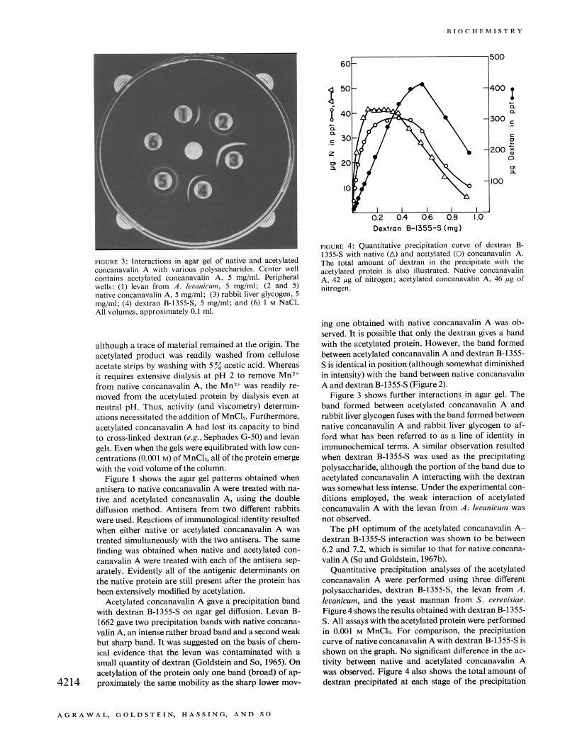
FIGURE 4: Quantitative precipitation curve of dextran B- 1355-S with native (A) and acetylated (0) concanavalin A. The total amount of dextran in the precipitate with the acetylated protein is also illustrated. Native concanavalin A, 42 pg of nitrogen; acetylated concanavalin A, 46 pg of nitrogen.
FIOURL 3: Interactions in agar gel of native and acetylated concanavalin A with various polysaccharides. Center well contains acetylated concanavalin A, 5 mg/ml. Peripheral wells: (1) levan from A. lrmnicum, 5 mg/ml; (2 and 5) native concanavalin A, 5 mglml; (3) rabbit liver glycogen, 5 mg/ml; (4) dextran B-1355-S 5 mg/ml; and (6) I M NaCI. All volumes, approximately 0.1 ml.
although a trace of material remained at the origin. The acetylated product was readily washed from cellulose acetate strips by washing with 5 % acetic acid. Whereas it requires extensive dialysis at pH 2 to remove Mn2+ from native concanavalin A, the MnZ+ was readily re- moved from the acetylated protein by dialysis even at neutral pH. Thus, activity (and viscometry) determin- ations necessitated the addition of MnC4. Furthermore, acetylated concanavalin A had lost its capacity to bind to cross-linked dextran (e.g., Sephadex G-50) and levan gels. Even when the gels were equilibrated with low con- centrations (0.001 M) of MnCI,, all of the protein emerge with the void volume of the column.
Figure 1 shows the agar gel patterns obtained when antisera to native concanavalin A were treated with na- tive and acetylated concanavalin A, using the double diffusion method. Antisera from two different rabbits were used. Reactions of immunological identity resulted when either native or acetylated concanavalin A was treated simultaneously with the two antisera. The same finding was obtained when native and acetylated con- canavalin A were treated with each of the antisera sep- arately. Evidently all of the antigenic determinants on the native protein are still present after the protein has been extensively modified by acetylation.
Acetylated concanavalin A gave a precipitation band with dextran B-1355s on agar gel diffusion. Levan B- 1662 gave two precipitation bands with native concana- valin A, an intense rather broad band and a second weak but sharp band. I t was suggested on the basis of chem- ical evidence that the levan was contaminated with a small quantity of dextran (Goldstein and So, 1965). On acetylation of the protein only one band (broad) of ap- proximately the same mobility as the sharp lower mov- 4214
ing one obtained with native concanavalin A was ob- served. It is possible that only the dextran gives a band with the acetylated protein. However, the band formed between acetylated concanavalin A and dextran B-1355- S is identical in position (although somewhat diminished in intensity) with the band between native concanavalin A and dextran B-13554 (Figure 2).
Figure 3 shows further interactions in agar gel. The band formed between acetylated concanavalin A and rabbit liver glycogen fuses with the band formed between native concanavalin A and rabbit liver glycogen to af- ford what has been referred to as a line of identity in immunochemical terms. A similar observation resulted when dextran B-1355-S was used as the precipitating polysaccharide, although the portion of the band due to acetylated concanavalin A interacting with the dextran was somewhat less intense. Under the experimental con- ditions employed, the weak interaction of acetylated concanavalin A with the levan from A. levanicum was not observed.
The pH optimum of the acetylated concanavalin A- dextran B-1355-S interaction was shown to be hetween 6.2 and 7.2, which is similar to that for native concana- valin A (So and Goldstein, 1967b).
Quantitative precipitation analyses of the acetylated concanavalin A were performed using three different polysaccharides, dextran B-1355-S, the levan from A. leumicum, and the yeast mannan from S. cereuisiae. Figure4 showstheresults obtained with dextranB-1355- S. All assays with the acetylated protein were performed in 0.001 M MnCL For comparison, the precipitation curve of native concanavalin A with dextran B-1355s is shown on the graph. No significant difference in the ac- tivity between native and acetylated concanavalin A was observed. Figure 4 also shows the total amount of dextran precipitated at each stage of the precipitation
A G R A W A L , G O L D S T E I N , H A S S I N G , A N D so
V O L . 7, N O . 1 2 , D E C E M B E R 1 9 6 8
- 0 0.2 0 4 0.6 0.0 I O
Dextran B-1355-Shq)
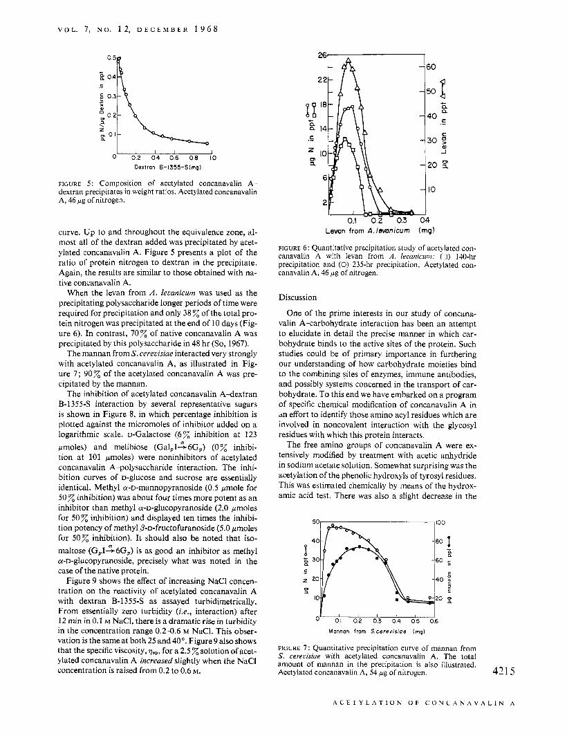
FIGURE 5: Composition of acetylated concanavalin A- dextran precipitates in weight ratios. Acetylated concanavalin A, 46 pg of nitrogen.
curve. Up to and throughout the equivalence zone, al- most all of the dextran added was precipitated by acet- ylated concanavalin A. Figure 5 presents a plot of the ratio of protein nitrogen to dextran in the precipitate. Again, the results are similar to those obtained with na- tive concanavalin A.
When the levan from A . 1el;anicurn was used as the precipitating polysaccharide longer periods of time were required for precipitation and only 38 % of tht total pro- tein nitrogen was precipitated at the end of 10 days (Fig- ure 6). In contrast, 7 0 z of native concanavalin A was precipitated by this polysaccharide in 48 hr (So, 1967).
The mannan from S. cerecisiae interacted very strongly with acetylated concanavalin A, as illustrated in Fig- ure 7; 90% of the acetylated concanavalin A was pre- cipitated by the mannan.
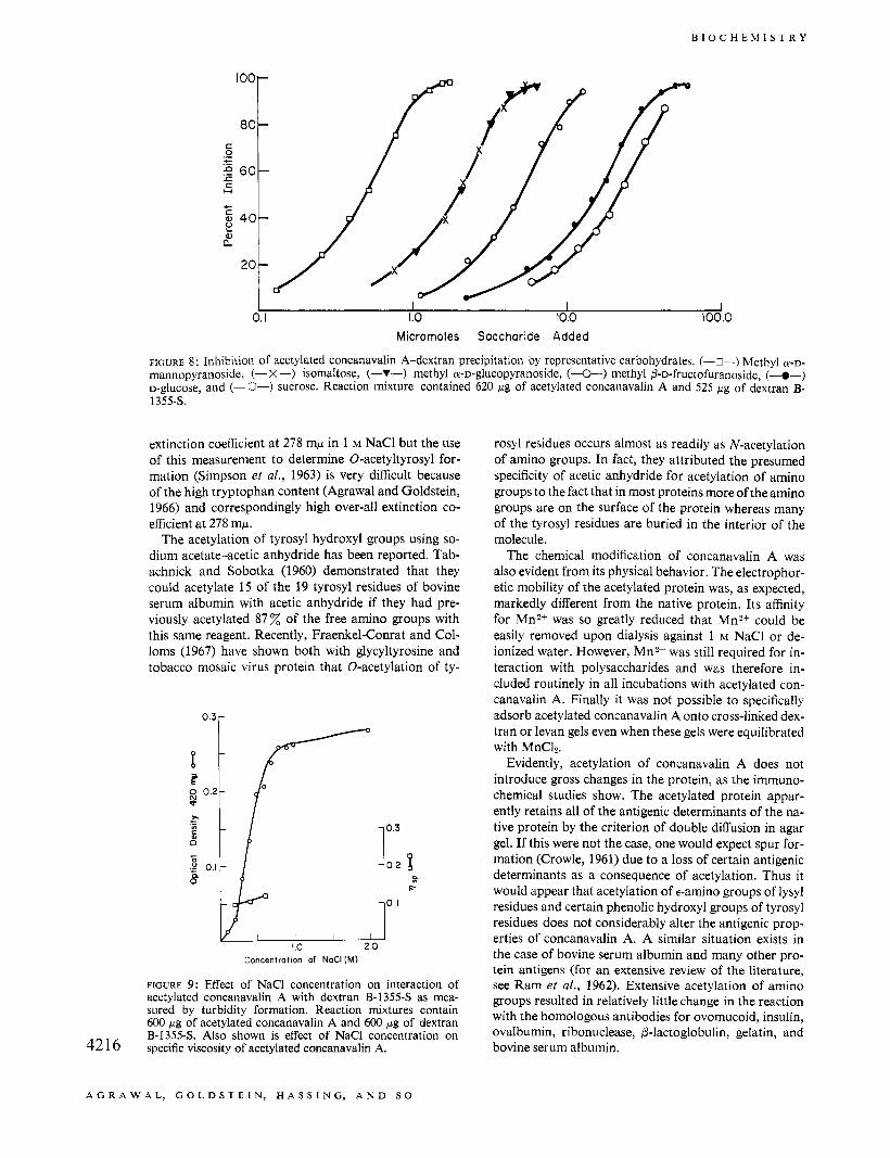
The inhibition of acetylated concanavalin A-dextran B-13.554 interaction by several representative sugars is shown in Figure 8, in which percentage inhibition is plotted against the micromoles of inhibitor added on a logarithmic scale. D-Galactose (6 % inhibition at 123 pmoles) and melibiose ( G a l P l ~ 6 G , ) (0% inhibi- tion at 101 pmoles) were noninhibitors of acetylated concanavalin A-polysaccharide interaction. The inhi- bition curves of D-glucose and sucrose are essentially identical. Methyl a-D-mannopyranoside (0.5 pmole for 50% inhibition) was about four times more potent as an inhibitor than methyl a-D-glucopyranoside (2.0 pmoles for 50% inhibition) and displayed ten times the inhibi- tion potency of methyl P-D-fructofuranoside (5.0 pmoles for 50% inhibition). It should also be noted that iso- maltose (Gp126Gp) is as good an inhibitor as methyl a-D-glucopyranoside, precisely what was noted in the case of the native protein.
Figure 9 shows the effect of increasing NaCl concen- tration on the reactivity of acetylated concanavalin A with dextran B-13558 as assayed turbidimetrically. From essentially zero turbidity (Le., interaction) after 12 min in 0.1 M NaCI, there is a dramatic rise in turbidity in the concentration range 0 . 2 4 . 6 M NaCl. This obser- vation is the same at both 25 and 40". Figure 9 also shows that the specific viscosity, p.,,, for a 2.5 solution of acet- ylated concanavalin A increased slightly when the NaCl concentration is raised from 0.2 to 0.6 M.
Leven from A./evmicurn (rng!
FIGURE 6: Quantitative precipitation study of acetylated con- canavalin A with levan from A . leaanicunz: (0) 140-hr precipitation and (0) 235-hr precipitation. Acetylated con- canavalin A, 46 pg of nitrogen.
Discussion
One of the prime interests in our study of concana- valin A-carbohydrate interaction has been an attempt to elucidate in detail the precise manner in which car- bohydrate binds to the active sites of the protein. Such studies could be of primary importance in furthering our understanding of how carbohydrate moieties bind to the combining sites of enzymes, immune antibodies, and possibly systems concerned in the transport of car- bohydrate. To this end we have embarked on a program of specific chemical modification of concanavalin A in dn effort to identify those amino acyl residues which are involved in noncovalent interaction with the glycosyl residues with which this protein interacts.
The free amino groups of concanavalin A were ex- tensively modified by treatment with acetic anhydride in sodium acetate solution. Somewhat surprising was the acetylation of the phenolic hydroxyls of tyrosyl residues. This was estimated chemically by means of the hydrox- amic acid test. There was also a slight decrease in the
O' 0.1 012 d3 014 015 26
Mannan from S.cerevisiae (mg)
FIGURE 7 : Quantitative precipitation curve of mannan from S. cererisiue with acetylated concanavalin A. The total amount of mannan in the precipitation is also illustrated. Acetylated concanavalin A, 54 pg of nitrogen. 421 5
A C E T Y L A T I O N O F C O N C A N A V A L I N A
B I O C H E M I S T R Y
60
+
a" 40/
I I I I 0. I 1.0 10.0 100.0
Micromoles Saccharide Added
FIGURE 8: Inhibition of acetylated concanavalin A-dextran precipitation by representative carbohydrates. (-s-) Methyl a - ~ - mannopyranoside, (- X-) isomaltose, (-Y-) methyl a-D-glucopyranoside, (&) methyl P-D-fructofuranoside, (-@-) D-glucose, and (-O-) sucrose. Reaction mixture contained 620 pg of acetylated concanavalin A and 525 pg of dextran B- 1355s.
extinction coefficient at 278 mp in 1 M NaCl but the use of this measurement to determine 0-acetyltyrosyl for- mation (Simpson et al., 1963) is very difficult because of the high tryptophan content (Agrawal and Goldstein, 1966) and correspondingly high over-all extinction co- efficient at 278 mp.
The acetylation of tyrosyl hydroxyl groups using so- dium acetate-acetic anhydride has been reported. Tab- achnick and Sobotka (1960) demonstrated that they could acetylate 15 of the 19 tyrosyl residues of bovine serum albumin with acetic anhydride if they had pre- viously acetylated 8 7 x of the free amino groups with this same reagent. Recently, Fraenkel-Conrat and Col- loms (1967) have shown both with glycyltyrosine and tobacco mosaic virus protein that 0-acetylation of ty-
0.3r
0.3
F
I .o 2 .o Concentration of NaCl (M)
FIGURE 9: Effect of NaCl concentration on interaction of acetylated concanavalin A with dextran B-1355-S as mea- sured by turbidity formation. Reaction mixtures contain 600 p g of acetylated concanavalin A and 600 pg of dextran B-1355-S. Also shown is effect of NaCl concentration on specific viscosity of acetylated concanavalin A. 42 16
rosy1 residues occurs almost as readily as N-acetylation of amino groups. In fact, they attributed the presumed specificity of acetic anhydride for acetylation of amino groups to the fact that in most proteins more of the amino groups are on the surface of the protein whereas many of the tyrosyl residues are buried in the interior of the molecule.
The chemical modification of concanavalin A was also evident from its physical behavior. The electrophor- etic mobility of the acetylated protein was, as expected, markedly different from the native protein. Its affinity for Mn2+ was so greatly reduced that Mn2+ could be easily removed upon dialysis against 1 M NaCl or de- ionized water. However, Mn2+ was still required for in- teraction with polysaccharides and was therefore in- cluded routinely in all incubations with acetylated con- canavalin A. Finally it was not possible to specifically adsorb acetylated concanavalin A onto cross-linked dex- tran or levan gels even when these gels were equilibrated with MnC12.
Evidently, acetylation of concanavalin A does not introduce gross changes in the protein, as the immuno- chemical studies show. The acetylated protein appar- ently retains all of the antigenic determinants of the na- tive protein by the criterion of double diffusion in agar gel. If this were not the case, one would expect spur for- mation (Crowle, 1961) due to a loss of certain antigenic determinants as a consequence of acetylation. Thus it would appear that acetylation of €-amino groups of lysyl residues and certain phenolic hydroxyl groups of tyrosyl residues does not considerably alter the antigenic prop- erties of concanavalin A. A similar situation exists in the case of bovine serum albumin and many other pro- tein antigens (for an extensive review of the literature, see Ram et al., 1962). Extensive acetylation of amino groups resulted in relatively little change in the reaction with the homologous antibodies for ovomucoid, insulin, ovalbumin, ribonuclease, @-lactoglobulin, gelatin, and bovine serum albumin.
A G R A W A L , G O L D S T E I N , H A S S I N G , A N D S O
V O L . 7, N O . 1 2 , D E C E M B E R 1 9 6 8
Agar gel diffusion studies indicated that acetylated concanavalin A still interacts strongly with rabbit liver glycogen. However, the reaction of the modified protein with dextran B-1355s is definitively diminished when compared to the interaction of native concanavalin A with this polysaccharide. The interaction of native con- canavalin A with the levan from Aerobacter lecanicurr was quite weak and the acetylated protein displays a greatly reduced affinity for this polysaccharide as can be seen from the agar gel studies.
Because of the extensive modification of the native protein and the corresponding changes produced in over-all charge distribution on the protein in the neu- tral pH range, it might be expected that the pH char- acteristics of the interaction of acetylated concanavalin A with polysaccharides would change somewhat. How- ever, examination of this effect showed that the pH op- timum was identical with that of native concanavalin A. Any charge effect that acetylation may have conferred on concanavalin A is apparently not sufficient to cause disruption of the saccharide combining sites.
Quantitative precipitation analyses of acetylated con- canavalin A with the three polysaccharides used in this study reveal a remarkable similarity to that observed for the native protein (So, 1967). The greatest change in reactivity occurred in the inter- action of the modified protein with the levan from A. lecanicuni. The low amount of nitrogen precipitated and the extensive time intervals required for this interaction show that acetylation of concanavalin A has diminished somewhat its capacity to react with this polysaccharide. However, since the native protein also interacted quite weakly with this levan, a slight reduction of an already weak interaction resulted in this low reactivity.
With the two more reactive polysaccharides, dextran B-13554 and the mannan from S. cereoishe, the pre- cipitation curves obtained were almost identical with those obtained with native concanavalin A. Also, the characteristically continuous variation in the ratio of protein nitrogen to carbohydrate in the precipitate (Fig- ure 5 ) remains the same as that for native concanavalin A.
Hapten inhibition studies conducted on the acetylated concanavalin A-dextran system indicate quite definitely that acetylation of the protein did not alter its specificity. The order of reactivity of the various inhibitors tested was identical with that found with native concanavalin A-dextran precipitation (So and Goldstein, 1967a), i.e., methyl a-D-mannopyranoside > methyl a-~-glucopy- ranoside > methyl Pa-fructofuranoside. However smaller quantities of these sugars were needed to effect the same percentage inhibition, indicating that the inter- action of polysaccharides with acetylated concanavalin A is weaker than that between native concanavalin A and polysaccharides. Thus, whereas it required 1.65 pmoles of methyl a-D-mannopyranoside to inhibit na- tive concanavalin A-dextran interaction by 50 % (So and Goldstein, 1968), it required only 0.5 pmole to achieve 50 inhibition of acetylated concanavalin A- dextran interaction.
Since the preparation of acetylated concanavalin A studied had an 84% reduction in ninhydrin color, con-
siderable free amino groups must have been acetylated and at neutral pH there was a considerable reduction of positive charges. Thus at pH 7 concanavalin A has a much higher net negative charge than in the native state.
When a macromolecule attains a considerable net charge, one would expect it to possess a lower electro- static free energy in an expanded configuration (Tan- ford, 1961). If this is indeed the case in the concanavalin A system, then the expanded configurations of the pro- tein could lead to a disruption of the integrity of the com- bining sites. However, the effect of repulsion by the net negative charges at neutral pH may be “masked” by an increase in the supporting electrolyte concentration. Thus, the expansion of bovine serum albumin at acid pH (as measured by an increase in the intrinsic viscosity) is initiated at a net positive charge of f 15 on the pro- tein molecule at ionic strength 0.01, but at an ionic strength of 0.15, a net positive charge of +35 is required to initiate the expansion of its configuration (Tanford, 1961). If at neutral pH the negative charge on concan- avalin A is “smeared” by a high salt concentration, then it would be expected that the protein would return to its native, preferred configuration. If this were the case, one might also expect that the combining sites on the pro- tein molecule would remain intact and that the protein would retain its activity. That this is in fact true can be seen from Figure 9.
The specific viscosity of acetylated concanavalin A was measured at a protein concentration of 2.5 in so- dium chloride solutions varying in ionic strength from 0.2 to 0.6 in order to ascertain if an increase in specific viscosity could be observed at the lower ionic strengths. The specific viscosity increased very slightly when the ionic strength was varied from 0.2 to 0.6. Since the ac- tivity of acetylated concanavalin A also increased dra- matically over this range of ionic strength, the specific vis- cosity apparently is not a sufficiently sensitive parameter for determining a marked change in the protein con- figuration. Lack of solubility of the protein prevented the use of a higher protein concentration or lower salt concentrations in these studies. If the loss of activity was due to a configurational change in acetylated concana- valin A due to electrostatic repulsions, the effect was not very great. An alternative explanation is that the region on the protein surface in close proximity to the combin- ing sites was disrupted locally but that the over-all con- figurational change of the protein in the range of ionic strength 0.2-1.0 is too small to detect by viscometric procedures.
In summary, these studies demonstrate that extensive acetylation of concanavalin A caused no gross disrup- tion of association of protein subunits or of the in- dividual combining sites. Despite the rigorous mod- ification, the protein is remarkably similar to native concanavalin A in its activity and in the nature of its antigenic determinants. This is strongly suggestive evi- dence for the nonparticipation of free amino groups and some of the tyrosyl residues in important structural fea- tures or in binding of carbohydrates at the combining site of concanavalin A.
Additional modification studies in progress should yield further information on the precise amino acyl res- 4217
A C E T Y L A T I O N O F C O N C A N A C A L I N A
B I O C H E M I S T K Y
idues which may be involved in concanavalin A-car- bohydrate interaction.
References
Agrawal, B. B. L. (1967), Ph.D. Thesis, The State
Agrawal, B. B. L., and Goldstein, 1. J. (1967a), Biochim.
Agrawal, B. B. L., and Goldstein, I. J. (1967b), Biochim.
Agrawal, N. B. L., and Goldstein, I. J. (1Y68), Arch.
Bose, R. J. (1964), Ph.D. Thesis, University of Min-
Cifonelli, J . A,, Montogomery, R., and Smith, F.
Crowle, A. J. (1961), Immunodiffusion, New York,
Dubois, M., Gilles, K. A., Hamilton, J. K., Rebers,
Fraenkel-Conrat, H. (1 953, Methods Enzymol. 4,
Fraenkel-Conrat, H., and Colloms, M. (1967), Bio-
Goldstein, I. J., Hollerman, C. E., and Merrick, J. M.
Goldstein, I. J., Hollerman, C. E., and Smith, E. E.
Goldstein, I. J., and lyer, R. N. (1966), Biochim.
Goldstein, I. J., and So, L. L. (1965), Arch. Biochem.
Harris, H., and Robson, E. B. (1963), Vox Sanguinis
Hestrin, S. (1949), J . Biol. Chem. 180,240. Kabat, E. A., and Meyer, M. M. (1961), Experimental
Immunochemistry, Springfield, Ill., C. C Thomas, p 803.
University of New York, Buffalo, N. Y.
Biophys. Acta 147,262.
Biophys. Acta 133,376.
Biochem. Biophys. 124,218.
nesota, Minneapolis, Minn.
(1956), J. Am. Chem. SOC. 78,2485.
N. Y., Academic, 69ff.
P. A., and Smith, F. (1956), Anal. Chem. 28,350.
247.
chemistry 6,2740.
(1965a), Biochim. Biophys. Acta 97,68.
(1965b), Biochemistry 4,876.
Biophys. Acta 121,197.
Biophys. 111,407.
8,348.
Leon, M. A. (1967), Science 158,1325. Manners, D. J., and Wright, A. (1962), J. Chem. SOC.,
4592. Nakamura, S., Tanaka, K., and Murakawa, S. (1960),
Nature 188,144. Nakamura, S., Tominaga, S., Katsuno, A,, and Mura-
kawa, S. (1965), Camp. Biochem. Physiol. 15, 435.
Olson, M. 0. J., and Liener, I. E. (1967), Biochemistry 6, 3801.
Poretz, R. D., and Goldstein, I. J. (1967a), 154th Na- tional Meeting of the American Chemical Society, Chicago, Ill., Sept 1967, Abstracts, D-48.
Poretz, R. D., and Goldstein, I. 3. (1967b), 154th National Meeting of the American Chemical Society, Chicago, Ill., Sept 1967, Abstracts, P-6.
Ram, J. S., Bier, M., and Maurer, P. H. (1962), Admn. Enzymol. 24,105.
Rosen, H. (1957), Arch. Biochem. Biophys. 67, 10. Simpson, R. T., Riordan, J. E., and Vallee, B. L. (1963),
Smith, E. E., and Goldstein, I. J. (1967), Arch. Biochem.
So, L. L. (1967), Ph.D. Thesis, The State University
So, L. L., and Goldstein, I. J . (1967a), J . Ztnmunol. 99,
So, L. L., and Goldstein, I. J. (1967b), J . Biol. Chem.
So, L. L., and Goldstein, I. J. (1968), J. Biol. Chem. 243,
Sumner, J. B., and Howell, S. F. (1935), J. Zmmunol.
Sumner, J. B., and Howell, S. F. (1936), J. Bacteriol.
Tabachnick, M., and Sobotka, H. (1960), J . Biol. Chem.
Tanford, C. (1961), Physical Chemistry of Macromole-
Biochemistry 2,616.
Biophys. 121,88.
of New York, Buffalo, N. Y.
158.
242,1617.
2003.
29,133.
32,227.
235,1051.
cules, New York, N. Y., Wiley, p 509 ff.
4218
A G K A W A L , G O L D S T E I N , H A S S I N G , A N D S O