Embed Size (px)
Citation preview
PROTEIN EXPRESSION AND
PURIFICATION
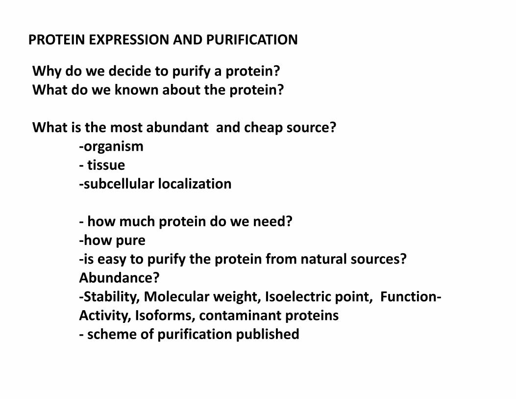
PROTEIN EXPRESSION AND PURIFICATION
Why do we decide to purify a protein?
What do we known about the protein?
What is the most abundant and cheap source?
-organism
- tissue
-subcellular localization
- how much protein do we need?
-how pure
-is easy to purify the protein from natural sources?
Abundance?
-Stability, Molecular weight, Isoelectric point, Function-
Activity, Isoforms, contaminant proteins
- scheme of purification published
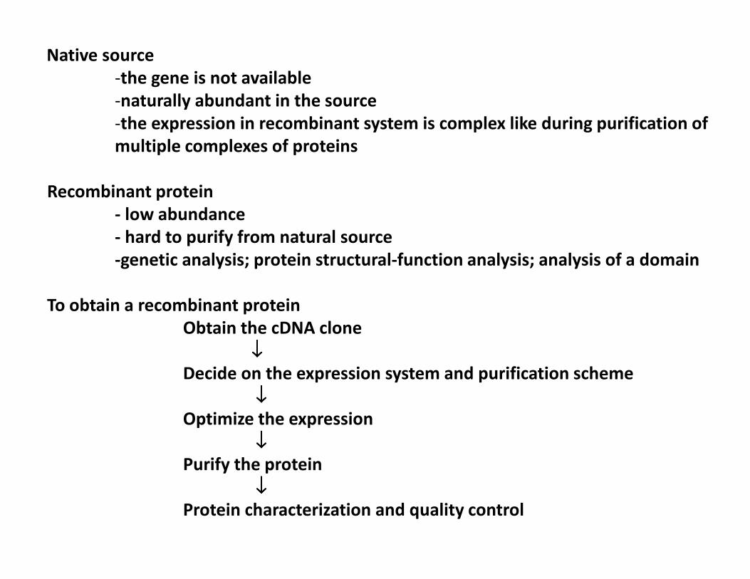
Native source
-the gene is not available
-naturally abundant in the source
-the expression in recombinant system is complex like during purification of
multiple complexes of proteins
Recombinant protein
- low abundance
- hard to purify from natural source
-genetic analysis; protein structural-function analysis; analysis of a domain
To obtain a recombinant protein
Obtain the cDNA clone
↓↓↓↓
Decide on the expression system and purification scheme
↓↓↓↓
Optimize the expression
↓↓↓↓
Purify the protein
↓↓↓↓
Protein characterization and quality control
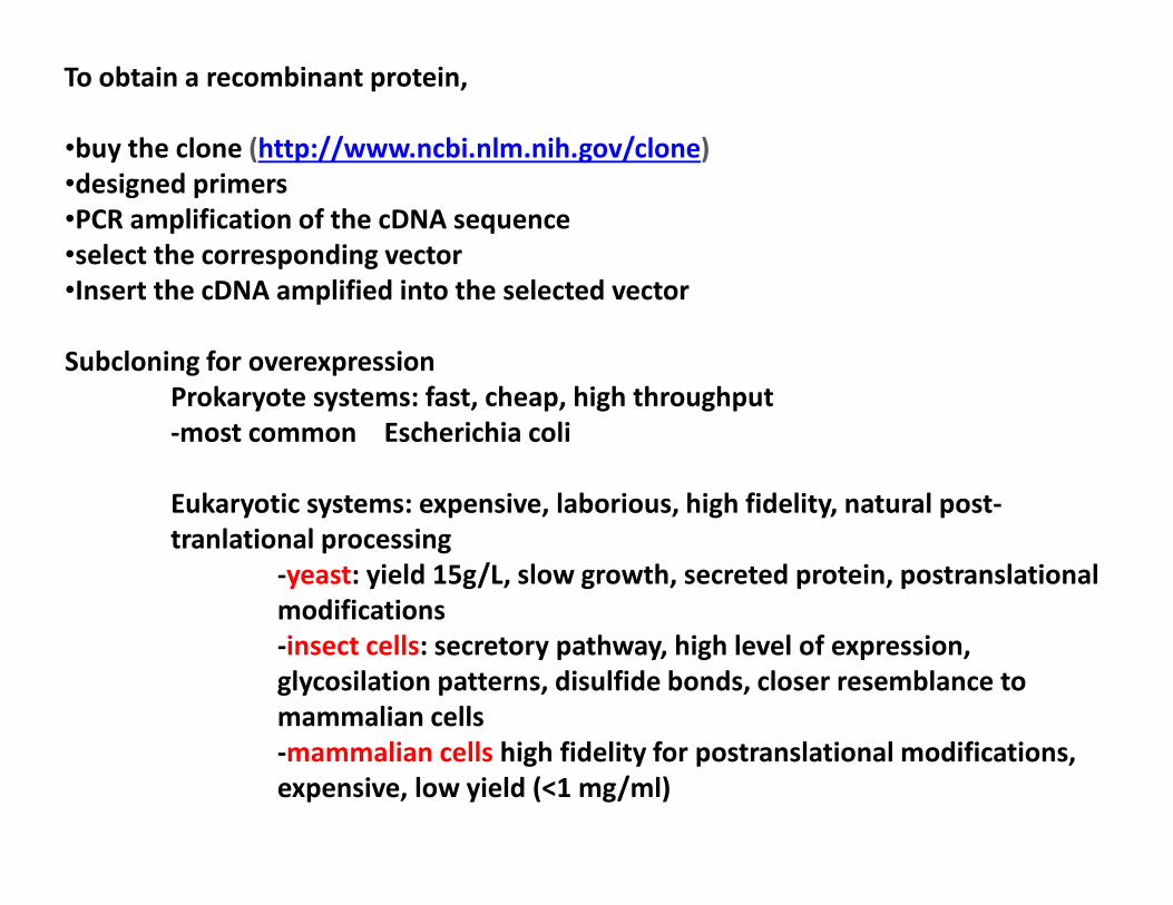
To obtain a recombinant protein,
•buy the clone (http://www.ncbi.nlm.nih.gov/clone)
•designed primers
•PCR amplification of the cDNA sequence
•select the corresponding vector
•Insert the cDNA amplified into the selected vector
Subcloning for overexpression
Prokaryote systems: fast, cheap, high throughput
-most common Escherichia coli
Eukaryotic systems: expensive, laborious, high fidelity, natural post-
tranlational processing
-yeast: yield 15g/L, slow growth, secreted protein, postranslational
modifications
-insect cells: secretory pathway, high level of expression,
glycosilation patterns, disulfide bonds, closer resemblance to
mammalian cells
-mammalian cells high fidelity for postranslational modifications,
expensive, low yield (<1 mg/ml)
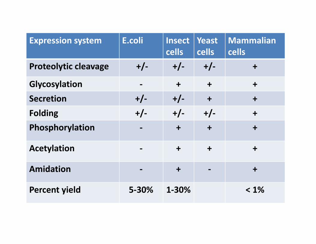
Expression system E.coli Insect
cells
Yeast
cells
Mammalian
cells
Proteolytic cleavage +/- +/- +/- +
Glycosylation - + + +
Secretion +/- +/- + +
Folding +/- +/- +/- +
Phosphorylation - + + +
Acetylation - + + +
Amidation - + - +
Percent yield 5-30% 1-30% < 1%

Prokaryotic cells
Antibiotic to select cell
transformed
↓↓↓↓
Induction of protein
expression. Addition of
inducer: IPTG, lactose
↓↓↓↓
Protein expression
↓↓↓↓
Centrifugation to collect
the cells
↓↓↓↓
Cell lyses
↓↓↓↓
Protein purification
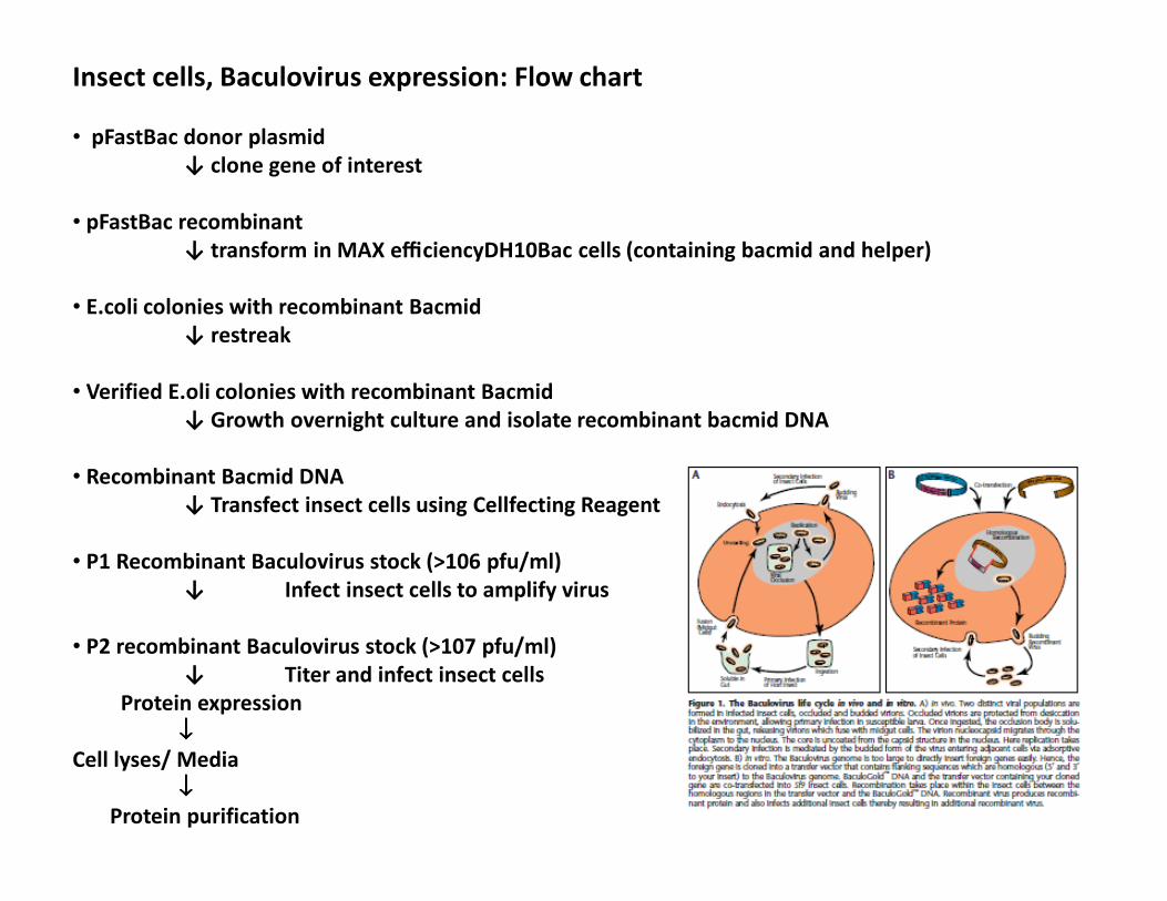
Insect cells, Baculovirus expression: Flow chart
• pFastBac donor plasmid
↓ clone gene of interest
• pFastBac recombinant
↓ transform in MAX efficiencyDH10Bac cells (containing bacmid and helper)
• E.coli colonies with recombinant Bacmid
↓ restreak
• Verified E.oli colonies with recombinant Bacmid
↓ Growth overnight culture and isolate recombinant bacmid DNA
• Recombinant Bacmid DNA
↓ Transfect insect cells using Cellfecting Reagent
• P1 Recombinant Baculovirus stock (>106 pfu/ml)
↓ Infect insect cells to amplify virus
• P2 recombinant Baculovirus stock (>107 pfu/ml)
↓ Titer and infect insect cells
Protein expression
↓↓↓↓
Cell lyses/ Media
↓↓↓↓
Protein purification
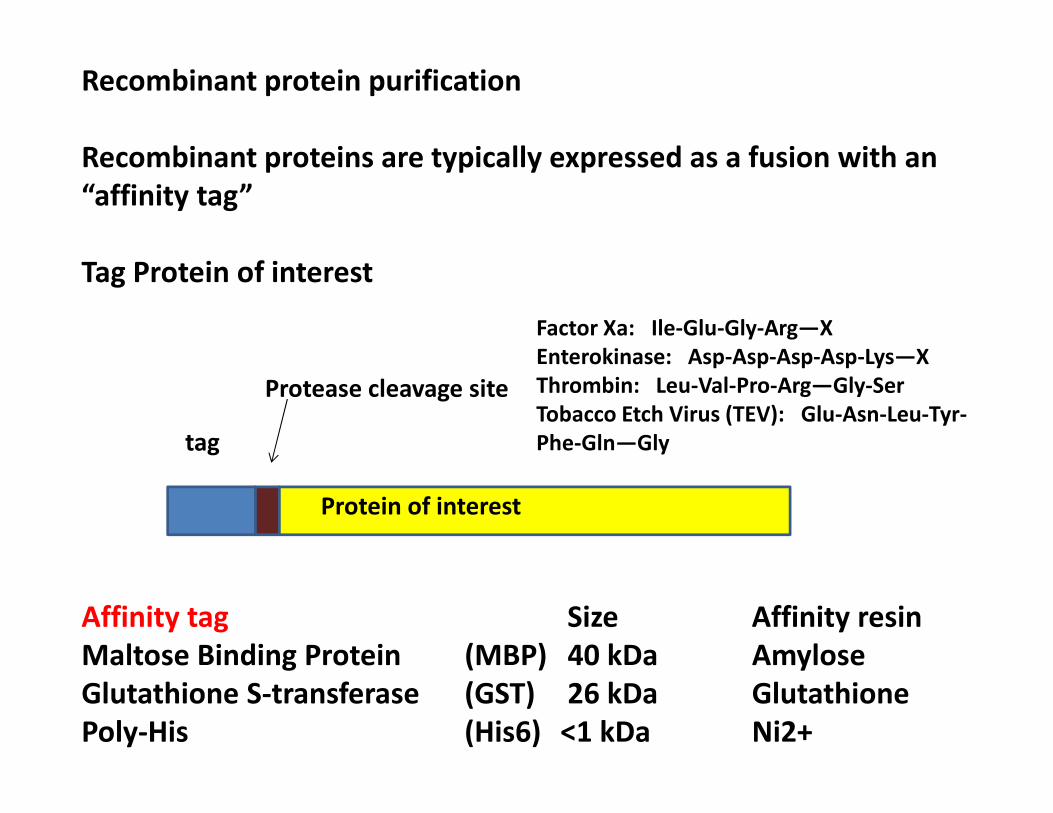
Recombinant protein purification
Recombinant proteins are typically expressed as a fusion with an
“affinity tag”
Tag Protein of interest
Affinity tag Size Affinity resin
Maltose Binding Protein (MBP) 40 kDa Amylose
Glutathione S-transferase (GST) 26 kDa Glutathione
Poly-His (His6) <1 kDa Ni2+
tag
Protease cleavage site
Protein of interest
Factor Xa: Ile-Glu-Gly-Arg—X
Enterokinase: Asp-Asp-Asp-Asp-Lys—X
Thrombin: Leu-Val-Pro-Arg—Gly-Ser
Tobacco Etch Virus (TEV): Glu-Asn-Leu-Tyr-
Phe-Gln—Gly
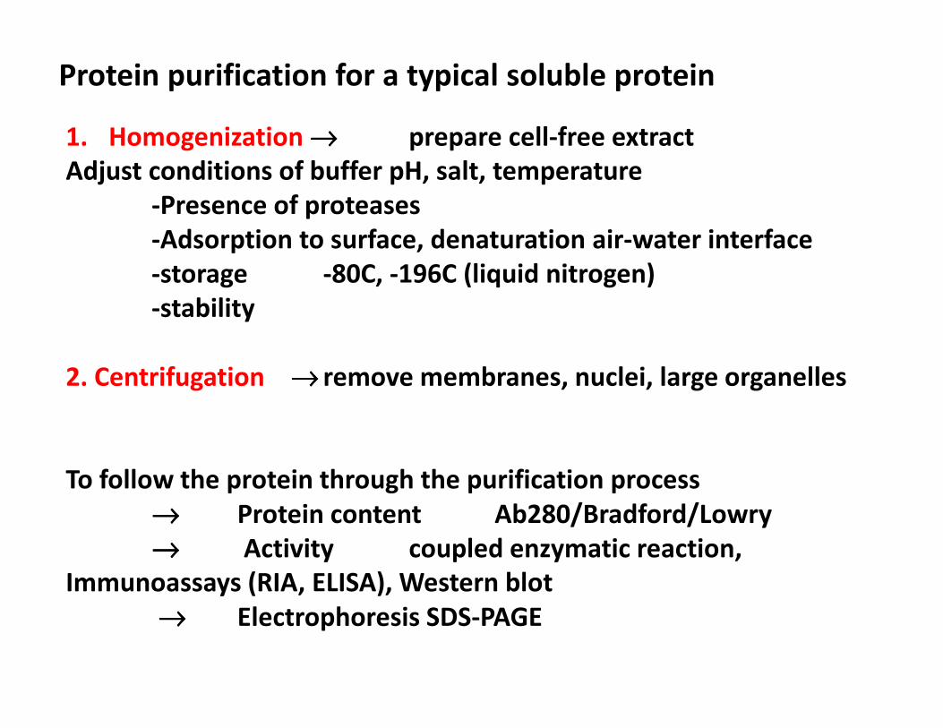
Protein purification for a typical soluble protein
1. Homogenization →→→→ prepare cell-free extract
Adjust conditions of buffer pH, salt, temperature
-Presence of proteases
-Adsorption to surface, denaturation air-water interface
-storage -80C, -196C (liquid nitrogen)
-stability
2. Centrifugation →→→→ remove membranes, nuclei, large organelles
To follow the protein through the purification process
→→→→ Protein content Ab280/Bradford/Lowry
→→→→ Activity coupled enzymatic reaction,
Immunoassays (RIA, ELISA), Western blot
→→→→ Electrophoresis SDS-PAGE
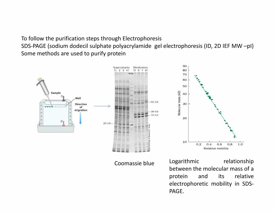
To follow the purification steps through Electrophoresis
SDS-PAGE (sodium dodecil sulphate polyacrylamide gel electrophoresis (ID, 2D IEF MW –pI)
Some methods are used to purify protein
Logarithmic relationship
between the molecular mass of a
protein and its relative
electrophoretic mobility in SDS-
PAGE.
Coomassie blue
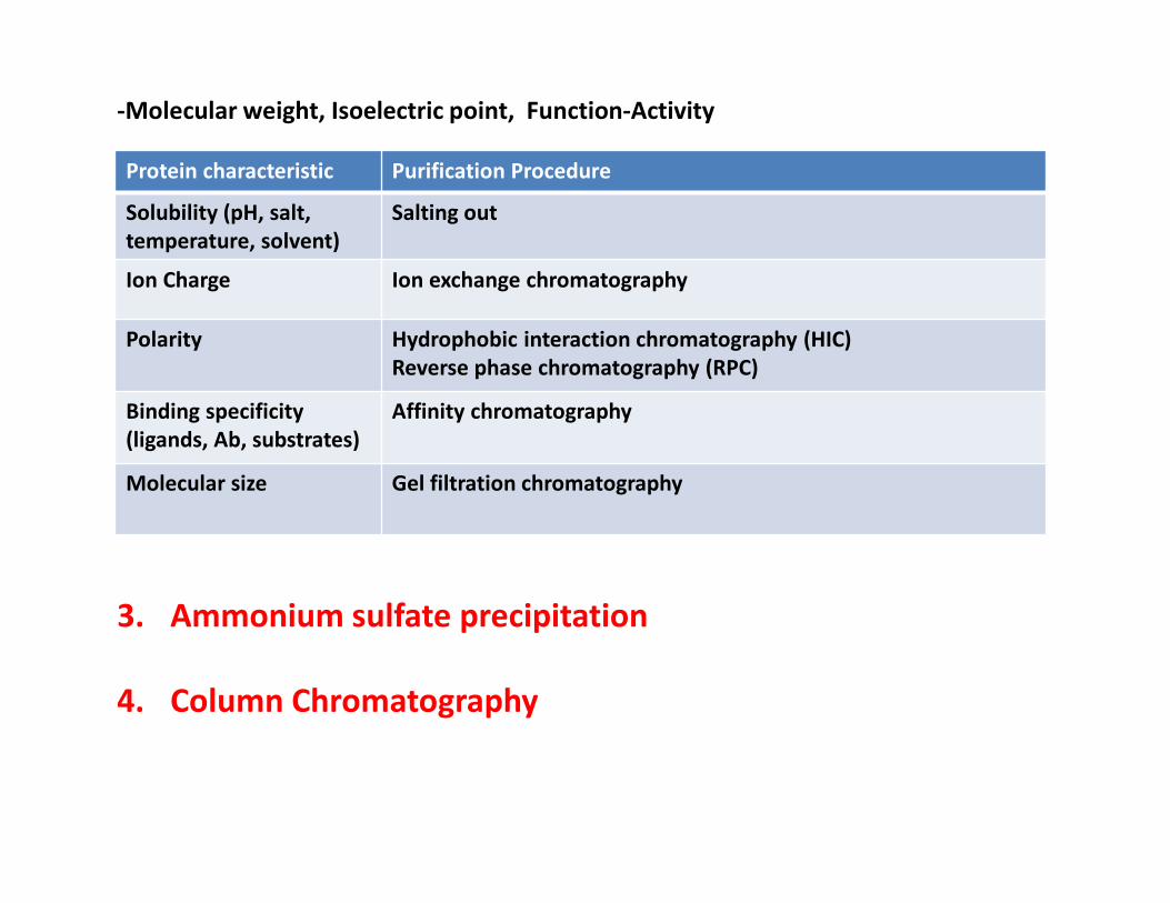
3. Ammonium sulfate precipitation
4. Column Chromatography
Protein characteristic Purification Procedure
Solubility (pH, salt,
temperature, solvent)
Salting out
Ion Charge Ion exchange chromatography
Polarity Hydrophobic interaction chromatography (HIC)
Reverse phase chromatography (RPC)
Binding specificity
(ligands, Ab, substrates)
Affinity chromatography
Molecular size Gel filtration chromatography
-Molecular weight, Isoelectric point, Function-Activity
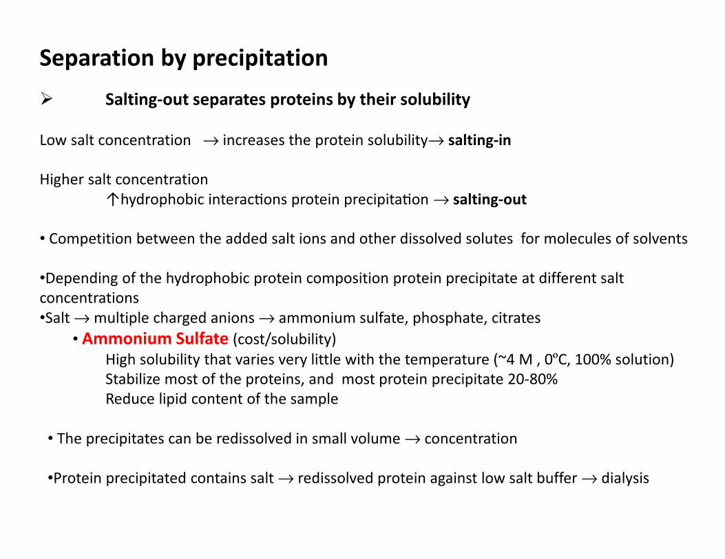
Separation by precipitation
� Salting-out separates proteins by their solubility
Low salt concentration → increases the protein solubility→ salting-in
Higher salt concentration
↑hydrophobic interac-ons protein precipita-on → salting-out
• Competition between the added salt ions and other dissolved solutes for molecules of solvents
•Depending of the hydrophobic protein composition protein precipitate at different salt
concentrations
•Salt → multiple charged anions → ammonium sulfate, phosphate, citrates
• Ammonium Sulfate (cost/solubility)
High solubility that varies very little with the temperature (~4 M , 0ºC, 100% solution)
Stabilize most of the proteins, and most protein precipitate 20-80%
Reduce lipid content of the sample
• The precipitates can be redissolved in small volume → concentration
•Protein precipitated contains salt → redissolved protein against low salt buffer → dialysis
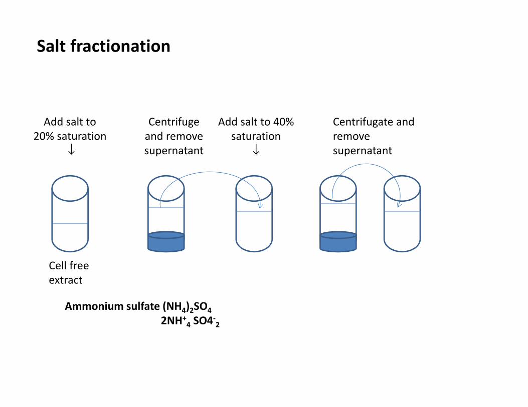
Salt fractionation
Add salt to
20% saturation
↓
Centrifuge
and remove
supernatant
Add salt to 40%
saturation
↓
Centrifugate and
remove
supernatant
Cell free
extract
Ammonium sulfate (NH4)2SO4
2NH+4 SO4-
2
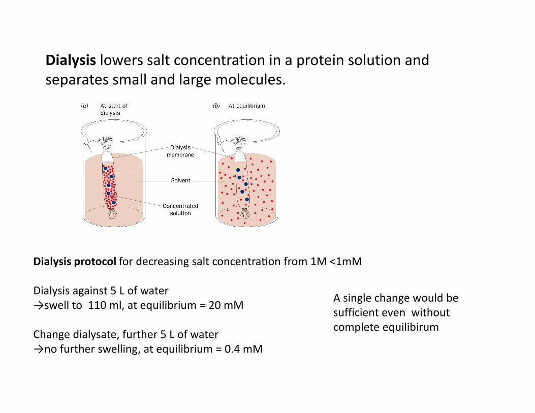
Dialysis lowers salt concentration in a protein solution and
separates small and large molecules.
Dialysis protocol for decreasing salt concentra-on from 1M ˂1mM
Dialysis against 5 L of water
→swell to 110 ml, at equilibrium = 20 mM
Change dialysate, further 5 L of water
→no further swelling, at equilibrium = 0.4 mM
A single change would be
sufficient even without
complete equilibirum
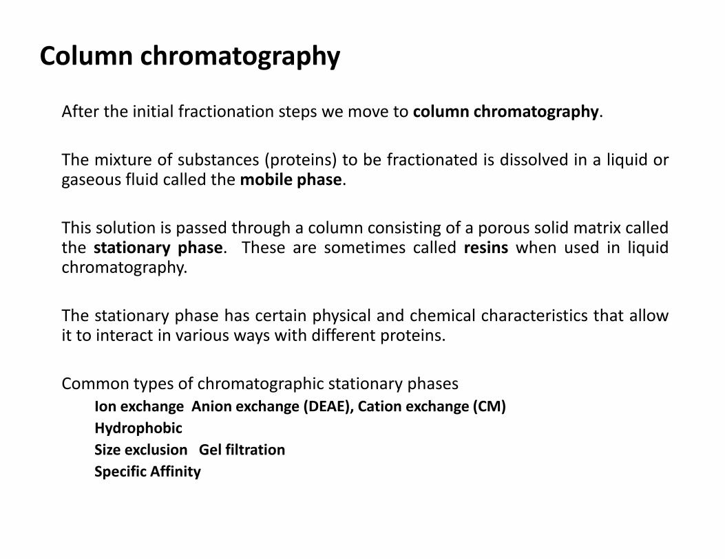
Column chromatography
After the initial fractionation steps we move to column chromatography.
The mixture of substances (proteins) to be fractionated is dissolved in a liquid orgaseous fluid called the mobile phase.
This solution is passed through a column consisting of a porous solid matrix calledthe stationary phase. These are sometimes called resins when used in liquidchromatography.
The stationary phase has certain physical and chemical characteristics that allowit to interact in various ways with different proteins.
Common types of chromatographic stationary phases
Ion exchange Anion exchange (DEAE), Cation exchange (CM)
Hydrophobic
Size exclusion Gel filtration
Specific Affinity
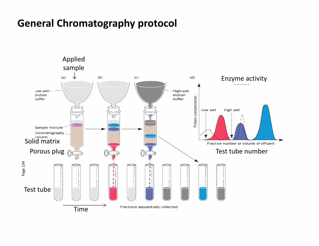
General Chromatography protocolP
ag
e 1
34
Applied
sample
↓
Solid matrix
Porous plug
Test tube
Time
Test tube number
Enzyme activity
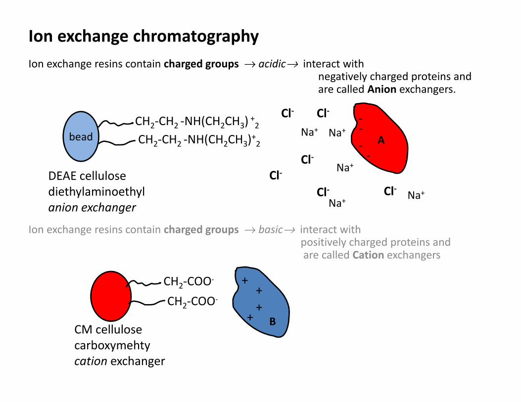
Ion exchange chromatography
Ion exchange resins contain charged groups → acidic→ interact with negatively charged proteins and are called Anion exchangers.
bead
DEAE cellulose
diethylaminoethyl
anion exchanger
CH2-CH2 -NH(CH2CH3) +2
CM cellulose
carboxymehty
cation exchanger
CH2-COO-
CH2-COO-
++
++
CH2-CH2 -NH(CH2CH3)+2
Ion exchange resins contain charged groups → basic→ interact with positively charged proteins and are called Cation exchangers
--
--
A
Cl-
Na+Na+
Na+Na+
Na+Cl-
Cl-
Cl-Cl-
Cl-
B
CM cellulose
cation exchanger
CH2-COO-
CH2-COO-
++
++
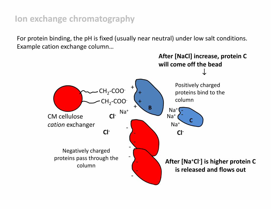
Positively charged
proteins bind to the
column
For protein binding, the pH is fixed (usually near neutral) under low salt conditions.
Example cation exchange column…
--
--
--
--
Negatively charged
proteins pass through the
column
Cl-Na+
Na+
Cl-Cl-
--
C
B
Na+
Na+
After [NaCl] increase, protein C
will come off the bead
↓↓↓↓
After [Na+Cl-] is higher protein C
is released and flows out
Ion exchange chromatography
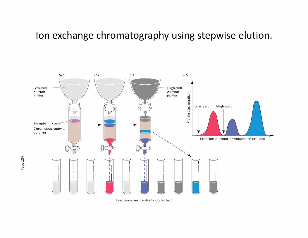
Ion exchange chromatography using stepwise elution.
Pa
ge
13
4
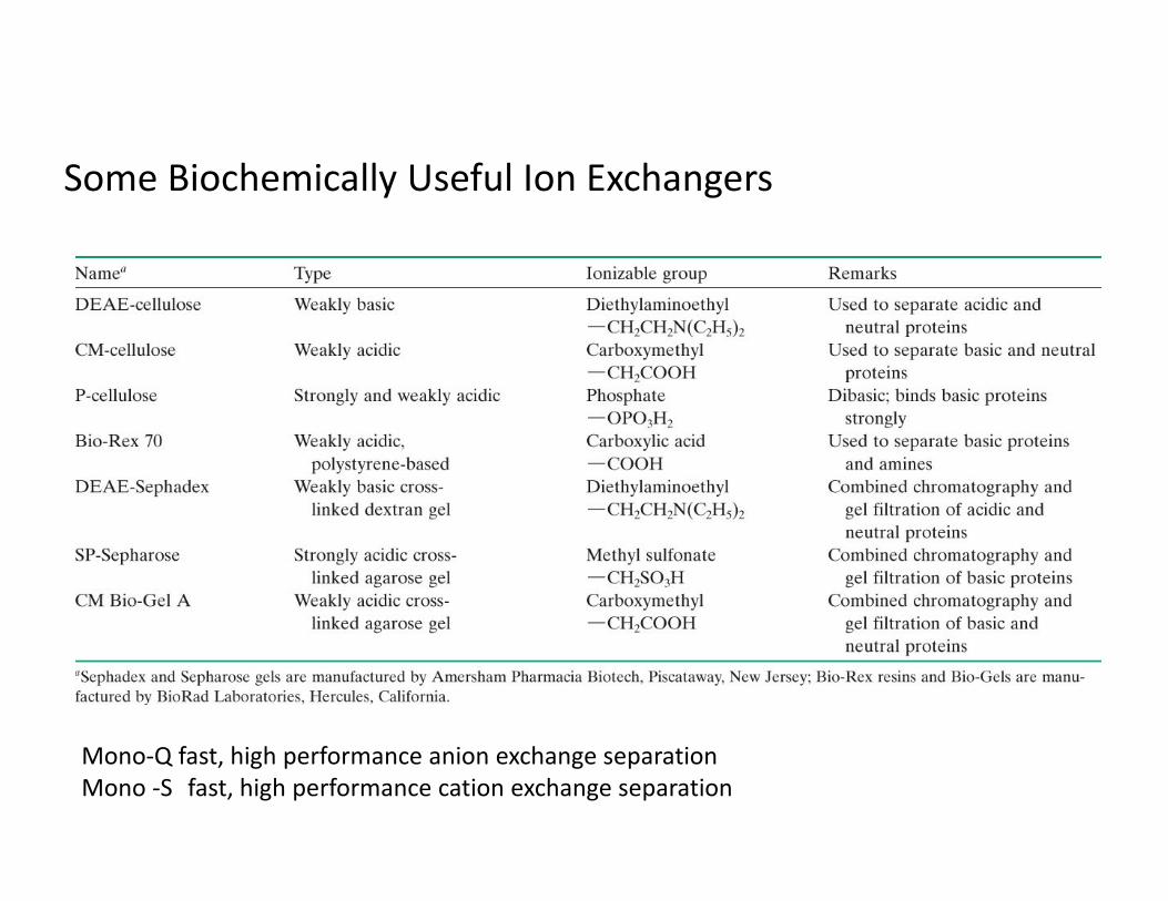
Some Biochemically Useful Ion Exchangers
Mono-Q fast, high performance anion exchange separation
Mono -S fast, high performance cation exchange separation
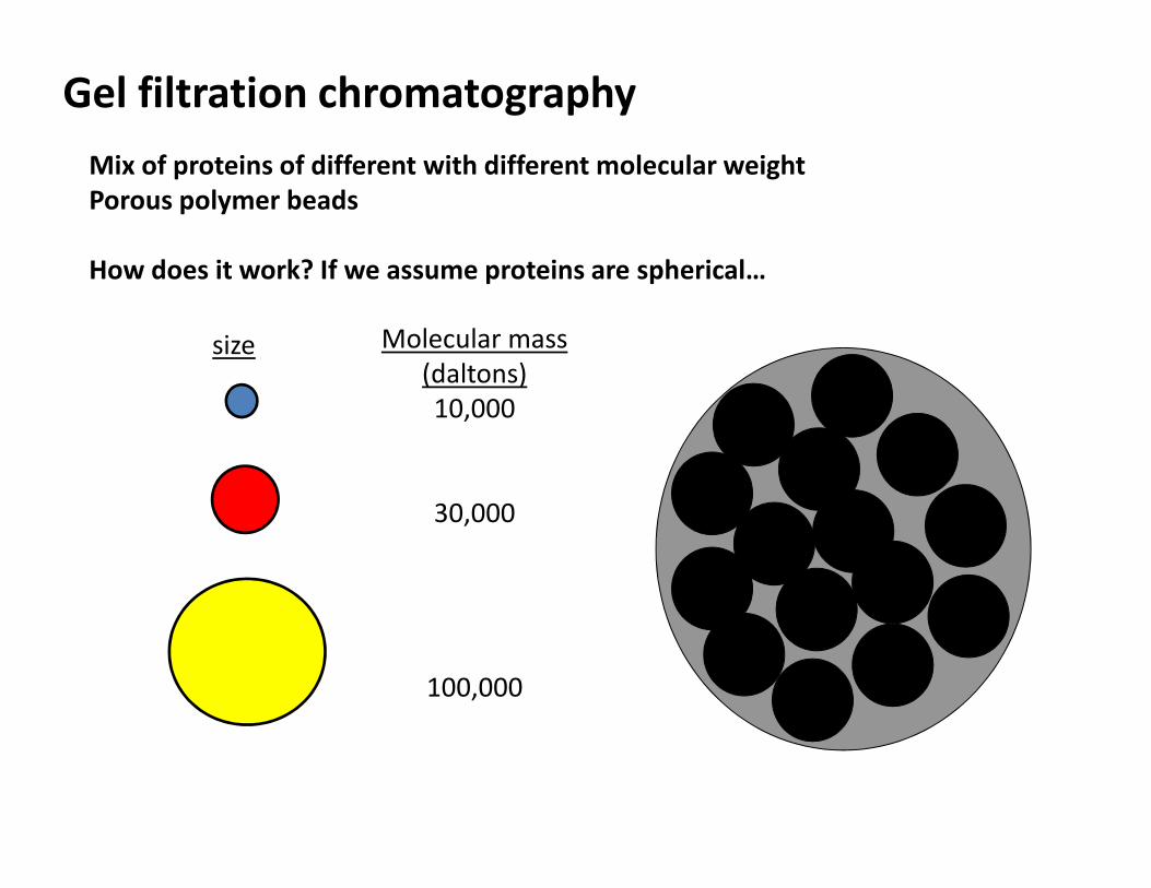
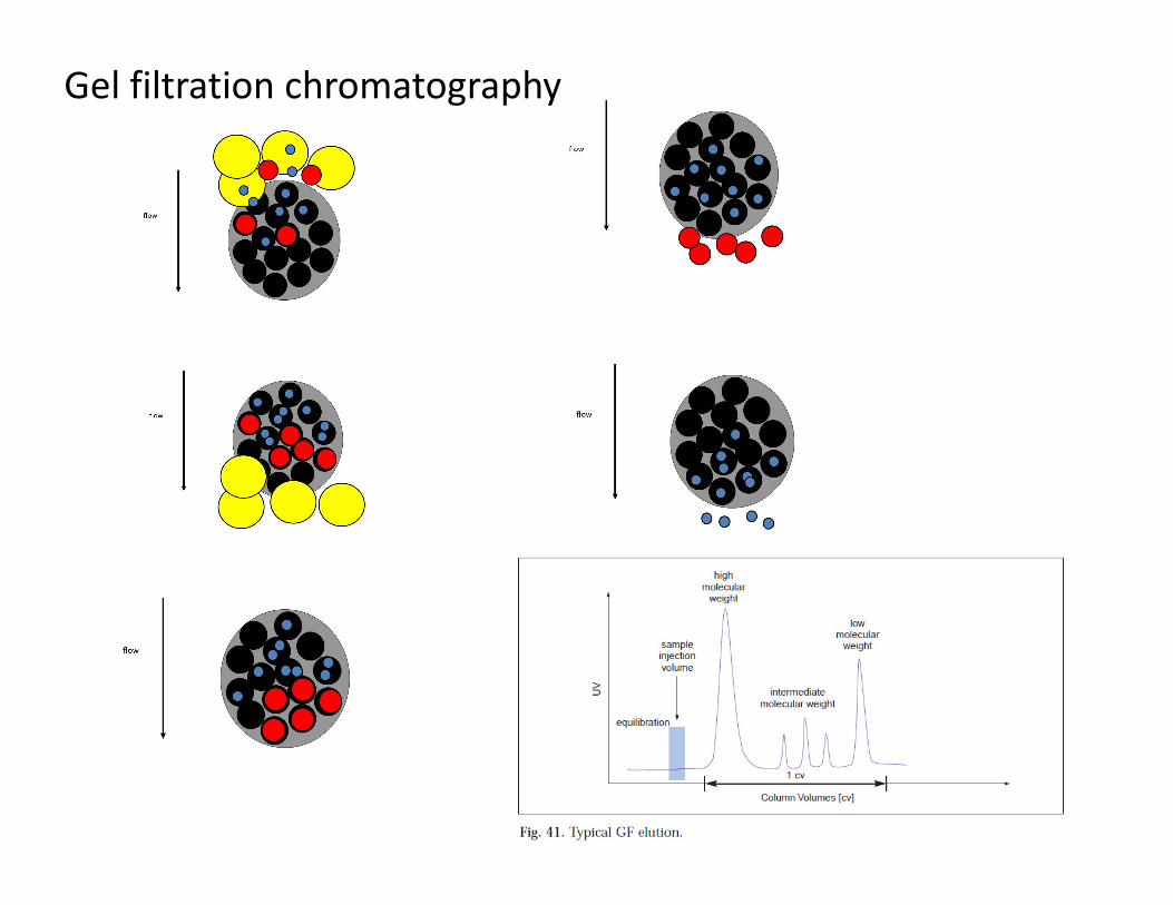
Gel filtration chromatography
Mix of proteins of different with different molecular weight
Porous polymer beads
How does it work? If we assume proteins are spherical…
size Molecular mass
(daltons)
10,000
30,000
100,000
Gel filtration chromatography
• The molecular mass of the smallest molecule unable to penetrate thepores of the gel is at the exclusion limit.
• The exclusion limit is a function of molecular shape, since elongatedmolecules are less likely to penetrate a gel pore than other shapes.
• Behavior of the molecule on the gel can be quantitatively characterized.
Total bed volume of the columnVt = Vx + V0
Vx = volume occupied by gel beads
V0 = volume of solvent space surrounding gel; Typically 35%
Gel filtration chromatography
• Elution volume (Ve) is the volume of a solvent required to elute a givensolute from the column after it has first contacted the gel.
• Relative elution volume (Ve/V0) is the behavior of a particular solute on agiven gel that is independent of the size of the column.
• This effectually means that molecules with molecular masses rangingbelow the exclusion limit of a gel will elute from a gel in the order of theirmolecular masses with the largest eluting first.
Gel filtration chromatography
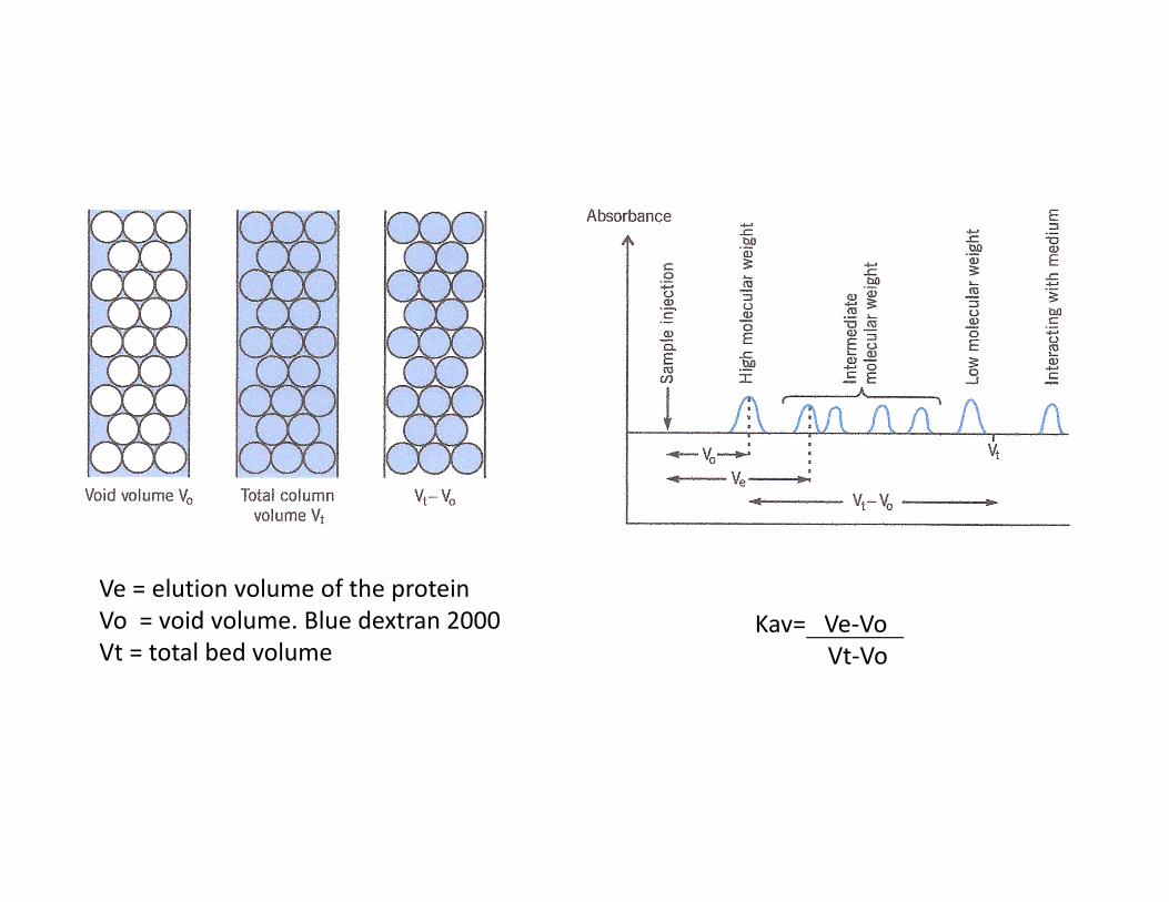
Kav= Ve-Vo
Vt-Vo
Ve = elution volume of the protein
Vo = void volume. Blue dextran 2000
Vt = total bed volume
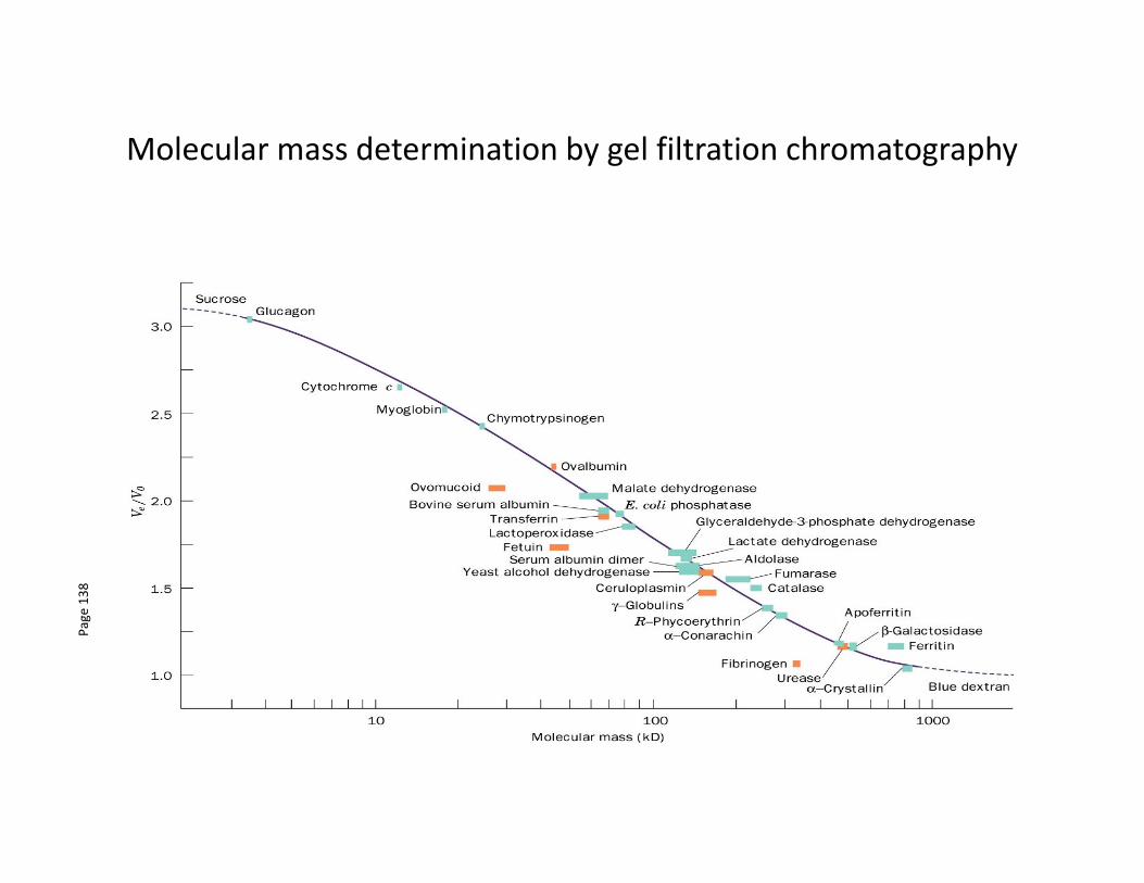
Molecular mass determination by gel filtration chromatography
Pa
ge
13
8
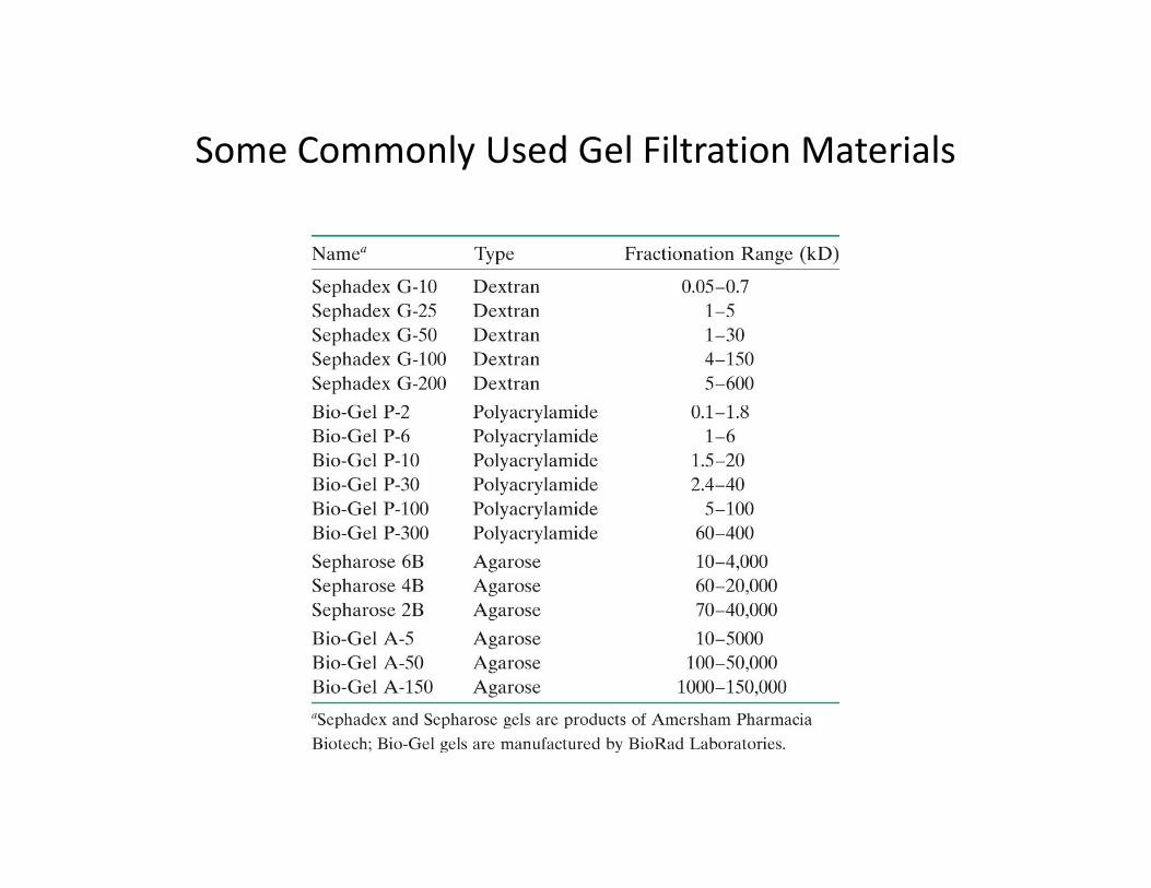
Some Commonly Used Gel Filtration Materials
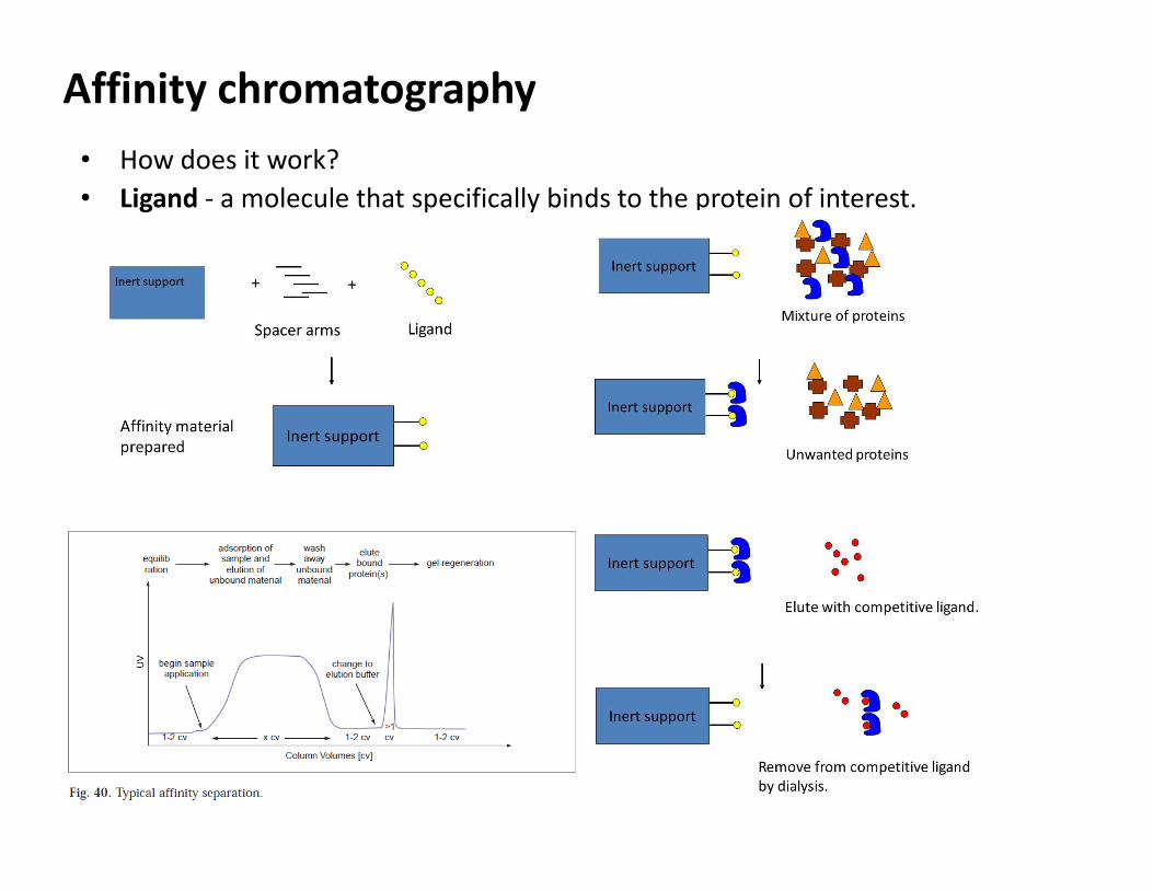
Affinity chromatography
• How does it work?
• Ligand - a molecule that specifically binds to the protein of interest.
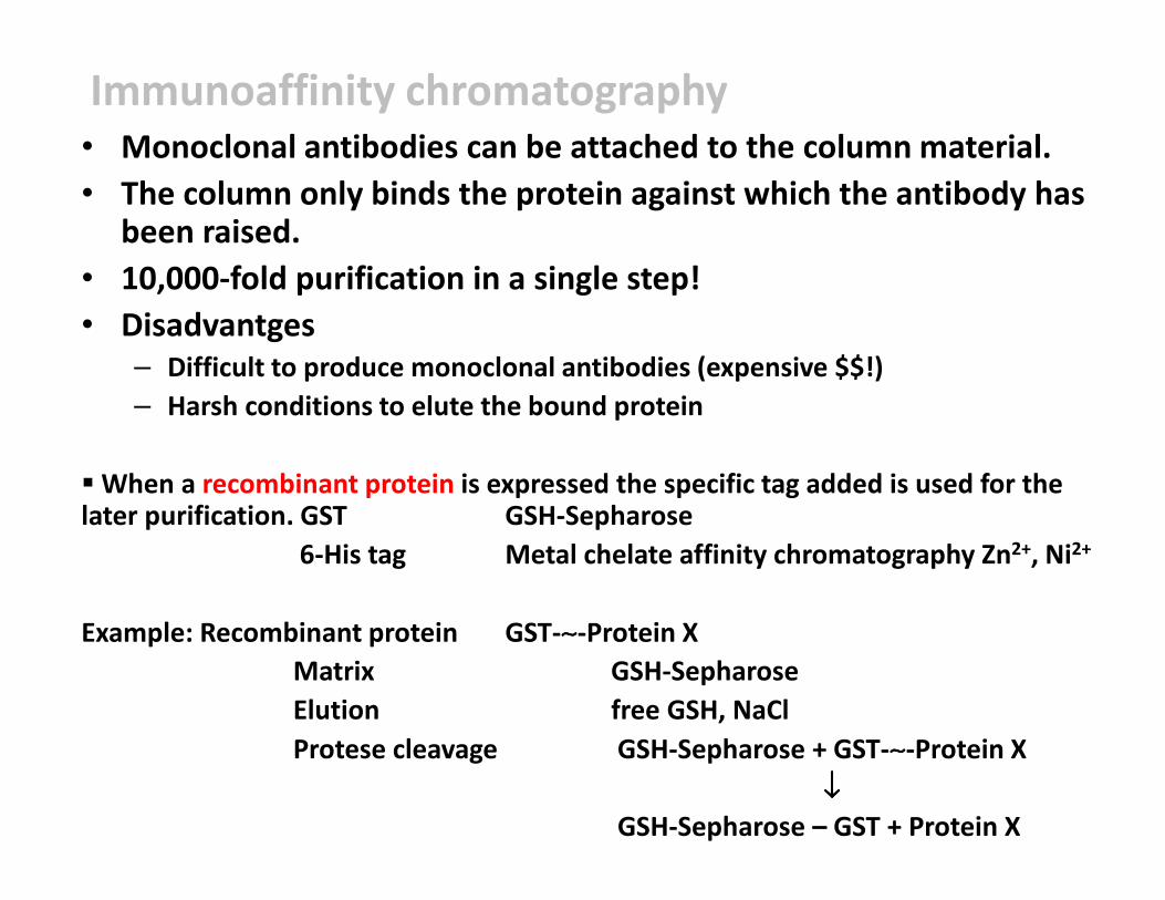
Immunoaffinity chromatography• Monoclonal antibodies can be attached to the column material.
• The column only binds the protein against which the antibody has been raised.
• 10,000-fold purification in a single step!
• Disadvantges– Difficult to produce monoclonal antibodies (expensive $$!)
– Harsh conditions to elute the bound protein
� When a recombinant protein is expressed the specific tag added is used for the later purification. GST GSH-Sepharose
6-His tag Metal chelate affinity chromatography Zn2+, Ni2+
Example: Recombinant protein GST-∼∼∼∼-Protein X
Matrix GSH-Sepharose
Elution free GSH, NaCl
Protese cleavage GSH-Sepharose + GST-∼∼∼∼-Protein X
↓↓↓↓
GSH-Sepharose – GST + Protein X
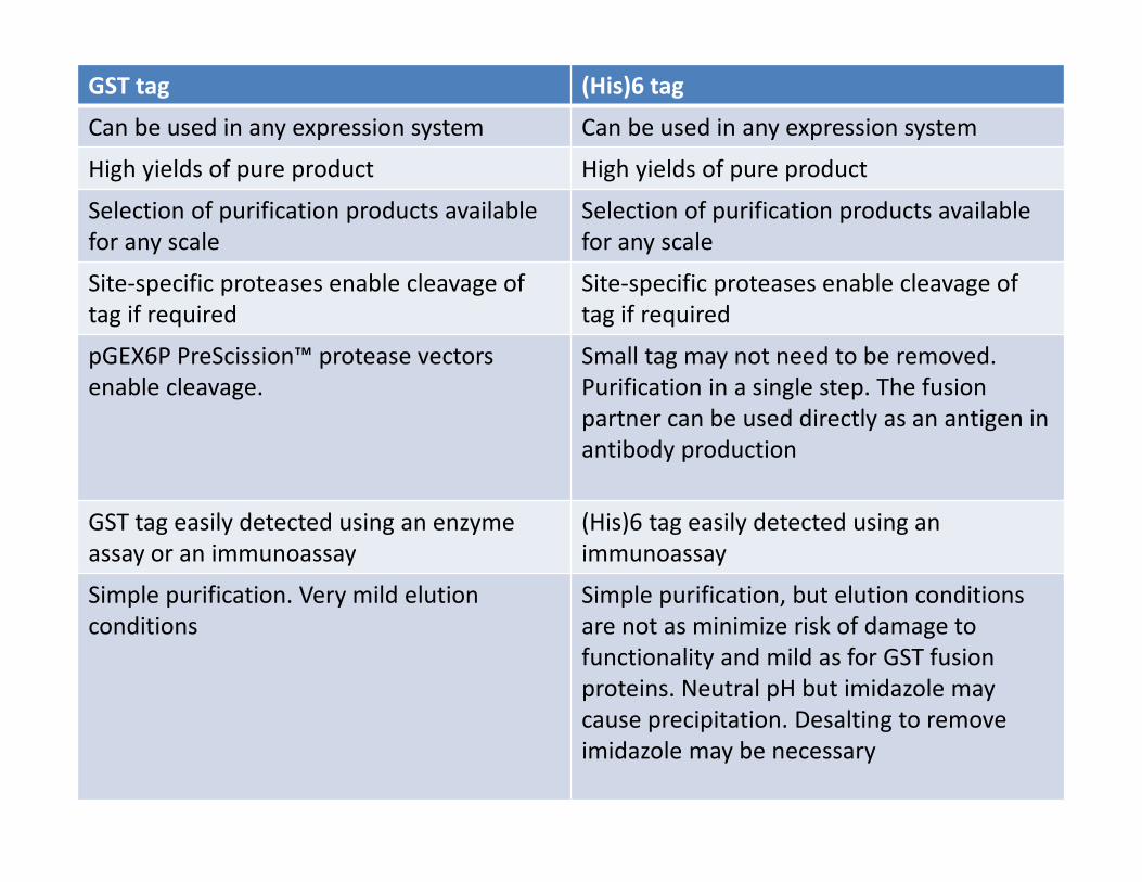
GST tag (His)6 tag
Can be used in any expression system Can be used in any expression system
High yields of pure product High yields of pure product
Selection of purification products available
for any scale
Selection of purification products available
for any scale
Site-specific proteases enable cleavage of
tag if required
Site-specific proteases enable cleavage of
tag if required
pGEX6P PreScission™ protease vectors
enable cleavage.
Small tag may not need to be removed.
Purification in a single step. The fusion
partner can be used directly as an antigen in
antibody production
GST tag easily detected using an enzyme
assay or an immunoassay
(His)6 tag easily detected using an
immunoassay
Simple purification. Very mild elution
conditions
Simple purification, but elution conditions
are not as minimize risk of damage to
functionality and mild as for GST fusion
proteins. Neutral pH but imidazole may
cause precipitation. Desalting to remove
imidazole may be necessary
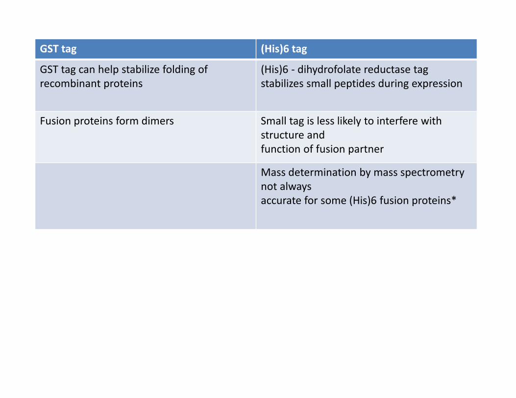
GST tag (His)6 tag
GST tag can help stabilize folding of
recombinant proteins
(His)6 - dihydrofolate reductase tag
stabilizes small peptides during expression
Fusion proteins form dimers Small tag is less likely to interfere with
structure and
function of fusion partner
Mass determination by mass spectrometry
not always
accurate for some (His)6 fusion proteins*
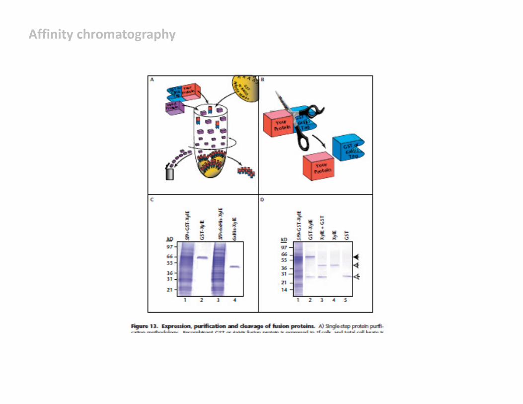
Affinity chromatography
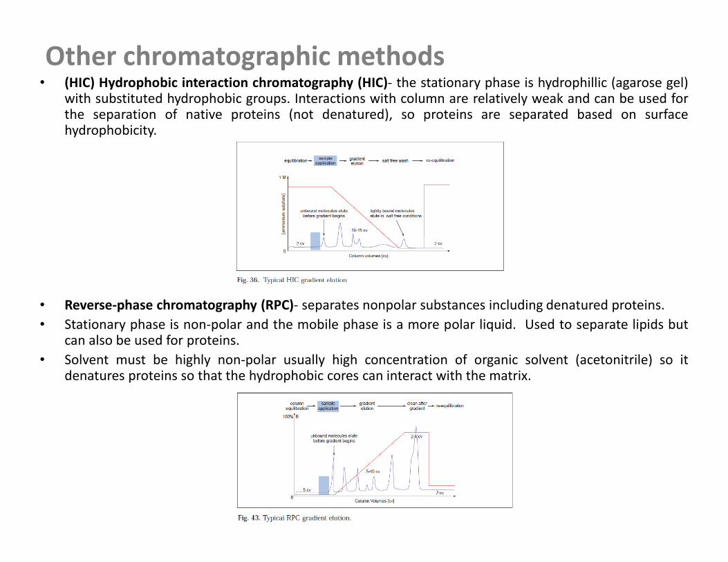
Other chromatographic methods• (HIC) Hydrophobic interaction chromatography (HIC)- the stationary phase is hydrophillic (agarose gel)
with substituted hydrophobic groups. Interactions with column are relatively weak and can be used forthe separation of native proteins (not denatured), so proteins are separated based on surfacehydrophobicity.
• Reverse-phase chromatography (RPC)- separates nonpolar substances including denatured proteins.
• Stationary phase is non-polar and the mobile phase is a more polar liquid. Used to separate lipids butcan also be used for proteins.
• Solvent must be highly non-polar usually high concentration of organic solvent (acetonitrile) so itdenatures proteins so that the hydrophobic cores can interact with the matrix.
Other chromatographic methods
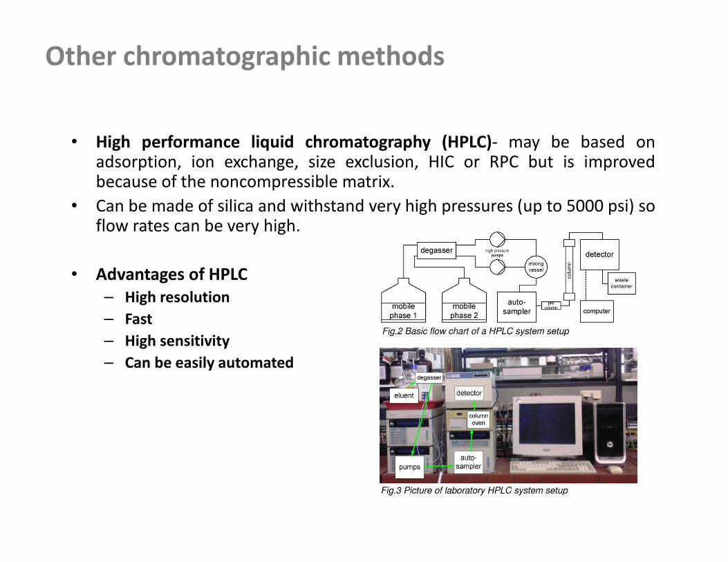
• High performance liquid chromatography (HPLC)- may be based onadsorption, ion exchange, size exclusion, HIC or RPC but is improvedbecause of the noncompressible matrix.
• Can be made of silica and withstand very high pressures (up to 5000 psi) soflow rates can be very high.
• Advantages of HPLC
– High resolution
– Fast
– High sensitivity
– Can be easily automated
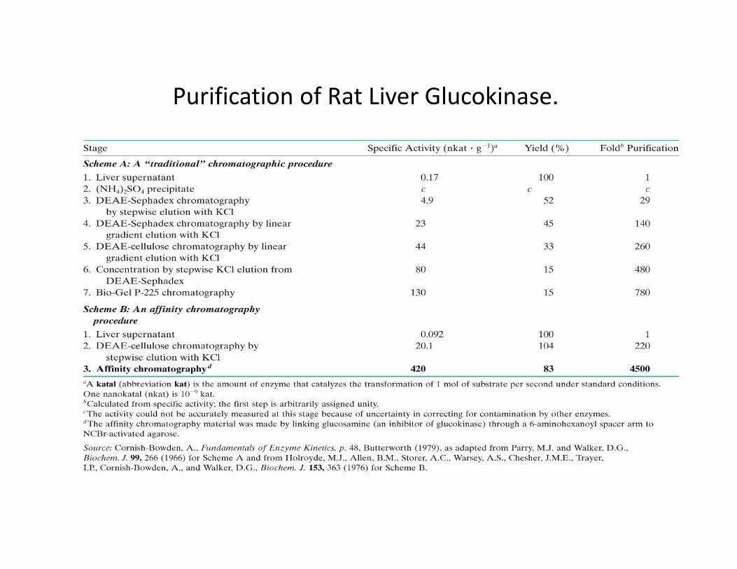
Purification of Rat Liver Glucokinase.
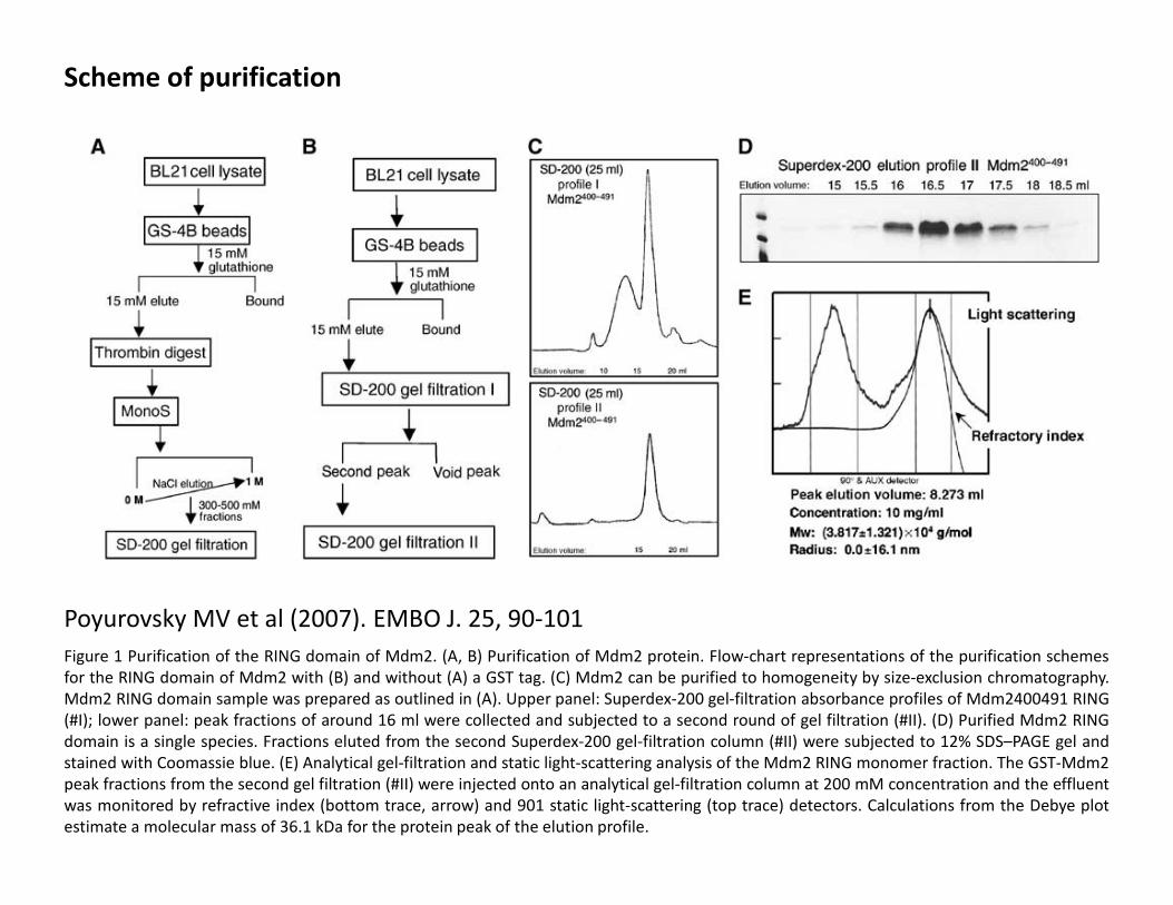
Scheme of purification
Poyurovsky MV et al (2007). EMBO J. 25, 90-101
Figure 1 Purification of the RING domain of Mdm2. (A, B) Purification of Mdm2 protein. Flow-chart representations of the purification schemes
for the RING domain of Mdm2 with (B) and without (A) a GST tag. (C) Mdm2 can be purified to homogeneity by size-exclusion chromatography.
Mdm2 RING domain sample was prepared as outlined in (A). Upper panel: Superdex-200 gel-filtration absorbance profiles of Mdm2400491 RING
(#I); lower panel: peak fractions of around 16 ml were collected and subjected to a second round of gel filtration (#II). (D) Purified Mdm2 RING
domain is a single species. Fractions eluted from the second Superdex-200 gel-filtration column (#II) were subjected to 12% SDS–PAGE gel and
stained with Coomassie blue. (E) Analytical gel-filtration and static light-scattering analysis of the Mdm2 RING monomer fraction. The GST-Mdm2
peak fractions from the second gel filtration (#II) were injected onto an analytical gel-filtration column at 200 mM concentration and the effluent
was monitored by refractive index (bottom trace, arrow) and 901 static light-scattering (top trace) detectors. Calculations from the Debye plot
estimate a molecular mass of 36.1 kDa for the protein peak of the elution profile.
References
-Protein purification, Principles and Practice. Scope R.K (1982). Springer-
Verlag. New York Heidelberg Berlin
-Baculovirus Expression Vector System Manual. Introduction Manual. 6th
Edition. URL://www.pharmigen .com
-Methods in Molecular Biology. Vol 235: E.coli Plasmid vectors. Edited by
Casali N. and preston A.. Hmana Press Inc. Totowa, NJ
-Biochemistry. Voet, Voet.
- Handbooks from Amersham Biosciences