Embed Size (px)
Citation preview
Cristina Isabel Caniço Escrevente
Dissertation presented to obtain the Ph.D. degree in BiologyInstituto de Tecnologia Química e Biológica | Universidade Nova de Lisboa
Oeiras,October, 2011
Insert here an image with rounded corners
Protein glycosylation of exosomes from ovarian carcinoma cells
Structures and biological roles
Cristina Isabel Caniço Escrevente
Dissertation presented to obtain the Ph.D. degree in BiologyInstituto de Tecnologia Química e Biológica | Universidade Nova de Lisboa
Oeiras, October, 2011
Protein glycosylation of exosomes from ovarian carcinoma cellsStructures and biological roles
ii
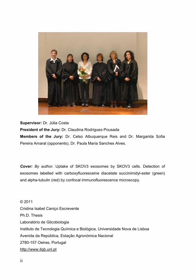
Supervisor: Dr. Júlia Costa
President of the Jury: Dr. Claudina Rodriguez-Pousada
Members of the Jury: Dr. Celso Albuquerque Reis and Dr. Margarida Sofia
Pereira Amaral (opponents), Dr. Paula Maria Sanches Alves.
Cover: By author. Uptake of SKOV3 exosomes by SKOV3 cells. Detection of
exosomes labelled with carboxyfluoresceine diacetate succinimidyl-ester (green)
and alpha-tubulin (red) by confocal immunofluorescence microscopy.
© 2011
Cristina Isabel Caniço Escrevente
Ph.D. Thesis
Laboratório de Glicobiologia
Instituto de Tecnologia Química e Biológica, Universidade Nova de Lisboa
Avenida da República, Estação Agronómica Nacional
2780-157 Oeiras, Portugal
http://www.itqb.unl.pt

iii
Acknowledgments
This PhD Thesis would not have been possible without the support and
assistance of a number of people to whom I would like to express my sincere
gratitude.
First, I would like to acknowledge my supervisor Dr. Júlia Costa for giving
me the opportunity to work on such an interesting project, and for providing me the
scientific support that made this Thesis possible. Thank you for accepting me as a
PhD student and for the encouragement and thoughtful guidance.
A warm thanks to Professor Peter Altevogt for the helpful suggestions that
enriched the content of this thesis, for the guidance and support during all this
years, and for welcoming me in his laboratory.
I am grateful to Dr. Paula Alves from the Animal Cell Technology Unit for
providing me the opportunity to work with the flow cytometer and all the TCA team
for the help.
I am thankful to Dr. Harald Conradt and Sebastian Kandzia from
GlycoThera, Germany, for their contribution to my work, and particularly for the
help given in the glycosylation profiling.
A special thanks to all my lab colleagues, present and former, for the
friendly and supportive atmosphere that contributed to the final outcome of this
Thesis. I am thankful to Ricardo, for being the “man” of the lab, for the endless
patience, the critical opinions, and also for the proofreading of this Thesis. I thank
Rita for being a good companionship in the last two years and Carla for the good
advices. Your support and friendship was very important. I would like to thank Eda
for being present since day one. Your sincere friendship, your practical demeanor,
and the continuous support given throughout all these years were priceless.
Catarina Gomes, I am grateful for your true friendship and all the good moments
shared together. I thank Catarina Brito for the friendship, the generosity, and
willingness to answer to all my doubts, not only then, when you were a member of
the lab, but also now. I thank Vanessa for being present in the beginning and for
teaching me so many important things about the science world, and finally,
Angelina for the helpful discussions.

iv
I would like to acknowledge all my good friends inside and outside the
science world. Although many times distant in space they were always present in
mind. Thank you, for being present whenever I needed. I believe that the world is
big but friendships are bigger.
In portuguese.... Não podia deixar de agradecer à minha família que
sempre acreditou em mim e me deu apoio. Obrigado, padrinhos, primas e primos,
por existirem e estarem ao meu lado em todos momentos. Obrigado aos meus
avós, Palmira e José, Emília e Joaquim, por terem feito parte da minha vida. Sei
que ficariam orgulhosos de mim.
Agradeço o apoio dos meus sogros, Teresa e Abílio, e dos meus novos
tios, Alice e Fernandes. Obrigado por me terem recebido de braços abertos na
vossa família.
Um obrigado infinito aos meus pais por me terem deixado seguir os meus
sonhos e terem acreditado que podia chegar onde cheguei. Espero não vos ter
desiludido ao ter escolhido este caminho.
Tenho de agradecer ao Pedro, mais do que um companheiro, um amigo.
Obrigado por teres escolhido Coimbra para estudar e, sobretudo, obrigado por
teres decidido partilhar a tua vida comigo. A tua paciência ilimitada é, sem dúvida,
a tua maior virtude.
Por último, dedico esta tese à minha filha Carolina, que apesar de ainda
não ter completado um ano já faz parte do mundo da ciência. Durante nove meses
trabalhou comigo no laboratório, e durante os seus primeiros meses partilhou a
minha atenção com a escrita desta tese. Obrigado por seres uma boa menina e
me teres deixado trabalhar de vez em quando. Obrigado pelos beijos molhados,
sorrisos e gargalhadas nos dias mais difíceis. Obrigado, simplesmente e acima de
tudo, por existires e fazeres parte da minha vida.
To Fundação para a Ciência e Tecnologia (FCT) and Fundo Social Europeu (FSE)
for financial support (SFRH/BD/30622/2006).
v
Summary
Exosomes are small membrane vesicles that are secreted by
several cell types including tumour cells. They are formed intracellularly by
an inward budding of the membrane of endosomal compartments which are
converted to multivesicular bodies. Exosomes are then released into the
extracellular environment after fusion of the multivesicular bodies with the
plasma membrane. Upon internalization by other cells they may transfer
proteins and RNA among cells. Tumour-derived exosomes can promote
angiogenesis, cell proliferation, tumour cell invasion and immune evasion.
These vesicles have been found in biological fluids such as malignant
ascites and blood and can therefore be used not only to identify potential
biomarkers of disease but also in vaccination.
Alterations in protein glycosylation are often associated with
tumourigenesis; therefore glycan structures represent ideal targets for
tumour specific diagnosis and new anti-tumour therapeutic strategies.
This research focused on the characterization and investigation of
the biological role of protein N-glycosylation from ovarian tumour derived
exosomes, with particular emphasis on the glycoprotein disintegrin and
metalloprotease 10 (ADAM10).
Chapter 2 details the investigation of the role of N-glycosylation on
ADAM10 intracellular localization, activity and sorting to exosomes. To
accomplish this objective, individual ADAM10 N-glycosylation site mutants
S269A, T280A, S441A, T553A were constructed using the QuickChange
site directed mutagenesis approach. Western blot analysis of the ovarian
carcinoma SKOV3 cells stably transfected with wild-type and N-
glycosylation mutants of ADAM10 showed that all potential glycosylation
sites were occupied by high-mannose and/or complex oligosaccharides.
Mutant T280A was found to accumulate in the endoplasmic reticulum as
vi
the non-processed precursor of the enzyme, whilst S441A showed
increased susceptibility to proteolysis. Study of ADAM10 in vivo activity
towards the cell adhesion molecule L1, in HEK293 cells and mouse
embryonic fibroblasts derived from the ADAM10 knockout mouse revealed
that the mutation of ADAM10 N-glycosylation sites did not completely
abolish enzyme activity. Accordingly, all mutants exhibited proteolytic
activity towards proTNF-alpha in vitro. However, a reduction in the
shedding levels was found, especially for T280A. On the other hand, all
ADAM10 N-glycosylation mutants cleaved to their mature form were
efficiently sorted into exosomes. Overall, these results showed the
importance of N-glycosylation for ADAM10 processing, protection from
proteolysis, and full-enzyme activity.
Exosomes can interact and be internalized by target cells; however,
the mechanisms involved in this process are still poorly understood.
Chapter 3 describes that SKOV3 cells were found to internalize SKOV3
derived exosomes labelled with carboxyfluoresceine diacetate succinimidyl-
ester, by immunofluorescence microscopy, via an energy-dependent
process. Partial colocalization with the endosome marker EEA1 and
inhibition by chlorpromazine suggested the involvement of clathrin-
dependent endocytosis. On the other hand, an uptake inhibition in the
presence of 5-ethyl-N-isopropyl amiloride, cytochalasin D and methyl-beta-
cyclodextrin suggested the involvement of additional endocytic pathways.
Furthermore, proteins present in exosomes and at SKOV3 cell surface
were at least partially required for internalization, as shown by the decrease
in the uptake levels following proteinase K treatment.
The analysis of the glycosylation profiles of SKOV3 exosomes using
lectins with different specificities revealed that they were enriched in
specific mannose- and sialic acid-containing glycoproteins that were not
present in cellular extracts and that may constitute exosome markers. The
effect of glycosylation on exosome uptake was also assessed. Sialic acid
vii
removal led to an increase in uptake efficiency whilst cell incubation with
monosaccharides D-galactose, α-L-fucose, α-D-mannose, D-N-
acetylglucosamine and the disaccharide β-lactose reduced exosome
uptake to a level comparable to that of the control D-glucose.
Chapter 4 addresses the further characterization of the N-glycan
profile of vesicles secreted by ovarian tumour cells. A detailed structure
analysis of the N-glycans of SKOV3 and OVM secreted vesicles,
microsomal and plasma membrane fractions was performed by high
performance anion exchange chromatography with pulsed amperometric
detection (HPAEC-PAD) and matrix assisted laser desorption/ionization
time-of-flight mass spectrometry (MALDI-TOF-MS). Glycans of the di-, tri-
and tetraantennary type with proximal fucose were found in secreted
vesicles of SKOV3 and OVM cells, as well as in the plasma membrane and
microsomal fraction. SKOV3 cells were found to contain predominantly
proximally fucosylated complex partially agalactosylated glycan structures
in all fractions. Moreover, the terminal GalNAcβ4GlcNAc (LacdiNAc) motif
was also found in SKOV3 secreted vesicles. OVM secreted vesicles
contained predominantly complex intact glycans of the di-, tri- and
tetraantennary types.
In conclusion, this research has contributed to the knowledge of
protein glycosylation present in vesicles secreted by ovarian tumour cells.
This information should provide the basis for further exploration of the
mechanisms of protein sorting and exosome biogenesis. Moreover, the
identification of specific glycoproteins and N-glycans in exosomes might
open new perspectives in biomarker discovery and cancer vaccination.

viii
ix
Resumo
Exossomas são pequenas vesículas membranares secretadas por
diversos tipos de células, incluindo células tumorais. Eles são formados
intracelularmente, pela invaginação da membrana de compartimentos
endossomais que dão origem aos corpos multivesiculares. A fusão dos
corpos multivesiculares com a membrana plasmática resulta na libertação
dos exossomas para o ambiente extracelular. Após internalização por
outras células, os exossomas podem transferir proteínas e RNA entre
células. Exossomas secretados por células tumorais podem promover a
angiogénese, proliferação celular, invasão tumoral e evasão do sistema
imunitário. Estas vesículas foram encontradas em fluidos biológicos, tais
como, ascites e sangue, e como tal, podem ser usadas para identificar
potenciais biomarcadores de doenças e também em vacinação.
Proteínas com glicosilação alterada encontram-se frequentemente
associadas com o processo de tumorigénese; como tal, estruturas
glicosídicas representam alvos ideais para o diagnóstico específico de
tumores e para o desenvolvimento de novas estratégias terapêuticas anti-
tumorais.
Esta Tese visou a caracterização e investigação do papel biológico
da N-glicosilação em proteínas presentes em exossomas secretados por
células do carcinoma do ovário, com particular ênfase na glicoproteína
desintegrina e metaloprotease ADAM10.
O Capítulo 2 descreveu a investigação do papel da N-glicosilação
na localização intracelular, actividade e transporte para os exossomas da
proteína ADAM10. Para alcançar esse objectivo foi utilizada a metodologia
de mutagénese dirigida para construir mutantes em que os locais de N-
glicosilação foram individualmente eliminados: S269A, T280A, S441A,
T553A. A análise por Western blot de células de carcinoma do ovário
SKOV3, transfectadas estavelmente com o tipo selvagem ou com os

x
mutantes de N-glicosilação da ADAM10, mostrou que todos os potenciais
locais de N-glicosilação se encontravam ocupados com oligossacáridos do
tipo oligomanose ou complexo. O mutante T280A foi encontrado
acumulado no reticulo endoplasmático, na forma precursora não
processada da enzima, enquanto o mutante S441A mostrou um aumento
na susceptibilidade à proteólise. O estudo da actividade in vivo da
ADAM10 monitorizando a molécula de adesão celular L1 como substrato,
em células HEK293 e em fibroblastos de ratinhos silenciados para a
ADAM10, revelou que a mutação individual dos diferentes locais de N-
glicosilação da ADAM10 não aboliu por completo a actividade da enzima.
Da mesma forma, todos os mutantes exibiram actividade proteolítica in
vitro usando o péptido proTNF-alpha como substrato. No entanto, foi
observada uma redução nos níveis de clivagem, especialmente com o
mutante T280A. Por outro lado, todos os mutantes de N-glicosilação da
ADAM10 que foram processados para a forma matura foram
eficientemente transportados para os exossomas. No global, estes
resultados mostram a importância da N-glicosilação no processamento,
protecção contra a proteólise e actividade enzimática da ADAM10.
Os exossomas podem interagir e ser internalizados por células-
alvo, no entanto, os mecanismos envolvidos nesse processo ainda são
pouco compreendidos. O Capítulo 3 mostrou, por microscopia de
fluorescência, a internalização de exossomas de células SKOV3 marcados
com succinimidil éster diacetato de carboxifluoresceína por células SKOV3,
através de um mecanismo dependente de energia. A colocalização parcial
com o marcador de endossomas EEA1 e a inibição com clorpromazina
sugeriu o envolvimento da via de endocitose mediada por clatrina. Por
outro lado, a inibição da internalização na presença de 5-etil-N-isopropil
amiloride, citocalasina D e metil-beta-ciclodextrina sugeriu o envolvimento
de outras vias endocíticas. Para além disso, foram necessárias, pelo
menos parcialmente, proteínas presentes nos exossomas e na superfície

xi
das células SKOV3 para a internalização, como mostrado pela diminuição
nos níveis de internalização após tratamento com proteinase K.
A análise do perfil de glicosilação dos exossomas de células
SKOV3, usando lectinas com diferentes especificidades, revelou que eles
eram enriquecidos em glicoproteínas específicas com manose e ácido
siálico. Essas glicoproteínas não se encontravam nos extractos celulares
podendo constituir marcadores de exossomas. Adicionalmente, foi
investigado o papel da glicosilação na internalização de exossomas. A
remoção do ácido siálico levou a uma tendência para um aumento na
eficiência da internalização, enquanto que a incubação com os
monossacáridos D-galactose, α-L-fucose, α-L-manose, D-N-
acetiglucosamina e o dissacárido β-lactose reduziu a internalização para
níveis comparáveis ao do controlo com D-glucose.
O Capítulo 4 visou a caracterização pormenorizada do perfil de N-
glicanos presentes em células do carcinoma do ovário. A análise detalhada
da estrutura dos N-glicanos presentes nas vesículas secretadas, fracção
microssomal e membrana plasmática de células SKOV3 e OVM foi
efectuada por cromatografia de troca iónica de elevada “performance” com
detecção amperométrica pulsada (HPAEC-PAD) e por espectometria de
massa com “matrix assisted laser desorption/ionization time-of-flight”
(MALDI-TOF-MS). Glicanos do tipo bi-, tri- e tetra-ramificados com fucose
próxima foram encontrados nas vesículas secretadas pelas células SKOV3
e OVM, bem como na membrana plasmática e fracção microssomal. Todas
as fracções das células SKOV3 apresentaram, predominantemente,
estruturas glicosídicas parcialmente agalactosiladas com fucose próxima.
Para além disso, o motivo terminal GalNAcβ4GlcNAc (LacdiNAc) também
foi encontrado nas vesículas secretadas pelas células SKOV3. As
vesículas secretadas pelas células OVM apresentaram
predominantemente glicanos intactos do tipo complexo bi-, tri- e
tetraramificados.

xii
Em conclusão, este estudo contribuiu para o conhecimento da
glicosilação de proteínas presentes em vesículas secretadas por células do
carcinoma do ovário. Esta informação poderá fornecer uma base para a
exploração futura dos mecanismos de selecção de proteínas e biogénese
dos exossomas. Além disso, a identificação de glicoproteínas e N-glicanos
específicos nos exossomas poderá abrir novas perspectivas na descoberta
de biomarcadores e na vacinação contra o cancro.
xiii
Table of contents
Thesis Outline xvii
List of Figures xix
List of Tables xxi
Abbreviations xxiii
Amino Acid nomenclature xxv
Chapter 1- General introduction .............................................................. 1
1.1. Ovarian cancer .......................................................................................... 3
1.2. Protein glycosylation .................................................................................. 4
1.2.1. N-glycosylation ................................................................................... 5
1.2.2. O-glycosylation ................................................................................... 7
1.2.3. Glycosylation in cancer ...................................................................... 8
1.3. Exosomes ................................................................................................ 11
1.3.1. Biogenesis and secretion ................................................................. 12
1.3.2. Composition ..................................................................................... 14
1.3.3. Function ............................................................................................ 15
1.3.4. Tumour cell-derived exosomes ........................................................ 17
1.3.5. Exosomes in diagnosis and therapeutics ......................................... 18
1.4. ADAM10 .................................................................................................. 19
1.4.1. Structure ........................................................................................... 20
1.4.2. Activity .............................................................................................. 22
1.4.3. Role in cancer .................................................................................. 25
1.5. Aims of this Thesis work .......................................................................... 27
Chapter 2 - Functional role of N-glycosylation from ADAM10 in
processing, localization and activity of the enzyme ............................ 29
2.1. Summary ................................................................................................. 31
2.2. Introduction .............................................................................................. 32
2.3. Materials and methods ............................................................................ 34
xiv
2.3.1. DNA constructs ................................................................................ 34
2.3.2. Cell culture and protein expression ................................................. 35
2.3.3. Isolation of membrane vesicles ....................................................... 36
2.3.4. In vivo cleavage assay .................................................................... 36
2.3.5. SDS-PAGE and Western blot analysis ............................................ 37
2.3.6. Protein deglycosylation .................................................................... 38
2.3.7. Immunoprecipitation ........................................................................ 38
2.3.8. Confocal immunofluorescence microscopy ..................................... 39
2.3.9. Peptide cleavage assay ................................................................... 40
2.3.10. ADAM10 structure ......................................................................... 40
2.4. Results .................................................................................................... 41
2.4.1. The N-glycosylation sites of hADAM10 are occupied ..................... 41
2.4.2. Intracellular localization of bADAM10 N-glycosylation mutants ...... 44
2.4.3. In vivo and in vitro activity of bADAM10wt and N-glycosylation
mutants ...................................................................................................... 47
2.4.4. Characterization of ADAM10 from exosomes ................................. 52
2.4.5. Modelling of the 3D structure of bADAM10 metalloprotease
domain ....................................................................................................... 55
2.5. Discussion ............................................................................................... 57
2.6. Acknowledgements ................................................................................. 60
Chapter 3 - Interaction and uptake of exosomes by ovarian
carcinoma cells ....................................................................................... 63
3.1. Summary ................................................................................................. 65
3.2. Introduction ............................................................................................. 67
3.3. Materials and methods ............................................................................ 69
3.3.1. Cell culture ....................................................................................... 69
3.3.2. Isolation of secreted membrane vesicles ........................................ 69
3.3.3. Glycoprotein detection using lectins and immunoblot ..................... 70
3.3.4. Glycosidase treatment ..................................................................... 71
3.3.5. Uptake of SKOV3 exosomes by SKOV3 cells ................................. 71
3.3.6. Immunofluorescence microscopy .................................................... 72

xv
3.3.7. Flow cytometry ................................................................................. 72
3.4. Results ..................................................................................................... 73
3.4.1. Uptake of SKOV3 exosomes by SKOV3 cells ................................. 73
3.4.2. Proteins are required for exosomes uptake ..................................... 77
3.4.3. Enrichment of specific glycoproteins in exosomes and relevance
in uptake ..................................................................................................... 78
3.5. Discussion ............................................................................................... 82
3.6. Acknowledgements.................................................................................. 85
Chapter 4 - N-glycosylation of secreted vesicles from ovarian
tumour cells ............................................................................................ 87
4.1. Summary ................................................................................................. 89
4.2. Introduction .............................................................................................. 91
4.3. Materials and methods ............................................................................ 93
4.3.1. Cell culture......................................................................................... 93
4.3.2. Isolation of cell-secreted vesicles ...................................................... 93
4.3.3. Isolation of microsomal and plasma membrane enriched
fractions ....................................................................................................... 94
4.3.4. Glycoprotein detection using lectins and immunoblot ....................... 95
4.3.5. Total N-glycans isolation ................................................................... 96
4.3.6. Analysis of desialylated N-glycans by HPAEC-PAD ......................... 97
4.3.7. Analysis of N-glycans by MALDI-TOF-MS ........................................ 98
4.4. Results ..................................................................................................... 99
4.4.1. Enrichment of specific glycoproteins in ovarian carcinoma
secreted vesicles ......................................................................................... 99
4.4.2. Structural analysis of desialylated N-glycans from ovarian tumour
secreted vesicles, plasma membrane and microsomal fractions .............. 103
4.5. Discussion ............................................................................................. 115
4.6. Acknowledgements................................................................................ 118
xvi
Chapter 5 - General discussion and conclusions ............................... 119
5.1. General discussion and perspectives ................................................... 121
5.1.1. Exosomes specific glycoproteins/glycoforms ................................ 122
5.1.2. Internalization of exosomes by target cells ................................... 125
5.1.3. Exosomes from ovarian tumour cells as biomarkers and
potential therapeutic targets .................................................................... 127
5.2. General conclusions ............................................................................. 132
Supplementary material ....................................................................... 133
Supplementary Figure 1 ............................................................................... 135
Supplementary Figure 2 ............................................................................... 138
References ............................................................................................ 141
References ................................................................................................... 143

xvii
Thesis Outline
Exosomes are secreted by several cell types, including ovarian
tumour cells and are found in biological fluids such as blood and malignant
ascites. The role of these vesicles in tumour progression has been studied
but the role of protein glycosylation is not known.
The major objective of this work was to characterize and investigate
the biological role of protein N-glycosylation from ovarian tumour derived
exosomes. Furthermore, the role of N-linked glycans in ADAM10
localization, activity and sorting to exosomes has also been investigated.
This dissertation begins with a general introduction to ovarian
cancer biology, which is followed by an overview about protein
glycosylation, with focus on the N-glycosylation biosynthetic pathway and
abnormal glycosylation properties found in cancer cells. Exosome
biogenesis, composition and function are described in detail and particular
relevance is given to tumour-derived exosomes as potential biomarkers
and therapeutic targets. Finally, a brief characterization of the
metalloprotease and disintegrin ADAM10 is given, including protein
structure, activity and role in cancer.
In Chapter 2, the role of N-linked oligosaccharides on ADAM10
intracellular localization, activity and sorting to exosomes was investigated.
Chapter 3 describes the mechanisms involved in the uptake of
labelled exosomes from SKOV3 cells by SKOV3 cells. Furthermore, the
glycosylation profile of these secreted vesicles was analysed by lectin blot
using different lectins.
In Chapter 4, the glycosylation of ovarian tumour secreted vesicles,
microsomal and plasma membrane enriched fractions were compared by
lectin blot analysis. The N-glycan structures from glycoproteins present in
xviii
these fractions were also analysed in detail by HPAEC-PAD and MALDI-
TOF-MS.
Finally, Chapter 5 consists of a general discussion with future
perspectives, and conclusions of the work presented in this Thesis.

xix
List of Figures
Figure 1 Page 7 N-glycosylation processing and representation of the
different N-glycan types.
Figure 2 Page 13 Exosome biogenesis and different types of
membrane vesicles.
Figure 3 Page 21 Domain structure of ADAM10.
Figure 4 Page 24 ADAM10 substrates.
Figure 5 Page 42 Schematic representation of ADAM10 and N-
glycosylation mutants.
Figure 6 Page 44 Western blot analysis of ADAM10 and N-
glycosylation mutants from SKOV3 cells.
Figure 7 Page 46 Colocalization of bADAM10wt and T280A mutant,
with markers of the secretory pathway by confocal
immunofluorescence microscopy.
Figure 8 Page 48 L1 cleavage by bADAM10wt and N-glycosylation
mutants in HEK293 cells.
Figure 9 Page 49 L1 cleavage in MEFs ADAM10-/- transiently
overexpressing pcDNA3-L1 and pcDNA3.1-LacZ,
bADAM10wt or each of the N-glycosylation mutants.
Figure 10 Page 51 Measurement of the proteolytic activity of
bADAM10wt and S269A, T280A N-glycosylation
mutants using a fluorescent TNF-alpha peptide
cleavage assay.
Figure 11 Page 53 Western blot analysis of endogenous hADAM10
from SKOV3 cell extracts and exosomes.
Figure 12
Figure 13
Page 55
Page 56
Western blot analysis of bADAM10wt and N-
glycosylation mutants from stably transfected
SKOV3 cells.
Representation of the three-dimensional model for
bADAM10 metalloprotease domain.
xx
List of Figures (continued)
Figure 14 Page 74 Uptake of SKOV3 exosomes by SKOV3 cells.
Figure 15 Page 76 Path of internalization of exosomes in SKOV3 cells.
Figure 16 Page 77 Effect of proteinase K treatment in SKOV3
exosomes uptake.
Figure 17 Page 79 Western blot and lectin detection of glycoproteins
from SKOV3 cellular extracts (Ext) and secreted
vesicles (Ves).
Figure 18 Page 80 Con A, SNA and MAL lectin detection of
glycoproteins from HEK293 and H4 cellular extract
(Ext) and secreted vesicles (Ves).
Figure 19
Figure 20
Figure 21
Figure 22
Figure 23
Figure 24
Figure 25
Page 81
Page 101
Page 102
Page 104
Page 105
Page 108
Page 109
Effects of neuraminidase and sugars on SKOV3
exosomes uptake.
Western blot and lectin detection of glycoproteins
from SKOV3 cellular extracts (Ext), microsomal
fraction (MF), plasma membrane fraction (PM),
intracellular fraction (IC), cytoplasmic fraction (Cyt)
and secreted vesicles (Ves).
SNA and MAL lectin detection of glycoproteins from
OVM, m130 and GG ovarian carcinoma cell lines.
HPAEC-PAD analysis of the desialylated N-glycans
released from SKOV3 secreted vesicles, plasma
membrane and microsomal fractions.
HPAEC-PAD analysis of the desialylated N-glycans
released from OVM secreted vesicles, plasma
membrane and microsomal fractions.
MALDI-TOF-MS analysis of the total desialylated N-
glycans from SKOV3 cells.
MALDI-TOF-MS analysis of the total desialylated N-
glycans from OVM cells.

xxi
List of Tables
Table 1 Page 110 Observed mass signals (m/z) by MALDI-TOF-MS
analysis. Proposed composition and compatible
structure of the major desialylated N-glycans from
SKOV3 cells.
Table 2 Page 113 Observed mass signals (m/z) by MALDI-TOF-MS
analysis. Proposed composition and compatible
structure of the major desialylated N-glycans from
OVM cells.

xxii

xxiii
Abbreviations
Abbreviation Full form
ACN acetonitrile
ADAM a disintegrin and metalloprotease
BCA bicinchoninic acid
BMS Brystol meyer squibb
BSA bovine serum albumin
CFSE carboxyfluoresceine diacetate succinimidyl-ester
Con A concanavalin A
dHex deoxyhexose
DMEM Dulbecco's modified Eagle's medium
EndoH endoglycosidase H
EEA1 early endosome antigen 1
EIPA 5-ethyl-N-isopropyl amiloride
ER endoplasmic reticulum
ERGIC endoplasmic reticulum-Golgi intermediate
compartment
ESCRT endosomal sorting complex required for transport
Exos exosomes
Fuc fucose
Gal galactose
GalNAc N-acetylgalactosamine
GlcNAc N- acetylglucosamine
Glc glucose
GM130 cis-Golgi matrix protein of 130 kDa
HEK human embryonic kidney
Hex hexose
HexNAc N-acetylhexosamine

xxiv
Abbreviation Full form
HPAEC-PAD high performance anion exchange chromatography
with pulsed amperometric detection
HRP horseradish peroxidase
LacNAc N-acetyllactosamine
LAMP1 lysosomal-associated membrane protein
MAL Maackia amurensis lectin
MALDI-TOF-MS matrix assisted laser desorption/ionization time-of-
flight mass spectrometry
Man mannose
MEFs mouse embryonic fibroblasts
MVBs multivesicular bodies
NeuAc N-acetylneuraminic acid
PBS phosphate-buffered saline
PCR polymerase chain reaction
PNGase F peptide-N-glycosidase F
PVDF polyvinyledene difluoride
SDS-PAGE sodium deodecyl sulphate - polyacrylamide gel
electrophoresis
sLea sialyl-Lewis
a
sLex sialyl-Lewis
x
SNA Sambuccus nigra lectin
TBS tris-buffered saline
TFA trifluoroacetic acid
TGN trans-Golgi network
TNF-α tumour necrosis factor α
Tris tris(hydroxymethyl)aminomethane
WFA Wisteria floribunda lectin
wt wild-type
xxv
Amino Acid nomenclature
Abbreviations Amino acid name
Ala A Alanine
Arg R Arginine
Asn N Asparagine
Asp D Aspartate (Aspartic Acid)
Cys C Cysteine
Gln Q Glutamine
Glu E Glutamate (Glutamic Acid)
Gly G Glycine
His H Histidine
Ile I Isoleucine
Leu L Leucine
Lys K Lysine
Met M Methionine
Phe F Phenylalanine
Pro P Proline
Ser S Serine
Thr T Threonine
Trp W Tryptophan
Try T Tyrosine
Val V Valine

xxvi

Chapter 1
General introduction

Chapter 1
2
General introduction
3
1. General introduction
1.1. Ovarian cancer
Ovarian cancer is the leading cause of death from gynaecological
malignancies in the Western world. The high death rate associated with this
disease is mainly due to the absence of specific symptoms, rapid growth
rate and to lack of an effective screening, and as a result most patients are
diagnosed at an advanced stage when the disease is already disseminated
throughout the abdominal cavity.
Ovarian cancers are a heterogeneous group of neoplasms and are
traditionally divided into three major categories according to their presumed
histogenesis: sex cord-stromal, germ cell, and epithelial tumours. Epithelial
ovarian tumours, which represent 90% of malignant ovarian tumours, are
further grouped into histological types: serous, mucinous, endometrioid,
clear cell, transitional cell tumours, carcinosarcoma, mixed epithelial
tumour, undifferentiated cancers, and others (reviewed in Scully, 1987;
Kaku et al., 2003).
The biological mechanisms of transformation in ovarian cancer are
poorly understood. Mutations in the BRCA1 and BRCA2 tumour suppressor
genes are responsible for a great number of hereditary ovarian cancers
(Holschneider and Berek, 2000), however, the majority of ovarian cancers
are sporadic, in which the disease arises from a multistep process requiring
a large number of genetic changes involving both activation of oncogenes
and loss of tumour-suppressor genes (Cvetkovic, 2003).
Ovarian cancers diagnosed while still limited to the ovaries (stage I),
allow that 90% of patients can be cured with conventional surgery and

Chapter 1
4
chemotherapy. Cure rates decrease substantially after tumour metastasis
to the pelvic organs (stage II), the abdomen (stage III) or beyond the
peritoneal cavity (stage IV) (Bast et al., 2009). Early detection of ovarian
cancer might significantly improve the overall survival rate. Nevertheless,
given the heterogeneity of the disease, it is unlikely that any single marker
will be sufficiently sensitive to provide an effective initial screen. Probably,
the combination of multiple serum markers may help achieve the sensitivity
and specificity required for the screening of ovarian cancer (Badgwell and
Bast, 2007).
1.2. Protein glycosylation
Glycosylation is the major post-translational modification of proteins
and lipids and is estimated to be present in more than half of all proteins in
nature. Glycosylation of proteins can be of the N- or O-linkage type
depending on glycans site of attachment. An N-glycan is a sugar chain
covalently linked via an N-acetylglucosamine (GlcNAc) residue to an
asparagine (Asn) residue of a polypeptide chain within the consensus
peptide sequence Asn-X-Ser/Thr. N-glycans share a common
pentasaccharide core region and can be generally divided into three main
types: high mannose, complex, and hybrid. An O-glycan is typically linked
to the polypeptide via an N-acetylgalactosamine (GalNac) residue to a
serine (Ser) or threonine (Thr) residue and can be extended into a variety
of different structural core classes (Varki et al., 2009).
The function of the glycans presented in glycoconjugates can be
crucial for the growth, development, and survival of cells and organisms.
Glycans have been shown to participate in recognition events and host-
pathogen interactions such as bacterial and viral infections. They are
involved in protein folding and sorting, vesicular trafficking, cell signalling,

General introduction
5
cell adhesion and motility, organogenesis and immunological defence
(reviewed in Varki, 1993; Ohtsubo and Marth, 2006). Additionally,
glycosylation can affect protein plasma residence time, it provides steric
protection from proteases, and it plays a role on enzyme activity, substrate
targeting and specificity (reviewed in Skropeta, 2009).
1.2.1. N-glycosylation
The biosynthesis of N-linked oligosaccharides occurs in the
endoplasmic reticulum (ER) and Golgi complex (Figure 1, A). During the
synthesis of N-linked glycans a 14-saccharide “core” unit is assembled as a
membrane bound dolichylpyrophosphate precursor by enzymes located on
both sides of the ER membrane. The structure of this precursor is common
to most eukaryotes and contains 3 glucose, 9 mannose, and 2 GlcNAc
residues (Glc3Man9GlcNAc2). The completed core oligosaccharide is
transferred from the dolichylpyrophosphate carrier to the nascent
polypeptide chain, and is coupled through an N-glycosidic bond to the side
chain of an asparagine residue. The oligosaccharyltransferase responsible
for this transfer is a complex enzyme with its active site in the ER lumen
and that recognizes a specific conformation of the glycosylation sequon
(Asn-X-Ser/Thr), where X can be any amino acid except proline. Of all
sequons, it has been estimated that 90% are glycosylated (Gavel and
Heijne, 1990). Before exiting the ER, a terminal glucose, and mannose
residues are removed from the N-glycan by ER glucosidases and
mannosidases. When the glycoprotein moves to the Golgi complex, the
glycan chains undergo further trimming of mannoses before further
extension of the glycan branches. Subsequent step-wise adding of sugars
to different positions of the extending glycan is catalyzed by a number of
Chapter 1
6
glycosyltransferases, each adds a particular monosaccharide through a
specific glycosidic bond (Helenius and Aebi, 2001; Varki et al., 2009).
N-glycan structures are generally classified into three principal
categories: high mannose, complex and hybrid (Figure 1, B), all of them
share a common tri-mannosyl (Man3GlcNAc2) core structure. The high
mannose glycans have 5 to 9 mannose (Man5–9GlcNAc2) sugars. Those
with two GlcNAc’s attached to the tri-mannosyl core are called complex
type. The hybrid type are a combination of high mannose and complex
glycans, and have at least three mannose sugars, but only one GlcNAc on
one nonreducing mannose (Helenius and Aebi, 2001; Varki et al., 2009).
The removal of N-glycans from proteins, either individually or in
combination has revealed their role in many cellular processes (reviewed in
Skropeta, 2009) and total inhibition of N-glycosylation lead to death in early
embryogenesis (Marek et al., 1999). Nevertheless, lysosomal targeting is
probably the best characterized role of N-linked glycosylation, the binding
of mannose 6-phosphate on N-linked oligosaccharides in the trans-Golgi
mediate the targeting of glycoproteins to the lysosome. Attachment of N-
glycans is also an essential requirement for some glycoproteins to exit the
secretory pathway. The ER is known to have a primary quality control
function by ensuring the fidelity and proper native conformation of proteins
that cross this compartment (Helenius and Aebi, 2001).

General introduction
7
Figure 1. N-glycosylation processing and representation of the different N-glycan types. (A) N-linked oligosaccharides processing involves a series of steps that occur in different compartments of the secretory pathway. (B) Schematic representation of high mannose, complex and hybrid N-glycans. ER, endoplasmatic reticulum; TGN, trans-Golgi network. Adapted from Taylor and Drickmer, 2006; Varki et al., 2009.
1.2.2. O-glycosylation
In O-linked glycosylation, monosaccharide residues are sequentially
added onto the protein, in the Golgi apparatus. There is no consensus
sequence and there are several possible O-glycosidic linkages.
The main forms of O-glycans on higher eukaryotic secretory
proteins are mucin-type glycans and proteoglycans (Ungar, 2009). Mucin

Chapter 1
8
synthesis starts with the addition of an GalNAc residue to a Ser/Thr side
chain. This is followed by the formation of one of four distinct core
structures by the addition of Gal and GlcNAc residues in various linkages.
These core structures are the basis for the synthesis of a large variety of
different glycans containing largely Gal, GlcNAc and NeuAc sugars. The
hallmark of proteoglycans is the extensive modification of sugar residues
with sulfate to create charged surfaces that interact with the extracellular
matrix. Their glycan chains are linked to serine hydroxyls through a
tetrasaccharide comprising xylose-Gal-Gal-glucuronic acid (GlcA) residues.
The main proteoglycan oligosaccharides are repetitive structures formed by
GlcNAc-GlcA or GlcNAc-iduronic acid dimers often containing N-linked (on
GlcNAc only) or O-linked sulfate groups (Ungar, 2009).
1.2.3. Glycosylation in cancer
Glycosylation alterations are typical for all kinds of cancers,
including ovarian cancer, and include increased and/or reduced expression
of certain structures, the persistence of incomplete or truncated structures,
the accumulation of precursors, and the appearance of novel structures.
Some of these alterations affect tumour progression by increasing cells
detachment from the extracellular matrix, cell motility, adhesion to activated
endothelial cells or metastasis. Glycoconjugates released by tumours have
been detected in the blood stream and have been used in diagnosis as
cancer markers and targets for therapeutic approaches (Varki et al., 2009;
Peracaula et al., 2008).
Described changes in glycosylation comprise increased β1–
6GlcNAc branching of N-glycans; changes in the amount, linkage, and
acetylation of sialic acids; reexpression of N-glycolylneuraminic acid;
overexpression of specific carbohydrate antigens that lead to terminal
General introduction
9
structures like sialyl-Lewisx (sLex), sialyl-Lewisa (sLea), or polysialic acid;
expression of selectin ligands; altered expression and enhanced shedding
of glycosphingolipids; increased expression of galectins and
polylactosamines; altered expression of the ABH(O) blood-group-related
structures; alterations in sulfation of GAGs; increased expression of
hyaluronan; or loss of expression of glycophospholipid anchors. In many
cases the formation of these aberrant structures depends on altered
regulation of one or more key glycosyltransferases or glycosidases (Varki
et al., 2009; Peracaula et al., 2008).
The most widely used tumour marker in ovarian cancer is CA125
(MUC16), a large mucin protein containing both N- and O-glycosylation.
CA125 is increased in 80-90% of patients with advanced stage ovarian
cancer, but it is only elevated in 50% of patients with stage I disease.
Moreover, CA125 lacks specificity since it is not expressed in all forms of
ovarian cancer and might be elevated in various benign conditions (Gupta
and Lis, 2009; Jelovac and Armstrong, 2011). Since aberrant glycosylation
also occurs in ovarian cancer, several studies have been performed to
identify abnormal oligosaccharides present on the glycoproteins of ovarian
cancer cell lines and human serum from ovarian cancer patients. Cell
surface antigens such as Lewisy, sLex, sialyl-Tn, and Tn antigens were
found in primary ovarian carcinomas and their metastases (Davidson et al.,
2000). Furthermore, mucinous tumours were found to intensively express
sialyl-Tn, Lewisa, and sLea, whereas serous and endometrioid tumours
strongly expressed Lewisy and H-type 2 antigen (Federici et al., 1999).
Bisecting glycans have also been described in tissues of endometrioid
ovarian cancers (Abbott et al., 2008).
Specific changes in the serum of ovarian cancer patients include
increase of core fucosylated, agalactosylated complex type diantennary
glycans originated from immunoglobulin G, along with the sLex determinant
(Saldova et al., 2007). Alterations in fucosylation and branching in α1-acid
Chapter 1
10
glycoprotein have also been proposed as potential biomarkers of ovarian
cancer (Imre et al., 2008). Furthermore, an increase in sialylation with a
shift in sialic acid linkage from α2,3- to α2,6- was observed in ovarian
cancer serum glycoproteins (Saldova et al., 2007).
Alterations in the functions of the glycosyltransferases responsible
for the formation of these antigens are also a consequence of ovarian
tumour development and have been reported in several studies. These
modifications include alterations in mRNA levels of α2,3- and α2,6-
sialyltransferases in the tissues of serous ovarian cancer (Wang et al.,
2005), elevated expression of N-acetylglucosaminyltransferase V in
mucinous ovarian carcinoma tissues (Takahashi et al., 2009), elevated α6-
fucosyltransferase in serous adenocarcinomas (Takahashi et al., 2000),
and elevated α3/4-fucosyltransferase activities in ovarian cancer
(Chandrasekaran et al., 1992).
Ovarian carcinoma cell lines, SKOV3, GG, OVM, and m130 were
also characterized with respect to their glycosylation using anti-
carbohydrate antibodies and lectins (Escrevente et al., 2006). These cell
lines contained variable amounts of Lewis carbohydrate motifs and
glycoproteins recognized by Concanavalin A, a lectin that binds α-mannosyl
containing-branched glycans predominantly of the high mannose followed
by hybrid and diantennary complex structures to a lower extent, and
Sambucus nigra lectin that binds structures containing terminal α2,6-linked
sialic acid. In addition, structure analysis of the N-linked glycans from total
glycoproteins derived from SKOV3 ovarian carcinoma cells confirmed the
abundance of high mannose and proximally fucosylated complex partially
agalactosylated glycan structures as well as the presence of the LacdiNAc
motif (Machado et al., 2011).
General introduction
11
1.3. Exosomes
Exosomes are small microvesicles that are released into the
extracellular environment by a variety of different cells. The release of
exosomes has been described for the first time for maturing reticulocytes
(Johnstone et al., 1987) but since then it has been observed for a multitude
of other hematopoietic cells, including B cells (Raposo et al., 1986),
dendritic cells (Théry et al., 1999), mast cells (Raposo et al., 1997), and T-
cells (Blanchard et al., 2002). In addition, they can be released by cells with
different origins such as intestinal epithelial cells (van Niel et al., 2001),
kidney cells (Hoorn et al., 2005), neurons (Faure et al., 2006) and tumour
cells (Andre et al., 2002). Furthermore, exosomes have been detected in
various bodily fluids such as blood plasma (Caby et al., 2005), urine
(Pisitkun et al., 2004), saliva (Ogawa et al., 2008), breast milk (Admyre et
al., 2007), and malignant pleural effusions (Bard et al., 2004).
Exosomes from different origins all share certain common
characteristics, including size (40-100 nm), structure (lipid-bilayer), shape
(cup-shaped morphology), density in sucrose gradient (1.13-1.19 g/ml) and
general protein and lipid composition (Simons and Raposo, 2009; Théry et
al., 2009).
Exosomes can be isolated based on their size, density and/or
biochemical properties. The most commonly used procedure is based on
differential sedimentation and involves several centrifugations to remove
dead cells, large debris and other cellular contaminants, followed by a final
high-speed ultracentrifugation to pellet small vesicles. This pellet contains
exosomes, which can be further purified by a sucrose density gradient
(Théry et al, 2006) to remove contaminating vesicles of distinct sizes and
densities that are also produced by healthy (Figure 2) or apoptotic/ dying
cells (Théry et al., 2009). Other methods including membrane ultrafiltration

Chapter 1
12
(Lamparski et al., 2002) and immunoaffinity capture (Clayton et al., 2001)
can also be used depending on the sample source and applications
intended.
1.3.1. Biogenesis and secretion
The uptake of material from the exterior of the cells occurs via
endocytosis. The endocytic vesicles are then transported to early
endosomes and recycled back to the plasma membrane, or delivered to
late endosomes together with other proteins (Figure 2). In late endosomes,
proteins are sorted into intraluminal vesicles by inward budding of the
limiting membrane into the endosomal lumen, forming multivesicular
endosomes, classified on the basis of their morphology as multivesicular
bodies (MVBs). Consequently, proteins are incorporated into the
invaginating membrane, thus maintaining the same topological orientation
as the plasma membrane while cytosolic components are engulfed (Février
and Raposo, 2004).
The mechanisms underlying sorting of proteins into intraluminal
vesicles at the endosomal-limiting membrane are largely unknown. One
mechanism described involves the recognition of ubiquitinated proteins by
the Endosomal Sorting Complex Required for Transport (ESCRT). The
ESCRT machinery is organized into four heteromeric protein complexes.
The ESCRT-0, -I and –II complexes recognize and sequester ubiquitinated
proteins in the endosomal membrane, whereas the ESCRT-III complex
seems to be responsible for membrane budding (reviewed in Lakkaraju and
Rodriguez-Boulan, 2008; Simons and Raposo, 2009). However, some
proteins present in the exosomes are not ubiquitinated, suggesting that
other mechanisms such as oligomerization or partitioning of protein into
lipid raft domains may be involved (de Gassart et al., 2003; Lakkaraju and
General introduction
13
Rodriguez-Boulan, 2008; Schorey and Bhatnagar, 2008). Recently, an
ESCRT-independent mechanism of protein sorting into MVBs involving the
sphingolipid ceramide has been described (Trajkovik et al., 2008).
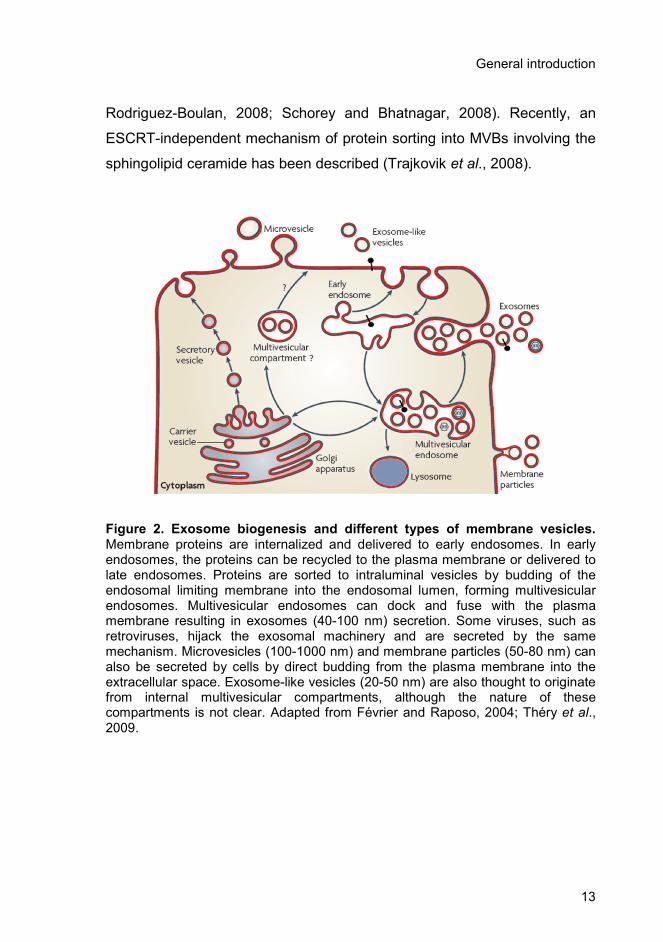
Figure 2. Exosome biogenesis and different types of membrane vesicles. Membrane proteins are internalized and delivered to early endosomes. In early endosomes, the proteins can be recycled to the plasma membrane or delivered to late endosomes. Proteins are sorted to intraluminal vesicles by budding of the endosomal limiting membrane into the endosomal lumen, forming multivesicular endosomes. Multivesicular endosomes can dock and fuse with the plasma membrane resulting in exosomes (40-100 nm) secretion. Some viruses, such as retroviruses, hijack the exosomal machinery and are secreted by the same mechanism. Microvesicles (100-1000 nm) and membrane particles (50-80 nm) can also be secreted by cells by direct budding from the plasma membrane into the extracellular space. Exosome-like vesicles (20-50 nm) are also thought to originate from internal multivesicular compartments, although the nature of these compartments is not clear. Adapted from Février and Raposo, 2004; Théry et al., 2009.
Chapter 1
14
In a final step, multivesicular endosomes can either fuse with
lysosomes for protein degradation or with the plasma membrane, resulting
in the release of exosomes (Figure 2). The mechanism that leads to
exosomes secretion, though still poorly understood, appears to involve two
Ras family monomeric proteins Rab27A and Rab27B (Ostrowski et al.,
2010).
In addition to exosomes other vesicles may be secreted from
mammalian cells and they have been classified based on their biogenesis
and physiochemical properties, namely diameter (Figure 2) (Théry et al.,
2009).
1.3.2. Composition
Exosomes contain a variety of molecules, including proteins, RNA
and lipids reflecting the cell type from which they originate and possibly
their functional role.
Although the protein content of exosomes varies depending on their
source, the proteomic analysis of exosomes from a wide variety of cells and
body fluids has identified several proteins that are well conserved and are
commonly used as exosomal markers. Nearly all exosomes contain
proteins involved in membrane transport and fusion (RabGTPases,
annexins, flotillin), in MVBs biogenesis (Alix, TSG101), metabolic enzymes
(peroxidases, pyruvate and lipid kinases) and cytoplasmic proteins (tubulin,
actin). Moreover, exosomes are enriched with heat shock proteins (hsp70
and 90), integrins and tetraspanins (CD63, CD9, CD81) (reviewed in
Simpon et al., 2008, Schorey and Bhatnagar, 2008; Simons and Raposo,
2009; Mathivanan and Simpson, 2010) and were shown to carry active
proteases, such as ADAM10 and ADAM17 (Stoeck et al., 2006).

General introduction
15
Recent studies have demonstrated that exosomes also carry and
deliver mRNA and small RNAs, including microRNA (Valadi et al., 2007;
Skog et al., 2008). A compendium of exosomes RNAs and proteins is
available at “Exocarta” (http://exocarta.ludwig.edu.au).
Moreover, exosomes display a specific lipid composition different
from the parental cells. They are particularly enriched in raft-lipids such as
cholesterol, sphingolipids, ceramide and glycerophospholipids with long
and saturated fatty-acyl chains (Subra et al., 2007; Trajkovik et al., 2008).
1.3.3. Function
Exosomes were originally observed as a mechanism to remove
unnecessary proteins, namely the transferrin receptor during reticulocytes
maturation (Johnstone et al., 1987). However, since then many other
functions have been proposed such as intercellular communication,
induction of immune responses, and transfer of infectious agents, among
several others.
The ability of exosomes to interact with cells has important
biological implications, such as cell-cell communication, and the delivery of
proteins and genetic material conferring new functional properties to the
recipient cells. Several types of interaction between exosomes and cells
have already been proposed based on indirect evidence and in vitro
studies. Exosomes can associate with the plasma membrane through
ligand-receptor interactions (Théry et al., 2009) or lipids, such as
phosphatidylserine (Keller et al., 2009). The process of internalization can
occur through direct fusion of the exosomes with the plasma membrane,
releasing the exosomal content into the cell cytoplasm. Alternatively,
exosomes can enter the cells by receptor-mediated endocytosis and later
fuse with the limiting membrane of the endosome releasing the exosomal

Chapter 1
16
content to be recycled to the cell surface or to be degraded in the lysosome
(Théry et al., 2009; Coccusi et al., 2009). Exosome uptake was shown to
occur via clathrin-mediated endocytosis in dendritic cells (Morelli et al.,
2004), as well as phagocytosis in monocytes and macrophages (Feng et
al., 2010).
One of the most studied roles of secreted exosomes is their
powerful effect on the immune system. Exosomes secreted from different
sources can have both an activating or a tolerogenic or even inhibitory
effect on immune cells. In general, exosomes produced by antigen-
presenting cells such as dendritic cells, macrophages and B cells are
immune activating. They activate directly or indirectly immune effector
mechanisms such as cytotoxicity, antibody and cytokine production, and
priming of T cells. On the other side, exosomes produced by epithelial cells
and the great majority of tumours are immune inhibitory, playing an
important role on tumour dissemination (Théry et al., 2009).
Exosomes can also participate in the propagation of infectious
agents. Février et al. showed that the prion protein and the scrapie forms of
the protein (PrPc and PrPsc, respectively) can be secreted in association
with exosomes (Février et al., 2005). Moreover, a role for exosomes in the
intercellular trafficking of human immunodeficiency virus (HIV-1) infectivity
has also been suggested (Gould et al., 2003; Wiley and Gummuluru, 2006;
Izquierdo-Useros et al., 2008).
Furthermore, exosomes have been proposed as a novel platform for
ADAM-mediated cleavage. The proteolysis of cell adhesion transmembrane
molecules like L1 and CD44 by ADAMs can occur in MVBs and exosomes,
in addition to the plasma membrane (Stoeck et al., 2006; Keller et al.,
2006).
General introduction
17
1.3.4. Tumour cell-derived exosomes
The biological significance of exosomes secretion by tumours, and
the presence of these vesicles in malignant effusions such as ascites, is not
clear. Tumours reported to release exosomes include cancers of the ovary,
breast, oral cavity, colorectum, brain, bladder, prostate, and melanomas
(reviewed in Zhang and Grizzle, 2011).
Tumour-secreted vesicles, including exosomes, can contribute to
tumour invasion and progression by transferring aggressive cancerous
phenotypes (Al-Nedawi et al., 2008), modifying the tumour
microenvironment (Castellana et al., 2009), conferring multi-drug resistance
(Shedden et al., 2003; Safaei et al., 2005), and promoting angiogenesis,
tumour growth and metastasis (reviewed in Muralidharan-Chari et al.,
2010).
Some reports point to a role of cancer exosomes in immune
activation (reviewed in Théry et al., 2009). Wolfers et al, (Wolfers et al.,
2001) showed that tumour-derived exosomes were able to transfer
antigens from tumour cells to dendritic cells and, therefore, function in
antigen-cross presentation on syngeneic and allogeneic established mouse
tumours. However, recent studies point to the increasing evidence that
cancer exosomes might instead suppress the immune system facilitating
tumour growth and invasion (reviewed in Valenti et al., 2007; Zhang and
Grizzle, 2011). This can occur through several mechanisms such as
induction of myeloid-derived suppressor cells (Iero et al., 2008) and
inhibition of dendritic cell maturation (Yu et al., 2007) or T cell function
(Clayton et al., 2007).
The role of exosomes in immune suppression and ovarian tumour
progression has been investigated. Ascites-derived exosomes induced
apoptosis of cells of the immune system (Peng et al., 2011) and resulted in
Chapter 1
18
the growth of an ovarian carcinoma in vivo, after systemic administration
(Keller et al., 2009). Moreover, exosomes derived from ovarian carcinoma
cell lines and patient’s ascites were shown to act as platforms for
ectodomain shedding, being an important source of soluble adhesion
molecules like L1 and CD44 that could regulate tumour cell function in an
autocrine/paracrine fashion (Gutwein et al., 2005; Stoeck et al., 2006), and
exhibited gelatinolytic activity, responsible for increasing tumour invasion
into the stroma (Runz et al., 2007. Exosomes derived from human ovarian
carcinoma cells have also been implicated in drug resistance, acting as a
mechanism of efflux for the drug cisplatin (Safaei et al., 2005).
1.3.5. Exosomes in diagnosis and therapeutics
The presence of exosomes in biological fluids such as, blood
plasma (Caby et al., 2005) and urine (Pisitkun et al., 2004), and the fact
that they can be easily collected using non-invasive methods holds
significant potential. Exosomes can be used for obtaining novel and
complex sets of biomarkers for the early detection and diagnosis of
diseases, for determining prognosis, and for therapeutic interventions such
as the development of vaccines.
Urinary exosomes have been extensively studied having in view the
discovery of potential disease biomarkers of bladder (Welton et al., 2010)
and prostate cancer (Nilsson et al., 2009). Similarly, exosomes collected
from blood plasma have also been investigated. MicroRNA profiling of
circulating exosomes was explored as a diagnostic marker for biopsy
profiling and to screen asymptomatic populations with ovarian (Taylor and
Gercel-Taylor, 2008) and lung cancer (Rabinowits et al., 2009). Blood
derived-exosomes of women with ovarian cancer contained high levels of
claudin-4, a membrane protein whose expression is elevated in ovarian

General introduction
19
cancer (Li et al., 2009), and exosomes derived from sera and malignant
ascites showed the presence of tumour progression related proteins (L1,
ADAM10, CD24, and EpCAM) (Gutwein et al., 2005; Runz et al., 2007).
Since both tumour and immune cells are able to secrete exosomes,
the application of these vesicles in immunotherapy has become a
promising area of research in cancer (reviewed in Tan et al., 2010),
infectious (Beauvillain et al., 2009) and autoimmune diseases (Kim et al.,
2005). The ability of exosomes released from dendritic cells, pulsed with
tumour antigens to stimulate T-cells and to induce the eradication of
tumours in mice (Zitvogel et al., 1998) has led to their use as therapeutic
agents for the stimulation of anti-tumoral immune responses. Exosomes
have already been used in phase I clinical trials with patients showing
advanced stage melanomas (Escudier et al., 2005), non-small cell lung
carcinomas (Morse et al., 2005), and colorectal cancer (Dai et al., 2008).
1.4. ADAM10
ADAM (A Disintegrin And Metalloprotease) 10 is a type I
transmembrane glycoprotein that belongs to the ADAMs family and to the
zinc-dependent metalloproteinases (metzincins) superfamily.
Mammalian ADAM10 was originally isolated from bovine brain,
based on its ability to cleave myelin based protein in in vitro preparations
(Chantry et al., 1989), but later work showed that it is widely expressed
among different tissues and several species.
The gene locus for human ADAM10 was matched to chromosome
15 at position 15q21.3-q23 (Yamazaki et al., 1997) and codes for a protein
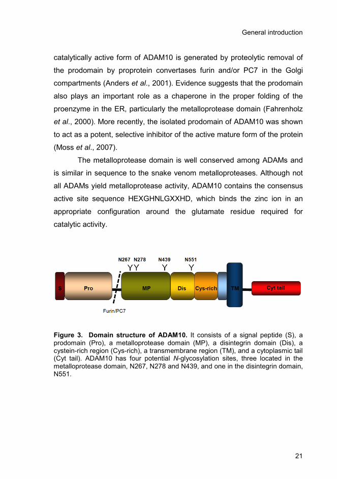
with 748 amino acids that has four potential N-glycosylation sites, N267,
N278, N439 and N551 (Figure 3).

Chapter 1
20
ADAM10 is synthesized in the rough endoplasmic reticulum and is
maturated in the late Golgi compartments. Subcellular localization analysis
revealed that human ADAM10 is mainly located at the plasma membrane
(Gutwein et al., 2003) but it also colocalizes with Golgi markers and it was
found in secreted vesicles identified as exosomes (Lammich et al., 1999;
Gutwein et al., 2003).
The major role of ADAM10 is the proteolytic cleavage of
transmembrane proteins with important physiological and pathological
roles. The severe phenotype of ADAM10-/- embryos implies that ADAM10
has a crucial role on early vertebrate embryogenesis. In fact, the knockout
of ADAM10 in mice is embryonic-lethal at 9.5 days and showed evidence of
pronounced defects of the developing central nervous system, somites, and
cardiovascular system due to disrupted Notch signalling (Hartmann et al.,
2002).
1.4.1. Structure
ADAM10 is characterized by a conserved domain structure common
to other ADAMs consisting of an N-terminal signal sequence that directs
the protein to the secretory pathway, followed by a prodomain, a
metalloprotease and disintegrin domains, a cystein-rich region, a
transmembrane domain, and finally an SH3-enriched cytoplasmic tail
(Wolfsberg et al., 1995) (Figure 3).
ADAMs structural domains have been shown to play specific roles.
ADAM10 prodomain keeps the metalloprotease inactive, through a “cystein
switch” mechanism. The Zn2+ is coordinated by three histidines in the
catalytic site, the free sulfhydryl group of the cystein residue provides a
fourth coordination site and inhibits the entrance of the water molecule
which is responsible for hydrolysis (Schlondorff and Blobel, 1999). The
General introduction
21
catalytically active form of ADAM10 is generated by proteolytic removal of
the prodomain by proprotein convertases furin and/or PC7 in the Golgi
compartments (Anders et al., 2001). Evidence suggests that the prodomain
also plays an important role as a chaperone in the proper folding of the
proenzyme in the ER, particularly the metalloprotease domain (Fahrenholz
et al., 2000). More recently, the isolated prodomain of ADAM10 was shown
to act as a potent, selective inhibitor of the active mature form of the protein
(Moss et al., 2007).
The metalloprotease domain is well conserved among ADAMs and
is similar in sequence to the snake venom metalloproteases. Although not
all ADAMs yield metalloprotease activity, ADAM10 contains the consensus
active site sequence HEXGHNLGXXHD, which binds the zinc ion in an
appropriate configuration around the glutamate residue required for
catalytic activity.
Figure 3. Domain structure of ADAM10. It consists of a signal peptide (S), a prodomain (Pro), a metalloprotease domain (MP), a disintegrin domain (Dis), a cystein-rich region (Cys-rich), a transmembrane region (TM), and a cytoplasmic tail (Cyt tail). ADAM10 has four potential N-glycosylation sites, three located in the metalloprotease domain, N267, N278 and N439, and one in the disintegrin domain, N551.
Chapter 1
22
ADAMs disintegrin and cystein-rich domains were described as
being directly involved in cell-cell adhesion processes through interactions
with integrins and other receptors (White, 2003). Although no disintegrin
activity has been reported for ADAM10, Janes et al. demonstrated that
these domains are essential for ephrinA5/EphA3 substrate recognition
(Janes et al., 2006). The formation of the ephrinA5/EphA3 complex creates
a high affinity binding site for the cysteine-rich domain of ADAM10 which
leads to a conformational switch that consents ADAM10 metalloprotease
domain to cleave EphA5 in trans.
ADAMs cytoplasmic tails were proposed to be involved in the
regulation of the metalloprotease activity, oligomerization, signalling and/or
control of maturation and subcellular localization. ADAMs tails contain
binding site motifs for SH3 domain-containing proteins and potential sites
for phosphorylation. There are a number of adaptor proteins such as
MAD2, Lck, Eve-1, PACSIN3 and synapse associated protein-97 with
binding capacity to the cytoplasmic tail of ADAM10 (reviewed in Edwards et
al., 2008). Moreover, a proline-rich stretch in the cytoplasmic domain of
ADAM10 has been shown to be important for the correct basolateral
localization of the protein in polarized epithelial cells promoting is
metalloprotease activity (Wild-bode et al., 2006). Similarly, the binding of
synapse associated protein-97 to ADAM10 cytoplasmic tail is also
responsible for the correct localization of ADAM10 in synaptic membranes
(Marcello et al., 2007).
1.4.2. Activity
The metalloprotease activity of ADAM10 is responsible for the
proteolytic cleavage of several transmembrane proteins and release of their
extracellular domain. This process is referred to as ectodomain shedding

General introduction
23
and occurs both constitutively and in response to a variety of stimuli,
including intracellular calcium concentration, phorbol esters and protein
kinase C activation (Pandiella and Massague, 1991; Reiss and Saftig,
2009).
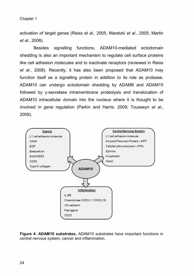
ADAM10 has more than 40 known protein substrates with important
roles in development, cell signalling and disease pathologies, such as
central nervous system disorders, inflammation and cancer (Figure 4)
(Pruessmeyer and Ludwig, 2009). ADAM10 substrates include the cell
adhesion molecule L1 (Mechtersheimer et al., 2001), pro-tumour necrosis
factor α (Lunn et al., 1997), type IV collagen (Millichip et al., 1998), amyloid
precursor protein (APP) (Lammich et al., 1999), ephrin-A2 (Hattori et al.,
2000), epidermal growth factor receptor (EGFR) (Yan et al., 2002), Notch
(Dallas et al., 1999; Hartmann et al., 2002), pro-heparin-binding epidermal
growth factor (Lemjabbar and Basbaum, 2002), fractalkine (Hundhausen et
al., 2003), CD44 (Nagano et al., 2004), N-cadherin (Reiss et al., 2005),
betacellulin (Sanderson et al., 2005), and the low-affinity immunoglobulin E
receptor CD23 (Weskamp et al., 2006), among others.
ADAM10 mediated proteolysis leads to the release of soluble
factors like growth factors and chemokines, and is associated with
extracellular signalling events. The released peptides can bind to their
receptors and mediate signals in an autocrine or paracrine fashion.
Moreover, ADAM10 mediated shedding can also play a role in intracellular
signalling by regulated intramembrane proteolysis (RIP) (Brown et al.,
2000). The ectodomain shedding of transmembrane proteins can promote
further proteolysis of the remaining transmembrane fragment by the γ-
secretase complex, for type I transmembrane proteins, or by the signal
peptide peptidase like proteins (SPPLs), for type II transmembrane
proteins, leading to the intracellular release of soluble substrate fragments,
which might be translocated into the nucleus leading to the transcriptional
Chapter 1
24
activation of target genes (Reiss et al., 2005, Maretzki et al., 2005; Martin
et al., 2008).
Besides signalling functions, ADAM10-mediated ectodomain
shedding is also an important mechanism to regulate cell surface proteins
like cell adhesion molecules and to inactivate receptors (reviewed in Reiss
et al., 2008). Recently, it has also been proposed that ADAM10 may
function itself as a signalling protein in addition to its role as protease.
ADAM10 can undergo ectodomain shedding by ADAM9 and ADAM15
followed by γ-secretase intramembrane proteolysis and translocation of
ADAM10 intracellular domain into the nucleus where it is thought to be
involved in gene regulation (Parkin and Harris, 2009; Tousseyn et al.,
2009).
Figure 4. ADAM10 substrates. ADAM10 substrates have important functions in central nervous system, cancer and inflammation.

General introduction
25
The factors that determine the substrate selectivity of ADAM10 are
still not fully understood. Substrates have highly variable cleavage sites and
there are no identified cleavage sequences. Moreover, many of ADAM10
substrates are also cleaved by other proteases, such as ADAM17 that
shares high homology both in sequence and in substrate specificity with
ADAM10. There is evidence that localization of both enzyme and substrate
at particular subcellular microdomains is important for recognition and
cleavage (Tellier et al., 2006). The secondary structure of the substrate
justamembrane stalk is also an important factor for recognition (Seals and
Courtneidge, 2003) and recent studies suggest that ADAM10 recognize
preferentially cleavage sequences with large residues, primarily leucine, but
also aromatic residues, while ADAM17 prefers smaller aliphatic residues
(Caescu et al., 2009).
Regulation of ADAMs activity might occur at different levels, such as
transcriptional control, alternative splicing, post-translational modifications,
interaction with other proteins, cellular localization, and substrate
availability (reviewed in Huovilla et al., 2005). Furthermore, ADAM10
activity can also be inhibited by tissue inhibitors of matrix
metalloproteinases (TIMPs) 1 and 3 (Amour et al., 2000). More recently,
synthetic inhibitors have also been developed mainly to be used as
therapeutic agents to block ADAM10 activity in cancer (Moss et al., 2008).
1.4.3. Role in cancer
The sheddase activity of ADAM10 plays an important role in cellular
signalling pathways and is critical for the survival of cancer cells. ADAM10
was found upregulated in several tumours, such as uterine, and ovarian
carcinomas (Fogel et al., 2003), human haematological malignancies (Wu
et al., 1997), neuroblastomas (Yavari et al., 1998), prostate cancer

Chapter 1
26
(McCulloch et al., 2004), gastric carcinoma cells (Tanida et al., 2004), colon
cancers (Gavert et al., 2005) and oral squamous cell carcinoma (Ko et al.,
2005).
ADAM10 contributes to tumorigenesis and metastasis through
cleavage and activation of several membrane proteins such as growth
factors and cell adhesion molecules, and by regulating the adhesion and
motility of cells, as a result of its role on extracellular matrix degradation
(reviewed in Reiss et al., 2006). ADAM10-mediated L1 release was
reported to enhance tumour dissemination by increasing cell migration in
ovarian and uterine carcinomas (Fogel et al., 2003). Moreover the
ectodomain shedding of cell adhesion molecules like L1 and CD44 can
enable cells to escape from the surrounding tissue and mediate cell
signalling that can promote cell proliferation, survival, and motility (Gutwein
et al., 2003; Nagano et al., 2004).
As mentioned before, ADAM10 can also be found in secreted
vesicles, identified as exosomes (Gutwein et al., 2003). Moreover, ADAM10
present in exosomes from ovarian carcinoma cells and ascites fluid was
found to be functionally active and able to cleave both L1 and CD44
(Stoeck et al., 2006). These findings suggest that exosomes serve as a
major platform for ectodomain shedding and as a vehicle for the export of
soluble molecules that might have a role on tumour progression.
Since ectodomain shedding has been implicated as a pathological
mechanism in cancer, ADAM10 inhibition has been explored as a target for
cancer therapeutic drugs. Several inhibitors for ADAM10 have been
described, but the lack of selectivity has contributed to adverse side effects
and to the failure in clinical trials (reviewed in Reiss et al., 2006). Moreover,
ADAM10 inhibition could have a profound negative effect in situations
where ADAM10-mediated cleavage has a protective role, for example in
Alzheimer’s disease where ADAM10 cleavage of APP leads to a decrease
in amyloide plaque formation.
General introduction
27
1.5. Aims of this Thesis work
Exosomes are small membrane vesicles that are secreted by
several cell types, including ovarian tumour cells, and have been found in
biological fluids such as blood and malignant ascites. They can contribute
to tumour invasion and progression by several mechanisms, and they are
potential targets for the discovery of new disease biomarkers and for
therapeutic interventions such as the development of vaccines.
Glycosylation is one of the most important post-translational
modifications and alterations in cell surface glycosylation are often related
to malignant transformation.
To date, little was known about the N-glycosylation profile of
glycoproteins that are sorted to exosomes. The major goal of this work was
to characterize and investigate the biological role of N-glycosylation from
proteins present in exosomes of ovarian tumour cell lines. Furthermore, the
glycoprotein metalloprotease ADAM10 has been studied in detail.
ADAM10 is a transmembrane protein with four potential N-
glycosylation sites that is found in exosomes secreted from ovarian
carcinoma cell lines. The first objective of this thesis work was to mutate
each individual N-glycosylation site of ADAM10 and to understand the role
played by N-glycosylation on ADAM10 intracellular localization, in vivo and
in vitro activities. Furthermore, the presence of wild-type and mutant forms
of ADAM10 in exosomes from ovarian carcinoma cells was also monitored,
in order to evaluate if N-glycosylation site occupancy was required for
ADAM10 sorting to exosomes.
The mechanisms of exosomes interaction and uptake by target cells
are still poorly understood. Therefore, the second objective consisted of
exploring the mechanisms of internalization of labelled exosomes by
SKOV3 ovarian tumour cells. Several endocytic pathways were explored
Chapter 1
28
using endocytosis inhibitors and colocalization studies by
immunofluorescence microscopy. Furthermore the roles of cell surface
proteins and exosomes surface proteins, as well as, oligosaccharides were
investigated in the uptake. In addition, the glycosylation profile of the
exosomes was analysed and compared with total cellular extracts using
lectins with different specificities.
Since the exosomes had specific glycosylation patterns, as detected
with lectins, the third objective consisted of the detailed structure analysis
of the N-glycans from the secreted vesicles of SKOV3 and OVM ovarian
carcinoma cell lines, as well as plasma membrane and microsomal
fractions. The techniques used were HPAEC-PAD and MALDI-TOF-MS.
Chapter 2
Functional role of N-glycosylation from ADAM10
in processing, localization and activity of the
enzyme
The majority of the work presented in this chapter has been included in the
following manuscript:
Escrevente C, Morais VA, Keller S, Soares CM, Altevogt P, Costa J (2008).
Functional role of N-glycosylation from ADAM10 in processing, localization and
activity of the enzyme. Biochim Biophys Acta. 1780: 905-13.

Chapter 2
30

Functional role of ADAM10 N-glycosylation
31
2. Functional role of N-glycosylation from ADAM10 in
processing, localization and activity of the enzyme
2.1. Summary
A disintegrin and metalloprotease 10 (ADAM10) is a type I
transmembrane glycoprotein with four potential N-glycosylation sites (N267,
N278, N439, and N551) that cleaves several plasma membrane proteins. In
this work, ADAM10 was found to contain high mannose and complex
glycans. Individual N-glycosylation site mutants S269A, T280A, S441A,
T553A were constructed, and results indicated that all sites were occupied.
T280A was found to accumulate in the endoplasmic reticulum as the non-
processed precursor of the enzyme. Furthermore, it exhibited only residual
levels of metalloprotease activity in vivo towards the L1 cell adhesion
molecule, as well as in vitro, using a proTNF-alpha peptide as substrate.
S441A showed increased ADAM10 susceptibility to proteolysis. Mutation of
N267, N439, and N551 did not completely abolish enzyme activity,
however, reduced levels were found.
ADAM10 is sorted into secretory vesicles, the exosomes. Here, a
fraction of ADAM10 from exosomes was found to contain more processed
N-linked glycans than the cellular enzyme.
In conclusion, N-glycosylation is crucial for ADAM10 processing and
resistance to proteolysis, and results suggest that it is required for full-
enzyme activity.
Chapter 2
32
2.2. Introduction
ADAM (a disintegrin and metalloprotease) 10 is a type I
transmembrane glycoprotein that belongs to the ADAMs protein family. It is
characterized by a conserved domain structure, consisting of an N-terminal
signal sequence that directs the protein to the secretory pathway followed
by a prodomain, a metalloprotease, a disintegrin domain, a cystein-rich
region, a transmembrane domain, and an SH3- enriched cytoplasmic tail
(Wolfsberg et al., 1995). In order to be catalytically active ADAM10
prodomain has to be cleaved by proprotein convertases furin and/or PC7 in
the Golgi compartments (Anders et al., 2001; Schlondorff and Blobel, 1999;
Seals and Courtneidge, 2003).
ADAM10 metalloprotease domain is known to mediate the
proteolytic cleavage of transmembrane proteins in their juxtamembrane
region resulting in the release of the extracellular domain as a soluble form
(shedding). ADAM10 can cleave a variety of proteins with importance in
development, cell signalling and disease, such as the cell adhesion
molecule L1 (Mechtersheimer et al., 2001), pro-tumour necrosis factor-
alpha (Lunn et al., 1997), type IV collagen (Millichip et al., 1998), amyloid
precursor protein (Lammich et al., 1999), ephrin-A2 (Hattori et al., 2000),
epidermal growth factor receptor (Yan et al., 2002), notch (Dallas et al.,
1999; Hartmann et al., 2002), pro-heparin-binding epidermal growth factor
(Lemjabbar and Basbaum, 2002), fractalkine (Hundhausen et al., 2003),
CD44 (Nagano et al., 2004), N-cadherin (Reiss et al.; 2005), betacellulin
(Sanderson et al., 2005), and low-affinity immunoglobulin E receptor CD23
(Weskamp et al., 2006), among others. Moreover, ADAM10 is
overexpressed in tumours, such as uterine and ovarian carcinomas (Fogel
et al., 2003), human haematological malignancies (Wu et al., 1997),
neuroblastomas (Yavari et al., 1998), prostate cancer (McCulloch et al.,

Functional role of ADAM10 N-glycosylation
33
2004), gastric carcinoma cells (Tanida et al., 2007), colon cancers (Gavert
et al., 2005), and oral squamous cell carcinoma (Ko et al., 2007),
suggesting a role in tumour progression and dissemination.
ADAMs disintegrin and cystein-rich domains were described as
being directly involved in cell-cell adhesion processes through interactions
with integrins and other receptors (White, 2003).
ADAM10 can be found in various cellular compartments. It has been
shown to colocalize with Golgi markers and was found on the cell surface
(Lammich et al., 1999; Gutwein et al., 2003). More recently, it was also
detected in secreted vesicles identified as exosomes (Gutwein et al., 2003).
Exosomes are small membrane vesicles (30-100 nm diameter) secreted by
various cell types as a consequence of fusion of multivesicular late
endosomes/lysosomes with the plasma membrane. Exosomes secretion
has first been reported in a variety of cells, including cancer cells, neurons,
B lymphocytes, T lymphocytes, dendritic cells, mast cells, and platelets
(Caby et al., 2005). Exosomes secreted from tumour cells could promote
cellular invasion and migration during metastasis (Stoeck et al., 2006).
ADAM10, like other ADAMs, is N-glycosylated. It has four potential
N-glycosylation sites (N-X-S/T, X≠Pro), three located in the metalloprotease
domain (N267, N278, and N439) and one in the disintegrin domain (N551).
Glycosylation is one of the most important post-translational modifications
in newly synthesized proteins which may influence the physicochemical
and biological properties of glycoproteins. N-glycosylation has been related
with protein folding and quality control of glycoproteins in the endoplasmic
reticulum with consequences for processing and trafficking (reviewed in
Helenius and Aebi, 2004; Morais et al., 2003), targeting in the secretory
pathway, namely to the lysosome (via the mannose 6-phosphate receptor)
(Hauri et al., 2000), dynamics of plasma membrane proteins (Cai et al.,
2005; Brooks et al., 2006; Turner et al., 2006), cell-cell interactions, e.g.,
Chapter 2
34
during metastases formation (Dube et al., 2005), or inflammation (Dube et
al., 2005; Lowe et al., 2003), among others.
In cancer, aberrant glycosylation, particularly of cell surface
glycoconjugates, is a hallmark associated with the disease (Dube et al.,
2005). Altered glycosylation is the consequence of changes in
glycosyltransferase expression and localization along the secretory
pathway and also depends on the availability of sugar nucleotide donors in
the Golgi lumen.
In the present work, the four potential N-glycosylation sites of
bADAM10 were found to be occupied. By mutating each of those sites we
observed that glycans from N278 were crucial for the intracellular
processing of bADAM10, with the enzyme being accumulated as the
precursor in the endoplasmic reticulum. Concerning N439, the N-glycans
protected against proteolytic cleavage. N-glycans from each mutant were
required for full in vivo activity. On the other hand, a fraction of human
endogenous ADAM10 from exosomes was found to contain more
processed N-linked glycans than the cellular counterpart.
2.3. Materials and methods
2.3.1. DNA constructs
The plasmid pcDNA3-ADAM10wt (Mechtersheimer et al., 2001)
coding for wild-type bovine ADAM10 with the HA tag at the C-terminus was
used as template for the construction of the N-glycosylation mutants:
pbADAM10S269A, pbADAM10T280A, pbADAM10S441A, and
pbADAM10T553A. Site-directed mutagenesis was done using
Functional role of ADAM10 N-glycosylation
35
QuickChange cloning techniques (Stratagene, La Jolla, CA, USA), following
the manufacturer's instructions. The serine or threonine from the N-
glycosylation site (N-X-S/T) were replaced by an alanine residue following a
strategy previously used (Morais et al., 2003; Morais et al., 2006).
The PCR reactions were carried out with 50-115 ng pcDNA3-
ADAM10wt as template, 2.5 mM dNTPs mix, 2.5 U/µl Pfu Turbo DNA
Polymerase and 10 µM of mutagenic primers. The primers used to obtain
pbADAM10S269A, pbADAM10T280A, pbADAM10S441A, and
pbADAM10T553A were, respectively as follows: 5′GGA ATT CGT AAC ATC
GCT TTC ATG GTG AAA CGC ATA AG3′, 5′GC ATA AGA ATC AAC ACA
GCT GCT GAT GAG AAG G3′, 5′GT AGT ATT AGA AAT ATA GCT CAA
GTT CTT GAG AAG AAG3′, 5′GAT CCT AAA CCG AAC TTC GCA GAC
TGT AAT AGA CAT ACG3′. Standard molecular biology techniques were
used for PCR product restriction and analysis. Mutations of the bADAM10
N-glycosylation sites were confirmed by automatic DNA sequencing
analysis.
2.3.2. Cell culture and protein expression
Human ovarian cancer SKOV3 cells, human embryonic kidney
HEK293 cells and mouse embryonic fibroblasts (MEFs) were grown at 37
°C, 5% CO2 in Dulbecco's Modified Eagle Medium (DMEM) (Sigma)
supplemented with 10% fetal calf serum (Gibco), 1% penicillin/streptomycin
solution (Gibco).
SKOV3 and HEK293 were transfected with each of the plasmids
indicated in 2.3.1 or pcDNA3-L1 by the calcium phosphate technique using
5 µg of plasmid DNA and selection of stably-transfected cells was
performed using 2 and 1 mg/ml geneticin sulphate G-418 (Gibco),
respectively. For transient expression, HEK293 cells and MEFs were
Chapter 2
36
transfected with 2 or 2.5 µg of plasmid DNA, respectively, using
Lipofectamine 2000 (Invitrogen), following manufacturer's instructions.
2.3.3. Isolation of membrane vesicles
Confluent SKOV3 cells were cultivated for 24 h in serum-free
medium. When indicated, cells were incubated with 10 µM TAPI-0
(Calbiochem) for 16 h. Supernatant was collected and centrifuged, at 4 °C,
for 10 min, at 500 × g followed by 20 min at 10,000 × g to remove dead
cells and cell debris, respectively. Membrane vesicles were collected by
centrifugation at 100,000 × g, for 2 h, at 4 °C.
For sucrose-density fractionation, membrane vesicles resuspended
in 0.25 M sucrose were loaded on the top of a step gradient comprising
different sucrose concentrations: 2, 1.3, 1.16, 0.8, 0.5, and 0.25 M as
previously described (Stoeck et al., 2006). The gradients were centrifuged
at 100,000 × g for 2.5 h. Twelve 1 ml fractions were collected from the top
of the gradient and precipitated with chloroform/methanol (1:4, v/v).
Samples were solubilized in sample buffer with or without β-
mercaptoethanol for SDS-PAGE and Western blot analysis.
2.3.4. In vivo cleavage assay
HEK293 cells were stably-transfected with pcDNA3-L1 (HEK-L1),
and the selected stable cell lines were then transiently-transfected with
pcDNA3-ADAM10wt or one of the N-glycosylation mutants. MEFs were
transiently-double transfected with pcDNA3-L1 and pcDNA3.1-LacZ,
pcDNA3-ADAM10wt, or one of the N-glycosylation mutants. Forty-eight
hours after transfection, supernatant corresponding to a 24 h-production in

Functional role of ADAM10 N-glycosylation
37
serum-depleted medium, was centrifuged at 500 x g, 10 min and 10.000 x
g, 20 min. When indicated, MEFs were incubated with the ADAM17
inhibitor Bristol Meyer Squib (BMS) (kindly provided by Dr. J. Arribas) for 24
h in serum-depleted medium. Cell supernatant was analysed by Western
blot.
2.3.5. SDS-PAGE and Western blot analysis
Total protein lysates were obtained by solubilization of centrifuged
cells in RIPA buffer (50 mM Tris-HCl pH 7.5, 150 mM NaCl, 0.1% SDS, 1%
sodium deoxycholate, 1% Triton X-100, 1:50 complete protease inhibitors
cocktail (Roche), for 10 min. Protein was precipitated with ethanol and
solubilized in reducing SDS-PAGE sample buffer. Endogenous hADAM10
analysis was performed in non-reducing SDS-PAGE. For SDS-PAGE 7.5%
acrylamide gels or pre-casted 4-12% acrylamide Bis-Tris gels (Invitrogen)
were used. Western blot was performed using the following primary
antibodies: rabbit anti-ADAM10 antibody AB387 (1:1000) raised against
amino acids 732-748 of ADAM10 of human origin produced and purified by
Davids Biotechnologies, mouse anti-ADAM10 monoclonal antibody
MAB1427 (1:1000) (R&D), mouse anti-HA tag 6E2 monoclonal antibody
(1:1000) (Cell Signalling), mouse anti-L1 11A (1:3), mouse antihuman CD9
(1:5000), mouse anti-annexin I monoclonal antibody (1:5000) (BD
Biosciences), and mouse anti-ubiquitin P4D1 monoclonal antibody (1:1000)
(Santa Cruz Biotechnology). Secondary antibodies were sheep anti-mouse
IgG coupled to HRP (1:4000) or donkey anti-rabbit IgG coupled to HRP
(Amersham) (1:3000). Antibody detection was accomplished with ECL Plus
Western Blotting Detection Reagent (GE Healthcare).

Chapter 2
38
2.3.6. Protein deglycosylation
For glycosidase hydrolysis cell lysates (approximately 1x10⁶ cells)
were incubated with endoglycosidase H (Endo H) (New England Biolabs)
and peptide: N-glycosidase F (PNGase F) (New England Biolabs or
Roche), according to supplier's protocol. Briefly, cell lysates were
denatured in glycoprotein denaturing buffer (0.5% SDS with or without 1%
β-mercaptoethanol) at 100 °C for 10 min, and incubated with
deglycosylation enzyme (PNGase F or Endo H) in the corresponding buffer.
Hydrolysis was carried out at 37 °C for 3 h or overnight. The samples were
further analyzed by SDS-PAGE and Western blot.
2.3.7. Immunoprecipitation
Exosomes were lysed with BOG buffer (20 mM Tris-HCl pH 8.0, 50
mM β-octylglucopyranoside (BOG), 1 mM phenylmethylsulphonylfluoride
(PMSF), 10 mM sodium fluoride, 10 mM sodium orthovanadate, 1:50
complete protease inhibitors cocktail (Roche) for 30 min at 4 °C with
occasional vortexing.
Pre-cleared lysates were incubated for 1 h at 4 °C with a rabbit anti-
HA.11 polyclonal antibody (Covance) previously coupled to protein A/G
Plus agarose beads (Santa Cruz Biotechnologies). Immunoprecipitated
proteins were washed three times with BOG buffer and were eluted at 100
°C for 5 min with sample buffer. Samples were analysed by SDS-PAGE
and Western blot.
Functional role of ADAM10 N-glycosylation
39
2.3.8. Confocal immunofluorescence microscopy
Cells were grown on glass coverslips until they reached 80%
confluence, they were washed with PBS containing 0.5 mM MgCl2, fixed
with 4% paraformaldehyde and permeabilized with methanol/acetone (1:1)
solution for 10 min at -20 °C. Blocking was done with 1% BSA in PBS for 1
h and primary and secondary antibodies were incubated at room
temperature for 2 and 1 h, respectively. Antibody solutions were prepared
in blocking solution and washes were performed with PBS. Coverslips were
mounted in Airvol and examined on a Leica Confocal (SP2+AOBS)
microscope. Primary antibodies were: rabbit anti-HA.11 (1:1000)
(Covance), goat IgG anti-calnexin C-20 (1:500) (Santa Cruz), mouse IgG
anti-ERGIC53 (1:1000) (Alexis Biochemicals), mouse IgG anti-GM130
(1:200) (BD Biosciences), mouse IgG anti-EEA1 (1:100) (BD Biosciences),
and mouse IgG anti-LAMP1 H4A3 (1:100) (BD Biosciences).
Secondary antibodies were: donkey anti-rabbit IgG AlexaFluor 594
(1:500), donkey anti-goat IgG AlexaFluor 488 (1:500), and donkey anti-
mouse IgG AlexaFluor 488 (1:500) (Molecular Probes).
For each picture, laser intensities and amplifier gains were adjusted
in order to avoid pixel saturation. Each fluorophore used was excited
independently and sequential detection was performed. Each picture
consisted of a z-series of 20 images of 1024-1024 pixel resolution with a
pinhole of 1.0 airy unit. Colocalization analysis was performed using the
open source Image J version 1.37 (http://rsb.info.nih.gov/ij/).

Chapter 2
40
2.3.9. Peptide cleavage assay
ADAM10-HA immunoprecipitates in 90 µl were incubated with 10 µl
(final concentration 50 mM) fluorescent quenching peptide Abz-Leu-Ala-
Gln-Ala-Val-Arg-Ser-Ser-Ser-Arg-Dap(dnp)-NH2 (Peptide International,
Louisville, Kentucky, USA). Cleavage was followed in a fluorescent plate
reader (Fluoroskan Ascent FL Thermo-Electron Corporation) using filters
for excitation at 320 nm and emission at 405 nm.
2.3.10. ADAM10 structure
Comparative modelling methods (Sali et al., 1993) were used to
derive the structure of the metalloprotease domain of bovine ADAM10 on
basis of the same domain from human ADAM17, the closest homologue
with three-dimensional structure determined (Maskos et al., 1998) (PDB
code 1BKC). These two domains have 39% identity, which should be
enough for deriving a model for ADAM10 with reasonable quality (Sanchez
et al., 1997).
The program MODELLER (Sali et al., 1993) version 7.7 was used in
the comparative modelling procedures. Resulting structures were analysed
with PROCHECK (Laskowski et al., 1993). A large number of models were
calculated, and the one having the lowest value of the objective function
was selected. Problems on the alignment were corrected and new models
were calculated in an iterative manner. A Ramachandran plot analysis of
the final model showed it to have 87.0% residues in most favored regions,
12.5% in additional allowed regions and 0.5% (1 residue) in disallowed
regions. This residue is Cys 344, which is involved in a disulphide bridge
with Cys 451, and corresponds to a cystein residue in the ADAM17

Functional role of ADAM10 N-glycosylation
41
template (Cys 365) that is also located in a disallowed zone of the
Ramachandran plot.
2.4. Results
2.4.1. The N-glycosylation sites of hADAM10 are occupied
ADAM10 contains four potential N-glycosylation sites in the mature
form of the protein (Figure 5). Three of these sites are located in the
metalloprotease domain: N267, N278 and N439 and one in the disintegrin
domain: N551. Endogenous human ADAM10 from SKOV3 ovarian
carcinoma cells appeared as a major band with an apparent molecular
mass of approximately 68 kDa (Figure 6, A). By enzymatic digestion with
Endo H, that removes N-linked glycans of the high mannose and hybrid
type, we observed that the hADAM10 mature form underwent a downward
shift of approximately 5 kDa. On the other hand, hydrolysis with PNGase F,
that removes both high mannose, hybrid, and complex type N-linked
glycans, reduced the molecular mass by 9 kDa to the expected value for
the hADAM10 core protein without the prodomain and with no post-
translational modifications (59 kDa) (Figure 6, A). These results indicated
that ADAM10 contained high mannose as well as complex N-glycans.
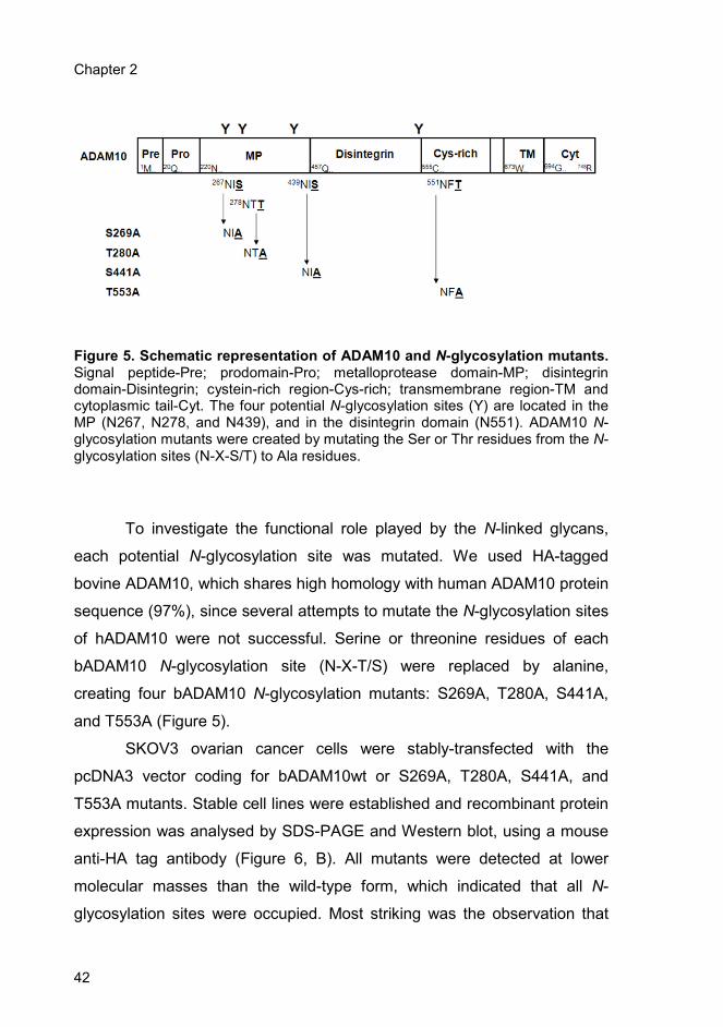
Chapter 2
42
Figure 5. Schematic representation of ADAM10 and N-glycosylation mutants. Signal peptide-Pre; prodomain-Pro; metalloprotease domain-MP; disintegrin domain-Disintegrin; cystein-rich region-Cys-rich; transmembrane region-TM and cytoplasmic tail-Cyt. The four potential N-glycosylation sites (Y) are located in the MP (N267, N278, and N439), and in the disintegrin domain (N551). ADAM10 N-glycosylation mutants were created by mutating the Ser or Thr residues from the N-glycosylation sites (N-X-S/T) to Ala residues.
To investigate the functional role played by the N-linked glycans,
each potential N-glycosylation site was mutated. We used HA-tagged
bovine ADAM10, which shares high homology with human ADAM10 protein
sequence (97%), since several attempts to mutate the N-glycosylation sites
of hADAM10 were not successful. Serine or threonine residues of each
bADAM10 N-glycosylation site (N-X-T/S) were replaced by alanine,
creating four bADAM10 N-glycosylation mutants: S269A, T280A, S441A,
and T553A (Figure 5).
SKOV3 ovarian cancer cells were stably-transfected with the
pcDNA3 vector coding for bADAM10wt or S269A, T280A, S441A, and
T553A mutants. Stable cell lines were established and recombinant protein
expression was analysed by SDS-PAGE and Western blot, using a mouse
anti-HA tag antibody (Figure 6, B). All mutants were detected at lower
molecular masses than the wild-type form, which indicated that all N-
glycosylation sites were occupied. Most striking was the observation that
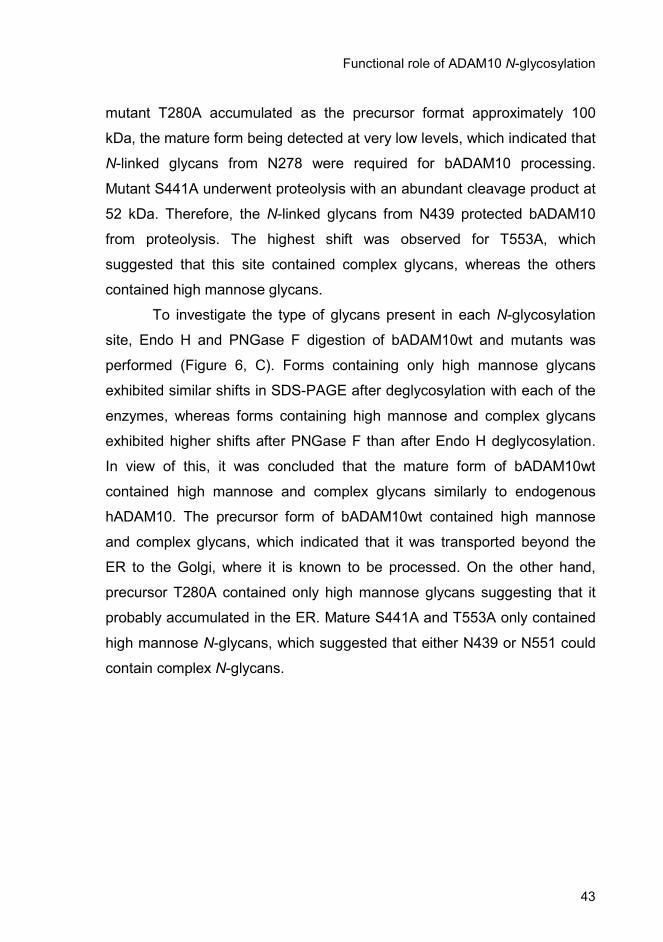
Functional role of ADAM10 N-glycosylation
43
mutant T280A accumulated as the precursor format approximately 100
kDa, the mature form being detected at very low levels, which indicated that
N-linked glycans from N278 were required for bADAM10 processing.
Mutant S441A underwent proteolysis with an abundant cleavage product at
52 kDa. Therefore, the N-linked glycans from N439 protected bADAM10
from proteolysis. The highest shift was observed for T553A, which
suggested that this site contained complex glycans, whereas the others
contained high mannose glycans.
To investigate the type of glycans present in each N-glycosylation
site, Endo H and PNGase F digestion of bADAM10wt and mutants was
performed (Figure 6, C). Forms containing only high mannose glycans
exhibited similar shifts in SDS-PAGE after deglycosylation with each of the
enzymes, whereas forms containing high mannose and complex glycans
exhibited higher shifts after PNGase F than after Endo H deglycosylation.
In view of this, it was concluded that the mature form of bADAM10wt
contained high mannose and complex glycans similarly to endogenous
hADAM10. The precursor form of bADAM10wt contained high mannose
and complex glycans, which indicated that it was transported beyond the
ER to the Golgi, where it is known to be processed. On the other hand,
precursor T280A contained only high mannose glycans suggesting that it
probably accumulated in the ER. Mature S441A and T553A only contained
high mannose N-glycans, which suggested that either N439 or N551 could
contain complex N-glycans.
Chapter 2
44
Figure 6. Western blot analysis of ADAM10 and N-glycosylation mutants from SKOV3 cells. (A) Deglycosylation of endogenous hADAM10 (1x10
6
cells/lane) with Endo H and PNGase F. Analysis was performed under non-reducing conditions using a mouse anti-ADAM10 antibody MAB1427. (B) Analysis of cells stably transfected with either bADAM10wt or N-glycosylation mutants under reducing conditions using a mouse anti-HA tag antibody. (C) Deglycosylation of bADAM10wt and bADAM10 N-glycosylation mutants with Endo H and PNGase F. Precursor (p) or mature (m) ADAM10 are indicated with closed arrowheads. Corresponding deglycosylated bands are shown with open arrowheads and the asterisk indicates bADAM10 cleavage product.
2.4.2. Intracellular localization of bADAM10 N-glycosylation
mutants
In agreement with previous reports (Dallas et al., 1999; Reiss et al.,
2005), the analysis of the localization of endogenous hADAM10 by confocal
microscopy of SKOV3 ovarian carcinoma cells showed predominant
localization of the protein at the perinuclear region and at the cell surface
Functional role of ADAM10 N-glycosylation
45
where it appeared as punctuated dots evenly distributed at the plasma
membrane (results not shown).
Since it has been reported that removal of N-glycosylation sites from
proteins can affect protein transport and intracellular localization (Martina et
al., 1998), bADAM10wt and mutants were colocalized with markers of the
secretory pathway using confocal immunofluorescence microscopy. The
markers used were: endoplasmic reticulum (ER) - calnexin, ER - Golgi
intermediate compartment (ERGIC) - ERGIC53, Golgi - GM130, early
endosomal antigen-1 - EEA1, and late endosome/lysosome-associated
membrane protein 1 - LAMP1 (Figure 7).
Bovine ADAM10wt colocalized with the EEA1 marker (Figure 7, D),
which indicated that it was present in the endosomes. Low levels of protein
were also found to colocalize with calnexin (Figure 7, A), and to a lower
extent with GM130 (Figure 7, C) and ERGIC53 (Figure 7, B), probably
corresponding to protein in traffick through the ER, Golgi and ERGIC. No
colocalization with LAMP1 was observed (Figure 7, E). All mutants, with the
exception of T280A, showed a similar staining profile (results not shown).
T280A mutant showed higher colocalization with calnexin (Figure 7, F).
These results showed that this mutant was retained in the ER and agreed
with T280A precursor accumulation and its sensitivity to both Endo H and
PNGase F.
Moreover, membrane protein biotinylation confirmed that most
mature hADAM10 and bADAM10wt mature forms were localized at the
plasma membrane. All N-glycosylation mutants showed comparable
expression levels, at the cell surface, compared with bADAM10wt, with the
exception of T280A that was almost undetectable (results not shown).

Chapter 2
46
Figure 7. Colocalization of bADAM10wt and T280A mutant, with markers of the secretory pathway by confocal immunofluorescence microscopy. SKOV3 cells stably-transfected with bADAM10wt (red) were fixed in 1:1 methanol/acetone and double labelled with rabbit anti-HA tag antibody and one of the following markers (green): (A) goat anti-calnexin; (B) mouse anti-ERGIC53; (C) mouse anti-GM130; (D) mouse anti-EEA1 or (E) mouse anti-LAMP1. (F) T280A mutant (red) was double labelled with rabbit anti-HA tag antibody and goat anti-calnexin (green). Secondary antibodies were donkey anti-rabbit IgG AlexaFluor 594, donkey anti-mouse IgG AlexaFluor 488 and donkey anti-goat IgG AlexaFluor 488. Scale bars, 10 µm.
Functional role of ADAM10 N-glycosylation
47
2.4.3. In vivo and in vitro activity of bADAM10wt and N-
glycosylation mutants
The shedding activity of bADAM10wt and glycosylation mutants
towards the L1 cell adhesion molecule was investigated. L1 is a 220 kDa
type I membrane glycoprotein of the immunoglobulin family that is
overexpressed in many human tumour cell lines, such as neuroblastomas,
melanomas (Fogel et al., 2003), colon (Gavert et al., 2005), and ovarian
carcinomas (Fogel et al., 2003), as well as in neural, hematopoietic, and
certain epithelial cells. L1 cleavage by ADAM10 occurs in intracellular
compartments, predominantly at the plasma membrane, and exosomes,
and results in two distinct fragments: a fragment of approximately 32 kDa
that remains attached to the cell membrane and a soluble fragment of
approximately 200 kDa that is released and can be detected in the
supernatant.
HEK293 cells were selected for this study since they show low
expression levels of endogenous hADAM10 and L1, and almost no L1
shedding (Figure 8, Ctr). Therefore, HEK293 cells were stably transfected
with pcDNA3-L1 (HEK-L1), and the selected stable cell lines were then
transiently transfected with bADAM10wt and glycosylation mutants. Forty
eight hours after transfection, L1 from the cell supernatants corresponding
to a 24 h-production in serum depleted medium, was analyzed by Western
blot and a representative blot is shown (Figure 8, upper panel). As controls
L1 and bADAM10 from the corresponding cell extracts were also analyzed
by Western blot. Mature full-length L1 probably at the plasma membrane
was detected (Figure 8, middle panel). Bovine ADAM10wt and mutants
were detected as mature form and precursor (Figure 8, lower panel).
Mutant T280A exhibited very low levels of mature form.
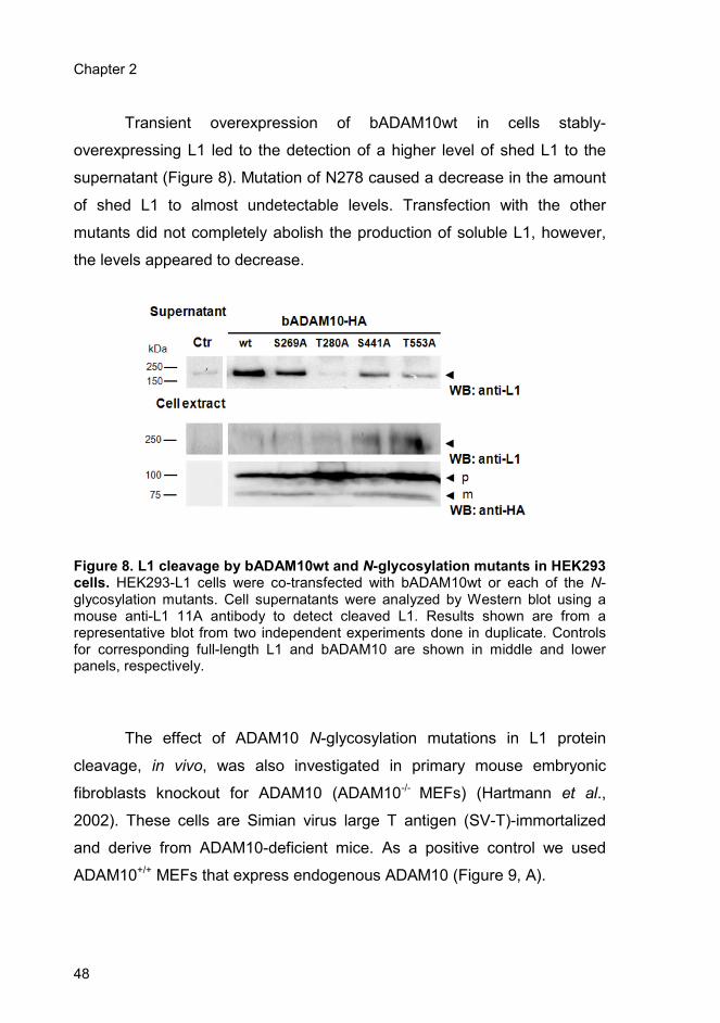
Chapter 2
48
Transient overexpression of bADAM10wt in cells stably-
overexpressing L1 led to the detection of a higher level of shed L1 to the
supernatant (Figure 8). Mutation of N278 caused a decrease in the amount
of shed L1 to almost undetectable levels. Transfection with the other
mutants did not completely abolish the production of soluble L1, however,
the levels appeared to decrease.
Figure 8. L1 cleavage by bADAM10wt and N-glycosylation mutants in HEK293 cells. HEK293-L1 cells were co-transfected with bADAM10wt or each of the N-glycosylation mutants. Cell supernatants were analyzed by Western blot using a mouse anti-L1 11A antibody to detect cleaved L1. Results shown are from a representative blot from two independent experiments done in duplicate. Controls for corresponding full-length L1 and bADAM10 are shown in middle and lower panels, respectively.
The effect of ADAM10 N-glycosylation mutations in L1 protein
cleavage, in vivo, was also investigated in primary mouse embryonic
fibroblasts knockout for ADAM10 (ADAM10-/- MEFs) (Hartmann et al.,
2002). These cells are Simian virus large T antigen (SV-T)-immortalized
and derive from ADAM10-deficient mice. As a positive control we used
ADAM10+/+ MEFs that express endogenous ADAM10 (Figure 9, A).

Functional role of ADAM10 N-glycosylation
49
MEFs were transiently-double transfected with pcDNA3-L1 and the
empty vector pcNA3.1-LacZ, bADAM10wt, or one of the N-glycosylation
mutants. Forty-eight hours after transfection, the supernatant
corresponding to a 24 h-production in serum-depleted medium, was
collected and analysed by Western blot (Figure 9, B). As controls L1 and
bADAM10 from the corresponding cell extracts were also analyzed by
Western blot (Figure 9, B).
Figure 9. L1 cleavage in ADAM10-/- MEFs transiently overexpressing pcDNA3-
L1 and pcDNA3.1-LacZ, bADAM10wt or each of the N-glycosylation mutants. (A) Analysis of endogenous mADAM10 protein in ADAM10
-/- and ADAM10
+/+ MEFs
using the polyclonal antibody rabbit anti mouse ADAM10 AB387. (B) L1 cleavage in MEFs in the absence or presence of 5 µM of the ADAM17 inhibitor, BMS. MEFs supernatants were analyzed by Western blot using a mouse anti-L1 11A antibody to detect cleaved L1. Controls for corresponding full-length L1 and bADAM10 are shown in middle and lower panels, respectively.
Chapter 2
50
Although, L1 cleavage levels in ADAM10-/- MEFs were lower than in
ADAM10+/+ MEFs, the production of soluble L1 was not completely
abolished (Figure 9, B). It has been described that in wt MEFs two ADAM
protein family members can cleave L1: while ADAM10 cleaves L1
constitutively, ADAM17 needs to be induced by phorbol-12-myristate-13-
acetate (PMA) or methyl-β-cyclodextrin (MCD) (Maretzy et al., 2005).
Nevertheless, it is possible that in absence of ADAM10, ADAM17 can
compensate and contribute to the constitutive L1 cleavage.
Since there was still cleavage of L1 in ADAM10-/- MEFs, that could
possibly be attributed to ADAM17 (Gutwein et al., 2005; Maretzky et al.,
2005), L1 cleavage assay was done in the presence of 5 µM ADAM17
inhibitor - BMS. After incubation with BMS a decrease in L1 cleavage levels
was observed both for ADAM10-/- and ADAM10+/+ MEFS. In addition, in
agreement with the results obtained above using HEK293 cells (Figure 8),
mutation of N278 caused a decrease in the amount of shed L1 (Figure 9,
B).
Since the in vivo activity is influenced by the catalytic activity of
ADAM10 as well as its intracellular localization, we determined the
proteolytic activity using a cell-free assay for mutants T280A and S269A
that had the highest and the lowest alterations of in vivo activity,
respectively. HEK293 cells were transiently doubly-transfected with
bADAM10wt or with S269A and T280A mutants solubilized in lysis buffer
and subjected to immunoprecipitation with anti-HA antibody to isolate the
proteins. After washing, the precipitated proteins were incubated with the
fluorescent quenching peptide Abz-Leu-Ala-Gln-Ala-Val-Arg-Ser-Ser-Ser-
Arg-Dap(dnp)-NH2 containing the ADAM cleavage site of TNF-alpha.
Bovine ADAM10wt and glycosylation mutant forms were efficiently
purified from cell lysates of HEK293 cells and were able to cleave the TNF-
alpha peptide in a time dependent fashion. A reduced cleavage of the
peptide to approximately 60% of the value obtained for bADAM10wt was
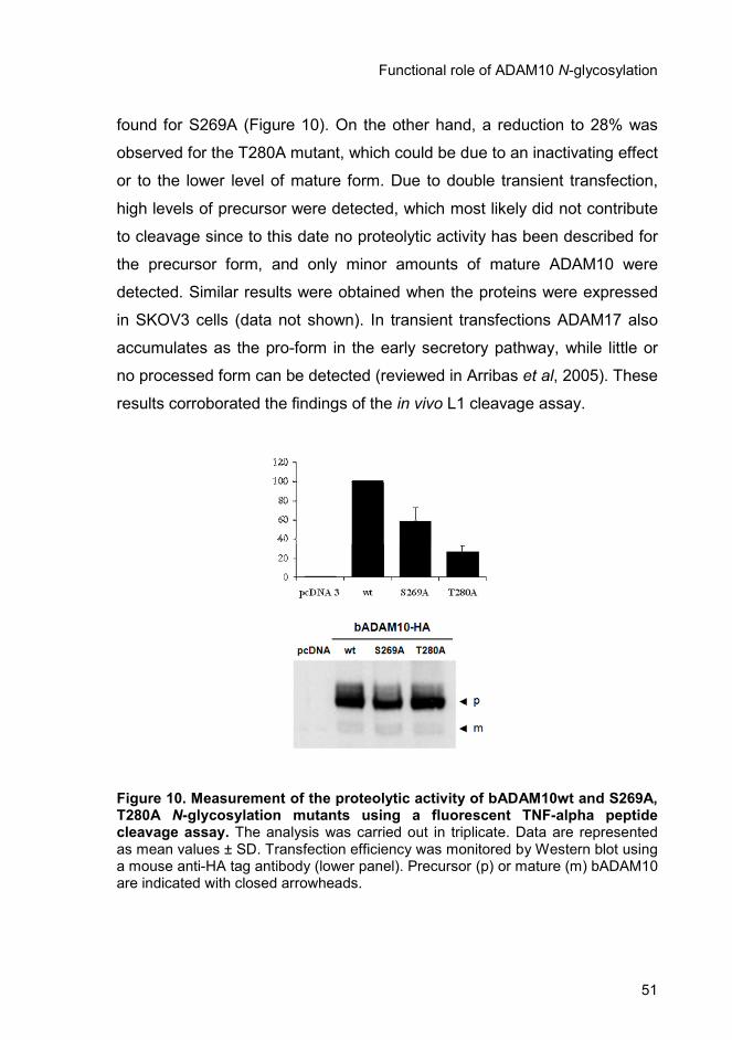
Functional role of ADAM10 N-glycosylation
51
found for S269A (Figure 10). On the other hand, a reduction to 28% was
observed for the T280A mutant, which could be due to an inactivating effect
or to the lower level of mature form. Due to double transient transfection,
high levels of precursor were detected, which most likely did not contribute
to cleavage since to this date no proteolytic activity has been described for
the precursor form, and only minor amounts of mature ADAM10 were
detected. Similar results were obtained when the proteins were expressed
in SKOV3 cells (data not shown). In transient transfections ADAM17 also
accumulates as the pro-form in the early secretory pathway, while little or
no processed form can be detected (reviewed in Arribas et al, 2005). These
results corroborated the findings of the in vivo L1 cleavage assay.
Figure 10. Measurement of the proteolytic activity of bADAM10wt and S269A, T280A N-glycosylation mutants using a fluorescent TNF-alpha peptide cleavage assay. The analysis was carried out in triplicate. Data are represented as mean values ± SD. Transfection efficiency was monitored by Western blot using a mouse anti-HA tag antibody (lower panel). Precursor (p) or mature (m) bADAM10 are indicated with closed arrowheads.
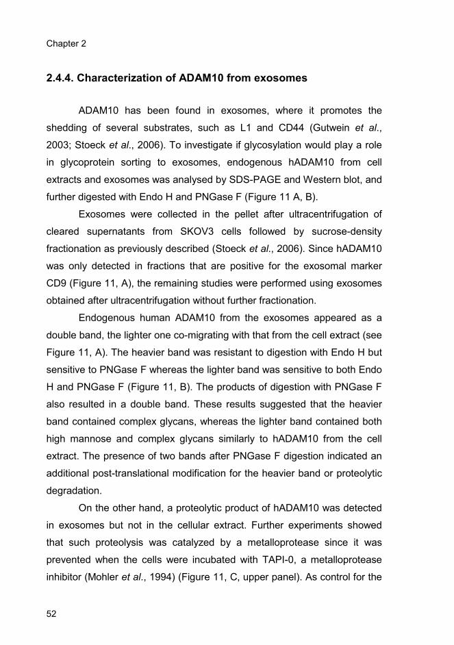
Chapter 2
52
2.4.4. Characterization of ADAM10 from exosomes
ADAM10 has been found in exosomes, where it promotes the
shedding of several substrates, such as L1 and CD44 (Gutwein et al.,
2003; Stoeck et al., 2006). To investigate if glycosylation would play a role
in glycoprotein sorting to exosomes, endogenous hADAM10 from cell
extracts and exosomes was analysed by SDS-PAGE and Western blot, and
further digested with Endo H and PNGase F (Figure 11 A, B).
Exosomes were collected in the pellet after ultracentrifugation of
cleared supernatants from SKOV3 cells followed by sucrose-density
fractionation as previously described (Stoeck et al., 2006). Since hADAM10
was only detected in fractions that are positive for the exosomal marker
CD9 (Figure 11, A), the remaining studies were performed using exosomes
obtained after ultracentrifugation without further fractionation.
Endogenous human ADAM10 from the exosomes appeared as a
double band, the lighter one co-migrating with that from the cell extract (see
Figure 11, A). The heavier band was resistant to digestion with Endo H but
sensitive to PNGase F whereas the lighter band was sensitive to both Endo
H and PNGase F (Figure 11, B). The products of digestion with PNGase F
also resulted in a double band. These results suggested that the heavier
band contained complex glycans, whereas the lighter band contained both
high mannose and complex glycans similarly to hADAM10 from the cell
extract. The presence of two bands after PNGase F digestion indicated an
additional post-translational modification for the heavier band or proteolytic
degradation.
On the other hand, a proteolytic product of hADAM10 was detected
in exosomes but not in the cellular extract. Further experiments showed
that such proteolysis was catalyzed by a metalloprotease since it was
prevented when the cells were incubated with TAPI-0, a metalloprotease
inhibitor (Mohler et al., 1994) (Figure 11, C, upper panel). As control for the
Functional role of ADAM10 N-glycosylation
53
presence of exosomes the same blot was probed with anti-annexin I, an
exosomal marker (Figure 11, C, lower panel).
Figure 11. Western blot analysis of endogenous hADAM10 from SKOV3 cell extracts and exosomes. (A) Total protein from cell extracts (Ext; 1x10
6 cells/lane),
exosomes (Exo; 1.5x10⁷ cells), and exosomes fractionated by centrifugation in
sucrose gradient, were analysed under non-reducing conditions using the mouse anti-ADAM10 antibody MAB1427. Exosomal marker CD9 was detected as a control using a different concentration gel. (B) Deglycosylation of hADAM10, from exosomes obtained after ultracentrifugation, with Endo H and PNGase F. (C) Exosomes were incubated for 16 h with the metalloprotease inhibitor TAPI-0. Annexin I, an exosomal marker was detected as an input control. Precursor (p) or mature (m) ADAM10 are indicated with closed arrowheads. Corresponding deglycosylated bands are shown with open arrowheads and asterisk indicates ADAM10 cleavage product.

Chapter 2
54
For comparison, recombinant bADAM10wt and mutants
overexpressed in SKOV3 cells were analysed (Figure 12, A). Precursor
T280A was not found in the exosomes, which is in agreement with the
previous results indicating that it is retained in the ER. Mature forms of
bADAM10wt, S269A, S441A, and T553A were found in the exosomes,
however, they were not detected as a double band, contrary to endogenous
hADAM10, probably due to the low levels of bADAM10 expression. In
addition, they all migrated at a slightly higher molecular mass than their
cellular counterparts. The increased mass estimation could be due to post-
translational modifications, such as N-glycosylation or ubiquitination.
Immunoprecipitation of bADAM10wt followed by Western blot analysis
using an anti-ubiquitin antibody allowed the identification of three bands
(Figure 12, B). The heavier band co-migrated with the mature form of
bADAM10wt, therefore suggesting that it was ubiquitinated. The other two
bands possibly corresponded to other ubiquitinated proteins that may
interact with bADAM10wt. As controls total cellular and exosomal proteins
were also analysed (Figure 12, B). Exosomal proteins were heavily
ubiquitinated in comparison with cellular proteins. High proteolytic activity
from the exosomes hindered PNGase F digestion analysis.
Proteolytic cleavage also occurred for exosomal bADAM10wt and
mutants similarly to endogenous hADAM10. This cleavage was particularly
evident for mutant S441A. These results further support the importance of
the N-linked glycans from N439 to protect the protein against proteolysis.
Deglycosylation of exosomal bADAM10 remained inconclusive due to high
level of protein degradation.
Detection of CD9, an exosomal marker, revealed no differences in
the amount of secreted vesicles produced in all the four N-glycosylation
mutants when compared with the bADAM10wt (results not shown).

Functional role of ADAM10 N-glycosylation
55
Figure 12. Western blot analysis of bADAM10wt and N-glycosylation mutants from stably transfected SKOV3 cells. (A) Total protein from cell extracts (Ext; 1x10
6 cells/lane) and exosomes (Exo; 1.5x10
7 cells) were analysed using a mouse
anti-HA tag antibody. Precursor (p) or mature (m) bADAM10 are indicated with closed arrowheads and asterisk indicates bADAM10 cleavage product. (B) Protein ubiquitination of cell extract (100 µg total protein), corresponding exosomes (70 µg total protein) and immunoprecipitated exosomal bADAM10wt. Analysis was performed using mouse anti-Ubiquitin P4D1 (Ub) and mouse anti-HA tag antibodies (HA). Mature (m) bADAM10 is indicated with closed arrowhead and asterisk indicates ubiquitinated proteins that interact with bADAM10.
2.4.5. Modelling of the 3D structure of bADAM10
metalloprotease domain
A model of bADAM10 metalloprotease domain based on its
homology with ADAM17 (39% identity) was constructed to investigate the
structural basis for the decreased in vitro activity of S269A, the absence of
T280A processing and the susceptibility of S441A to proteolytic

Chapter 2
56
degradation, as well as significantly decreased in vivo activities of T280A
and S441A (Figure 13). Both N267 and N278 glycosylation sites are
located in exposed zones before and after beta-strands, respectively, in
loop regions as expected. The N267 residue has one glycosylation
counterpart in ADAM17 (N264) in a spatially related region (not sequence
related). The N278 has no counterpart on ADAM17. Surprisingly, the N439
glycosylation site is located inside an alpha-helix. In a related spatial zone
in ADAM17, there is potential glycosylation site N452, which is, however,
located at the beginning of the helix and not inside it as in the case of
ADAM10.
Figure 13. Representation of the three-dimensional model for bADAM10 metalloprotease domain, rendered using cartoons. The potential N-glycosylation residues are highlighted using CPK models and labelled. Figure produced using PyMol (DeLano, Palo Alto, 2002).

Functional role of ADAM10 N-glycosylation
57
2.5. Discussion
Glycosylation is an important post-translational modification that
plays an important role in a number of physiological and biochemical
properties of a glycoprotein including stability, folding, intracellular
trafficking, or activity (Helenius et al., 2003). However, N-linked
glycosylation does not occur at every potential site and the role played by
glycosylation in different proteins is highly variable and depends on the
individual protein.
ADAM10 is a glycoprotein with four potential N-glycosylation sites:
N267, N278, N439, and N551 but the functional role of ADAM10 N-
glycosylation is unknown. To investigate if N-glycosylation has any role on
ADAM10 processing, stability, activity, and intracellular localization, the four
potential sites were mutated and characterized individually.
Our results showed that all four bADAM10 N-glycosylation sites
were occupied with high mannose or complex glycans. Moreover, the
mutation of a single N-glycosylation consensus sequence at N278
profoundly affected the efficient processing and trafficking of bADAM10. It
is well known that the interaction of glycoproteins with the lectin
chaperones from the ER, calnexin or calreticulin, is required for their correct
folding, reviewed in (Helenius et al., 2003; Morais et al., 2003). The time
that a certain glycoprotein remains in the calnexin/calreticulin folding cycle
in the ER depends on the protein, and it can vary from minutes to hours. If
glycoproteins fail to attain the correct fold, they can either be accumulated
in the ER as aggregates, be translocated to the cytosolic face of the ER
and be degraded by the proteasome, or they can be transported to the
Golgi apparatus and divert to the lysosome where they are degraded
(Helenius et al., 2003;Trombeta et al., 2003). Our results suggest that the
N-linked glycans from N278 are important for the folding of the protein
Chapter 2
58
probably mediating the interaction with calnexin. In the absence of this
folding step ADAM10 would fail to attain its final conformation and would be
retained in the ER. As a consequence the protein would remain as an
inactive precursor protein not only because of impaired folding but also
because further proteolytic processing to the mature form, which takes
place in the Golgi, would not occur due to compartmentalization
constraints. In agreement, T280A mutant showed reduced levels of activity
both in vivo and in vitro. Since only the ADAM10 mature form is active
(Schlondorff and Blobel, 1999; Seals and Courtneidge, 2003) the residual
activity observed for this mutant is the result of the small amount of
detected mature form. The 3D-model of ADAM10 metalloprotease domain,
built using comparative modelling techniques based on the 3D structure of
ADAM17, showed that the N278 site is located in an exposed zone and,
therefore, has a good possibility of being occupied, which agrees with the
experimental results. In addition, results obtained by other authors
supported that ADAM10 N278 site is occupied in human platelets
(Lewandrowski et al., 2006). Therefore, the N-linked glycans probably
mediate interaction with calnexin.
Mutation of the N439 glycosylation site resulted in increased
susceptibility of bADAM10 to proteases. The 3D model of ADAM10 showed
that N439 was found in an alpha-helix, which is a rare event but has a
probability of occurring of 10.5% (Petrescu et al., 2004) that is a non-
negligible value. Therefore, mutation of this site probably induced a
conformation alteration that exposed a particular region susceptible to
protease degradation. Furthermore, since N439, as well as N267, are not
located near the active site of ADAM10 from the 3D model, it is probable
that mutating them did not have a direct effect on substrate binding, and
therefore would not abolish enzyme activity, but could cause alterations in
protein conformation, explaining the decreased activities. Changes in
conformation caused by mutation of glycosylation sites with consequences

Functional role of ADAM10 N-glycosylation
59
for activity have been described for other proteins, for example, the Kv3.1
voltage-gated K+ channel (Brooks et al., 2006) or the GABA-transporter 1
(Cai et al., 2005).
N551 is located at the end of the disintegrin domain, juxtaposed to
the Cys-rich domain. These domains have been described as mediators of
protein-protein interactions and regulators of ADAM protease specificity
(White, 2003). Furthermore, Janes et al. showed that the cystein-rich
domain is the major responsible for substrate recognition and binding of the
EphA3/ephrin complex (Janes et al., 2005). Even if the analysis of the
crystallographic structure of the bADAM10 disintegrin and cystein-rich
domains (PDB: 2ao7) showed that N551 is located away from the identified
substrate recognition site, it is possible that mutating this site caused
conformational changes of these domains leading to decreased substrate
recognition specificities. This could explain the decreased in vivo activity of
T553A.
Signals responsible for the sorting of proteins into vesicles to be
released as exosomes, within MVBs are still being elucidated. Mono-
ubiquitination of plasma membrane proteins acts as a signal for
endocytosis and targeting to the endosomes. Furthermore, the
sequestration of transmembrane proteins in exosomes could require
(mono)-ubiquitination and the ESCRT-I, -II and -III protein complexes
(reviewed in Keller et al., 2006; van Niel et al., 2006). Accordingly, in the
present work, a comparatively higher amount of ubiquitinated proteins was
found in the exosomes, and in particular, recombinant exosomal bADAM10
appeared to be ubiquitinated. Concerning endogenous exosomal
hADAM10, a fraction of the protein was detected at a higher molecular
mass, which could be explained by ubiquitination or another post-
translational modification. In addition, this fraction exclusively presented
complex N-glycans differently from total cellular hADAM10, which
contained a mixture of complex and high mannose oligosaccharides.

Chapter 2
60
Several possibilities can be admitted to explain this finding: i) more complex
type glycoforms of hADAM10 are preferentially enriched during sorting and
trafficking from the plasma membrane to the MVBs; ii) preferential
enrichment of complex type glycoforms occurs during glycoprotein sorting
into the exosomes; iii) increased proteolysis of less processed glycoforms
occurs in the exosomes, thus revealing more processed glycoforms.
Further experiments are required to clarify this matter, however, the low
amounts of protein makes it a difficult task.
An ADAM10 cleavage product was specifically found in exosomes
and it was identified as being the result of metalloprotease cleavage. It is
possible that the ADAM10 and the unidentified metalloprotease are
specifically sorted into the exosomes and only get in contact there.
In conclusion, the work described here provides evidence for the
functional role of N-glycans from each of the N-glycosylation sites from
ADAM10. They are required for the processing and activation of the
enzyme, to protect the protein from proteolytic degradation, and, finally,
they are required for full-enzyme activity.
2.6. Acknowledgements
We thank Dr. Harald S. Conradt (GlycoThera, Germany) for critical
reading of the manuscript. We thank the Cell Imaging Service (Instítuto
Gulbenkian de Ciência, Oeiras) for the use of the confocal microscope. We
gratefully acknowledge Dr. Joaquin Arribas (Vall d’Hebron University
Hospital Research Institute, Barcelona) for the ADAM17 inhibitor-BMS and
Dr. Paul Saftig (Christian-Albrechts-University, Kiel) for the ADAM10-/-
MEFS. This work was funded by projects Signalling and Traffic, Nº LSHG-
CT-2004-503228, and CellPROM, Nº. 500039-2, European Commission.
Functional role of ADAM10 N-glycosylation
61
CE had a Ph.D. fellowship from Fundação para a Ciência e a Tecnologia,
Portugal.

Chapter 2
62
Chapter 3
Interaction and uptake of exosomes by ovarian
carcinoma cells
Work presented in this chapter corresponds to the following manuscript:
Escrevente C, Keller S, Altevogt P, Costa J (2011). Interaction and uptake of
exosomes by ovarian carcinoma cells. BMC Cancer. 11:108.

Chapter 3
64
Interaction and uptake of exosomes
65
3. Interaction and uptake of exosomes by ovarian
carcinoma cells
3.1. Summary
Exosomes consist of membrane vesicles that are secreted by
several cell types, including tumours and have been found in biological
fluids. Exosomes interact with other cells and may serve as vehicles for the
transfer of protein and RNA among cells.
SKOV3 exosomes were labelled with carboxyfluoresceine diacetate
succinimidyl-ester and collected by ultracentrifugation. Uptake of these
vesicles, under different conditions, by the same cells from where they
originated was monitored by immunofluorescence microscopy and flow
cytometry analysis. Lectin analysis was performed to investigate the
glycosylation properties of proteins from exosomes and cellular extracts.
In this work, the ovarian carcinoma SKOV3 cell line has been shown
to internalize exosomes from the same cells via several endocytic
pathways that were strongly inhibited at 4 ºC, indicating their energy
dependence. Partial colocalization with the endosome marker EEA1 and
inhibition by chlorpromazine suggested the involvement of clathrin-
dependent endocytosis. Furthermore, uptake inhibition in the presence of
5-ethyl-N-isopropyl amiloride, cytochalasin D and methyl-beta-cyclodextrin
suggested the involvement of additional endocytic pathways. The uptake
required proteins from the exosomes and from the cells since it was
inhibited after proteinase K treatments. The exosomes were found to be
enriched in specific mannose- and sialic acid-containing glycoproteins.
Sialic acid removal caused a small but non-significant increase in uptake.
Chapter 3
66
Furthermore, the monosaccharides D-galactose, α-L-fucose, α-D-mannose,
D-N-acetylglucosamine and the disaccharide β-lactose reduced exosomes
uptake to a comparable extent as the control D-glucose.
In conclusion, exosomes are internalized by ovarian tumour cells via
various endocytic pathways and proteins from exosomes and cells are
required for uptake. On the other hand, exosomes are enriched in specific
glycoproteins that may constitute exosome markers. This work contributes
to the knowledge about the properties and dynamics of exosomes in
cancer.

Interaction and uptake of exosomes
67
3.2. Introduction
Exosomes are small membrane vesicles between 40-100 nm in
diameter that are secreted by various cell types, including tumour cells,
neurons, B- and T-lymphocytes, intestinal epithelial cells (Denzer et al.,
2000; Keller et al., 2006; Simpson et al., 2009), and in physiological fluids
(Simpson et al., 2009; van Niel et al., 2006).
Exosome biogenesis involves the inward budding of endosomes
into MVBs to form intraluminal vesicles that are then released to the
extracellular space. During this process transmembrane proteins are
incorporated into the invaginating membrane maintaining the same
topological orientation as the plasma membrane, while cytosolic
components are engulfed.
The molecular basis of protein sorting during exosome formation
appears to involve an ubiquitin-dependent mechanism and the endosomal
sorting complexes required for transport - ESCRT (Katzmman et al., 2001;
Lakkaraju and Rodriguez-Boulan, 2008). However, some proteins present
in the exosomes are not ubiquitinated, suggesting that other mechanisms
such as oligomerization or partitioning of protein into lipid raft domains may
be involved (Lakkaraju and Rodriguez-Boulan, 2008; de Gassart et al.,
2003; Schorey and Bhatnagar, 2008; Trajkovic et al., 2008).
Exosomes are released by MVBs fusion with the plasma membrane
and the mechanism appears to involve Rab27A and Rab27B (Ostrowski et
al., 2010). Recent studies have suggested their participation in different
physiological and/or pathological processes such as tumour progression,
stimulation of the immune system, coagulation and inflammation, and
intercellular transfer of infectious agents, such as proteins and RNA (van
Niel et al., 2006; Lakkaraju and Rodriguez-Boulan, 2008; Schorey and
Bhatnagar, 2008).
Chapter 3
68
Many of the functions described for exosomes depend on their
ability to specifically interact with a target cell and several types of
interaction have already been proposed based on indirect evidence and in
vitro studies. Exosomes can associate with the plasma membrane through
ligand-receptor interactions (Théry et al., 2009) or lipids, such as
phosphatidylserine (Keller et al., 2009). The process of internalization can
occur through direct fusion of the exosomes with the plasma membrane,
leading to the release of the exosomal content into the cell cytoplasm.
Alternatively, exosomes can enter the cells by receptor-mediated
endocytosis and later fuse with the limiting membrane of the endosome
releasing the exosomal content to be recycled to the cell surface or to be
degraded in the lysosome (Théry et al., 2009; Cocucci et al., 2009).
Exosome uptake was shown to occur via clathrin-mediated endocytosis in
dendritic cells (Morelli et al., 2004), as well as phagocytosis in monocytes
and macrophages (Feng et al., 2010).
Exosomes have a unique protein and lipid composition that varies
depending on the cells from which they originate. Nevertheless, as a
consequence of their endosomal origin nearly all exosomes contain
proteins involved in membrane transport and fusion (RabGTPases,
annexins, flotilin), in MVBs biogenesis (TSG101, Alix), heat shock proteins
(Hsc70, Hsc90), integrins, and tetraspanins (CD9, CD63, CD81, CD82)
(Simons and Raposo, 2009; Stoorvogel et al., 2002). In addition, they are
enriched in raft-lipids such as cholesterol, sphingolipids, and ceramide. In
exosomes, an enrichment of certain glycosylated motifs has also been
observed (Krishnamoorthy et al., 2009).
Glycosylation is a post-translational modification that plays an
important role in several properties of proteins including stability, folding,
intracellular trafficking, and recognition. Lectins and their interactions with
carbohydrates have been found to play a role in exosome uptake by
dendritic cells (Hao et al., 2007) and macrophages (Barres et al., 2010).
Interaction and uptake of exosomes
69
In the present work, the SKOV3 ovarian carcinoma cell line has
been shown to internalize exosomes derived from the same cells via
various endocytic pathways. Proteins from exosomes and from cells were
required for the uptake. On the other hand, exosomes were highly enriched
in specific glycosylated sialic acid- and mannose-containing glycoproteins,
and sialic acid removal caused a small though non-significant increase in
uptake.
3.3. Materials and methods
3.3.1. Cell culture
Human ovarian cancer SKOV3, embryonic kidney HEK293, and
neuroglioma H4 cell lines were grown in DMEM (Sigma) at 37 °C, 5% CO2
supplemented with 10% fetal calf serum (Gibco), 1% penicillin/streptomycin
solution (Gibco).
3.3.2. Isolation of secreted membrane vesicles
Confluent SKOV3, HEK293 and H4 cells were cultivated for 24 h in
serum-free medium. The supernatant was collected and centrifuged, at
500, 10,000 and 100,000 x g 10, 20 and 120 min, respectively, at 4 °C. The
pellet of the last centrifugation consisted of secreted membrane vesicles.
Sucrose-density fractionation was performed as described previously
(Stoeck et al., 2006).
Chapter 3
70
3.3.3. Glycoprotein detection using lectins and immunoblot
Cellular extracts were obtained by solubilization of centrifuged cells
in Triton X-100 buffer (50 mM Tris-HCl pH 7.5, 5 mM EDTA, 1% Triton X-
100, 0.02% complete protease inhibitors cocktail (Roche)), for 30 min. Total
protein concentration was determined by the bicinchoninic acid (BCA)
method.
Glycoproteins from total cellular extracts and secreted membrane
vesicles were stained after transfer to polyvinylidene difluoride (PVDF)
membrane with lectins. Concanavalin A (Con A) (Sigma), biotinylated
Sambucus nigra (SNA) and Maackia amurensis lectin (MAL) (Galab
Technologies) were used. Glycoproteins were fixed on the PVDF
membrane with 25% (v/v) 2-propanol and 10% (v/v) acetic acid for 5 min.
The membranes were blocked for 1 h with TBS, 0.1% Tween-20 (TTBS) for
Con A or with TTBS containing 2% BSA for SNA and MAL. For Con A
detection the membrane was incubated overnight with 25 µg/ml Con A in
TTBS containing 1 mM CaCl2 and 1 mM MgCl2 (TTBSS) followed by 1 h
incubation with 0.5 µg/ml horseradish peroxidase type I (Sigma) in TTBSS.
For SNA and MAL detection, membranes were incubated overnight in
TTBS with 0.5 or 5 µg/ml SNA or MAL lectin, respectively. Membranes
were further incubated for 1 h with 0.02 µg/ml streptavidin-peroxidase
(Sigma). Detection was performed with the Immobilon Western
chemiluminescent HRP substrate (Millipore).
Immunoblot was performed as previously described (Chapter 2).
The following antibodies were used: mouse anti-CD9 (1:5000) and mouse
anti-L1 (L1 11A) (1:3).
Interaction and uptake of exosomes
71
3.3.4. Glycosidase treatment
Hydrolysis of α2,3- and α2,6-linked NeuAc from total protein cellular
extracts and exosomes was carried out overnight at 37 ºC by the addition of
15 mU neuraminidase from Vibrio cholerae or from Arthrobacter urefaciens
(Roche) in 50 mM sodium acetate pH 5.5 containing 4 mM CaCl2, and 50
mM sodium acetate pH 5.0, respectively. For specific hydrolysis of α2,3-
linked NeuAc, 9 U neuraminidase from Streptococcus pneumonia
(Prozyme, Glyko) in 50 mM sodium phosphate pH 6.0 were used, for 1 h at
37 ºC.
3.3.5. Uptake of SKOV3 exosomes by SKOV3 cells
SKOV3 exosomes were labelled with carboxyfluoresceine diacetate
succinimidyl-ester (CFSE) (Invitrogen) as previously described (Keller et
al., 2009). Briefly, exosomes (20 µg) collected after a 100,000 x g
ultracentrifugation were incubated with 7.5 µM CFSE for 30 min at 37 ºC in
a final volume of 200 µl PBS containing 0.5% BSA. Labelled exosomes
(Exos-CFSE) were 65-fold diluted with DMEM supplemented with 10%
vesicles-free fetal calf serum and pelleted by ultracentrifugation for 16 h at
10,0000 x g, 12 ºC. Exos-CFSE were resuspended in DMEM and incubated
with SKOV3 cells at 37 or 4 ºC
When indicated Exos-CFSE or cells were treated for 30 min with
100 µg/ml proteinase K, or for 2 h with 15 mU neuraminidase from V.
cholerae or from A. urefaciens, before uptake. SKOV3 cells were also
incubated, 30 min prior to and during uptake, with the inhibitors 10 µg/ml
chlorpromazine, 5 µg/ml cytochalasin D, 50 µM 5-ethyl-N-isopropyl
amiloride (EIPA) or 2% methyl-beta-cyclodextrin, or with 150 mM of the

Chapter 3
72
monosaccharides D-glucose, D-galactose, α-L-fucose, α-D-mannose, D-N-
acetylglucosamine, and the disaccharide β-lactose (Sigma).
Uptake assays were always performed in the presence of the
compounds and analyzed after 2 or 4 h by immunofluorescence
microscopy or flow cytometry.
3.3.6. Immunofluorescence microscopy
Immunofluorescence microscopy was done as previously described
(Chapter 2). Primary antibodies were: mouse IgG anti-alpha-tubulin DM1A
(1:2000) (Sigma), mouse IgG anti-EEA1 (1:100) (BD Biosciences), mouse
IgG anti-LAMP1 H4A3 (1:100) (BD Biosciences) and mouse IgG anti-
caveolin-1 (1:50) (Santa Cruz). Secondary antibody was donkey anti-
mouse IgG AlexaFluor 594 (1:500) (Molecular Probes). Images were
acquired on a Leica DMRB microscope using a DFC340FX camera
coupled to the microscope, and Leica application suite V3.3.0 software. For
colocalization, images were acquired on a confocal SP5 microscope. Each
picture was acquired with laser intensities and amplifier gains adjusted to
avoid pixel saturation. Each fluorophore used was excited independently
and sequential detection was performed. Each picture consisted of a z-
series of 20 images of 1024-1024 pixel resolution with a pinhole of 1.0 airy
unit. Colocalization analysis was performed using the open source Image J
version 1.38 (http://rsb.info.nih.gov/ij/).
3.3.7. Flow cytometry
SKOV3 cells incubated with Exos-CFSE for 4 h at 37 or 4 ºC were
washed with PBS, detached using trypsin and resuspended in PBS with 2%
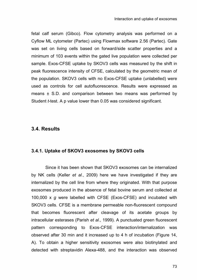
Interaction and uptake of exosomes
73
fetal calf serum (Gibco). Flow cytometry analysis was performed on a
Cyflow ML cytometer (Partec) using Flowmax software 2.56 (Partec). Gate
was set on living cells based on forward/side scatter properties and a
minimum of 103 events within the gated live population were collected per
sample. Exos-CFSE uptake by SKOV3 cells was measured by the shift in
peak fluorescence intensity of CFSE, calculated by the geometric mean of
the population. SKOV3 cells with no Exos-CFSE uptake (unlabelled) were
used as controls for cell autofluorescence. Results were expressed as
means ± S.D. and comparison between two means was performed by
Student t-test. A p value lower than 0.05 was considered significant.
3.4. Results
3.4.1. Uptake of SKOV3 exosomes by SKOV3 cells
Since it has been shown that SKOV3 exosomes can be internalized
by NK cells (Keller et al., 2009) here we have investigated if they are
internalized by the cell line from where they originated. With that purpose
exosomes produced in the absence of fetal bovine serum and collected at
100,000 x g were labelled with CFSE (Exos-CFSE) and incubated with
SKOV3 cells. CFSE is a membrane permeable non-fluorescent compound
that becomes fluorescent after cleavage of its acetate groups by
intracellular esterases (Parish et al., 1999). A punctuated green fluorescent
pattern corresponding to Exos-CFSE interaction/internalization was
observed after 30 min and it increased up to 4 h of incubation (Figure 14,
A). To obtain a higher sensitivity exosomes were also biotinylated and
detected with streptavidin Alexa-488, and the interaction was observed
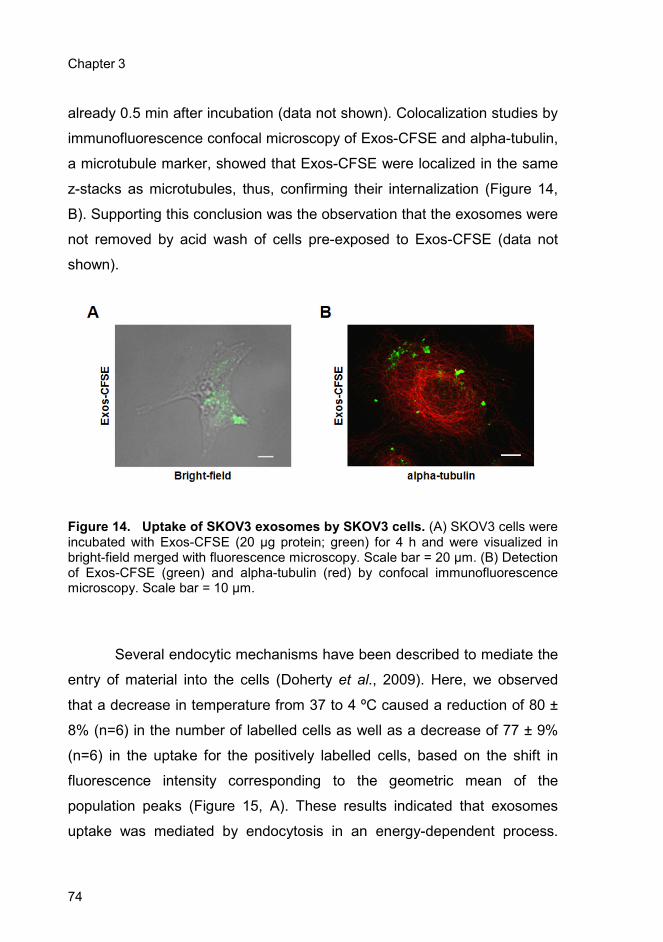
Chapter 3
74
already 0.5 min after incubation (data not shown). Colocalization studies by
immunofluorescence confocal microscopy of Exos-CFSE and alpha-tubulin,
a microtubule marker, showed that Exos-CFSE were localized in the same
z-stacks as microtubules, thus, confirming their internalization (Figure 14,
B). Supporting this conclusion was the observation that the exosomes were
not removed by acid wash of cells pre-exposed to Exos-CFSE (data not
shown).
Figure 14. Uptake of SKOV3 exosomes by SKOV3 cells. (A) SKOV3 cells were incubated with Exos-CFSE (20 µg protein; green) for 4 h and were visualized in bright-field merged with fluorescence microscopy. Scale bar = 20 µm. (B) Detection of Exos-CFSE (green) and alpha-tubulin (red) by confocal immunofluorescence microscopy. Scale bar = 10 µm.
Several endocytic mechanisms have been described to mediate the
entry of material into the cells (Doherty et al., 2009). Here, we observed
that a decrease in temperature from 37 to 4 ºC caused a reduction of 80 ±
8% (n=6) in the number of labelled cells as well as a decrease of 77 ± 9%
(n=6) in the uptake for the positively labelled cells, based on the shift in
fluorescence intensity corresponding to the geometric mean of the
population peaks (Figure 15, A). These results indicated that exosomes
uptake was mediated by endocytosis in an energy-dependent process.
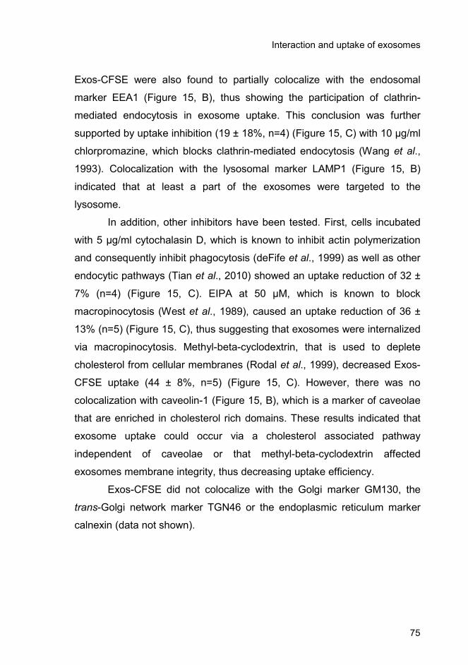
Interaction and uptake of exosomes
75
Exos-CFSE were also found to partially colocalize with the endosomal
marker EEA1 (Figure 15, B), thus showing the participation of clathrin-
mediated endocytosis in exosome uptake. This conclusion was further
supported by uptake inhibition (19 ± 18%, n=4) (Figure 15, C) with 10 µg/ml
chlorpromazine, which blocks clathrin-mediated endocytosis (Wang et al.,
1993). Colocalization with the lysosomal marker LAMP1 (Figure 15, B)
indicated that at least a part of the exosomes were targeted to the
lysosome.
In addition, other inhibitors have been tested. First, cells incubated
with 5 µg/ml cytochalasin D, which is known to inhibit actin polymerization
and consequently inhibit phagocytosis (deFife et al., 1999) as well as other
endocytic pathways (Tian et al., 2010) showed an uptake reduction of 32 ±
7% (n=4) (Figure 15, C). EIPA at 50 µM, which is known to block
macropinocytosis (West et al., 1989), caused an uptake reduction of 36 ±
13% (n=5) (Figure 15, C), thus suggesting that exosomes were internalized
via macropinocytosis. Methyl-beta-cyclodextrin, that is used to deplete
cholesterol from cellular membranes (Rodal et al., 1999), decreased Exos-
CFSE uptake (44 ± 8%, n=5) (Figure 15, C). However, there was no
colocalization with caveolin-1 (Figure 15, B), which is a marker of caveolae
that are enriched in cholesterol rich domains. These results indicated that
exosome uptake could occur via a cholesterol associated pathway
independent of caveolae or that methyl-beta-cyclodextrin affected
exosomes membrane integrity, thus decreasing uptake efficiency.
Exos-CFSE did not colocalize with the Golgi marker GM130, the
trans-Golgi network marker TGN46 or the endoplasmic reticulum marker
calnexin (data not shown).
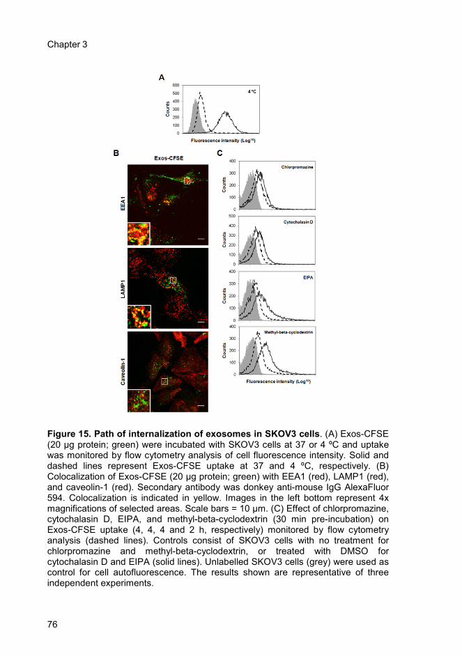
Chapter 3
76
Figure 15. Path of internalization of exosomes in SKOV3 cells. (A) Exos-CFSE (20 µg protein; green) were incubated with SKOV3 cells at 37 or 4 ºC and uptake was monitored by flow cytometry analysis of cell fluorescence intensity. Solid and dashed lines represent Exos-CFSE uptake at 37 and 4 ºC, respectively. (B) Colocalization of Exos-CFSE (20 µg protein; green) with EEA1 (red), LAMP1 (red), and caveolin-1 (red). Secondary antibody was donkey anti-mouse IgG AlexaFluor 594. Colocalization is indicated in yellow. Images in the left bottom represent 4x magnifications of selected areas. Scale bars = 10 µm. (C) Effect of chlorpromazine, cytochalasin D, EIPA, and methyl-beta-cyclodextrin (30 min pre-incubation) on Exos-CFSE uptake (4, 4, 4 and 2 h, respectively) monitored by flow cytometry analysis (dashed lines). Controls consist of SKOV3 cells with no treatment for chlorpromazine and methyl-beta-cyclodextrin, or treated with DMSO for cytochalasin D and EIPA (solid lines). Unlabelled SKOV3 cells (grey) were used as control for cell autofluorescence. The results shown are representative of three independent experiments.
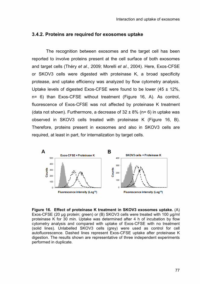
Interaction and uptake of exosomes
77
3.4.2. Proteins are required for exosomes uptake
The recognition between exosomes and the target cell has been
reported to involve proteins present at the cell surface of both exosomes
and target cells (Théry et al., 2009; Morelli et al., 2004). Here, Exos-CFSE
or SKOV3 cells were digested with proteinase K, a broad specificity
protease, and uptake efficiency was analyzed by flow cytometry analysis.
Uptake levels of digested Exos-CFSE were found to be lower (45 ± 12%,
n= 6) than Exos-CFSE without treatment (Figure 16, A). As control,
fluorescence of Exos-CFSE was not affected by proteinase K treatment
(data not shown). Furthermore, a decrease of 32 ± 8% (n= 6) in uptake was
observed in SKOV3 cells treated with proteinase K (Figure 16, B).
Therefore, proteins present in exosomes and also in SKOV3 cells are
required, at least in part, for internalization by target cells.
Figure 16. Effect of proteinase K treatment in SKOV3 exosomes uptake. (A) Exos-CFSE (20 µg protein; green) or (B) SKOV3 cells were treated with 100 µg/ml proteinase K for 30 min. Uptake was determined after 4 h of incubation by flow cytometry analysis and compared with uptake of Exos-CFSE with no treatment (solid lines). Unlabelled SKOV3 cells (grey) were used as control for cell autofluorescence. Dashed lines represent Exos-CFSE uptake after proteinase K digestion. The results shown are representative of three independent experiments performed in duplicate.

Chapter 3
78
3.4.3. Enrichment of specific glycoproteins in exosomes and
relevance in uptake
Glycan-lectin interactions have been suggested to play a role in the
uptake of exosomes by target cells (Hao et al., 2007; Barres et al., 2010),
therefore, we have investigated if glycans would play a role in exosome
uptake by SKOV3 cells.
First, glycoproteins of cellular extracts and secreted vesicles from
SKOV3 cells were detected with the lectins Con A (binds α-mannosyl
containing branched glycans predominantly of the high-mannose followed
by hybrid- and diantennary complex type structures to a lower extent)
(Bhattacharyya et al., 1989), SNA (recognizes NeuAcα2,6Gal/GalNAc) and
MAL (binds NeuAcα2,3Galβ1,4GlcNAc/Glc) (Knibbs et al., 1991).
The profile from secreted vesicles was distinct from that of cellular
extracts with the three lectins (Figure 17, A). Detection with SNA and MAL
was almost totally abolished after digestion with V. Cholerae and A.
urefaciens neuraminidases (cleave terminal α2,3- and α2,6-linked NeuAc),
whereas only MAL binding was abolished after S. pneumoniae (cleaves
only α2,3-linked NeuAc) digestion (Figure 17, B), thus confirming that SNA
and MAL binding to secreted vesicles glycoproteins was specific. The sialic
acid was not present in sialylated Lewis epitopes, since there was no
detection with anti-sialyl-Lewisa or anti-sialyl-Lewisx antibodies (data not
shown).
SKOV3 secreted vesicles are constituted by exosomes and
apoptotic vesicles, which can be fractionated by using a sucrose gradient,
as previously described (Stoeck et al., 2006; Escrevente et al., 2008). CD9
is a tetraspanin protein that has been used as exosome marker (Février
and Raposo, 2004; Lamparski et al., 2002). After sucrose gradient
separation of the secreted vesicles all the SNA and MAL binding was found
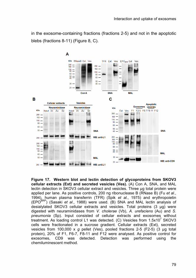
Interaction and uptake of exosomes
79
in the exosome-containing fractions (fractions 2-5) and not in the apoptotic
blebs (fractions 8-11) (Figure 8, C).
Figure 17. Western blot and lectin detection of glycoproteins from SKOV3 cellular extracts (Ext) and secreted vesicles (Ves). (A) Con A, SNA, and MAL lectin detection in SKOV3 cellular extract and vesicles. Three µg total protein were applied per lane. As positive controls, 200 ng ribonuclease B (RNase B) (Fu et al., 1994), human plasma transferrin (TFR) (Spik et al., 1975) and erythropoietin (EPO
BRP) (Sasaki et al., 1988) were used. (B) SNA and MAL lectin analysis of
desialylated SKOV3 cellular extracts and vesicles. Total proteins (3 µg) were digested with neuraminidases from V. cholerae (Vb), A. urefaciens (Au) and S. pneumonia (Sp). Input consisted of cellular extracts and exosomes without treatment. As loading control L1 was detected. (C) Vesicles from 1.5x10
7 SKOV3
cells were fractionated in a sucrose gradient. Cellular extracts (Ext), secreted vesicles from 100,000 x g pellet (Ves), pooled fractions 2-5 (F2-5) (3 µg total protein), 20% of F1, F6-7, F8-11 and F12 were analysed. As positive control for exosomes, CD9 was detected. Detection was performed using the chemiluminescent method.

Chapter 3
80
Specific patterns of protein glycosylation were also found for
exosomes from two other human cell lines, embryonic kidney HEK293 and
neuroglioma H4 cells (Figure 18).
To investigate a possible role for glycosylation in exosome uptake
Exos-CFSE were desialylated with V. cholerae and A. urefaciaens
sialidases and exosomes uptake was monitored by immunofluorescence
microscopy and flow cytometry analysis of cell fluorescence intensity.
Uptake efficiency of exosomes after neuraminidase treatment was slightly
increased (16 ± 14%, n= 6) when compared with exosomes incubated with
neuraminidase buffer (Figure 19, A), however, the observed increase was
not statistically significant using the Student t-test (p=0.0764). In addition,
increases in the uptake of exosomes incubated with neuraminidase (38 ±
13%, n=6) or neuraminidase buffer (23 ± 14%, n=6) were detected when
compared with Exos-CFSE without treatment (Figure 19, A).
Figure 18. Con A, SNA, and MAL lectin detection of glycoproteins from HEK293 and H4 cellular extract (Ext) and secreted vesicles (Ves). For the analysis, 3 µg of total protein were applied per lane. Detection was performed using the chemiluminescent method.
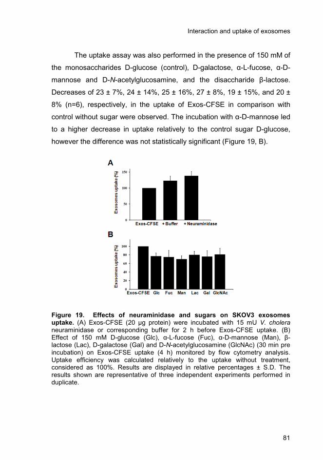
Interaction and uptake of exosomes
81
The uptake assay was also performed in the presence of 150 mM of
the monosaccharides D-glucose (control), D-galactose, α-L-fucose, α-D-
mannose and D-N-acetylglucosamine, and the disaccharide β-lactose.
Decreases of 23 ± 7%, 24 ± 14%, 25 ± 16%, 27 ± 8%, 19 ± 15%, and 20 ±
8% (n=6), respectively, in the uptake of Exos-CFSE in comparison with
control without sugar were observed. The incubation with α-D-mannose led
to a higher decrease in uptake relatively to the control sugar D-glucose,
however the difference was not statistically significant (Figure 19, B).
Figure 19. Effects of neuraminidase and sugars on SKOV3 exosomes uptake. (A) Exos-CFSE (20 µg protein) were incubated with 15 mU V. cholera neuraminidase or corresponding buffer for 2 h before Exos-CFSE uptake. (B) Effect of 150 mM D-glucose (Glc), α-L-fucose (Fuc), α-D-mannose (Man), β- lactose (Lac), D-galactose (Gal) and D-N-acetylglucosamine (GlcNAc) (30 min pre incubation) on Exos-CFSE uptake (4 h) monitored by flow cytometry analysis. Uptake efficiency was calculated relatively to the uptake without treatment, considered as 100%. Results are displayed in relative percentages ± S.D. The results shown are representative of three independent experiments performed in duplicate.

Chapter 3
82
3.5. Discussion
Exosomes are small membrane vesicles that are secreted by
several cell types, including tumours and they have been found in biological
fluids. They contain several membrane and cytoplasmic proteins and, in
cancer, they play a role in cell migration and metastases. They may also
transfer proteins associated with deregulated signaling pathways in cancer
(Al-Nedawi et al., 2008; Iero et al., 2007), therefore, contributing to the
propagation of transformed phenotype. Furthermore, the exosomes may
transfer mRNA and miRNA (Skog et al., 2008).
In the present work, we have found that the SKOV3 ovarian
carcinoma cell line internalizes exosomes derived from the same cells in an
energy-dependent process, via various endocytic pathways: colocalization
studies with organelle markers and incubation with inhibitors have shown
that the endocytosis pathway dependent on clathrin, macropinocytosis, and
a cholesterol associated pathway independent of caveolae were associated
with the uptake. Evidence from the literature showed that in dendritic cells
exosome uptake was also inhibited at 4 °C and with cytochalasin D, and
was further trafficked to the late endosomes/lysosomes (Morelli et al.,
2004). In phagocytic cells, more specifically macrophages, internalization
was also dependent on the actin cytoskeleton but not on pathways
involving caveolae, macropinocytosis or clathrin coated vesicles (Feng et
al., 2010). In the PC12 cell line exosomes uptake was also found to occur
via the endocytic pathway (Tian et al., 2010). After internalization,
exosomes could fuse with the endosomal membrane and deliver their
content to the cytoplasm of the target cell. For SKOV3 cells such process
could take place over a long period of time since internalized exosomes
were detected in the cells for at least 20 h (data not shown).
Interaction and uptake of exosomes
83
The initial recognition events that precede uptake may involve
several molecules, proteins (Simons and Raposo, 2009), and lipids (Keller
et al., 2009) have already been indicated as involved in this process. In this
work, the impairment observed for vesicles or cells treated with proteinase
K indicated that proteins from the extracellular surface from both target
cells and exosomes are required for internalization. Further studies to
identify which proteins play an important role in the recognition are
required.
Here we also found that exosomes from SKOV3 cells are
particularly enriched in specific glycoproteins with high mannose or
NeuAcα2,3/6-containing structures. In this context, other authors, using
lectin arrays, have also observed the enrichment in T-cells secreted
microvesicles of high-mannose and sialic acid-containing structures in
comparison with cell membranes (Krishnamoorthy et al., 2009).
Furthermore, an enrichment of more extensively glycosylated forms of PrPc
has been found in the exosomes in comparison with cell lysates (Vella et
al., 2007).
Removal of NeuAc from the exosomes with neuraminidase led to an
increase in the uptake, but non-significant. The removal of NeuAc
decreased the negative charge at the exosome surface exposing galactose
or N-acetylgalactosamine residues. This could lead to physico-chemical
alterations of the membrane or create new ligands for carbohydrate binding
proteins at the surface of the cells that would mediate the binding.
The monosaccharides D-galactose, α-L-fucose, α-D-mannose, D-N-
acetylglucosamine, and the disaccharide β-lactose reduced exosomes
uptake to a comparable extent as the control D-glucose probably due to
increased osmotic pressure that is known to reduce endocytosis (Oka et
al.1989). Therefore, these sugars do not play a major role in exosome
interaction and uptake in SKOV3 cells. This result is different from that
found in dendritic cells where exosome uptake was specifically inhibited by

Chapter 3
84
mannose and N-acetylglucosamine, the interaction being at least in part
mediated by a C-type lectin (Hao et al., 2007), or in macrophages where
lactose diminished exosome uptake probably through its action on galectin-
5 that mediated the process (Barres et al., 2010).
Here, certain glycoproteins were specifically detected in exosomes
and may constitute markers. It will be interesting to investigate if those
glycoproteins are selectively sorted to the exosomes or if only certain
glycoforms from the same protein are sorted into the exosomes. It can be
admitted that glycans by themselves may be important for glycoprotein
sorting into exosomes. Since protein oligomerization is known to promote
sorting into exosomes independently of the ESCRT machinery and
ubiquitination (Simons and Raposo, 2009), the glycans could mediate
oligomerization via interaction with lectins as previously described for the
transferrin receptor in reticulocytes (Vidal et al., 1997). Moreover, glycans
may interact with galectins at the cell surface, which may lead to their
enrichment in membrane lipid domains as previously described (Garner
and Baum, 2008). Considering that lipid microdomains play a role on
exosomes biogenesis (Simons and Raposo, 2009), glycans could in that
case also have a role on sorting to exosomes. Further studies are required
to clarify this matter.
Exosomes contain cell surface cancer antigens, which confers them
the potential for therapeutic approaches in cancer vaccination. Our
observation that they are particularly enriched in glycan epitopes urges the
need to further characterize the corresponding structures since one of the
major changes that occur in cancer is in cell surface glycosylation, which
has been largely used as biomarker. The knowledge of exosomes
glycosylation with the possibility for its modulation will open new
perspectives in cancer vaccination.
Interaction and uptake of exosomes
85
3.6. Acknowledgements
We thank the Cell Imaging Service (Instituto Gulbenkian de Ciência,
Portugal) for the use of the confocal microscope; Dr. Paula Alves and Dr.
Catarina Brito (Instítuto de Biologia Experimental e Tecnológica, Oeiras) for
the use of the flow cytometer; Dr. Rui Gardner (Instítuto Gulbenkian de
Ciência, Oeiras) for helpful discussion; Dr. Tiago Outeiro (Instítuto de
Medicina Molecular, Lisbon) for the gift of the H4 cell line.
This work was funded by projects Signalling and Traffic, No. LSHG-
CT-2004-503228, CellPROM, Nº 500039-2, European Commission, and
PIC/IC/82765/2007, PTDC/SAU-NEU/100724/2008, Fundação para a
Ciência e Tecnologia, Portugal. CE had a Ph.D. fellowship from Fundação
para a Ciência e a Tecnologia.

Chapter 3
86
Chapter 4
N-glycosylation of secreted vesicles from
ovarian tumour cells

Chapter 4
88
N-glycosylation of secreted vesicles
89
4. N-glycosylation of secreted vesicles from ovarian
tumour cells
4.1. Summary
Vesicles secreted from cells, including exosomes, are released by
several cell types, including ovarian tumour cells, and are found in
biological fluids such as malignant ascites and blood. They contain several
membrane and cytoplasmic proteins and they play a role in cancer
progression.
Aberrant glycosylation is an important aspect in malignant
transformation and consequently in ovarian cancer. Exosomes from ovarian
tumour cell lines have been shown to be enriched in specific glycoproteins
in comparison with cellular extracts. Nevertheless, detailed knowledge
about these glycan structures is not known at present.
In the present work, vesicles secreted from SKOV3 cells were
compared with plasma membrane and microsomal enriched fractions and
were found to contain specific glycoproteins that were distinctly recognized
by the lectins Con A, SNA, MAL and Wisteria floribunda (WFA).
Detailed structure analysis of the N-glycans from SKOV3 and OVM
secreted vesicles, microsomal, and plasma membrane fractions was
performed by HPAEC-PAD and MALDI-TOF-MS. Complex glycans of di-,
tri- and tetraantennary type with proximal fucose (Fuc) were found in all
fractions of both cell lines. High mannose structures were enriched in the
microsomal fraction of the two cell lines. Agalactosylated truncated
structures were found predominantly in SKOV3 cells, while the terminal
LacdiNAc motif was abundant in SKOV3-secreted vesicles.
Chapter 4
90
The results obtained contributed to the knowledge of the
glycosylation of vesicles secreted from ovarian tumour cells comparatively
to other cellular membrane enriched fractions. This might open new
perspective for the use of secreted vesicles as potential sources of tumour
markers.
N-glycosylation of secreted vesicles
91
4.2. Introduction
Many cell types, including tumour cells, secrete exosomes into the
extracellular space. These vesicles can also be found in the body fluids,
such as blood, urine, saliva, and malignant effusions (reviewed in van Niel
et al., 2006), and can contribute to tumour invasion and progression by
transferring genetic information that confers aggressive phenotypes (Al-
Nedawi et al., 2008), modifying the tumour microenvironment (Castellana et
al., 2009), conferring multi-drug resistance (Shedden et al., 2003; Safaei et
al., 2005), and promoting angiogenesis, tumour growth and metastasis
(reviewed in Muralidharan-Chari et al., 2010). Moreover, exosomes contain
proteins related with tumour progression (Gutwein et al., 2005; Runz et al.,
2007) that might be used as tumour markers and also constitute potential
targets for therapeutic approaches.
Ovarian tumour cells were found to release large amounts of
vesicles and the rate of their release has been shown to correlate with
tumour invasiveness (Ginestra et al., 1999). Moreover exosomes were also
found in blood (Taylor and Gercel-Taylor, 2008; Li et al., 2009) and
malignant ascites (Gutwein et al., 2005; Runz et al., 2007) of patients with
ovarian carcinoma.
Glycosylation alterations are typical for all kinds of cancers,
including ovarian carcinoma, and include several changes related to
terminal carbohydrate structures, such as incomplete synthesis and
modification of normally existing carbohydrates, and changes in the
carbohydrate core structures (Dabelsteen, 1996). Several studies have
already described specific glycosylation changes in the serum of ovarian
cancer patients. They include increased core fucosylated, agalactosylated
complex-type diantennary glycans originated from immunoglobulin G, along
with the sLex determinant (Saldova et al., 2007), increased sialylation with a

Chapter 4
92
shift in sialic acid linkage from α2,3- to α2,6- in ovarian cancer serum
glycoproteins (Saldova et al., 2007), and alterations in fucosylation and
branching in α1-acid glycoprotein (Imre et al., 2008).
Ovarian carcinoma cell lines, SKOV3, GG, OVM, and m130 were
already characterized with respect to their glycosylation using anti-
carbohydrate antibodies and lectins (Escrevente et al., 2006). The cells
contained variable amounts of Lewis carbohydrate motifs and all contained
glycoproteins recognized by Con A, a lectin that binds α-mannosyl
containing-branched glycans predominantly of the high mannose followed
by hybrid and diantennary complex structures to a lower extent
(Bhattacharyya et al., 1989), and SNA that binds structures containing
terminal α2,6-linked sialic acid (Knibbs et al., 1991). In addition, structure
analysis of the N-linked glycans from total glycoproteins derived from
SKOV3 ovarian carcinoma cells confirmed the abundance of high mannose
and proximally-fucosylated, complex type partially agalactosylated glycan
structures, as well as the presence of the LacdiNAc motif (Machado et al.,
2011).
SKOV3 cells exosomes have been shown to contain higher levels of
certain glycosylated sialic acid- and mannose-containing glycoproteins
when compared with total cellular extracts (Chapter 3). Nevertheless,
detailed knowledge about the N-glycan structures and their role is not
elucidated at present.
Lectin binding assays using Con A, SNA, MAL and WFA revealed
that secreted vesicles have a unique glycosylation profile when compared
with plasma membrane, and microsomal enriched fractions. The N-glycan
analysis from SKOV3 and OVM secreted vesicles, plasma membrane and
microsomal fractions by HPAEC-PAD and MALDI-TOF-MS showed that all
fractions of both cell lines contained complex glycans of di-, tri- and
tetraantennary type with proximal Fuc. Furthermore, high mannose type
structures were found to be enriched in the microsomal fraction of the two

N-glycosylation of secreted vesicles
93
cell lines. Agalactosylated truncated structures were found predominantly in
SKOV3 cells. Finally, the terminal LacdiNAc motif was abundant in
secreted vesicles of SKOV3 cells.
4.3. Materials and methods
4.3.1. Cell culture
Human ovarian cancer SKOV3, OVM, m130, and GG cell lines were
grown in DMEM (Sigma) at 37 °C, 5% CO2, supplemented with 10% fetal
calf serum (Gibco), and 1% penicillin/streptomycin solution (Gibco).
4.3.2. Isolation of cell-secreted vesicles
Cell-secreted vesicles were isolated as previously described
(Chapter 3). Briefly, confluent SKOV3, OVM, m130 and GG cells were
cultivated for 48 h in serum-free medium. The supernatant was collected
and centrifuged at 500, 10,000 and 100,000 x g, 10, 20 and 120 min,
respectively, at 4 °C. The pellet of the last centrifugation, consisting of
secreted membrane vesicles, was resuspended in PBS.
Chapter 4
94
4.3.3. Isolation of microsomal and plasma membrane enriched
fractions
The isolation of the microsomal membrane fraction from SKOV3
and OVM cells was accomplished by differential centrifugation. Cells were
pelleted by centrifugation, washed once with PBS, and resuspended in
homogenization buffer (1 mM NaHCO3, 0.2 mM CaCl2, 0.2 mM MgCl2,
1mM spermidine, pH 8). Cells were disrupted with a pellet pestle (Sigma)
and centrifuged at 600 x g, 10 min, 4 ºC. Supernatant was collected and
further centrifuged at 15,000 x g and 100,000 x g, for 10 and 120 min,
respectively, at 4 ºC. The pellet, consisting of the microsomal membrane
fraction was homogenised in PBS with a 25 gauge needle until complete
resuspension. The supernatant containing the cytosolic fraction was also
collected for further analysis.
The isolation of plasma membrane proteins from SKOV3 and OVM
cells was performed by cell surface biotinylation. SKOV3 cells were washed
with PBSCM (1 mM CaCl2, 1 mM MgCl2 in PBS) and biotinylated with 0.5
mg/ml sulfo-NHS-SS-biotin (Pierce) in PBSCM for 40 min at 4 ºC with
agitation (Sousa et al., 2003). After a two fold wash with PBSCM containing
50mM NH4Cl followed by DMEM with 10% FCS and 1% P/S, cells were
incubated with lysis buffer (1% Triton-X, 0.5% DOC, 0.1% SDS, 5 mM
EDTA, 0.02% complete protease inhibitors cocktail (Roche)) for 30 min at 4
ºC with agitation. After centrifugation the supernatants were incubated with
streptavidin-agarose beads (Sigma) for 2 h, at 4 ºC to pellet biotinylated
proteins. The agarose beads were pelleted and washed four times with
washing buffer (10 mM Tris-HCl buffer pH 8.0, 0.5 M NaCl, 1 mM EDTA
and 1% NP-40). Biotinylated proteins were eluted by incubation in 0.08%
Tris-HCl pH 6.8, 2% SDS, 10% glycerol, 5 min at 100 ºC. Non-biotinylated
proteins were also collected from the supernatant for further analysis.
N-glycosylation of secreted vesicles
95
4.3.4. Glycoprotein detection using lectins and immunoblot
Cellular extracts were obtained by solubilization of centrifuged cells
in Triton X-100 buffer (50 mM Tris-HCl pH 7.5, 5 mM EDTA, 1% Triton X-
100, 0.02% complete protease inhibitors cocktail (Roche)) for 30 min.
Secreted vesicles, plasma membrane, and microsomal fractions were
obtained as described above.
Total protein concentration was determined by the BCA method.
Glycoproteins were stained after transfer to PVDF membrane with lectins.
Con A (Sigma), biotinylated SNA, MAL (Galab Technologies), and WFA
(Vector Labs) were used. Glycoproteins were fixed on the PVDF membrane
with 25% (v/v) 2-propanol and 10% (v/v) acetic acid for 5 min. The
membranes were blocked for 1 h with TBS, 0.1% Tween 20 (TTBS) for Con
A, with TTBS containing 2% BSA for SNA, and MAL and with TBS, 0.15%
Tween 20, 2% BSA for WFA. For Con A detection the membrane was
incubated overnight with 25 µg/ml Con A in TTBS containing 1 mM CaCl2
and 1 mM MgCl2 (TTBSS) followed by 1 h incubation with 0.5 µg/ml
horseradish peroxidase type I (Sigma) in TTBSS. For SNA, MAL and WFA
detection, membranes were incubated overnight in TTBS with 0.5 µg/ml
SNA or 5 µg/ml MAL and WFA lectin. Membranes were further incubated
for 1 h with 0.02 µg/ml streptavidin-peroxidase (Sigma).
When indicated, membranes were incubated simultaneously with
the lectin and the corresponding competitive sugar, 0.2 M methyl-α-D-
mannopyranoside for Con A and 0.2 M N-acetyl-D-galactosamine for WFA.
Detection was performed with the Immobilon Western chemiluminescent
HRP substrate (Millipore).
Chapter 4
96
4.3.5. Total N-glycans isolation
Total N-glycans isolation from SKOV3 and OVM cell-secreted
vesicles, microsomal, and plasma membrane fractions were performed as
previously described (Machado et al., 2011). A total of 3.5 and 3 mg of total
proteins corresponding to secreted vesicles and microsomal fraction,
respectively, were solubilized for 30 min in PBS containing 2% (v/v)
Nonidet P-40 and 0.1% benzonase (Sigma), pH 7.2. For the isolation of N-
glycan from the plasma membrane, a solution containing 350 µg of
biotinylated proteins were used. Proteins were precipitated with 4 volumes
of ice-cold 100% ethanol for 2 h at −20 °C and dried in a Speed Vac
concentrator (Savant Instruments Inc.). The resulting pellet was incubated
for 90 min in 0.3 M Tris–HCl pH 8.3, 6 M urea, 1 mM dithioerythritol, diluted
in water and incubated overnight with 70 µg of trypsin (Merk) and 0.02%
sodium azide.
Trypsin inactivation was accomplished by 10 min incubation at 90
°C, followed by the addition of 0.02% complete protease inhibitor cocktail
during 1 h. Next, the sample solution was subjected to buffer exchange
using a 5 kDa Vivaspin concentrator (Sartorius) to PNGase F buffer (10
mM NaH2PO4, 5 mM ethylenediaminetetraacetic acid, pH 7.5). All
incubations were performed at room temperature unless otherwise noted.
The N-glycan release from total glycopeptides of secreted vesicles,
microsomal, and membrane fractions from SKOV3 cells were performed by
overnight incubation with 50 U PNGase F (Roche) at 37 °C. The released
N-glycans were separated from the glycoproteins by precipitation with 2.4
volumes of ice-cold 100% ethanol for 2 h at −20 °C. After centrifugation at
10,000 × g, for 10 min at 4 °C, the N-glycans in suspension were removed,
and two additional washes of the glycoprotein pellet were made with ice-

N-glycosylation of secreted vesicles
97
cold 50% ethanol. The supernatants containing N-glycans were pooled and
dried in a Speed Vac concentrator.
4.3.6. Analysis of desialylated N-glycans by HPAEC-PAD
The N-glycan samples were desalted using 100 mg Hypercarb
cartridges (Thermo) previously washed with 80% (v/v) acetonitrile (ACN)
with 0.1% trifluoroacetic acid (TFA), and six times with water. The N-
glycans solubilized in water were then loaded onto the column, washed two
times with water, and finally eluted with 40% ACN with 0.1% TFA. The
eluted glycans were neutralized with 2.5% (v/v) NH4OH and dried in a
Speed Vac concentrator. The desialylated N-glycans were prepared by
incubation with 5 mM H2SO4 for 90 min at 80 °C. Hydrolyzed N-glycans
were neutralized with 50 mM NaOH and dried in a Speed Vac concentrator.
The analysis of N-glycan structures by HPAEC-PAD was performed
using an ICS-3000 ion chromatography system (Dionex Corporation),
consisting of an AS autosampler, a DC detector/chromatography module,
and a DP dual pump. Samples of 20 µL were injected into an equilibrated
CarboPac PA200 column equipped with a guard column (3 × 50 mm,
Dionex). Released desialylated N-glycans were eluted by using a
concentration gradient of 0.15 M sodium hydroxide (solvent A) and 0.15 M
sodium hydroxide/0.6 M sodium acetate (solvent B), at a flow rate of 0.4
mL/ min. The gradient consisted of a 30 min isocratic run at 100% solvent
A, followed by increasing solvent B concentration (10 min to 15%, 2 min to
30%, 3 min to 100%). The column temperature was constant at 30 °C. The
electrochemical detection of the oligosaccharides was performed by
application of the detection potentials and durations as recommended by
the manufacturer.
Chapter 4
98
For subsequent MALDI-TOF-MS analysis pools or individual
fractions from the HPAEC-PAD analysis were collected after online
desalting using a carbohydrate membrane desalter device (CMD-I, Dionex).
Glycan fractions were dried in a Speed Vac concentrator for further
analysis.
4.3.7. Analysis of N-glycans by MALDI-TOF-MS
Glycans were analyzed with a Bruker ULTRAFLEX time-of-flight
(TOF/TOF) instrument in the linear positive ion mode. Native desialylated
oligosaccharides were analyzed using a matrix of 2,5-dihydroxybenzoic
acid as a UV-absorbing material. Samples of 1 µL at an approximate
concentration of 1–10 pmol/µL were mixed with equal amounts of the
respective matrix and sodium salt. This mixture was spotted onto a
stainless steel target and dried at room temperature before analysis.
Structures were tentatively assigned to various N-glycans based on their
observed masses compared with the calculated masses of known N-
glycans.
Mass spectrum data were analyzed with the Bruker DataAnalysis
version 4 (Bruker Daltonics). Further annotation of N-glycans and database
searches were performed using the GlycoWorkbench software (Ceroni et
al., 2008).

N-glycosylation of secreted vesicles
99
4.4. Results
4.4.1. Enrichment of specific glycoproteins in ovarian carcinoma
secreted vesicles
SKOV3-secreted vesicles and exosomes were found to be highly
enriched in certain glycosylated sialic acid- and mannose-containing
glycoproteins when compared with total cellular extracts (Chapter 3). To
investigate if secreted vesicles had a characteristic glycosylation profile that
was distinct from plasma membrane and microsomal fractions, the different
fractions were detected with lectins after Western blotting.
In this study, secreted vesicles isolated by ultracentrifugation of cell
supernatants at 100,000 x g were used for analysis since this fraction has
been shown to be enriched in exosomes (Théry et al., 2006). Previous
results from our laboratory have shown that MAL- and SNA-binding
glycoproteins were only detected in exosome-containing fractions after
sucrose density purification (Chapter 3).
The microsomal fraction was obtained by differential centrifugation
and it consisted of the 100,000 x g pellet after clarification of the cell extract
at 15,000 x g, while a plasma membrane enriched fraction was obtained by
cell surface protein biotinylation and separation with streptavidin-agarose.
As controls, a cytoplasmic fraction consisting of post-100,000 x g
supernatant, and an intracellular fraction containing proteins that were not
biotinylated were also analysed.
Glycoproteins of SKOV3 cellular extracts (Ext), secreted vesicles
(Ves), and all the other enriched fractions were detected with the lectins
Con A, SNA, MAL and WFA (recognizes the LacdiNAc motif,
GalNAcβ4GlcNAc). The glycosylation profile of SKOV3-secreted vesicles
Chapter 4
100
was distinct from all other fractions with all the lectins tested (Figure 20).
Con A was shown to strongly bind glycoproteins of SKOV3-secreted
vesicles when compared with other fractions, using the same amount of
total protein. The binding specificity was confirmed using 0.2 M of the
competitive sugar methyl-α-D-mannopyranoside.
In SKOV3 cell secreted vesicles three glycoproteins with sizes of
approximately 260 kDa and one with about 110 kDa were recognized by
WFA (indicated with asterisks in Figure 20). In the plasma membrane
enriched fraction only one specific glycoprotein at approximately 160 kDa
was recognized. The detection was specific since it was abolished in the
presence of 0.2 M of competitive sugar N-acetyl-galactosamine. The signal
detected in cellular extracts, microsomal, intracellular, and cytoplasmic
fractions with WFA was probably not specific since it was not inhibited after
incubation with the competitive sugar (Figure 20).
Previous results have shown that specific bands from SKOV3
exosomes were strongly recognized by SNA and MAL contrary to cellular
extracts (Chapter 3). In the present study, SKOV3-secreted vesicles
contained two major bands identified by SNA, one with an apparent
molecular mass of 260 kDa and the second between 80 and 110 kDa
(indicated with asterisks in Figure 20). MAL detected two bands with high
intensity in SKOV3-secreted vesicles, one band around 260 kDa and a
second band between 110 and 160 kDa (indicated with asterisks in Figure
20). These specific glycoproteins were not found in microsomal or plasma
membrane enriched fractions (Figure 20). Therefore, sialylated
glycoproteins identified were specific to secreted vesicles, and were not
found in other membrane fractions.
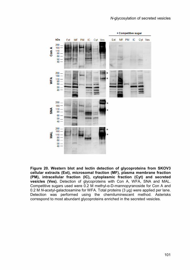
N-glycosylation of secreted vesicles
101
Figure 20. Western blot and lectin detection of glycoproteins from SKOV3 cellular extracts (Ext), microsomal fraction (MF), plasma membrane fraction (PM), intracellular fraction (IC), cytoplasmic fraction (Cyt) and secreted vesicles (Ves). Detection of glycoproteins with Con A, WFA, SNA and MAL. Competitive sugars used were 0.2 M methyl-α-D-mannopyranoside for Con A and 0.2 M N-acetyl-galactosamine for WFA. Total proteins (3 µg) were applied per lane. Detection was performed using the chemiluminescent method. Asterisks correspond to most abundant glycoproteins enriched in the secreted vesicles.

Chapter 4
102
To determine if there was also an enrichment of specific sialic acid-
containing glycoproteins in other ovarian carcinoma cell lines, secreted
vesicles from OVM, m130, and GG cells were analysed by Western blot
with SNA and MAL (Figure 21). SNA was found to bind specific
glycoproteins in m130 secreted vesicles, including one protein above 260
kDa, one with approximately 160 kDa and three proteins with apparent
molecular masses around 80 kDa (indicated with asterisks in Figure 21).
MAL detected one major glycoprotein in m130 and OVM secreted vesicles
with about 110 kDa (indicated with asterisks in Figure 21). No specific
enrichment was observed for GG cells with these lectins.
Figure 21. Western blot and SNA and MAL lectin detection of glycoproteins from OVM, m130 and GG ovarian carcinoma cell lines. For the analysis, three µg of total proteins from cellular extracts (Ext) and secreted vesicles (Ves) were applied per lane. Detection was performed using the chemiluminescent method. Asterisks correspond to most abundant glycoproteins enriched in the secreted vesicles.
N-glycosylation of secreted vesicles
103
4.4.2. Structural analysis of desialylated N-glycans from ovarian
tumour secreted vesicles, plasma membrane and microsomal
fractions
To further elucidate the glycan structures of secreted vesicles
comparatively to cellular membranes, the N-linked oligosaccharides of
SKOV3 and OVM cell-secreted vesicles, plasma membrane, and
microsomal enriched fractions were released with PNGase
F, desialylated by mild acid hydrolysis and were analyzed by HPAEC-PAD
(Figure 22, C, D, and E)
The majority of the peaks eluted in the region of the chromatogram
corresponding to high mannose: Man5GlcNAc2, Man6GlcNAc2,
Man7GlcNAc2, Man8GlcNAc2 and Man9GlcNAc2 (9-18 min) (Figure 22, A
and Figure 23, A) and complex N-glycans di-, tri-, and tetraantennary with
and without LacNAc repeats and proximal Fuc (12-21 min) (Figure 22, B
and Figure 23, B).
The HPAEC-PAD profile of secreted vesicles from SKOV3 and
OVM cells was distinct from those of plasma membrane and microsomal
fractions. For example, the microsomal fraction showed peaks with
retention times identical to those of high mannose glycans contrary to other
fractions (indicated with asterisks in Figure 22 and Figure 23).
Differences between the N-glycans profile of SKOV3 and OVM
vesicles were also detected. For example, OVM vesicles showed a
relatively larger peak co-eluting with tetraantennary glycans with proximal
Fuc (Standard D, Figure 22 and Figure 23).
SKOV3 and OVM plasma membrane and microsomal fractions
shared some similarities in their N-glycans profile, namely, a high intensity
peak with a retention time of approximately 21 min that was almost
undetected in the secreted vesicles (Figure 22 and Figure 23).
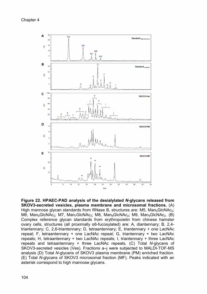
Chapter 4
104
Figure 22. HPAEC-PAD analysis of the desialylated N-glycans released from SKOV3-secreted vesicles, plasma membrane and microsomal fractions. (A) High mannose glycan standards from RNase B, structures are: M5, Man5GlcNAc2; M6, Man6GlcNAc2; M7, Man7GlcNAc2; M8, Man8GlcNAc2; M9, Man9GlcNAc2. (B) Complex reference glycan standards from erythropoietin from chinese hamster ovary cells, structures (all proximally α6-fucosylated) are: A, diantennary; B, 2,4-triantennary; C, 2,6-triantennary; D, tetraantennary; E, triantennary + one LacNAc repeat; F, tetraantennary + one LacNAc repeat; G, triantennary + two LacNAc repeats; H, tetraantennary + two LacNAc repeats; I, triantennary + three LacNAc repeats and tetraantennary + three LacNAc repeats. (C) Total N-glycans of SKOV3-secreted vesicles (Ves). Fractions a–j were subjected to MALDI-TOF-MS analysis (D) Total N-glycans of SKOV3 plasma membrane (PM) enriched fraction. (E) Total N-glycans of SKOV3 microsomal fraction (MF). Peaks indicated with an asterisk correspond to high mannose glycans.
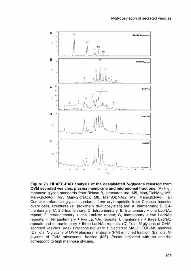
N-glycosylation of secreted vesicles
105
Figure 23. HPAEC-PAD analysis of the desialylated N-glycans released from OVM secreted vesicles, plasma membrane and microsomal fractions. (A) High mannose glycan standards from RNase B, structures are: M5, Man5GlcNAc2; M6, Man6GlcNAc2; M7, Man7GlcNAc2; M8, Man8GlcNAc2; M9, Man9GlcNAc2. (B) Complex reference glycan standards from erythropoietin from Chinese hamster ovary cells, structures (all proximally α6-fucosylated) are: A, diantennary; B, 2,4-triantennary; C, 2,6-triantennary; D, tetraantennary; E, triantennary + one LacNAc repeat; F, tetraantennary + one LacNAc repeat; G, triantennary + two LacNAc repeats; H, tetraantennary + two LacNAc repeats; I, triantennary + three LacNAc repeats and tetraantennary + three LacNAc repeats. (C) Total N-glycans of OVM-secreted vesicles (Ves). Fractions k-p were subjected to MALDI-TOF-MS analysis (D) Total N-glycans of OVM plasma membrane (PM) enriched fraction. (E) Total N-glycans of OVM microsomal fraction (MF). Peaks indicated with an asterisk correspond to high mannose glycans.

Chapter 4
106
Due to the high complexity of the samples and to obtain more
structural information, the total desialylated N-glycan pool released from
SKOV3 and OVM cell-secreted vesicles, plasma membrane, and
microsomal enriched fractions were analysed by MALDI-TOF-MS (Figure
24 and Figure 25). Furthermore, individual fractions of SKOV3 (a-j) (Figure
22, C) and OVM (k-p) (Figure 23, C) vesicles from the HPAEC-PAD profiles
were also collected after on-line desalting and were analysed by MALDI-
TOF-MS (Supplementary Figure 1 and Supplementary Figure 2). The
results of the observed m/z and possible glycan structures, compatible with
the proposed compositions and considering the N-glycosylation
biosynthetic pathway of mammalian cells, are indicated in Table I and
Table II for SKOV3 and OVM cells, respectively.
The major findings were as follows. Signals at m/z 1809.7, 2174.8
and 2539.9 were compatible with complex glycans of the di-, tri- and
tetraantennary type with proximal Fuc and they were detected for the
secreted vesicles as well as in the plasma membrane and microsomal
fractions. The results were found in the two cell lines (Figure 24 and Figure
25).
Signals at m/z 1419.5, 1581.6, 1743.6 and 1905.6, compatible with
high mannose structures (Man6GlcNAc2; Man7GlcNAc2; Man8GlcNAc2 and
Man9GlcNAc2, respectively) were predominantly found in the microsomal
fraction of the two cell lines (Figure 24, C and Figure 25, C).
Peaks at m/z 2012.5 and 1850.5 were compatible with
agalactosylated truncated structures (triantennary minus one Gal and two
Gal, respectively). Peaks at m/z 2377.6 and 2215.6 were compatible with
tetraantennary glycans less one Gal and two Gal, respectively. These
peaks were predominantly found in SKOV3 cells, in all fractions (Figure 24,
A-C and Table I). For OVM cells they were found in minor amounts and
only in the vesicles fraction (Figure 25, A-C and Table II).
N-glycosylation of secreted vesicles
107
Peaks at m/z 1850.5, 2215.6 and 2580.7 in SKOV3-secreted
vesicles could also correspond to complex glycans of the di-, tri- and
tetraantennary type bearing the LacdiNAc motif (Figure 24, A). This
hypothesis agrees with the detection of SKOV3-secreted vesicles with the
lectin WFA (Figure 20). These peaks were absent from OVM plasma
membrane and microsomal fractions and had a low intensity (1850.5,
2215.6) or were absent (2580.7) from OVM-secreted vesicles (Figure 25,
A).
The profiles for secreted vesicles were shown to be more
heterogeneous than in the other fractions for the two cell lines. For OVM
cells a comparatively higher amount of higher antennarity, tri- and
tetraantennary glycans, relatively to diantennary glycans was detected in
secreted vesicles versus plasma membrane or microsomal fractions
(Figure 23 and Figure 25).

Chapter 4
108
Figure 24. MALDI-TOF-MS analysis of the total desialylated N-glycans from SKOV3 cells. (A) secreted vesicles (Ves), (B) plasma membrane fraction (PM), (C) microsomal fraction (MF). Only one or two possible isomeric structures of selected peaks are presented. Graphical representations of glycans are consistent with the nomenclature of the Consortium for Functional Glycomics.

N-glycosylation of secreted vesicles
109
Figure 25. MALDI-TOF-MS analysis of the total desialylated N-glycans from OVM cells. (A) secreted vesicles (Ves), (B) plasma membrane fraction (PM), (C) microsomal fraction (MF). Only one or two possible isomeric structures of selected peaks are presented. Graphical representations of glycans are consistent with the nomenclature of the Consortium for Functional Glycomics.
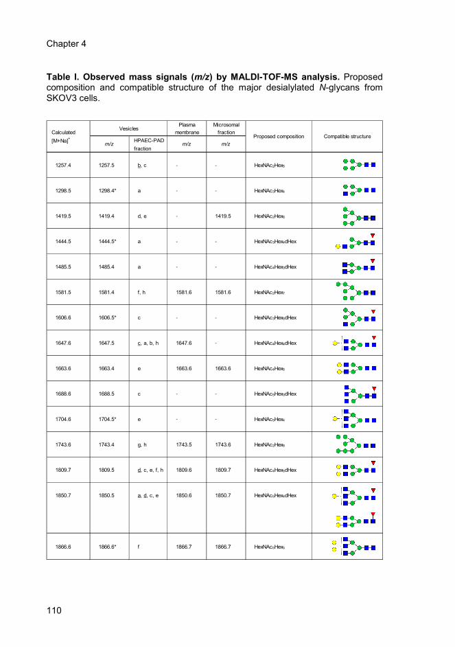
Chapter 4
110
Plasma Microsomal
Calculated membrane fraction
[M+Na]+
HPAEC-PAD
fraction
1257.4 1257.5 b, c - - HexNAc2Hex5
1298.5 1298.4* a - - HexNAc3Hex4
1419.5 1419.4 d, e - 1419.5 HexNAc2Hex6
1444.5 1444.5* a - - HexNAc3Hex4dHex
1485.5 1485.4 a - - HexNAc4Hex3dHex
1581.5 1581.4 f, h 1581.6 1581.6 HexNAc2Hex7
1606.6 1606.5* c - - HexNAc3Hex5dHex
1647.6 1647.5 c, a, b, h 1647.6 - HexNAc4Hex4dHex
1663.6 1663.4 e 1663.6 1663.6 HexNAc4Hex5
1688.6 1688.5 c - - HexNAc5Hex3dHex
1704.6 1704.5* e - - HexNAc5Hex4
1743.6 1743.4 g, h 1743.5 1743.6 HexNAc2Hex8
1809.7 1809.5 d, c, e, f, h 1809.6 1809.7 HexNAc4Hex5dHex
1850.7 1850.5 a, d, c, e 1850.6 1850.7 HexNAc5Hex4dHex
1866.6 1866.6* f 1866.7 1866.7 HexNAc5Hex5
m/z m/z
Proposed composition Compatible structure
Vesicles
m/z
Table I. Observed mass signals (m/z) by MALDI-TOF-MS analysis. Proposed composition and compatible structure of the major desialylated N-glycans from SKOV3 cells.
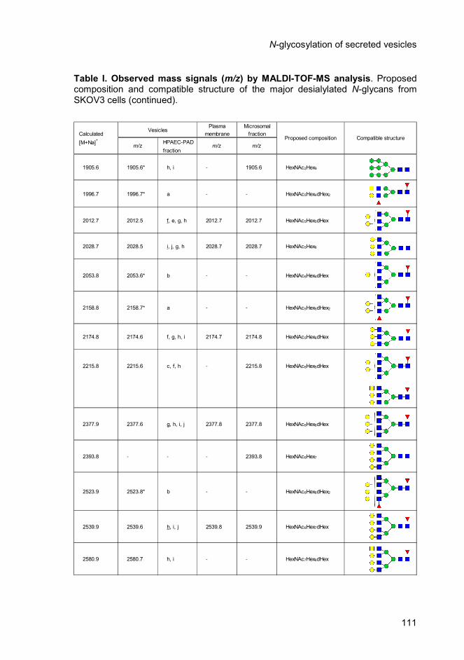
N-glycosylation of secreted vesicles
111
Plasma Microsomal
Calculated membrane fraction
[M+Na]+
HPAEC-PAD
fraction
1905.6 1905.6* h, i - 1905.6 HexNAc2Hex9
1996.7 1996.7* a - - HexNAc5Hex4dHex2
2012.7 2012.5 f, e, g, h 2012.7 2012.7 HexNAc5Hex5dHex
2028.7 2028.5 i, j, g, h 2028.7 2028.7 HexNAc5Hex6
2053.8 2053.6* b - - HexNAc6Hex4dHex
2158.8 2158.7* a - - HexNAc5Hex5dHex2
2174.8 2174.6 f, g, h, i 2174.7 2174.8 HexNAc5Hex6dHex
2215.8 2215.6 c, f, h - 2215.8 HexNAc6Hex5dHex
2377.9 2377.6 g, h, i, j 2377.8 2377.8 HexNAc6Hex6dHex
2393.8 - - - 2393.8 HexNAc6Hex7
2523.9 2523.8* b - - HexNAc6Hex6dHex2
2539.9 2539.6 h, i, j 2539.8 2539.9 HexNAc6Hex7dHex
2580.9 2580.7 h, i - - HexNAc7Hex6dHex
m/z m/z
Proposed composition Compatible structure
Vesicles
m/z
Table I. Observed mass signals (m/z) by MALDI-TOF-MS analysis. Proposed composition and compatible structure of the major desialylated N-glycans from SKOV3 cells (continued).
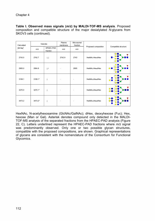
Chapter 4
112
Plasma Microsomal
Calculated membrane fraction
[M+Na]+
HPAEC-PAD
fraction
2743.0 2742.7 i, j 2742.9 2743 HexNAc7Hex7dHex
2905.0 2904.8 j, i - 2905 HexNAc7Hex8dHex
3108.1 3108.1* j - - HexNAc8Hex8dHex
3270.0 3270.1* j - - HexNAc8Hex9dHex
3473.2 3473.2* j - - HexNAc9Hex9dHex
m/z m/z
Proposed composition Compatible structure
Vesicles
m/z
Table I. Observed mass signals (m/z) by MALDI-TOF-MS analysis. Proposed composition and compatible structure of the major desialylated N-glycans from SKOV3 cells (continued).
HexNAc, N-acetylhexosamine (GlcNAc/GalNAc); dHex, deoxyhexose (Fuc); Hex, hexose (Man or Gal). Asterisk denotes compound only detected in the MALDI-TOF-MS analysis of the separated fractions from the HPAEC-PAD analysis (Figure 22, C). Letters underlined represent the HPAEC-PAD fractions where m/z signal was predominantly observed. Only one or two possible glycan structures, compatible with the proposed compositions, are shown. Graphical representations of glycans are consistent with the nomenclature of the Consortium for Functional Glycomics.
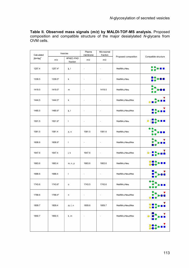
N-glycosylation of secreted vesicles
113
Plasma Microsomal
Calculated membrane fraction
[M+Na]+
HPAEC-PAD
fraction
1257.4 1257.4* k, l - - HexNAc2Hex5
1339.5 1339.5* k - - HexNAc4Hex3
1419.5 1419.5* m - 1419.5 HexNAc2Hex6
1444.5 1444.5* k - - HexNAc3Hex4dHex
1485.5 1485.6* k, l - - HexNAc4Hex3dHex
1501.5 1501.6* l - - HexNAc4Hex4
1581.5 1581.4 n, o 1581.5 1581.6 HexNAc2Hex7
1606.6 1606.6* l - - HexNAc3Hex5dHex
1647.6 1647.4 l, k 1647.6 - HexNAc4Hex4dHex
1663.6 1663.4 m, n, p 1663.6 1663.6 HexNAc4Hex5
1688.6 1688.4 l - - HexNAc5Hex3dHex
1743.6 1743.6* o 1743.5 1743.6 HexNAc2Hex8
1768.6 1768.4* n - - HexNAc3Hex6dHex
1809.7 1809.4 m, l, n 1809.6 1809.7 HexNAc4Hex5dHex
1850.7 1850.5 k, m - - HexNAc5Hex4dHex
Vesicles
Proposed composition Compatible structure
m/z m/z m/z
Table II. Observed mass signals (m/z) by MALDI-TOF-MS analysis. Proposed composition and compatible structure of the major desialylated N-glycans from OVM cells.
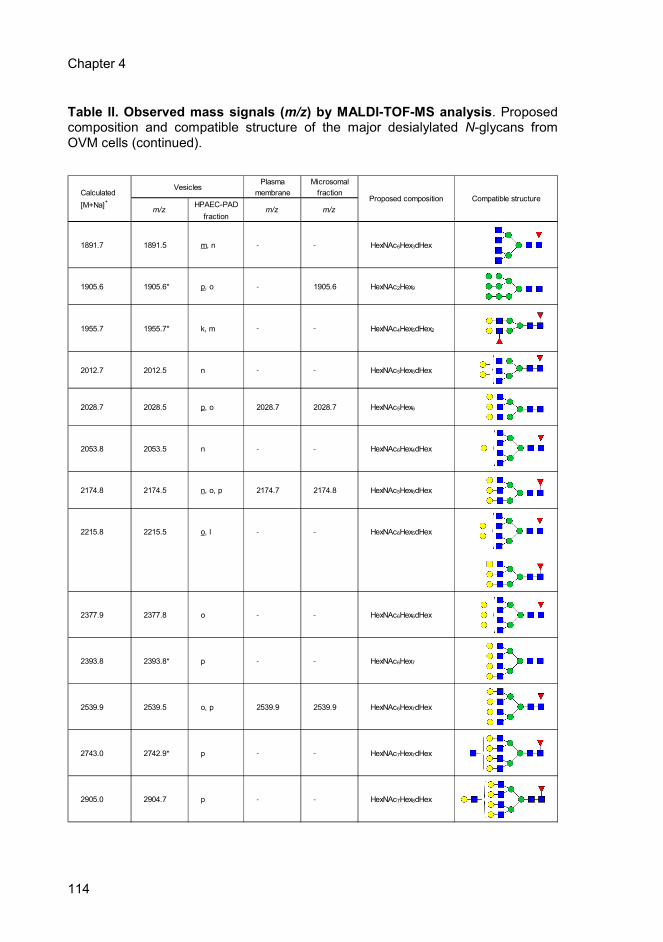
Chapter 4
114
Plasma Microsomal
Calculated membrane fraction
[M+Na]+
HPAEC-PAD
fraction
1891.7 1891.5 m, n - - HexNAc6Hex3dHex
1905.6 1905.6* p, o - 1905.6 HexNAc2Hex9
1955.7 1955.7* k, m - - HexNAc4Hex5dHex2
2012.7 2012.5 n - - HexNAc5Hex5dHex
2028.7 2028.5 p, o 2028.7 2028.7 HexNAc5Hex6
2053.8 2053.5 n - - HexNAc6Hex4dHex
2174.8 2174.5 n, o, p 2174.7 2174.8 HexNAc5Hex6dHex
2215.8 2215.5 o, l - - HexNAc6Hex5dHex
2377.9 2377.8 o - - HexNAc6Hex6dHex
2393.8 2393.8* p - - HexNAc6Hex7
2539.9 2539.5 o, p 2539.9 2539.9 HexNAc6Hex7dHex
2743.0 2742.9* p - - HexNAc7Hex7dHex
2905.0 2904.7 p - - HexNAc7Hex8dHex
Vesicles
Proposed composition Compatible structure
m/z m/z m/z
Table II. Observed mass signals (m/z) by MALDI-TOF-MS analysis. Proposed composition and compatible structure of the major desialylated N-glycans from OVM cells (continued).
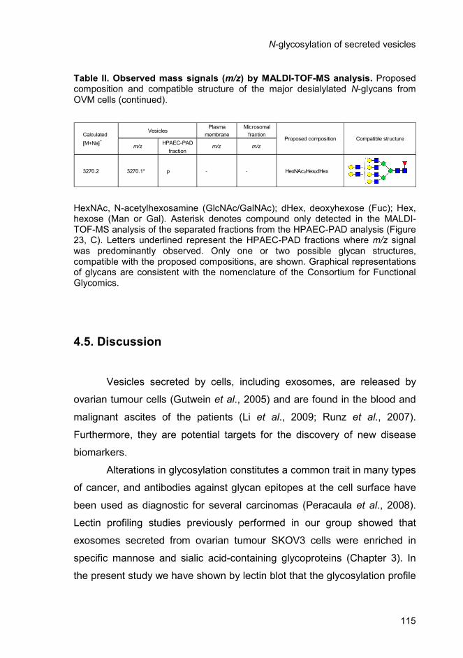
N-glycosylation of secreted vesicles
115
Plasma Microsomal
Calculated membrane fraction
[M+Na]+
HPAEC-PAD
fraction
3270.2 3270.1* p - - HexNAc8Hex9dHex
Vesicles
Proposed composition Compatible structure
m/z m/z m/z
Table II. Observed mass signals (m/z) by MALDI-TOF-MS analysis. Proposed composition and compatible structure of the major desialylated N-glycans from OVM cells (continued).
HexNAc, N-acetylhexosamine (GlcNAc/GalNAc); dHex, deoxyhexose (Fuc); Hex, hexose (Man or Gal). Asterisk denotes compound only detected in the MALDI-TOF-MS analysis of the separated fractions from the HPAEC-PAD analysis (Figure 23, C). Letters underlined represent the HPAEC-PAD fractions where m/z signal was predominantly observed. Only one or two possible glycan structures, compatible with the proposed compositions, are shown. Graphical representations of glycans are consistent with the nomenclature of the Consortium for Functional Glycomics.
4.5. Discussion
Vesicles secreted by cells, including exosomes, are released by
ovarian tumour cells (Gutwein et al., 2005) and are found in the blood and
malignant ascites of the patients (Li et al., 2009; Runz et al., 2007).
Furthermore, they are potential targets for the discovery of new disease
biomarkers.
Alterations in glycosylation constitutes a common trait in many types
of cancer, and antibodies against glycan epitopes at the cell surface have
been used as diagnostic for several carcinomas (Peracaula et al., 2008).
Lectin profiling studies previously performed in our group showed that
exosomes secreted from ovarian tumour SKOV3 cells were enriched in
specific mannose and sialic acid-containing glycoproteins (Chapter 3). In
the present study we have shown by lectin blot that the glycosylation profile

Chapter 4
116
of SKOV3-secreted vesicles was also different from other membrane
enriched fractions, namely plasma membrane and microsomal enriched
fractions. Our observations are in agreement with the results obtained
using lectin microarrays in T-cells microvesicles that showed enrichment in
high mannose and sialic acid-containing structures, as well as complex N-
glycans, and fucosylated structures in comparison with cell membranes
micellae (Krishnamoorthy et al., 2009). Furthermore, the results allowed the
identification of glycoproteins that are specific for secreted vesicles and
possibly constitute exosomes markers. Further studies are required to
purify and characterize those proteins by lectin affinity chromatography,
followed by peptide mass fingerprinting. Antibodies raised against the
polypeptide part will allow clarifying if specific proteins or protein glycoform
are enriched in exosomes.
In addition, lectin detection revealed that SKOV3- and OVM-
secreted vesicles had specific α2,3- and/or α2,6-linked NeuAc-containing
glycoproteins that were not detected in other fractions. Since altered
expression of certain sialic acid types or their linkages can have prognostic
significance in several human cancers (reviewed in Li et al., 2010) it will be
interesting to analyse the profile of native oligosaccharides from secreted
vesicles and from the other membrane enriched fractions.
In order to further elucidate the N-glycan structures in the secreted
vesicles from ovarian tumour cells, relatively to plasma membrane and
microsomal fractions, SKOV3 and OVM cells were analysed by HPAEC-
PAD followed by MALDI-TOF-MS. As a common feature the results
indicated the presence of glycans of the di-, tri- and tetraantennary type
with proximal Fuc in secreted vesicles of SKOV3 and OVM cells, as well as
in plasma membrane and microsomal fractions. The abundance of
proximally fucosylated complex partially agalactosylated glycan structures
in all fractions of SKOV3 cells was also detected.
N-glycosylation of secreted vesicles
117
A higher amount of high mannose structures was found in
microsomal fractions of both SKOV3 and OVM cell lines. Since the
endoplasmic reticulum is a major component of the microsomal fraction
(Michelsen and Hagen, 2009) these high mannose glycans probably
correspond to unprocessed structures characteristic of glycoproteins in
transit or resident in the endoplasmic reticulum.
Moreover, the results suggested the presence of the LacdiNAc motif
in SKOV3-secreted vesicles, which was in agreement with the WFA
detection of specific bands in secreted vesicles. Partially agalactosylated
glycans and the LacdiNAc motif have already been described in
endogenous cellular and supernatant glycoproteins from SKOV3 cells
(Machado et al., 2011). The LacdiNAc motif is associated with
glycoproteins secreted by tumour cells such as ribonuclease I found in
pancreatic tumour cells (Peracaula et al., 2003), plasminogen activator
from Bowes melanoma cells (Chan et al., 1991), and glycodelin-A that is
increased in many gynecological tumours (Horowitz et al., 2001).
Glycan profiles of secreted vesicles from the two cell lines studied
were more heterogeneous than the plasma membrane or the microsomal
fraction. The results also suggested a comparatively higher amount of more
processed glycans of the tri- and tetraantennary types in secreted vesicles
in comparison with plasma membrane and microsomal fractions for the
OVM cells. This effect could be due to more processed glycoforms of a
certain glycoprotein being enriched in exosomes, as suggested for
ADAM10 (Chapter 2). Alternatively, the enrichment of specific glycoproteins
with more processed glycans in exosomes would be plausible. To explore
these hypothesis further work is required in characterizing purer exosome
fractions, in exploring additional cell lines, and in improving glycan
separation, possibly after labelling, for quantitation.
From our knowledge this is the first study endeavouring the detailed
structural analysis of the N-glycans present in glycoproteins from ovarian
Chapter 4
118
tumour-secreted vesicles. In this work, secreted vesicles were isolated by
ultracentrifugation at 100,000 x g since this fraction has been shown to be
enriched in exosomes (Théry et al., 2006). Preliminary results using
Nanoparticle Tracking Analysis (Nanosight) revealed that the majority of the
isolated SKOV3- and OVM-secreted vesicles had average sizes of 134 and
113 nm, respectively. Nonetheless, vesicles with smaller and larger
dimensions were also detected (data not shown). Secreted vesicles can be
further purified using a sucrose gradient. Purified exosomes have a density
of 1.13-1.19 g/ml and have a size between 40-100 nm (Simons and
Raposo, 2009). Therefore, it would be interesting to analyse the N-glycans
from purified exosomes not only to confirm if N-glycan structures enriched
in the secreted vesicles are found in exosomes but also to investigate if
additional specific structures are found.
The knowledge of N-glycan structures present in purified exosomes
could provide the basis for further studies on the mechanisms involved in
exosomes biogenesis. In a more applied perspective, the identification of
specific N-glycans might give new insights for biomarkers discovery and
cancer vaccination.
4.6. Acknowledgements
We thank Sebastian Kandzia and Dr. Harald S. Conradt,
GlycoThera, Germany, for HPAEC-PAD and MALDI-TOF-MS analysis.
This work was funded by projects PIC/IC/82765/2007 and
PTDC/SAU-NEU/100724/2008, Fundação para a Ciência e a Tecnologia,
Portugal. CE had a Ph.D. fellowship from Fundação para a Ciência e a
Tecnologia.

Chapter 5
General discussion and conclusions

Chapter 5
120
General discussion and conclusions
121
5. General discussion and conclusions
5.1. General discussion and perspectives
Ovarian cancer has the highest mortality rate among the
gynecological malignancies in the Western world. Long-term survival has
not changed significantly in the last two decades, largely due to inadequate
diagnostic approaches and the lack of efficient treatments.
Aberrant protein glycosylation is an early event of oncogenic
transformation; therefore, glycan structures represent ideal targets for
tumour specific diagnosis and new anti-tumour therapeutic strategies. In
ovarian cancer a number of proteins were found to be aberrantly
glycosylated, including CA125 (Jankovic and Milutinovic, 2008), alpha-1-
proteinase inhibitor, haptoglobin (Turner et al., 1995), immunoglobulin G,
and other acute phase proteins (Saldova et al., 2007). Moreover, an
increased sialylation of proteins and deregulated sialylation pathways were
also observed (Aranganathan et al., 2005).
Exosomes are small membrane vesicles that are secreted by
several cell types, including ovarian tumour cells (Gutwein et al., 2005), and
have been found in vivo in several biological fluids, such as blood and
malignant ascites of ovarian cancer patients (Caby et al., 2005; Runz et al.,
2007). Exosomes have the ability to interact with other cells and were
shown to contribute to tumour progression by promoting immune escape
mechanisms, facilitating extracellular matrix degradation and transferring
proteins and genetic material among cells (reviewed in Muralidharan-Chari
et al., 2010).
Chapter 5
122
Although many studies have been carried out to identify the protein,
lipid and RNA composition of exosomes, little is known about the
glycosylation profile of these secreted vesicles and the potential role of N-
glycans in protein sorting to exosomes or in recognition and uptake of
exosomes by target cells.
In this context, the intent of this Thesis was to investigate the protein
glycosylation profile of vesicles secreted from ovarian tumour cells. In
addition, the biological role of N-glycans in ADAM10 protein sorting to
exosomes and in the internalization of SKOV3 exosomes by SKOV3 cells
was explored.
5.1.1. Exosome specific glycoproteins/glycoforms
Proteomic studies have shown that glycoproteins, similarly to other
proteins, are found in exosomes (Simpson et al., 2008). In addition, some
reports have described the presence of heavily glycosylated forms of some
proteins, such as FAS-L (Abrahams et al., 2003) and the prion precursor
protein (Vella et al., 2007), in secreted vesicles that are not detected in
cellular extracts. In this context, here the use of lectins with different
carbohydrate specificities revealed that vesicles secreted from SKOV3 and
OVM cells contained specific glycoproteins, in particular proteins containing
α2,3-linked sialic acid, that were not detected in cellular extracts, plasma
membrane or microsomal enriched fractions (Chapter 4). Vesicles secreted
from SKOV3 cells were also found to strongly bind Con A, which
recognizes predominantly high mannose glycans, compared with other
fractions (Chapter 3 and Chapter 4). Furthermore, secreted vesicles from
the two cell lines were found to contain a higher amount of N-glycans than
plasma membrane or microsomal fractions, as shown by the peak
intensities in the HPAEC-PAD profiles (Chapter 4). These results together
General discussion and conclusions
123
showed that specific glycoproteins and/or glycoforms were enriched in
ovarian tumour secreted vesicles. The molecular mechanisms underlying
this enrichment are still not known.
To date, several mechanisms of protein sorting to exosomes have
been proposed. The most studied involves the recognition of ubiquitinated
proteins by the ESCRT machinery (reviewed in Lakkaraju and Rodriguez-
Boulan, 2008; Simons and Raposo, 2009). In SKOV3 cells, this pathway
might account for the recruitment of some proteins to exosomes since
exosomal proteins were found to be heavily ubiquitinated in comparison
with cellular proteins (Chapter 2) and SKOV3 exosomes contained
TSG101, a protein that is involved in the ESCRT sorting machinery
(unpublished data). Overexpressed ADAM10-HA was also found to be
ubiquitinated in exosomes, however due to the low protein expression it
was not possible to determine if endogenous ADAM10 was also
ubiquitinated in exosomes (Chapter 2). To substantiate the possibility of
glycoproteins being sorted to exosomes via ESCRT, RNA interference
targeting different mRNAs coding for proteins involved in the ESCRT
machinery could be used and the presence of different glycoproteins
monitored in exosomes.
Nevertheless, not all proteins found in exosomes are ubiquitinated
and other sorting mechanisms might coexist in the same cells, depending
on the cargo protein or even for the same protein. Mechanisms involving
partitioning into cholesterol and sphingolipid-rich lipid microdomains (lipid-
rafts and tetraspanin webs) and higher-order oligomerization (aggregation
of oligomers) at the plasma membrane might be involved in protein sorting
to exosomes (de Gassart et al., 2003; Trajkovic et al., 2008; Lakkaraju et
al., 2008; Schorey et al., 2008). On the other hand, N-glycans have been
implicated in protein sorting mechanisms, for example, acting as apical
targeting signals for secretory proteins in epithelial cells (Scheiffele et al.,
1995). Moreover, they might interact with galectins at the cell surface
Chapter 5
124
leading to their enrichment in membrane lipid domains (Garner and Baum,
2008) and have been described to mediate oligomerization via interaction
with lectins (Vidal et al., 1997). Therefore, it is possible that glycans could
also play a role on protein sorting to exosomes and consequently the
presence of specific glycoproteins and/or glycoforms in secreted vesicles
could be the result of more complex type glycoforms being preferentially
sorted to the multivesicular bodies and/or to the exosomes. A glycan-
dependent protein sorting mechanism that targets proteins to microdomains
from where both microvesicles and HIV particles emerge has already been
proposed in T-cells (Krishnamoorthy et al., 2009). Further studies are still
required to understand if glycosylated proteins have specific mechanisms
of sorting to exosomes and if glycans play any role in this sorting process. It
is also feasible that different protein glycoforms are sorted to exosomes
through different mechanisms.
The enrichment of specific N-glycans in exosomes could also result
from the fact that more processed glycoforms are less susceptible to
proteolysis. Thus N-glycans could have a role in the protection of proteins
present in secreted vesicles. The role of N-glycans in protein stability and
steric protection of susceptible sites from attack by proteases is well known
(Varki, 1993; Opdenakker et al., 1993). This protection by N-glycans has
been observed for several proteins, including ADAM10, where the removal
of N439 glycosylation site exposed a particular region susceptible to
protease degradation (Chapter 2). Supporting the hypothesis that
exosomes are enriched in proteolytic activity are the observations that
active metalloproteases, such as the membrane type 1 matrix
metalloproteinase (Hakulinen et al., 2008) and ADAM10 (Stoeck et al.,
2006), are present in exosomes and are capable of cleaving protein
substrates. Accordingly, in SKOV3 exosomes, an ADAM10 proteolytic
product resulting from metalloprotease cleavage was found in exosomes
and not in cellular extracts (Chapter 2).
General discussion and conclusions
125
More studies are still necessary to fully elucidate the mechanisms
underlying the enrichment of specific glycoproteins/glycoforms in
exosomes.
5.1.2. Internalization of exosomes by target cells
Exosomes can interact and be internalized by target cells. They can
deliver cytokines, growth factors, receptors and genetic material, thus
conferring new functional properties to the recipient cells. The process of
internalization can occur through direct fusion of the exosomes with the
plasma membrane or alternatively, exosomes can enter the cells via
clathrin-mediated endocytosis, phagocytosis or macropinocitosys (Morelli,
et al., 2004; Feng et al., 2010; Fitzner et al., 2011). In Chapter 3, SKOV3
cell-derived exosomes were found to be internalized by SKOV3 cells in an
energy-dependent process, via various endocytic pathways. Incubation with
different endocytic inhibitors showed that a clathrin-mediated endocytosis
pathway, macropinocytosis and a cholesterol associated pathway
independent of caveolae were associated with the uptake.
The initial recognition events that precede uptake may involve
several molecules such as proteins (Chapter 3) (Simons and Raposo,
2009), lipids (Keller et al., 2009) or glycans (Hao et al., 2007) present at the
cell surface or in the exosomes membrane. Glycans have been shown to
bind specifically carbohydrate receptors (lectins) on other cell surfaces and
mediate several biological interactions such as cell-cell adhesion and host-
pathogen recognition (reviewed in Brooks, 2009). Many pathogens, such as
HIV and the influenza virus, are dependent on the recognition of specific
glycan epitopes for the process of infection to occur. Since virus and
exosomes might share similar biogenesis and internalization pathways
(Izquierdo-Useros et al., 2011), it is plausible that glycans could also have a
Chapter 5
126
role in exosomes uptake by target cells. Accordingly, studies have shown
that in dendritic cells exosome uptake was at least in part mediated by a C-
type lectin and was specifically inhibited by mannose and N-
acetylglucosamine (Hao et al., 2007). In macrophages, lactose was found
to reduce exosomes uptake probably through its action on galectin-5
(Barres et al., 2010).
In SKOV3 cells, NeuAc removal from exosomes caused a trend
towards an increase of uptake but sugar inhibition assays with fucose,
mannose, galactose, N-acetylglucosamine or lactose reduced exosomes
uptake to the same extent as the control glucose (Chapter 3). Overall these
results suggested that N-glycans did not play a major role in exosomes
uptake by ovarian tumour SKOV3 cells.
The observation that N-glycans are important for exosomes uptake
in antigen presenting cells such as dendritic cells or macrophages but not
in tumour cells, like SKOV3 cells, might be explained by the fact that
different cell types have different internalization mechanisms depending on
their function. Antigen presenting cells were shown to recognize pathogens
through specific carbohydrate-binding proteins, especially C-type lectins,
galectins and sialic acid-specific lectins of the Siglec family that recognize
specific glycan structures such as Lewisx, Lewisy , GlcNAc, high mannose
structures and sialic acid (reviewed in van Kooyk and Rabinovich, 2008;
Davicino et al., 2011). This interaction leads to antigen presentation and
signalling in antigen presenting cells, thus modulating immune responses.
Similarly dendritic cells were shown to internalize exosomes that can also
mediate immune responses either as activators or as inhibitors depending
on their source (Théry et al., 2009). It would be interesting to investigate the
role of N-glycans in the uptake of SKOV3 cell-derived exosomes by
dendritic cells and explore the potential differences between the
internalization mechanisms of antigen-presenting and tumour cells.
General discussion and conclusions
127
The interaction and internalization of tumour cell-derived exosomes
by the same tumour cells that produced them raises several questions,
namely the in vivo role of this phenomenon. Microvesicles secreted from
tumour cells can be internalized by normal cells and transfer RNA and
proteins that promote tumour growth (Skog et al., 2008). Similarly, tumour
cell-derived exosomes could also be internalized by the same tumour cells
to transfer information and facilitate tumour progression and dissemination.
For example, glioblastoma secreted vesicles were shown to transfer the
oncogenic form of the epidermal growth factor receptor, known as
EGFRvIII, between cells with an aggressive phenotype and glioblastoma
cells lacking EGFRvIII (Al-Nedawi et al., 2008) and exosomes from
melanoma cells were found to transfer the tumour-associated protein
caveolin-1 to melanoma cells that do not express this protein (Parolini et
al., 2009). Further investigation is required to clarify all the biological roles
of tumour exosomes uptake by the same cells that produce them. This
knowledge could help to unveil the role of secreted vesicles in tumour
progression and dissemination.
5.1.3. Exosomes from ovarian tumour cells as biomarkers and
potential therapeutic targets
A major goal in the field of ovarian cancer research is to identify
biomarkers for the early diagnosis of the disease. The ability to isolate
exosomes, in a non-invasive manner, from different physiological sources
such as blood, holds significant potential. Several studies have shown that
microRNA profile and proteins expressed in exosomes might be used as
markers for the early detection and diagnosis of diseases, for determining
Chapter 5
128
prognosis, and for prediction of therapeutic efficacy (Taylor and Gercel-
Taylor, 2008; Rabinowits et al., 2009; Li et al., 2009; Lu et al., 2009).
Cell surface glycosylation is one of the major changes that occur in
cancer cells. Alterations include the synthesis of highly branched and
heavily sialylated glycans, the premature termination of biosynthesis
resulting in the expression of uncompleted forms and the re-expression of
glycosidic antigens of fetal type (reviewed in Dall'Olio, 1996). As a result,
many of the markers used to detect tumours are glycoproteins and glycan
epitopes (Peracaula et al., 2008).
In this study, the analysis of different human ovarian tumour cell
lines and neuroglioma H4 cells revealed that secreted vesicles contained
specific glycoproteins, in particular high mannose and sialic acid-containing
proteins, which were not found in cellular extracts or other membrane
enriched fractions such as plasma membrane or microsomal fractions
(Chapter 3 and Chapter 4). In the future, the purification of these specific
glycoproteins, by lectin affinity chromatography, and their characterization
might help to establish if they constitute exosome markers. Additionally,
antibodies raised against these glycoproteins could be used to screen for
the presence of exosome markers in other cell lines.
Studies are still required to determine if the glycoproteins, detected
by lectin blot in secreted vesicles, are specific or enriched in tumour cell-
derived vesicles. If confirmed, their use as cancer markers could be
explored. Furthermore, given that the rate of exosomes release has been
shown to correlate with tumour invasiveness (Ginestra et al., 1999), it
would be interesting to explore the possibility of using these glycoproteins
to monitor disease progression.
The structural analysis of the glycan epitopes of ovarian tumour
cells by HPAEC-PAD and MALDI-TOF-MS (Chapter 4) suggested the
presence of the LacdiNAc motif in SKOV3 cells secreted vesicles (Chapter
4). The LacdiNAc motif has already been described in endogenous cellular

General discussion and conclusions
129
and secreted glycoproteins from SKOV3 cells (Machado et al., 2010) and it
has previously been reported in glycoproteins associated to tumour cells,
such as, ribonuclease I found in pancreatic tumour cells (Peracaula et al.,
2003), plasminogen activator from Bowes melanoma cells (Chan et al.,
1991) and glycodelin-A that is increased in many gynecological tumours
(Horowitz et al., 2001). Since exosomes can be collected from biological
fluids, the presence of the LacdiNac motif could be investigated in
exosomes from the blood or ascitic fluid of ovarian cancer patients to
determine the potential use of this motif as a diagnosis and/or prognosis
marker.
The use of exosomes in immunotherapy has also become a
promising area of research. Exosomes are good candidates for the
selective delivery of drugs, small molecules or agents for gene therapy
mainly due to their ability to evade immune recognition, cell-specificity,
efficient cargo delivery and low immunogenicity (reviewed in Tan et al.,
2010). Using nanotechnology, exosomes can also be engineered to carry
cytokines, DNA, RNA, adjuvants, labels, costimulatory signals, and gene
therapy vectors (reviewed in Tan et al., 2010).
Several studies have already shown the potential of exosomes to
carry and transfer information. Exosomes derived from murine mast cells
were found to deliver functional mRNAs to human mast cells (Valadi et al.,
2007) and the delivery of curcumin by exosomes was found to be more
effective in preventing septic shock than the delivery using liposomes (Sun
et al., 2010). Recently, purified exosomes, loaded with exogenous siRNA
and targeted to neurons, were shown to deliver siRNA specifically to mouse
brain, suggesting that exosomes might cross the blood-brain barrier
(Alvarez-Erviti et al., 2011).
The observation that B-cell derived exosomes could bear functional
MHC class II complexes leading to antigen specific T-cell activation in vitro
(Raposo et al., 1996) and the capacity of exosomes released from dendritic

Chapter 5
130
cells, pulsed with tumor antigens, to stimulate T-cells and to promote anti-
tumour immune responses in vivo (Zitvogel et al., 1998) has lead to clinical
studies exploiting the application of exosomes as therapeutic vaccines. To
date, three Phase I clinical trials have been conducted involving the
application of dendritic cell derived exosomes to elicit immune responses
against established tumours of colorectal, melanoma and non-small cell
lung carcinoma (Dai et al., 2008; Escudier et al., 2005; Morse et al., 2005).
In all studies, exosomes were proven to be safely administered but the
immune responses were very limited. In the future immune responses will
need to be enhanced by using proper adjuvants or by engineering
exosomes with tumour antigens to make them more recognizable to the
immune system (Tan et al., 2010). A Phase II clinical trial using dendritic
cell-derived exosomes, with enhanced bioactivity, in inoperable non small
cell lung cancer is currently in progress (ClinicalTrials.gov identifier:
NCT01159288).
The identification of glycoproteins and/or glycans enriched in
vesicles secreted by tumour cells may provide with specific and more
efficient targets for the development of future cancer vaccines. Several
carbohydrate-based vaccines have already been developed and some
have proceeded into clinical trials (reviewed in Vliegenthart, 2006;
Astronomo and Burton, 2010). As an example, the LacdiNAc motif that is
uncommon in mammals but that is frequently expressed by helminth
parasites was found to induce an immune response mediated by galectin-3
(van den Berg et al., 2004). Since the LacdiNAc is found in SKOV3
secreted vesicles, the use of this motif in the development of a vaccine
against ovarian cancer could be explored.
In a different approach, exosomes immune properties could be
influenced by changing their glycoprotein and/or glycan composition.
Exosomes could be modified to carry or overexpress specific glycoproteins
or glycan epitopes in order to increase their immunogenicity and enhance

General discussion and conclusions
131
possible immune responses against the tumour from where they originated.
This could be achieved with the genetic engineering of the corresponding
cell lines (overexpression of the target glycoprotein and/or engineering of
the glycosylation pathway) or with the modulation of exosome glycosylation
using glycosyltransferases in vitro. Additional studies are required to
understand the potential of the glycoproteins and glycans, identified in
secreted vesicles, in the induction of immune responses against tumour
cells.
The study performed in this Thesis used vesicles secreted from
ovarian tumour cells in culture; however, this approach has some obvious
limitations. Cells in culture have been selected through successive
passages and their exosomes may display a molecular composition distinct
from patient exosomes. Moreover, most human tumours are a
heterogeneous mixture of different cells that potentially release exosomes
as well as other vesicles. It would be interesting to perform a detailed N-
glycosylation analysis of exosomes derived from blood and malignant
ascites of patients with ovarian carcinoma to confirm and possibly identify
new glycoproteins and/or N-glycans that could be used as biomarkers
and/or therapeutic targets in ovarian cancer.

Chapter 5
132
5.2. General conclusions
The work described in this Thesis allowed a better understanding of
the protein glycosylation of vesicles secreted by ovarian tumour cells.
N-glycosylation was shown to have important functions in ADAM10
processing, protection from proteolysis, and full-enzyme activity.
Exosomes from SKOV3 ovarian tumour cells were shown to be
internalized by the same cell line that produced them via several endocytic
pathways but N-glycans had no major role in the internalization process.
SKOV3 secreted vesicles were found to be enriched in specific
mannose- and sialic acid-containing glycoproteins that were not present in
cellular extracts, plasma membrane or microsomal fractions.
The N-glycan profiles of cellular fractions and vesicles of SKOV3
and OVM cells showed some distinguishing features: the microsomal
fractions were abundant in high mannose glycans; the profiles of secreted
vesicles were more heterogeneous than those of plasma membrane or
microsomal fractions and contained complex type glycans, which for
SKOV3 cells were partially truncated; the LacdiNAc motif was found
enriched in SKOV3 vesicles.
In summary, this work contributed to a better knowledge of the
protein glycosylation of vesicles secreted by tumour cells and opens new
perspectives in the use of specific glycoproteins and/or N-glycans as
potential biomarkers and therapeutic targets.

Supplementary
material

Supplementary material
134
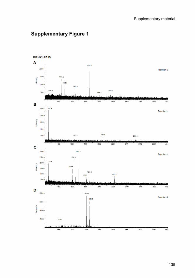
Supplementary material
135
Supplementary Figure 1

Supplementary material
136
Supplementary Figure 1 (continued)
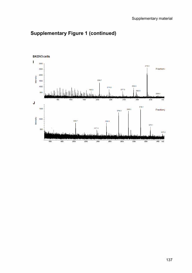
Supplementary material
137
Supplementary Figure 1 (continued)
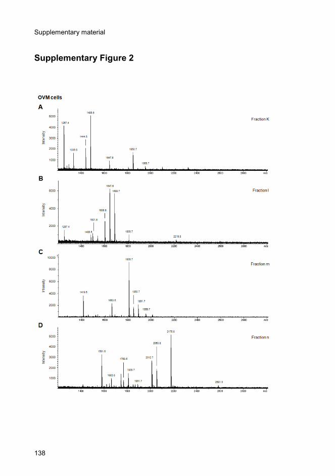
Supplementary material
138
Supplementary Figure 2

Supplementary material
139
Supplementary Figure 2 (continued)

Supplementary material
140
Supplementary Figure 1. MALDI/TOF-MS analysis of desialylated N-linked oligosaccharides from SKOV3 secreted vesicles, obtained from fractions a-j (Figure 22). Supplementary Figure 2. MALDI/TOF-MS analysis of desialylated N-linked oligosaccharides from OVM secreted vesicles, obtained from fractions k-p (Figure 23).
References

References
142

References
143
References
Abbott KL, Nairn AV, Hall EM, Horton MB, McDonald JF, Moremen KW, Dinulescu
DM, and Pierce M (2008). Focused glycomic analysis of the N-linked
glycan biosynthetic pathway in ovarian cancer. Proteomics. 8: 3210-3220.
Abrahams VM, Straszewski SL, Kamsteeg M, Hanczaruk B, Schwartz PE,
Rutherford TJ, and Mor G (2003). Epithelial Ovarian Cancer Cells Secrete
Functional Fas Ligand. Cancer Res. 63: 5573-5581.
Admyre C, Johansson SM, Qazi KR, Filen JJ, Lahesmaa R, Norman M, Neve EP,
Scheynius A, and Gabrielsson S (2007). Exosomes with immune
modulatory features are present in human breast milk. J Immunol. 179:
1969-1978.
Al-Nedawi K, Meehan B, Micallef J, Lhotak V, May L, Guha A, and Rak J (2008).
Intercellular transfer of the oncogenic receptor EGFRvIII by microvesicles
derived from tumour cells. Nat Cell Biol. 10: 619-624.
Alvarez-Erviti L, Seow Y, Yin H, Betts C, Lakhal S, and Wood MJ (2011). Delivery
of siRNA to the mouse brain by systemic injection of targeted exosomes.
Nat Biotechnol. 29: 341-345.
Amour A, Knight CG, Webster A, Slocombe PM, Stephens PE, Knauper V,
Docherty AJ, and Murphy G (2000). The in vitro activity of ADAM-10 is
inhibited by TIMP-1 and TIMP-3. FEBS Lett. 473: 275-279.
Anders A, Gilbert S, Garten W, Postina R, and Fahrenholz F (2001). Regulation of
the {alpha}-secretase ADAM10 by its prodomain and proprotein
convertases. FASEB J. 15: 1837-1839.
Andre F, Schartz NE, Chaput N, Flament C, Raposo G, Amigorena S, Angevin E,
and Zitvogel L (2002). Tumor-derived exosomes: a new source of tumor
rejection antigens. Vaccine. 20 Suppl 4: A28-31.
Aranganathan S, Senthil K, and Nalini N (2005). A case control study of
glycoprotein status in ovarian carcinoma. Clin Biochem. 38: 535-539.
References
144
Arribas, J., and S. Ruiz-Paz. (2005). ADAM 17: Regulation of Ectodomain
Shedding. Springer, Netherlands.
Astronomo RD, and Burton DR (2010). Carbohydrate vaccines: developing sweet
solutions to sticky situations? Nat Rev Drug Discov. 9: 308-324.
Badgwell D, and Bast RC, Jr. (2007). Early detection of ovarian cancer. Dis
Markers. 23: 397-410.
Bard MP, Hegmans JP, Hemmes A, Luider TM, Willemsen R, Severijnen LA, van
Meerbeeck JP, Burgers SA, Hoogsteden HC, and Lambrecht BN (2004).
Proteomic analysis of exosomes isolated from human malignant pleural
effusions. Am J Respir Cell Mol Biol. 31: 114-121.
Barres C, Blanc L, Bette-Bobillo P, Andre S, Mamoun R, Gabius H-J, and Vidal M
(2010). Galectin-5 is bound onto the surface of rat reticulocyte exosomes
and modulates vesicle uptake by macrophages. Blood. 115: 696-705.
Bast RC, Hennessy B, and Mills GB (2009). The biology of ovarian cancer: new
opportunities for translation. Nat Rev Cancer. 9: 415-428.
Beauvillain C, Juste MO, Dion S, Pierre J, and Dimier-Poisson I (2009). Exosomes
are an effective vaccine against congenital toxoplasmosis in mice.
Vaccine. 27: 1750-1757.
Bhattacharyya L, Khan MI, and Brewer CF (1988). Interactions of concanavalin A
with asparagine-linked glycopeptides: formation of homogeneous cross-
linked lattices in mixed precipitation systems. Biochemistry. 27: 8762-8767.
Blanchard N, Lankar D, Faure F, Regnault A, Dumont C, Raposo G, and Hivroz C
(2002). TCR activation of human T cells induces the production of
exosomes bearing the TCR/CD3/zeta complex. J Immunol. 168: 3235-
3241.
Brooks NL, Corey MJ, and Schwalbe RA (2006). Characterization of N-
glycosylation consensus sequences in the Kv3.1 channel. FEBS J. 273:
3287-3300.
References
145
Brooks S (2009). Strategies for Analysis of the Glycosylation of Proteins: Current
Status and Future Perspectives. Molecular Biotechnology. 43: 76-88.
Brown MS, Ye J, Rawson RB, and Goldstein JL (2000). Regulated intramembrane
proteolysis: a control mechanism conserved from bacteria to humans. Cell.
100: 391-398.
Caby M-P, Lankar D, Vincendeau-Scherrer C, Raposo G, and Bonnerot C (2005).
Exosomal-like vesicles are present in human blood plasma. Int. Immunol.
17: 879-887.
Caescu CI, Jeschke GR, and Turk BE (2009). Active-site determinants of substrate
recognition by the metalloproteinases TACE and ADAM10. Biochem J.
424: 79-88.
Cai, G., P.S. Salonikidis, J. Fei, W. Schwarz, R. Schulein, W. Reutter, and H. Fan.
(2005). The role of N-glycosylation in the stability, trafficking and GABA-
uptake of GABA-transporter 1. Terminal N-glycans facilitate efficientGABA-
uptake activity of theGABA transporter. FEBS J. 272:1625-1638.
Castellana D, Zobairi F, Martinez MC, Panaro MA, Mitolo V, Freyssinet JM, and
Kunzelmann C (2009). Membrane microvesicles as actors in the
establishment of a favorable prostatic tumoral niche: a role for activated
fibroblasts and CX3CL1-CX3CR1 axis. Cancer Res. 69: 785-793.
Ceroni A, Maass K, Geyer H, Geyer R, Dell A, and Haslam SM (2008).
GlycoWorkbench: a tool for the computer-assisted annotation of mass
spectra of glycans. J Proteome Res. 7: 1650-1659.
Chan AL, Morris HR, Panico M, Etienne AT, Rogers ME, Gaffney P, Creighton-
Kempsford L, and Dell A (1991). A novel sialylated N-acetylgalactosamine-
containing oligosaccharide is the major complex-type structure present in
Bowes melanoma tissue plasminogen activator. Glycobiology. 1: 173-185.
Chandrasekaran EV, Jain RK, and Matta KL (1992). Ovarian cancer alpha 1,3-L-
fucosyltransferase. Differentiation of distinct catalytic species with the
unique substrate, 3'-sulfo-N-acetyllactosamine in conjunction with other
synthetic acceptors. J Biol Chem. 267: 23806-23814.
References
146
Chantry A, Gregson NA, and Glynn P (1989). A novel metalloproteinase associated
with brain myelin membranes. Isolation and characterization. J Biol Chem.
264: 21603-21607.
Clayton A, Court J, Navabi H, Adams M, Mason MD, Hobot JA, Newman GR, and
Jasani B (2001). Analysis of antigen presenting cell derived exosomes,
based on immuno-magnetic isolation and flow cytometry. J Immunol
Methods. 247: 163-174.
Clayton A, Mitchell JP, Court J, Mason MD, and Tabi Z (2007). Human tumor-
derived exosomes selectively impair lymphocyte responses to interleukin-
2. Cancer Res. 67: 7458-7466.
Cocucci E, Racchetti G, and Meldolesi J (2009). Shedding microvesicles: artefacts
no more. Trends in Cell Biology. 19: 43-51.
Cvetkovic D (2003). Early events in ovarian oncogenesis. Reproductive Biology
and Endocrinology. 1: 68-74.
Dabelsteen E (1996). Cell surface carbohydrates as prognostic markers in human
carcinomas. J Pathol. 179: 358-369.
Dai S, Wei D, Wu Z, Zhou X, Wei X, Huang H, and Li G (2008). Phase I clinical trial
of autologous ascites-derived exosomes combined with GM-CSF for
colorectal cancer. Mol Ther. 16: 782-790.
Dall'olio F (1996). Protein glycosylation in cancer biology: an overview. Clin Mol
Pathol. 49: M126-135.
Dallas DJ, Genever PG, Patton AJ, Millichip MI, McKie N, and Skerry TM (1999).
Localization of ADAM10 and notch receptors in bone. Bone. 25: 9-15.
Davicino RC, Elicabe RJ, Di Genaro MS, and Rabinovich GA (2011). Coupling
pathogen recognition to innate immunity through glycan-dependent
mechanisms. Int Immunopharmacol.
Davidson B, Berner A, Nesland JM, Risberg B, Kristensen GB, Trope CG, and
Bryne M (2000). Carbohydrate antigen expression in primary tumors,
metastatic lesions, and serous effusions from patients diagnosed with
References
147
epithelial ovarian carcinoma: evidence of up-regulated Tn and Sialyl Tn
antigen expression in effusions. Hum Pathol. 31: 1081-1087.
de Gassart A, Geminard C, Fevrier B, Raposo G, and Vidal M (2003). Lipid raft-
associated protein sorting in exosomes. Blood. 102: 4336-4344.
DeFife KM, Jenney CR, Colton E, and Anderson JM (1999). Disruption of
filamentous actin inhibits human macrophage fusion. FASEB J. 13: 823-
832.
DeLano, W.L. (2002). The PyMOL Molecular Graphics System. DeLano Scientific,
Palo Alto.
Denzer K, Kleijmeer MJ, Heijnen HF, Stoorvogel W, and Geuze HJ (2000).
Exosome: from internal vesicle of the multivesicular body to intercellular
signaling device. J Cell Sci. 113: 3365-3374.
Edwards DR, Handsley MM, and Pennington CJ (2008). The ADAM
metalloproteinases. Mol Aspects Med. 29: 258-289.
Escrevente C, Machado E, Brito C, Reis CA, Stoeck A, Runz S, Marme A, Altevogt
P, and Costa J (2006). Different expression levels of alpha3/4
fucosyltransferases and Lewis determinants in ovarian carcinoma tissues
and cell lines. Int J Oncol. 29: 557-566.
Escudier B, Dorval T, Chaput N, Andre F, Caby MP, Novault S, Flament C,
Leboulaire C, Borg C, Amigorena S, Boccaccio C, Bonnerot C, Dhellin O,
Movassagh M, Piperno S, Robert C, Serra V, Valente N, Le Pecq JB,
Spatz A, Lantz O, Tursz T, Angevin E, and Zitvogel L (2005). Vaccination
of metastatic melanoma patients with autologous dendritic cell (DC)
derived-exosomes: results of thefirst phase I clinical trial. J Transl Med. 3:
10.
Fahrenholz F, Gilbert S, Kojro E, Lammich S, and Postina R (2000). Alpha-
secretase activity of the disintegrin metalloprotease ADAM 10. Influences
of domain structure. Ann N Y Acad Sci. 920: 215-222.
References
148
Faure J, Lachenal G, Court M, Hirrlinger J, Chatellard-Causse C, Blot B, Grange J,
Schoehn G, Goldberg Y, and Boyer V (2006). Exosomes are released by
cultured cortical neurones. Molecular and Cellular Neuroscience. 31: 642-
648.
Federici MF, Kudryashov V, Saigo PE, Finstad CL, and Lloyd KO (1999). Selection
of carbohydrate antigens in human epithelial ovarian cancers as targets for
immunotherapy: serous and mucinous tumors exhibit distinctive patterns of
expression. Int J Cancer. 81: 193-198.
Feng D, Zhao W-L, Ye Y-Y, Bai X-C, Liu R-Q, Chang L-F, Zhou Q, and Sui S-F
(2010). Cellular Internalization of Exosomes Occurs Through
Phagocytosis. Traffic. 11: 675-687.
Fevrier B, Vilette D, Laude H, and Raposo G (2005). Exosomes: a bubble ride for
prions? Traffic. 6: 10-17.
Février B, and Raposo G (2004). Exosomes: endosomal-derived vesicles shipping
extracellular messages. Current Opinion in Cell Biology. 16: 415-421.
Fitzner D, Schnaars M, van Rossum D, Krishnamoorthy G, Dibaj P, Bakhti M,
Regen T, Hanisch UK, and Simons M (2011). Selective transfer of
exosomes from oligodendrocytes to microglia by macropinocytosis. J Cell
Sci. 124: 447-458.
Fogel M, Gutwein P, Mechtersheimer S, Riedle S, Stoeck A, Smirnov A, Edler L,
Ben-Arie A, Huszar M, and Altevogt P (2003). L1 expression as a predictor
of progression and survival in patients with uterine and ovarian
carcinomas. The Lancet. 362: 869-875.
Fogel M, Mechtersheimer S, Huszar M, Smirnov A, Abu-Dahi A, Tilgen W,
Reichrath J, Georg T, Altevogt P, and Gutwein P (2003). L1 adhesion
molecule (CD 171) in development and progression of human malignant
melanoma. Cancer Letters. 189: 237-247.
Fu D, Chen L, and O'Neill RA (1994). A detailed structural characterization of
ribonuclease B oligosaccharides by 1H NMR spectroscopy and mass
spectrometry. Carbohydrate Research. 261: 173-186.

References
149
Gao Y, and Pimplikar SW (2001). The gamma -secretase-cleaved C-terminal
fragment of amyloid precursor protein mediates signaling to the nucleus.
PNAS. 98: 14979-14984.
Garner OB, and Baum LG (2008). Galectin-glycan lattices regulate cell-surface
glycoprotein organization and signalling. Biochem Soc Trans. 36: 1472-
1477.
Gavel Y, and von Heijne G (1990). Sequence differences between glycosylated
and non-glycosylated Asn-X-Thr/Ser acceptor sites: implications for protein
engineering. Protein Eng. 3: 433-442.
Gavert N, Conacci-Sorrell M, Gast D, Schneider A, Altevogt P, Brabletz T, and
Ben-Ze'ev A (2005). L1, a novel target of {beta}-catenin signaling,
transforms cells and is expressed at the invasive front of colon cancers. J.
Cell Biol. 168: 633-642.
Ginestra A, Miceli D, Dolo V, Romano FM, and Vittorelli ML (1999). Membrane
vesicles in ovarian cancer fluids: a new potential marker. Anticancer Res.
19: 3439-3445.
Gould SJ, Booth AM, and Hildreth JE (2003). The Trojan exosome hypothesis.
Proc Natl Acad Sci U S A. 100: 10592-10597.
Gupta D, and Lis CG (2009). Role of CA125 in predicting ovarian cancer survival -
a review of the epidemiological literature. J Ovarian Res. 2: 13.
Gutwein P, Mechtersheimer S, Riedle S, Stoeck A, Gast D, Joumaa S, Zentgraf H,
Fogel M, and Altevogt DP (2003). ADAM10-mediated cleavage of L1
adhesion molecule at the cell surface and in released membrane vesicles.
FASEB J. 17: 292-294.
Gutwein P, Stoeck A, Riedle S, Gast D, Runz S, Condon TP, Marme A, Phong M-
C, Linderkamp O, Skorokhod A, and Altevogt P (2005). Cleavage of L1 in
Exosomes and Apoptotic Membrane Vesicles Released from Ovarian
Carcinoma Cells. Clin Cancer Res. 11: 2492-2501.

References
150
Hakomori S (2004). Carbohydrate-to-carbohydrate interaction, through
glycosynapse, as a basis of cell recognition and membrane organization.
Glycoconjugate Journal. 21: 125-137.
Hakulinen J, Sankkila L, Sugiyama N, Lehti K, and Keski-Oja J (2008). Secretion of
active membrane type 1 matrix metalloproteinase (MMP-14) into
extracellular space in microvesicular exosomes. J Cell Biochem. 105:
1211-1218.
Hao S, Bai O, Li F, Yuan J, Laferte S, and Xiang J (2007). Mature dendritic cells
pulsed with exosomes stimulate efficient cytotoxic T-lymphocyte responses
and antitumour immunity. Immunology. 120: 90-102.
Hartmann D, de Strooper B, Serneels L, Craessaerts K, Herreman A, Annaert W,
Umans L, Lubke T, Lena Illert A, von Figura K, and Saftig P (2002). The
disintegrin/metalloprotease ADAM 10 is essential for Notch signalling but
not for {alpha}-secretase activity in fibroblasts. Hum. Mol. Genet. 11: 2615-
2624.
Hattori M, Osterfield M, and Flanagan JG (2000). Regulated Cleavage of a
Contact-Mediated Axon Repellent. Science. 289: 1360-1365.
Hauri, H., C. Appenzeller, F. Kuhn, and O. Nufer. (2000). Lectins and traffic in the
secretory pathway. FEBS Lett. 476:32-37.
Helenius A, and Aebi M (2001). Intracellular functions of N-linked glycans. Science.
291: 2364-2369.
Helenius A, and Aebi M (2004). Roles of N-Linked Glycans in the Endoplasmic
Reticulum. Annual Review of Biochemistry. 73: 1019-1049.
Holschneider CH, and Berek JS (2000). Ovarian cancer: Epidemiology, biology,
and prognostic factors. Seminars in Surgical Oncology. 19: 3-10.
Hoorn EJ, Pisitkun T, Zietse R, Gross P, Frokiaer J, Wang NS, Gonzales PA, Star
RA, and Knepper MA (2005). Prospects for urinary proteomics: exosomes
as a source of urinary biomarkers. Nephrology (Carlton). 10: 283-290.
References
151
Horowitz IR, Cho C, Song M, Flowers LC, Santanam N, Parthasarathy S, and
Ramachandran S (2001). Increased glycodelin levels in gynecological
malignancies. Int J Gynecol Cancer. 11: 173-179.
Hundhausen C, Misztela D, Berkhout TA, Broadway N, Saftig P, Reiss K,
Hartmann D, Fahrenholz F, Postina R, Matthews V, Kallen K-J, Rose-John
S, and Ludwig A (2003). The disintegrin-like metalloproteinase ADAM10 is
involved in constitutive cleavage of CX3CL1 (fractalkine) and regulates
CX3CL1-mediated cell-cell adhesion. Blood. 102: 1186-1195.
Huovila AP, Turner AJ, Pelto-Huikko M, Karkkainen I, and Ortiz RM (2005).
Shedding light on ADAM metalloproteinases. Trends Biochem Sci. 30:
413-422.
Iero M, Valenti R, Huber V, Filipazzi P, Parmiani G, Fais S, and Rivoltini L (2008).
Tumour-released exosomes and their implications in cancer immunity. Cell
Death Differ. 15: 80-88.
Imre T, Kremmer T, Heberger K, Molnar-Szollosi E, Ludanyi K, Pocsfalvi G,
Malorni A, Drahos L, and Vekey K (2008). Mass spectrometric and linear
discriminant analysis of N-glycans of human serum alpha-1-acid
glycoprotein in cancer patients and healthy individuals. J Proteomics. 71:
186-197.
Izquierdo-Useros N, Naranjo-Gomez M, Archer J, Hatch SC, Erkizia I, Blanco J,
Borras FE, Puertas MC, Connor JH, Fernandez-Figueras MT, Moore L,
Clotet B, Gummuluru S, and Martinez-Picado J (2009). Capture and
transfer of HIV-1 particles by mature dendritic cells converges with the
exosome-dissemination pathway. Blood. 113: 2732-2741.
Izquierdo-Useros N, Puertas MC, Borras FE, Blanco J, and Martinez-Picado J
(2011). Exosomes and retroviruses: the chicken or the egg? Cell Microbiol.
13: 10-17.
Janes PW, Saha N, Barton WA, Kolev MV, Wimmer-Kleikamp SH, Nievergall E,
Blobel CP, Himanen J-P, Lackmann M, and Nikolov DB (2005). Adam
References
152
Meets Eph: An ADAM Substrate Recognition Module Acts as a Molecular
Switch for Ephrin Cleavage In trans. Cell. 123: 291-304.
Jankovic MM, and Milutinovic BS (2008). Glycoforms of CA125 antigen as a
possible cancer marker. Cancer Biomark. 4: 35-42.
Jelovac D, and Armstrong DK (2011). Recent progress in the diagnosis and
treatment of ovarian cancer. CA Cancer J Clin. 61: 183-203.
Johnstone RM, Adam M, Hammond JR, Orr L, and Turbide C (1987). Vesicle
formation during reticulocyte maturation. Association of plasma membrane
activities with released vesicles (exosomes). Journal of Biological
Chemistry. 262: 9412-9420.
Kaku T, Ogawa S, Kawano Y, Ohishi Y, Kobayashi H, Hirakawa T, and Nakano H
(2003). Histological classification of ovarian cancer. Medical Electron
Microscopy. 36: 9-17.
Katzmann DJ, Babst M, and Emr SD (2001). Ubiquitin-Dependent Sorting into the
Multivesicular Body Pathway Requires the Function of a Conserved
Endosomal Protein Sorting Complex, ESCRT-I. Cell. 106: 145-155.
Keller S, Sanderson MP, Stoeck A, and Altevogt P (2006). Exosomes: From
biogenesis and secretion to biological function. Immunology Letters. 107:
102-108.
Keller S, König A-K, Marmé F, Runz S, Wolterink S, Koensgen D, Mustea A,
Sehouli J, and Altevogt P (2009). Systemic presence and tumor-growth
promoting effect of ovarian carcinoma released exosomes. Cancer Letters.
278: 73-81.
Kim SH, Lechman ER, Bianco N, Menon R, Keravala A, Nash J, Mi Z, Watkins SC,
Gambotto A, and Robbins PD (2005). Exosomes derived from IL-10-
treated dendritic cells can suppress inflammation and collagen-induced
arthritis. J Immunol. 174: 6440-6448.
Knibbs RN, Goldstein IJ, Ratcliffe RM, and Shibuya N (1991). Characterization of
the carbohydrate binding specificity of the leukoagglutinating lectin from
References
153
Maackia amurensis. Comparison with other sialic acid-specific lectins.
Journal of Biological Chemistry. 266: 83-88.
Ko SY, Lin SC, Wong YK, Liu CJ, Chang KW, and Liu TY (2007). Increase of
disintergin metalloprotease 10 (ADAM10) expression in oral squamous cell
carcinoma. Cancer Lett. 245: 33-43.
Krishnamoorthy L, Bess JW, Preston AB, Nagashima K, and Mahal LK (2009).
HIV-1 and microvesicles from T cells share a common glycome, arguing
for a common origin. Nat Chem Biol. 5: 244-250.
Lakkaraju A, and Rodriguez-Boulan E (2008). Itinerant exosomes: emerging roles
in cell and tissue polarity. Trends in Cell Biology. 18: 199-209.
Lammich S, Kojro E, Postina R, Gilbert S, Pfeiffer R, Jasionowski M, Haass C, and
Fahrenholz F (1999). Constitutive and regulated alpha-secretase cleavage
of Alzheimer's amyloid precursor protein by a disintegrin metalloprotease.
Proc Natl Acad Sci U S A. 96: 3922-3927.
Lamparski HG, Metha-Damani A, Yao J-Y, Patel S, Hsu D-H, Ruegg C, and Le
Pecq J-B (2002). Production and characterization of clinical grade
exosomes derived from dendritic cells. Journal of Immunological Methods.
270: 211-226.
Laskowski, R.A., M.W. MacArthur, D.S. Moss, and J.M. Thornton. (1993).
PROCHECK: a program to check the stereochemical quality of protein
structures. J. Appl. Crystallogr. 26:283-291.
Lemjabbar H, and Basbaum C (2002). Platelet-activating factor receptor and
ADAM10 mediate responses to Staphylococcus aureus in epithelial cells.
Nat. Med. 8: 41-46.
Lewandrowski, U., J. Moebius, U. Walter, and A. Sickmann. (2006). Elucidation of
N-glycosylation sites on human platelet proteins: a glycoproteomic
approach. Mol. Cell. Proteomics. 5:226-233.

References
154
Li J, Sherman-Baust CA, Tsai-Turton M, Bristow RE, Roden RB, and Morin PJ
(2009). Claudin-containing exosomes in the peripheral circulation of
women with ovarian cancer. BMC Cancer. 9: 244.
Li M, Song L, and Qin X (2010). Glycan changes: cancer metastasis and anti-
cancer vaccines. J Biosci. 35: 665-673.
Lu Q, Zhang J, Allison R, Gay H, Yang WX, Bhowmick NA, Frelix G, Shappell S,
and Chen YH (2009). Identification of extracellular delta-catenin
accumulation for prostate cancer detection. Prostate. 69: 411-418.
Lunn CA, Fan X, Dalie B, Miller K, Zavodny PJ, Narula SK, and Lundell D (1997).
Purification of ADAM 10 from bovine spleen as a TNF[alpha] convertase.
FEBS Letters. 400: 333-335.
Machado E, Kandzia S, Carilho R, Altevogt P, Conradt HS, and Costa J (2011). N-
Glycosylation of total cellular glycoproteins from the human ovarian
carcinoma SKOV3 cell line and of recombinantly expressed human
erythropoietin. Glycobiology. 21: 376-386.
Marcello E, Gardoni F, Mauceri D, Romorini S, Jeromin A, Epis R, Borroni B,
Cattabeni F, Sala C, Padovani A, and Di Luca M (2007). Synapse-
associated protein-97 mediates alpha-secretase ADAM10 trafficking and
promotes its activity. J Neurosci. 27: 1682-1691.
Marek KW, Vijay IK, and Marth JD (1999). A recessive deletion in the GlcNAc-1-
phosphotransferase gene results in peri-implantation embryonic lethality.
Glycobiology. 9: 1263-1271.
Maretzky T, Schulte M, Ludwig A, Rose-John S, Blobel C, Hartmann D, Altevogt P,
Saftig P, and Reiss K (2005). L1 Is Sequentially Processed by Two
Differently Activated Metalloproteases and Presenilin/{gamma}-Secretase
and Regulates Neural Cell Adhesion, Cell Migration, and Neurite
Outgrowth. Mol. Cell. Biol. 25: 9040-9053.
Martin L, Fluhrer R, Reiss K, Kremmer E, Saftig P, and Haass C (2008). Regulated
intramembrane proteolysis of Bri2 (Itm2b) by ADAM10 and
SPPL2a/SPPL2b. J Biol Chem. 283: 1644-1652.

References
155
Martina, J.A., J.L. Daniotti, and H.J. Maccioni. (1998). Influence of N-glycosylation
and N-glycan trimming on the activity and intracellular traffic of GD3
synthase. J. Biol. Chem. 273:3725-3731.
Maskos, K., C. Fernandez-Catalan, R. Huber, G.P. Bourenkov, H. Bartunik, G.A.
Ellestad, P. Reddy, M.F. Wolfson, C.T. Rauch, B.J. Castner, R. Davis, H.R.
Clarke, M. Petersen, J.N. Fitzner, D.P. Cerretti, C.J. March, R.J. Paxton,
R.A. Black, and W. Bode. (1998). Crystal structure of the catalytic domain
of human tumor necrosis factor-alphaconverting enzyme. Proc. Natl. Acad.
Sci. U. S. A. 95:3408-3412.
Mathivanan S, Ji H, and Simpson RJ (2010). Exosomes: extracellular organelles
important in intercellular communication. J Proteomics. 73: 1907-1920.
McCulloch DR, Akl P, Samaratunga H, Herington AC, and Odorico DM (2004).
Expression of the Disintegrin Metalloprotease, ADAM-10, in Prostate
Cancer and Its Regulation by Dihydrotestosterone, Insulin-Like Growth
Factor I, and Epidermal Growth Factor in the Prostate Cancer Cell Model
LNCaP. Clin Cancer Res. 10: 314-323.
Mechtersheimer S, Gutwein P, Agmon-Levin N, Stoeck A, Oleszewski M, Riedle S,
Postina R, Fahrenholz F, Fogel M, Lemmon V, and Altevogt P (2001).
Ectodomain shedding of L1 adhesion molecule promotes cell migration by
autocrine binding to integrins. J. Cell Biol. 155: 661-674.
Michelsen U, and von Hagen J (2009). Chapter 19 Isolation of Subcellular
Organelles and Structures. in Methods in Enzymology. (Richard, R. B., and
Murray, P. D. eds.). Academic Press. pp 305-328.
Millichip MI, Dallas DJ, Wu E, Dale S, and McKie N (1998). The Metallo-Disintegrin
ADAM10 (MADM) from Bovine Kidney Has Type IV Collagenase Activityin
Vitro. Biochemical and Biophysical Research Communications. 245: 594-
598.
Mohler KM, Sleath PR, Fitzner JN, Cerretti DP, Alderson M, Kerwar SS, Torrance
DS, Otten-Evans C, Greenstreet T, Weerawarna K, and et al. (1994).
References
156
Protection against a lethal dose of endotoxin by an inhibitor of tumour
necrosis factor processing. Nature. 370: 218-220.
Morais, V.A., M.T. Costa, and J. Costa. (2003). N-glycosylation of recombinant
human fucosyltransferase III is required for its in vivo folding in mammalian
and insect cells. Biochim. Biophys. Acta. 1619:133-138.
Morais, V.A., C. Brito, D.S.C. Pijak, A.S, R.R. Fortna, T. Li, P.C. Wong, R.W.
Doms, and J. Costa. (2006). N-glycosylation of human nicastrin is required
for interaction with the lectins from the secretory pathway calnexin and
ERGIC-53. Biochim. Biophys. Acta. 1762:802-810.
Morelli AE, Larregina AT, Shufesky WJ, Sullivan MLG, Stolz DB, Papworth GD,
Zahorchak AF, Logar AJ, Wang Z, Watkins SC, Falo LD, Jr., and Thomson
AW (2004). Endocytosis, intracellular sorting, and processing of exosomes
by dendritic cells. Blood. 104: 3257-3266.
Morse MA, Garst J, Osada T, Khan S, Hobeika A, Clay TM, Valente N, Shreeniwas
R, Sutton MA, Delcayre A, Hsu DH, Le Pecq JB, and Lyerly HK (2005). A
phase I study of dexosome immunotherapy in patients with advanced non-
small cell lung cancer. J Transl Med. 3: 9.
Moss ML, Bomar M, Liu Q, Sage H, Dempsey P, Lenhart PM, Gillispie PA, Stoeck
A, Wildeboer D, Bartsch JW, Palmisano R, and Zhou P (2007). The
ADAM10 prodomain is a specific inhibitor of ADAM10 proteolytic activity
and inhibits cellular shedding events. J Biol Chem. 282: 35712-35721.
Moss ML, Stoeck A, Yan W, and Dempsey PJ (2008). ADAM10 as a target for anti-
cancer therapy. Curr Pharm Biotechnol. 9: 2-8.
Muralidharan-Chari V, Clancy JW, Sedgwick A, and D'Souza-Schorey C (2010).
Microvesicles: mediators of extracellular communication during cancer
progression. J Cell Sci. 123: 1603-1611.
Nagano O, Murakami D, Hartmann D, de Strooper B, Saftig P, Iwatsubo T,
Nakajima M, Shinohara M, and Saya H (2004). Cell-matrix interaction via
CD44 is independently regulated by different metalloproteinases activated

References
157
in response to extracellular Ca2+ influx and PKC activation. J. Cell Biol.
165: 893-902.
Nilsson J, Skog J, Nordstrand A, Baranov V, Mincheva-Nilsson L, Breakefield XO,
and Widmark A (2009). Prostate cancer-derived urine exosomes: a novel
approach to biomarkers for prostate cancer. Br J Cancer. 100: 1603-1607.
Obregon C, Rothen-Rutishauser B, Gerber P, Gehr P, and Nicod LP (2009). Active
Uptake of Dendritic Cell-Derived Exovesicles by Epithelial Cells Induces
the Release of Inflammatory Mediators through a TNF-{alpha}-Mediated
Pathway. Am J Pathol. 175: 696-705.
Ogawa Y, Kanai-Azuma M, Akimoto Y, Kawakami H, and Yanoshita R (2008).
Exosome-like vesicles with dipeptidyl peptidase IV in human saliva. Biol
Pharm Bull. 31: 1059-1062.
Ohtsubo K, and Marth JD (2006). Glycosylation in cellular mechanisms of health
and disease. Cell. 126: 855-867.
Oka JA, Christensen MD, and Weigel PH (1989). Hyperosmolarity inhibits
galactosyl receptor-mediated but not fluid phase endocytosis in isolated rat
hepatocytes. J Biol Chem. 264: 12016-12024.
Opdenakker G, Rudd PM, Ponting CP, and Dwek RA (1993). Concepts and
principles of glycobiology. FASEB J. 7: 1330-1337.
Ostrowski M, Carmo NB, Krumeich S, Fanget I, Raposo G, Savina A, Moita CF,
Schauer K, Hume AN, Freitas RP, Goud B, Benaroch P, Hacohen N,
Fukuda M, Desnos C, Seabra MC, Darchen F, Amigorena S, Moita LF, and
Thery C (2010). Rab27a and Rab27b control different steps of the
exosome secretion pathway. Nat Cell Biol. 12: 19-30.
Pandiella A, and Massague J (1991). Multiple signals activate cleavage of the
membrane transforming growth factor-alpha precursor. J Biol Chem. 266:
5769-5773.
Parish CR (1999). Fluorescent dyes for lymphocyte migration and proliferation
studies. Immunol Cell Biol. 77: 499-508.
References
158
Parkin E, and Harris B (2009). A disintegrin and metalloproteinase (ADAM)-
mediated ectodomain shedding of ADAM10. J Neurochem. 108: 1464-
1479.
Parolini I, Federici C, Raggi C, Lugini L, Palleschi S, De Milito A, Coscia C, Iessi E,
Logozzi M, Molinari A, Colone M, Tatti M, Sargiacomo M, and Fais S
(2009). Microenvironmental pH Is a Key Factor for Exosome Traffic in
Tumor Cells. Journal of Biological Chemistry. 284: 34211-34222.
Peng P, Yan Y, and Keng S (2011). Exosomes in the ascites of ovarian cancer
patients: origin and effects on anti-tumor immunity. Oncol Rep. 25: 749-
762.
Peracaula R, Royle L, Tabares G, Mallorqui-Fernandez G, Barrabes S, Harvey DJ,
Dwek RA, Rudd PM, and de Llorens R (2003). Glycosylation of human
pancreatic ribonuclease: differences between normal and tumor states.
Glycobiology. 13: 227-244.
Peracaula R, Barrabes S, Sarrats A, Rudd PM, and de Llorens R (2008). Altered
glycosylation in tumours focused to cancer diagnosis. Dis Markers. 25:
207-218.
Petrescu, A.J., A.L. Milac, S.M. Petrescu, R.A. Dwek, and M.R. Wormald. (2004).
Statistical analysis of the protein environment of N-glycosylation sites:
implications for occupancy, structure, and folding. Glycobiology. 14:103-
114.
Pisitkun T, Shen RF, and Knepper MA (2004). Identification and proteomic profiling
of exosomes in human urine. Proc Natl Acad Sci U S A. 101: 13368-
13373.
Pruessmeyer J, and Ludwig A (2009). The good, the bad and the ugly substrates
for ADAM10 and ADAM17 in brain pathology, inflammation and cancer.
Semin Cell Dev Biol. 20: 164-174.
Rabinowits G, Gercel-Taylor C, Day JM, Taylor DD, and Kloecker GH (2009).
Exosomal microRNA: a diagnostic marker for lung cancer. Clin Lung
Cancer. 10: 42-46.
References
159
Raposo G, Nijman HW, Stoorvogel W, Liejendekker R, Harding CV, Melief CJ, and
Geuze HJ (1996). B lymphocytes secrete antigen-presenting vesicles. J
Exp Med. 183: 1161-1172.
Raposo G, Tenza D, Mecheri S, Peronet R, Bonnerot C, and Desaymard C (1997).
Accumulation of major histocompatibility complex class II molecules in
mast cell secretory granules and their release upon degranulation. Mol Biol
Cell. 8: 2631-2645.
Reiss K, Maretzky T, Ludwig A, Tousseyn T, de Strooper B, Hartmann D, and
Saftig P (2005). ADAM10 cleavage of N-cadherin and regulation of cell-cell
adhesion and beta-catenin nuclear signalling. EMBO J. 24: 742-752.
Reiss K, Ludwig A, and Saftig P (2006). Breaking up the tie: disintegrin-like
metalloproteinases as regulators of cell migration in inflammation and
invasion. Pharmacol Ther. 111: 985-1006.
Reiss K, and Saftig P (2009). The "a disintegrin and metalloprotease" (ADAM)
family of sheddases: physiological and cellular functions. Semin Cell Dev
Biol. 20: 126-137.
Ristorcelli E, Beraud E, Verrando P, Villard C, Lafitte D, Sbarra V, Lombardo D,
and Verine A (2008). Human tumor nanoparticles induce apoptosis of
pancreatic cancer cells. FASEB J. 22: 3358-3369.
Rodal SK, Skretting G, Garred O, Vilhardt F, van Deurs B, and Sandvig K (1999).
Extraction of cholesterol with methyl-beta-cyclodextrin perturbs formation
of clathrin-coated endocytic vesicles. Mol Biol Cell. 10: 961-974.
Runz S, Keller S, Rupp C, Stoeck A, Issa Y, Koensgen D, Mustea A, Sehouli J,
Kristiansen G, and Altevogt P (2007). Malignant ascites-derived exosomes
of ovarian carcinoma patients contain CD24 and EpCAM. Gynecol Oncol.
107: 563-571.
Safaei R, Larson BJ, Cheng TC, Gibson MA, Otani S, Naerdemann W, and Howell
SB (2005). Abnormal lysosomal trafficking and enhanced exosomal export
of cisplatin in drug-resistant human ovarian carcinoma cells. Mol Cancer
Ther. 4: 1595-1604.
References
160
Saldova R, Royle L, Radcliffe CM, Abd Hamid UM, Evans R, Arnold JN, Banks RE,
Hutson R, Harvey DJ, Antrobus R, Petrescu SM, Dwek RA, and Rudd PM
(2007). Ovarian cancer is associated with changes in glycosylation in both
acute-phase proteins and IgG. Glycobiology. 17: 1344-1356.
Sanchez, R., and A. Sali. (1997). Advances in comparative protein-structure
modelling. Curr. Opin. Struct. Biol. 7:206-214.
Sanderson MP, Erickson SN, Gough PJ, Garton KJ, Wille PT, Raines EW, Dunbar
AJ, and Dempsey PJ (2005). ADAM10 Mediates Ectodomain Shedding of
the Betacellulin Precursor Activated by p-Aminophenylmercuric Acetate
and Extracellular Calcium Influx. J. Biol. Chem. 280: 1826-1837.
Sasaki H, Ochi N, Dell A, and Fukuda M (1988). Site-specific glycosylation of
human recombinant erythropoietin: analysis of glycopeptides or peptides at
each glycosylation site by fast atom bombardment mass spectrometry.
Biochemistry. 27: 8618-8626.
Schauer R (2009). Sialic acids as regulators of molecular and cellular interactions.
Current Opinion in Structural Biology. 19: 507-514.
Scheiffele P, Peranen J, and Simons K (1995). N-glycans as apical sorting signals
in epithelial cells. Nature. 378: 96-98.
Schlondorff J, and Blobel CP (1999). Metalloprotease-disintegrins: modular
proteins capable of promoting cell-cell interactions and triggering signals
by protein-ectodomain shedding. J Cell Sci. 112 ( Pt 21): 3603-3617.
Schorey JS, and Bhatnagar S (2008). Exosome Function: From Tumor
Immunology to Pathogen Biology. Traffic. 9: 871-881.
Scully RE (1987). Classification of human ovarian tumors. Environ Health
Perspect. 73: 15-25.
Seals DF, and Courtneidge SA (2003). The ADAMs family of metalloproteases:
multidomain proteins with multiple functions. Genes Dev. 17: 7-30.

References
161
Shedden K, Xie XT, Chandaroy P, Chang YT, and Rosania GR (2003). Expulsion
of small molecules in vesicles shed by cancer cells: association with gene
expression and chemosensitivity profiles. Cancer Res. 63: 4331-4337.
Simons M, and Raposo G (2009). Exosomes--vesicular carriers for intercellular
communication. Curr Opin Cell Biol. 21: 575-581.
Simpson RJ, Jensen SS, and Lim JW (2008). Proteomic profiling of exosomes:
current perspectives. Proteomics. 8: 4083-4099.
Simpson RJ, Lim JWE, Moritz RL, and Mathivanan S (2009). Exosomes: proteomic
insights and diagnostic potential. Expert Review of Proteomics. 6: 267-283.
Skog J, Wurdinger T, van Rijn S, Meijer DH, Gainche L, Curry WT, Carter BS,
Krichevsky AM, and Breakefield XO (2008). Glioblastoma microvesicles
transport RNA and proteins that promote tumour growth and provide
diagnostic biomarkers. Nat Cell Biol. 10: 1470-1476.
Skropeta D (2009). The effect of individual N-glycans on enzyme activity. Bioorg
Med Chem. 17: 2645-2653.
Sousa VL, Brito C, Costa T, Lanoix J, Nilsson T, and Costa J (2003). Importance of
Cys, Gln, and Tyr from the Transmembrane Domain of Human alpha 3/4
Fucosyltransferase III for Its Localization and Sorting in the Golgi of Baby
Hamster Kidney Cells. J. Biol. Chem. 278: 7624-7629.
Spik G, Bayard B, Fournet B, Strecker G, Bouquelet S, and Montreuil J (1975).
Studies on glycoconjugates. LXIV. Complete structure of two carbohydrate
units of human serotransferrin. FEBS Letters. 50: 296-299.
Stoeck A, Keller S, Riedle S, Sanderson MP, Runz S, Le Naour F, Gutwein P,
Ludwig A, Rubinstein E, and Altevogt P (2006). A role for exosomes in the
constitutive and stimulus-induced ectodomain cleavage of L1 and CD44.
Biochem J. 393: 609-618.
Stoorvogel W, Kleijmeer MJ, Geuze HJ, and Raposo G (2002). The Biogenesis
and Functions of Exosomes. Traffic. 3: 321-330.

References
162
Subra C, Laulagnier K, Perret B, and Record M (2007). Exosome lipidomics
unravels lipid sorting at the level of multivesicular bodies. Biochimie. 89:
205-212.
Sun D, Zhuang X, Xiang X, Liu Y, Zhang S, Liu C, Barnes S, Grizzle W, Miller D,
and Zhang HG (2010). A novel nanoparticle drug delivery system: the anti-
inflammatory activity of curcumin is enhanced when encapsulated in
exosomes. Mol Ther. 18: 1606-1614.
Takahashi N, Yamamoto E, Ino K, Miyoshi E, Nagasaka T, Kajiyama H, Shibata K,
Nawa A, and Kikkawa F (2009). High expression of N-
acetylglucosaminyltransferase V in mucinous tumors of the ovary. Oncol
Rep. 22: 1027-1032.
Takahashi T, Ikeda Y, Miyoshi E, Yaginuma Y, Ishikawa M, and Taniguchi N
(2000). alpha1,6fucosyltransferase is highly and specifically expressed in
human ovarian serous adenocarcinomas. Int J Cancer. 88: 914-919.
Tan A, De La Pena H, and Seifalian AM (2010). The application of exosomes as a
nanoscale cancer vaccine. Int J Nanomedicine. 5: 889-900.
Tanida S, Joh T, Itoh K, Kataoka H, Sasaki M, Ohara H, Nakazawa T, Nomura T,
Kinugasa Y, and Ohmoto H (2004). The mechanism of cleavage of EGFR
ligands induced by inflammatory cytokines in gastric cancer cells.
Gastroenterology. 127: 559-569.
Tanious FA, Veal JM, Buczak H, Ratmeyer LS, and Wilson WD (1992). DAPI (4',6-
diamidino-2-phenylindole) binds differently to DNA and RNA: minor-groove
binding at AT sites and intercalation at AU sites. Biochemistry. 31: 3103-
3112.
Taylor DD, and Gercel-Taylor C (2008). MicroRNA signatures of tumor-derived
exosomes as diagnostic biomarkers of ovarian cancer. Gynecol Oncol.
110: 13-21.
Taylor ME, and Drickamer K (2006). Introduction to Glycobiology. Second ed.
Oxford University Press, New York.

References
163
Tellier E, Canault M, Rebsomen L, Bonardo B, Juhan-Vague I, Nalbone G, and
Peiretti F (2006). The shedding activity of ADAM17 is sequestered in lipid
rafts. Exp Cell Res. 312: 3969-3980.
Thery C, Regnault A, Garin J, Wolfers J, Zitvogel L, Ricciardi-Castagnoli P, Raposo
G, and Amigorena S (1999). Molecular characterization of dendritic cell-
derived exosomes. Selective accumulation of the heat shock protein
hsc73. J Cell Biol. 147: 599-610.
Thery C, Amigorena S, Raposo G, and Clayton A (2006). Isolation and
characterization of exosomes from cell culture supernatants and biological
fluids. Curr Protoc Cell Biol. Chapter 3: Unit 3 22.
Thery C, Ostrowski M, and Segura E (2009). Membrane vesicles as conveyors of
immune responses. Nat Rev Immunol. 9: 581-593.
Tousseyn T, Thathiah A, Jorissen E, Raemaekers T, Konietzko U, Reiss K, Maes
E, Snellinx A, Serneels L, Nyabi O, Annaert W, Saftig P, Hartmann D, and
De Strooper B (2009). ADAM10, the rate-limiting protease of regulated
intramembrane proteolysis of Notch and other proteins, is processed by
ADAMS-9, ADAMS-15, and the gamma-secretase. J Biol Chem. 284:
11738-11747.
Trajkovic K, Hsu C, Chiantia S, Rajendran L, Wenzel D, Wieland F, Schwille P,
Brugger B, and Simons M (2008). Ceramide Triggers Budding of Exosome
Vesicles into Multivesicular Endosomes. Science. 319: 1244-1247.
Trombetta, E.S., and A.J. Parodi. (2003). Quality control and protein folding in the
secretory pathway. Annu. Rev. Cell Dev. Biol. . 19:649-676.
Turner GA, Goodarzi MT, and Thompson S (1995). Glycosylation of alpha-1-
proteinase inhibitor and haptoglobin in ovarian cancer: evidence for two
different mechanisms. Glycoconj J. 12: 211-218.
Ungar D (2009). Golgi linked protein glycosylation and associated diseases. Semin
Cell Dev Biol. 20: 762-769.
References
164
Valadi H, Ekstrom K, Bossios A, Sjostrand M, Lee JJ, and Lotvall JO (2007).
Exosome-mediated transfer of mRNAs and microRNAs is a novel
mechanism of genetic exchange between cells. Nat Cell Biol. 9: 654-659.
Valenti R, Huber V, Iero M, Filipazzi P, Parmiani G, and Rivoltini L (2007). Tumor-
Released Microvesicles as Vehicles of Immunosuppression. Cancer Res.
67: 2912-2915.
van den Berg TK, Honing H, Franke N, van Remoortere A, Schiphorst WE, Liu FT,
Deelder AM, Cummings RD, Hokke CH, and van Die I (2004). LacdiNAc-
glycans constitute a parasite pattern for galectin-3-mediated immune
recognition. J Immunol. 173: 1902-1907.
van Kooyk Y, and Rabinovich GA (2008). Protein-glycan interactions in the control
of innate and adaptive immune responses. Nat Immunol. 9: 593-601.
van Niel G, Raposo G, Candalh C, Boussac M, Hershberg R, Cerf-Bensussan N,
and Heyman M (2001). Intestinal epithelial cells secrete exosome-like
vesicles. Gastroenterology. 121: 337-349.
van Niel G, Porto-Carreiro I, Simoes S, and Raposo G (2006). Exosomes: A
Common Pathway for a Specialized Function. J Biochem. 140: 13-21.
Varki A (1993). Biological roles of oligosaccharides: all of the theories are correct.
Glycobiology. 3: 97-130.
Varki A, Cummings RD, Esko JD, Freeze HH, Stanley P, Bertozzi CR, Hart GW,
and Etzler ME (2009). Essentials of Glycobiology. Second ed. Cold Spring
Harbor Laboratory Press, Cold Spring Harbor, (NY).
Vella LJ, Sharples RA, Lawson VA, Masters CL, Cappai R, and Hill AF (2007).
Packaging of prions into exosomes is associated with a novel pathway of
PrP processing. The Journal of Pathology. 211: 582-590.
Vidal M, Mangeat P, and Hoekstra D (1997). Aggregation reroutes molecules from
a recycling to a vesicle-mediated secretion pathway during reticulocyte
maturation. J Cell Sci. 110 ( Pt 16): 1867-1877.

References
165
Vigerust DJ, and Shepherd VL (2007). Virus glycosylation: role in virulence and
immune interactions. Trends in Microbiology. 15: 211-218.
Vliegenthart JF (2006). Carbohydrate based vaccines. FEBS Lett. 580: 2945-2950.
Wang PH, Lee WL, Juang CM, Yang YH, Lo WH, Lai CR, Hsieh SL, and Yuan CC
(2005). Altered mRNA expressions of sialyltransferases in ovarian cancers.
Gynecol Oncol. 99: 631-639.
Welton JL, Khanna S, Giles PJ, Brennan P, Brewis IA, Staffurth J, Mason MD, and
Clayton A (2010). Proteomics analysis of bladder cancer exosomes. Mol
Cell Proteomics. 9: 1324-1338.
Weskamp G, Ford JW, Sturgill J, Martin S, Docherty AJP, Swendeman S,
Broadway N, Hartmann D, Saftig P, Umland S, Sehara-Fujisawa A, Black
RA, Ludwig A, Becherer JD, Conrad DH, and Blobel CP (2006). ADAM10
is a principal 'sheddase' of the low-affinity immunoglobulin E receptor
CD23. Nat Immunol. 7: 1293-1298.
West MA, Bretscher MS, and Watts C (1989). Distinct endocytotic pathways in
epidermal growth factor-stimulated human carcinoma A431 cells. J Cell
Biol. 109: 2731-2739.
White JM (2003). ADAMs: modulators of cell-cell and cell-matrix interactions. Curr
Opin Cell Biol. 15: 598-606.
Wild-Bode C, Fellerer K, Kugler J, Haass C, and Capell A (2006). A Basolateral
Sorting Signal Directs ADAM10 to Adherens Junctions and Is Required for
Its Function in Cell Migration. Journal of Biological Chemistry. 281: 23824-
23829.
Wiley RD, and Gummuluru S (2006). Immature dendritic cell-derived exosomes
can mediate HIV-1 trans infection. Proc Natl Acad Sci U S A. 103: 738-
743.
Wolfers J, Lozier A, Raposo G, Regnault A, Thery C, Masurier C, Flament C,
Pouzieux S, Faure F, Tursz T, Angevin E, Amigorena S, and Zitvogel L
References
166
(2001). Tumor-derived exosomes are a source of shared tumor rejection
antigens for CTL cross-priming. Nat Med. 7: 297-303.
Wolfsberg TG, Primakoff P, Myles DG, and White JM (1995). ADAM, a novel family
of membrane proteins containing A Disintegrin And Metalloprotease
domain: multipotential functions in cell-cell and cell- matrix interactions. J.
Cell Biol. 131: 275-278.
Wu E, Croucher PI, and McKie N (1997). Expression of Members of the Novel
Membrane Linked Metalloproteinase Family ADAM in Cells Derived from a
Range of Haematological Malignancies. Biochemical and Biophysical
Research Communications. 235: 437-442.
Yamazaki K, Mizui Y, Sagane K, and Tanaka I (1997). Assignment of a disintegrin
and metalloproteinase domain 10 (Adam10) gene to mouse chromosome
9. Genomics. 46: 528-529.
Yan Y, Shirakabe K, and Werb Z (2002). The metalloprotease Kuzbanian
(ADAM10) mediates the transactivation of EGF receptor by G protein-
coupled receptors. J. Cell Biol. 158: 221-226.
Yavari R, Adida C, Bray-Ward P, Brines M, and Xu T (1998). Human
metalloprotease-disintegrin Kuzbanian regulates sympathoadrenal cell fate
in development and neoplasia. Hum. Mol. Genet. 7: 1161-1167.
Yu S, Liu C, Su K, Wang J, Liu Y, Zhang L, Li C, Cong Y, Kimberly R, Grizzle WE,
Falkson C, and Zhang HG (2007). Tumor exosomes inhibit differentiation
of bone marrow dendritic cells. J Immunol. 178: 6867-6875.
Zhang HG, and Grizzle WE (2011). Exosomes and cancer: a newly described
pathway of immune suppression. Clin Cancer Res. 17: 959-964.
Zitvogel L, Regnault A, Lozier A, Wolfers J, Flament C, Tenza D, Ricciardi-
Castagnoli P, Raposo G, and Amigorena S (1998). Eradication of
established murine tumors using a novel cell-free vaccine: dendritic cell-
derived exosomes. Nat Med. 4: 594-600.


![The Role of Exosomes in Bone Remodeling: …downloads.hindawi.com/journals/dm/2019/9417914.pdfregulation [35]. 3.2. Exosomes from Osteoblasts. Ample data suggest that exosomes shed](https://img.pdfslide.net/doc/110x75/5f03c0c07e708231d40a9922/the-role-of-exosomes-in-bone-remodeling-regulation-35-32-exosomes-from-osteoblasts.jpg)