Embed Size (px)
Citation preview
REVIEW Open Access
Proteinase 3; a potential target in chronicobstructive pulmonary disease and otherchronic inflammatory diseasesHelena Crisford1,3* , Elizabeth Sapey1 and Robert A. Stockley2
Abstract
Chronic Obstructive Pulmonary Disease (COPD) is a common, multifactorial lung disease which results in significantimpairment of patients’ health and a large impact on society and health care burden. It is believed to be the resultof prolonged, destructive neutrophilic inflammation which results in progressive damage to lung structures. Duringthis process, large quantities of neutrophil serine proteinases (NSPs) are released which initiate the damage andcontribute towards driving a persistent inflammatory state.Neutrophil elastase has long been considered the key NSP involved in the pathophysiology of COPD. However, inrecent years, a significant role for Proteinase 3 (PR3) in disease development has emerged, both in COPD and otherchronic inflammatory conditions. Therefore, there is a need to investigate the importance of PR3 in diseasedevelopment and hence its potential as a therapeutic target. Research into PR3 has largely been confined to its roleas an autoantigen, but PR3 is involved in triggering inflammatory pathways, disrupting cellular signalling, degradingkey structural proteins, and pathogen response.This review summarises what is presently known about PR3, explores its involvement particularly in thedevelopment of COPD, and indicates areas requiring further investigation.
Keywords: Proteinase 3/myeloblastin, Serine proteinases, Chronic obstructive pulmonary disease, Lungs, Inflammation
BackgroundThe serine proteinase Proteinase 3 (PR3) is an enzymereleased during neutrophilic inflammation and is capableof cleaving many targets including key structural proteinsof the lung. Chronic Obstructive Pulmonary Disease(COPD) is an inflammatory condition associated withneutrophilic inflammation. For this reason neutrophilelastase (NE) has long been considered to be a central,proteinase in the pathophysiology as it can replicate manyof the structural changes of the disease and hence a poten-tial target for therapeutic manipulation, PR3,another keyneutrophil serine proteinase has largely been ignored,even though it may have an important additional role in
the lung as well as other human diseases [1]. This reviewsummarises the current literature to provide an update onthe potential role of PR3 in health and disease, with aprimary focus on COPD.
Proteinase 3PR3, alternatively referred to as myeloblastin, azurophilgranule protein-7 or p29b, is a highly abundant neutro-phil protein which is genetically transcribed in primitivemyeloid and monocytic progenitor cells, and expressedin cells of granulocyte and monocyte linage, especiallyneutrophils but including mast cells and basophils [2–5]and in the neutrophil, it is mainly located within theprimary azurophil granules of the mature cell but is alsopresent in specific granules, secretory vesicles, and onthe cell surface [6, 7]. It is expressed constitutively onthe membrane by naïve neutrophils in peripheral bloodof healthy individuals (known as “constitutive” PR3) andis secreted into extracellular medium by activated
* Correspondence: [email protected] of Inflammation and Ageing, University of Birmingham, Edgbaston,Birmingham B15 2GW, UK3Institute of Inflammation and Ageing, College of Medical and DentalSciences, Centre for Translational Inflammation Research, University ofBirmingham Research Laboratories, Queen Elizabeth Hospital Birmingham,Mindelsohn Way, Birmingham B15 2WB, UKFull list of author information is available at the end of the article
© The Author(s). 2018 Open Access This article is distributed under the terms of the Creative Commons Attribution 4.0International License (http://creativecommons.org/licenses/by/4.0/), which permits unrestricted use, distribution, andreproduction in any medium, provided you give appropriate credit to the original author(s) and the source, provide a link tothe Creative Commons license, and indicate if changes were made. The Creative Commons Public Domain Dedication waiver(http://creativecommons.org/publicdomain/zero/1.0/) applies to the data made available in this article, unless otherwise stated.
Crisford et al. Respiratory Research (2018) 19:180 https://doi.org/10.1186/s12931-018-0883-z
neutrophils following granule translocation to the cellmembrane (known as “induced” PR3) [8–11].It is encoded by the gene PRTN3 which is located at hu-
man chromosome 19p13.3 and spans 6.57 kb pairs includ-ing 5 exons and 4 introns. The gene consists of 222 aminoacids that fold to form the 29 kDa glycoprotein PR3 [4].PR3 is classified within the family of “chymotrypsin”-
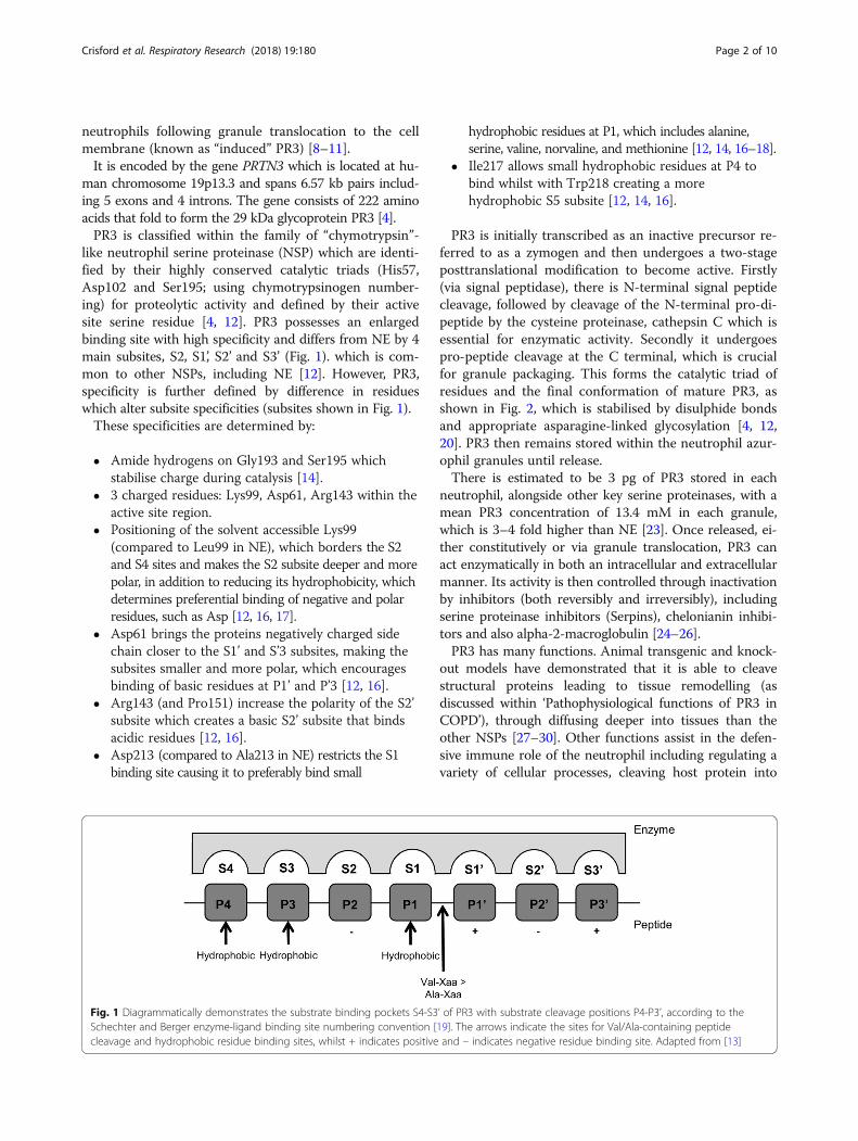
like neutrophil serine proteinase (NSP) which are identi-fied by their highly conserved catalytic triads (His57,Asp102 and Ser195; using chymotrypsinogen number-ing) for proteolytic activity and defined by their activesite serine residue [4, 12]. PR3 possesses an enlargedbinding site with high specificity and differs from NE by 4main subsites, S2, S1’, S2’ and S3’ (Fig. 1). which is com-mon to other NSPs, including NE [12]. However, PR3,specificity is further defined by difference in residueswhich alter subsite specificities (subsites shown in Fig. 1).These specificities are determined by:
� Amide hydrogens on Gly193 and Ser195 whichstabilise charge during catalysis [14].
� 3 charged residues: Lys99, Asp61, Arg143 within theactive site region.
� Positioning of the solvent accessible Lys99(compared to Leu99 in NE), which borders the S2and S4 sites and makes the S2 subsite deeper and morepolar, in addition to reducing its hydrophobicity, whichdetermines preferential binding of negative and polarresidues, such as Asp [12, 16, 17].
� Asp61 brings the proteins negatively charged sidechain closer to the S1’ and S’3 subsites, making thesubsites smaller and more polar, which encouragesbinding of basic residues at P1’ and P’3 [12, 16].
� Arg143 (and Pro151) increase the polarity of the S2’subsite which creates a basic S2’ subsite that bindsacidic residues [12, 16].
� Asp213 (compared to Ala213 in NE) restricts the S1binding site causing it to preferably bind small
hydrophobic residues at P1, which includes alanine,serine, valine, norvaline, and methionine [12, 14, 16–18].
� Ile217 allows small hydrophobic residues at P4 tobind whilst with Trp218 creating a morehydrophobic S5 subsite [12, 14, 16].
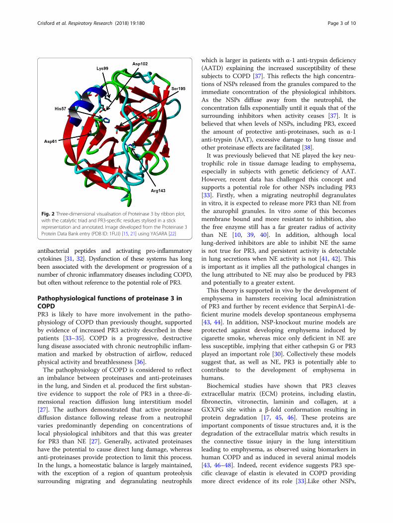
PR3 is initially transcribed as an inactive precursor re-ferred to as a zymogen and then undergoes a two-stageposttranslational modification to become active. Firstly(via signal peptidase), there is N-terminal signal peptidecleavage, followed by cleavage of the N-terminal pro-di-peptide by the cysteine proteinase, cathepsin C which isessential for enzymatic activity. Secondly it undergoespro-peptide cleavage at the C terminal, which is crucialfor granule packaging. This forms the catalytic triad ofresidues and the final conformation of mature PR3, asshown in Fig. 2, which is stabilised by disulphide bondsand appropriate asparagine-linked glycosylation [4, 12,20]. PR3 then remains stored within the neutrophil azur-ophil granules until release.There is estimated to be 3 pg of PR3 stored in each
neutrophil, alongside other key serine proteinases, with amean PR3 concentration of 13.4 mM in each granule,which is 3–4 fold higher than NE [23]. Once released, ei-ther constitutively or via granule translocation, PR3 canact enzymatically in both an intracellular and extracellularmanner. Its activity is then controlled through inactivationby inhibitors (both reversibly and irreversibly), includingserine proteinase inhibitors (Serpins), chelonianin inhibi-tors and also alpha-2-macroglobulin [24–26].PR3 has many functions. Animal transgenic and knock-
out models have demonstrated that it is able to cleavestructural proteins leading to tissue remodelling (asdiscussed within ‘Pathophysiological functions of PR3 inCOPD’), through diffusing deeper into tissues than theother NSPs [27–30]. Other functions assist in the defen-sive immune role of the neutrophil including regulating avariety of cellular processes, cleaving host protein into
Fig. 1 Diagrammatically demonstrates the substrate binding pockets S4-S3’ of PR3 with substrate cleavage positions P4-P3’, according to theSchechter and Berger enzyme-ligand binding site numbering convention [19]. The arrows indicate the sites for Val/Ala-containing peptidecleavage and hydrophobic residue binding sites, whilst + indicates positive and – indicates negative residue binding site. Adapted from [13]
Crisford et al. Respiratory Research (2018) 19:180 Page 2 of 10
antibacterial peptides and activating pro-inflammatorycytokines [31, 32]. Dysfunction of these systems has longbeen associated with the development or progression of anumber of chronic inflammatory diseases including COPD,but often without reference to the potential role of PR3.
Pathophysiological functions of proteinase 3 inCOPDPR3 is likely to have more involvement in the patho-physiology of COPD than previously thought, supportedby evidence of increased PR3 activity described in thesepatients [33–35]. COPD is a progressive, destructivelung disease associated with chronic neutrophilic inflam-mation and marked by obstruction of airflow, reducedphysical activity and breathlessness [36].The pathophysiology of COPD is considered to reflect
an imbalance between proteinases and anti-proteinasesin the lung, and Sinden et al. produced the first substan-tive evidence to support the role of PR3 in a three-di-mensional reaction diffusion lung interstitium model[27]. The authors demonstrated that active proteinasediffusion distance following release from a neutrophilvaries predominantly depending on concentrations oflocal physiological inhibitors and that this was greaterfor PR3 than NE [27]. Generally, activated proteinaseshave the potential to cause direct lung damage, whereasanti-proteinases provide protection to limit this process.In the lungs, a homeostatic balance is largely maintained,with the exception of a region of quantum proteolysissurrounding migrating and degranulating neutrophils
which is larger in patients with α-1 anti-trypsin deficiency(AATD) explaining the increased susceptibility of thesesubjects to COPD [37]. This reflects the high concentra-tions of NSPs released from the granules compared to theimmediate concentration of the physiological inhibitors.As the NSPs diffuse away from the neutrophil, theconcentration falls exponentially until it equals that of thesurrounding inhibitors when activity ceases [37]. It isbelieved that when levels of NSPs, including PR3, exceedthe amount of protective anti-proteinases, such as α-1anti-trypsin (AAT), excessive damage to lung tissue andother proteinase effects are facilitated [38].It was previously believed that NE played the key neu-
trophilic role in tissue damage leading to emphysema,especially in subjects with genetic deficiency of AAT.However, recent data has challenged this concept andsupports a potential role for other NSPs including PR3[33]. Firstly, when a migrating neutrophil degranulatesin vitro, it is expected to release more PR3 than NE fromthe azurophil granules. In vitro some of this becomesmembrane bound and more resistant to inhibition, alsothe free enzyme still has a far greater radius of activitythan NE [10, 39, 40]. In addition, although locallung-derived inhibitors are able to inhibit NE the sameis not true for PR3, and persistent activity is detectablein lung secretions when NE activity is not [41, 42]. Thisis important as it implies all the pathological changes inthe lung attributed to NE may also be produced by PR3and potentially to a greater extent.This theory is supported in vivo by the development of
emphysema in hamsters receiving local administrationof PR3 and further by recent evidence that SerpinA1-de-ficient murine models develop spontaneous emphysema[43, 44]. In addition, NSP-knockout murine models areprotected against developing emphysema induced bycigarette smoke, whereas mice only deficient in NE areless susceptible, implying that either cathepsin G or PR3played an important role [30]. Collectively these modelssuggest that, as well as NE, PR3 is potentially able tocontribute to the development of emphysema inhumans.Biochemical studies have shown that PR3 cleaves
extracellular matrix (ECM) proteins, including elastin,fibronectin, vitronectin, laminin and collagen, at aGXXPG site within a β-fold conformation resulting inprotein degradation [17, 45, 46]. These proteins areimportant components of tissue structures and, it is thedegradation of the extracellular matrix which results inthe connective tissue injury in the lung interstitiumleading to emphysema, as observed using biomarkers inhuman COPD and as induced in several animal models[43, 46–48]. Indeed, recent evidence suggests PR3 spe-cific cleavage of elastin is elevated in COPD providingmore direct evidence of its role [33].Like other NSPs,
Fig. 2 Three-dimensional visualisation of Proteinase 3 by ribbon plot,with the catalytic triad and PR3-specific residues stylised in a stickrepresentation and annotated. Image developed from the Proteinase 3Protein Data Bank entry (PDB ID: 1FUJ) [15, 21] using YASARA [22]
Crisford et al. Respiratory Research (2018) 19:180 Page 3 of 10
PR3 can also affect mucus clearance by damaging bron-chial epithelium and cilia [16]. In addition, PR3 is ableto induce mucus production from submucosal glandserous cells and PR3 activity has been implicated in thisrole in cystic fibrosis (CF) [49]. The net result is excessmucus production in the airways and impaired mucusclearance, which is also a feature of chronic bronchitis,and therefore PR3 is likely to have a similar role inCOPD. AATD is a genetic cause of emphysema andchronic bronchitis (in about 30% of patients) and is theresult of mutations resulting in little/no production offunctional AAT protein. PR3 has a lower association ratewith AAT than NE, which means that, in patients withAATD, PR3 is even more poorly regulated, causing agreater proteinase/anti-proteinase imbalance than withNE, and hence potentially mediates more damage to thelungs [4, 27, 50].As well as causing direct tissue damage, PR3 is also
potentially involved in amplifying the inflammationassociated with COPD as with other chronic inflamma-tory diseases.PR3 is known to modulate a variety of cytokine
functions, which impact processes such as metabolismand inflammasome generation [51–53]. The enzymefacilitates an increased production and/or modulationof proinflammatory cytokines and the reduction ofanti-inflammatory cytokine production as summarisedin Table 1. Many of these cytokines have been impli-cated in a number of inflammatory diseases, whichsupports a putative role of PR3 in chronic inflamma-tory conditions in general as well as COPD with andwithout AATD.All these cytokines can act through autocrine, para-
crine and endocrine pathways to activatepro-inflammatory cascade responses and upregulatepro-inflammatory genes and transcription factors leadingto an inflammatory state [65]. The products of these keyinflammatory pathways can further induce feedbackloops to enhance chronic inflammation [66–68]. There-fore (similarly to NE) PR3 can potentially play multipleroles in the initiation and amplification as well as theresolution of inflammation, at least as demonstrated invitro.More recently, PR3 has been also found to degrade
the anti-inflammatory mediator progranulin (PGRN),resulting in generation of granulin (GRN) peptides invitro [32, 68–70]. PGRN degradation causes increasedneutrophil infiltration, activation of reactive oxidative spe-cies, pro-inflammatory cytokine production andanti-inflammatory pathway inhibition, sustaining aninflammatory state in other inflammatory disease [71].GRN molecules are also known to accumulate and releasethe chemoattractant interleukin (IL)-8 amplifying neutro-phil recruitment [70, 72]. In clinically-stable COPD, the
concentration of PR3 in airway secretions is a strongerpredictor of PGRN levels than NE, because of its greaterneutrophil concentration and hence greater secretionactivity [69].PR3 is also able to act in a pro-inflammatory manner by
interacting with the complement pathway. It is able tofragment the neutrophil surface complement component5a (C5a) receptor, resulting in the loss of the N-terminusand an inability to bind C5a [73]. In CF, the lack of C5aRsignalling contributes towards inefficient clearance of mi-crobial infections in vitro and also inactivates signallingand stimulates neutrophils to degranulate [73]. Thisresults in a cycle of dysfunctional neutrophils therebyperpetuating the bacterial-stimulated inflammatory signalsand further neutrophil recruitment. Although there is nodirect evidence, it is likely that C5aR inhibition by PR3also has a role in COPD with elevated levels of C5a in thesputum of patients and correlations with circulating C5a,physiological gas transfer and the degree of emphysema[74]. Further research is clearly indicated to determine therelevance of this mechanism in COPD.Despite the potential to impede bacterial clearance,
it has also been reported that PR3 itself possessesbactericidal properties through cleavage of the pro-microbicidal protein hCAP-18 (human cathelicidin)into the antibacterial peptide, mucus inducer and neu-trophil chemo-attractant LL-37 [51, 75–78]. Furthermore,levels of LL-37 in sputum are related to disease severity inpatients with COPD suggesting an indirect role for PR3which is worthy of further investigation [79].In addition, PR3 can adhere to neutrophil extracellular
traps (NETs) contributing towards the destruction ofbacterial virulence factors [80–82]. However, manyrespiratory-relevant bacteria, such as Streptococcuspneumoniae and Haemophilus influenzae, have evolvedNET evasion mechanisms which may overcome thispotential clearance mechanism [83, 84]. It has also beennoted that patients with Pseudomonas aeruginosa infec-tion are more susceptible to poor outcome when lackingsufficient PR3 inhibition and patients with AATD are atparticularly high risk of respiratory infection and lungdamage as other natural proteinase inhibitors are unableto compensate for low AAT levels [27, 85]. This is againamplified by the greater neutrophil PR3 content and thefact that the other major lung inhibitor of serine protein-ases, secretory leukocyte proteinase inhibitor (SLPI), doesnot inhibit PR3 [86].However, PR3 is also able to inactivate SLPI, by cleaving
at the Ala-16 site within the N-terminal and preventingSLPI/enzymes complex formation which would indirectlyamplify the local activity of other serine proteinases suchas NE [86].Analysis of biopsied lung tissue, from patients with
severe emphysema, has shown that cytosolic PR3
Crisford et al. Respiratory Research (2018) 19:180 Page 4 of 10
interrupts the initiation of anti-inflammatory mecha-nisms and promotes an apoptotic environment, inducingdeath of lung epithelial cells which has been implicatedin the pathophysiology of emphysema by a further indir-ect route [87].An additional mechanism implicated in the pathophysi-
ology of COPD involves the receptor for advanced glycationend-products (RAGE) and soluble RAGE. In prostate cancercell lines, PR3 has been shown to bind to RAGE both pro-moting cell activation and preventing its cleavage which es-calates inflammation [88, 89]. Furthermore, decreased levelsof sRAGE have been implicated in emphysema development[89, 90]. Clearly the relevance of this alternative functionalso needs to be explored in relation to COPD.
Pathophysiological functions of proteinase 3 inother diseasesThe actions discussed above are not just relevant toCOPD but are relevant to the pathophysiology of manyother diseases. PR3 also has many additional roles whichcan lead to, or amplify other disease states (see Fig. 3).As noted in Table 1, PR3 has both a direct and indirect
effect on many cytokines and hence can have furtherdownstream influences on diseases beyond or associatedwith COPD, as outlined in Table 2.However, although the effects of dysregulation of these
cytokines are also implicated in other diseases, PR3 has
not been directly studied in relation to theirpathophysiology.In addition, the interaction between PR3 and PGRN
also likely has wider impact than in COPD, through afurther role in inflammatory conditions involving PGRN,including lipopolysaccharide-induced acute lung injury,dermatitis and inflammatory arthritis (in murinemodels), as well as a reported genetic link betweenloss-of-function mutations in PGRN and the develop-ment of neurodegenerative disease [99–104].It is also suspected that PR3, alongside other NSPs,
could have a role in ECM breakdown affecting thepathophysiology of diseases in other organs, such asaneurysms due to vascular remodelling as shown inporcine vasculature; however the relevance has not yetbeen investigated in detail in humans [105].PR3 has a role in the efficacy of neutrophil transmigration
through interaction with the cell surface receptor NB1(CD177) which acts with PECAM-1 (CD31) duringtrans-endothelial migration of neutrophils [106, 107]. In CF,this is supported by a positive relationship between PR3activity and neutrophil migration effectiveness [20]. Theinteraction of PR3 with NB1 and PECAM-1 is confirmed invitro in endothelial cells, where it inhibits activation andupregulation of these adhesion molecules [108].There is also evidence that PR3 is associated with distor-
tion of cellular signalling pathways and the development of
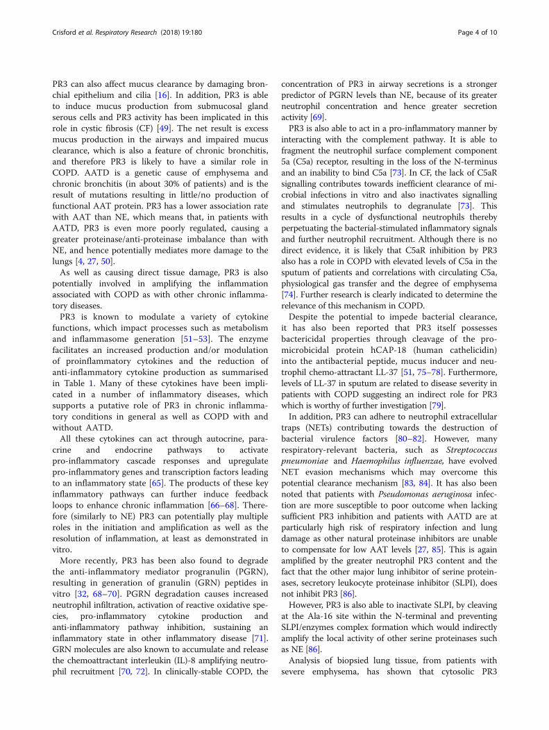
Fig. 3 Summary of the actions of Proteinase 3 (PR3), as outlined in this review, which likely impact on COPD and other systemic diseases. Theprocesses with a putative central role in the pathophysiology of emphysema are highlighted in bold
Crisford et al. Respiratory Research (2018) 19:180 Page 5 of 10
autonomous cell growth. In leukaemia, early expression ofPR3 during haematopoiesis is able to induce factor-inde-pendent growth and overexpression of PR3 in myeloid leu-kaemia cells prevents their differentiation into monocytoidcells supporting this mechanism [109–111].Alternatively to its pro-apoptotic role in COPD, PR3
may paradoxically prevent apoptosis in granulomatosis
with polyangiitis (GPA) by associating with calreticulin,through co-externalisation with phosphatidylserine byphospholipid flip-flop via phospholipid scramblase 1(PLSCR1), to override the ‘eat me’ signalling [112].Finally, PR3 also has a role as an autoantigen in
many diseases, including GPA and idiopathic interstitialpneumonias, and is the target of cytoplasmic (c)-anti-neutrophil cytoplasmic antibodies, also referred to asPR3-ANCA in vasculitis [113–115]. Development ofdisease is dependent on ability of PR3 to associate withthe cell membrane [112]. The binding of PR3-ANCAwith cell associated PR3 initiates a cascade whichamplifies inflammation and results in local cellular andtissue damage [9, 114, 116–118].It was suspected that PR3-ANCA formation may have a
role in COPD development, as more patients with COPDwere found to be antinuclear antibody positive than healthycontrols [119]. However, despite a reported associationwith emphysema-dominant disease and lower body massindex, no clear pathophysiological relationship has beenestablished [119]. These wide ranging pro-inflammatoryeffects of PR3 in other conditions therefore may be alsorelevant in the pathophysiology of COPD, both directly bytissue damage and indirectly through other multiple path-ways of inflammation.
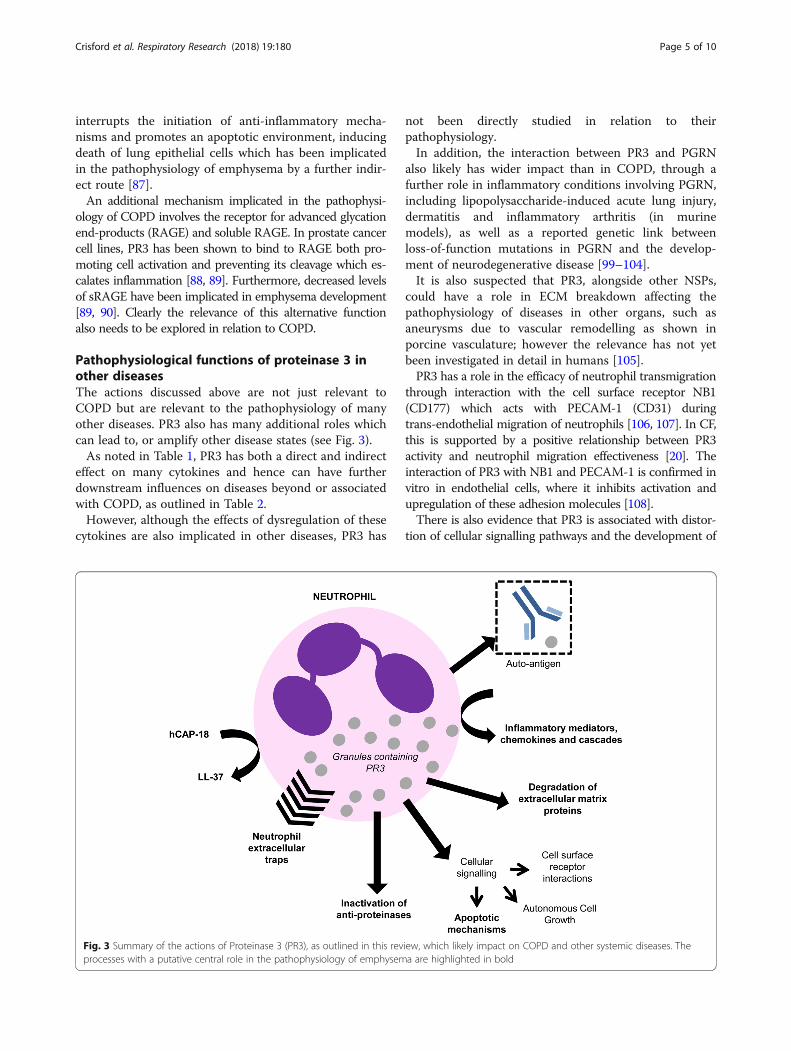
Table 1 Summary of the cytokines affected by PR3, with the PR3 action on cytokines and the resulting response. The processesrelevant to the pathophysiology of COPD are highlighted in bold
Cytokine Role of PR3 Action of cytokine References
Interleukin(IL)-1β
Proteolytically activates extracellular pro-formsto be cleaved into active counterparts byCaspase 1 in inflammasomes
• ↑ neutrophil activation and recruitment• canonical NFκB signalling• ↑ cyclooxygenases [44] and prostaglandinE production
• pushes towards T helper cell (Th)17 differentiation
[31, 54–56]
IL-18 • Induces interferon (IFN)-γ and Fas ligand, ↑ differentiationto Th1, Th2 or Th17 responses(dependant on accompanying signals)
[55, 57]
Tumour necrosisfactor (TNF)-α
Cleaves precursor to bioactive form (via twohypothesised cleavage sites at Ala15-Leu16or Val77-Arg78)
• Activates the caspase and MEK cascades,and PI-3-kinase and canonical NFκB pathway
• Activates Etk = ↑cellular adhesion, migrationand propagation
• ↑ neutrophil chemotaxis• Upregulation of pro-inflammatory genes e.g.IL-8, CCL2, CXCL10, COX-2, and pro-coagulants
• Recruits apoptosis-inhibiting molecules• ↓ signalling by cIAP-mediated ubiquitination
[54, 58]
IL-6 Functionally inactivates and degrades thesoluble IL-6 receptor (sIL-6R) – exactmechanisms unknown
• Disrupts trans-signalling activity• Prevents apoptosis• ↑ neutrophil recruitment and infiltration
[59, 60]
IL-8 (CXCL8) Truncates stored IL-8 (77) into the 10-fold morepotent chemo-attractant IL-8 (70) throughcleavage of an Ala-Lys bond
• ↑ respiratory burst• Potentiates inflammatory disease cycle• Drives neutrophil chemotaxis
[61]
IL-17 (CTLA8) Stimulation increases cytokine production • Directs towards a dominant Th17 environment• T cell hypo-responsiveness
[62]
IL-32 Processes activating cytokines IL-1β, TNF-αand IFN-γ directly or indirectly; cleaves IL-32at IL-32α to a more bioactive form
• Activates canonical NFκB and MAPK cascades• ↑ production of cytokines incl. TNF-α, IL-8and CXCL2 production
[63, 64]
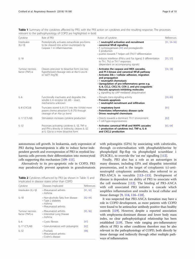
Table 2 Cytokines influenced by PR3 (as shown in Table 1) andimplicated in disease states other than COPD
Cytokine Diseases Implicated References
Interleukin (IL)-1β • Rheumatoid arthritis• Asthma
[91, 92]
IL-18 • Non-alcoholic fatty liver disease• Type 2 diabetes• Asthma• Rheumatoid arthritis
[92–94]
Tumour necrosisfactor (TNF)-α
• Rheumatoid arthritis• Interstitial Lung Disease• Asthma
[95, 96]
IL-6 • Cystic fibrosis [97]
IL-17 (CTLA8) • Granulomatosis with polyangiitis [62]
IL-32 • Psoriasis• Rheumatoid arthritis• Crohn’s disease
[98]
Crisford et al. Respiratory Research (2018) 19:180 Page 6 of 10
PR3 as a therapeutic target in COPDThere is considerable theoretical evidence and cell-basedand animal-model data to support the role of PR3 in thedevelopment of COPD. However, as yet, PR3 activity inCOPD has been poorly characterised.To study PR3 in COPD requires the ability to quantify
active (uninhibited) PR3 accurately and distinguish itfrom other NSPs to determine its specific functionwithin biological samples. Reagents for free PR3 activityhave only lately become available and until recently,detection required immunofluorescent staining of biopsyspecimens, which if positive was followed by aPR3-ANCA specific enzyme-linked immunosorbentassay (ELISA) [120, 121]. Indeed, this was the inter-nationally accepted method for diagnosing PR3-ANCA.Direct PR3 assays have been proposed as a biomarker todetermine PR3 presence and production for assisting adiagnosis [122, 123]; however, like immunofluorescencetechniques, they do not distinguish the active PR3 fromPR3 which has been inactivated by its inhibitors. Asimilar challenge was seen for the measurement of NEactivity and a novel approach to this has been the devel-opment of NE specific footprint, which may also be amore relevant approach for PR3 activity in vivo [124].Whilst there is increasing interest in modifying NSP
activity in conditions which predominantly featureneutrophilic inflammation, these have primarily focusedon reducing the activity of NE and PR3 has not generallybeen considered as a relevant target in COPD.The detection of PR3 activity, directly or indirectly,
would improve our understanding of its role in COPDand individual patient’s disease activity. It would alsopotentially allow earlier diagnosis of diseases where PR3activity was relevant (including COPD) before extensivedamage has occurred. Understanding the role of PR3,might therefore allow earlier interventions and thera-peutic strategies to be developed with PR3 as a valid tar-get in COPD. Specific inhibitors might serve to reducedisease severity, mortality and the long-term healthburdens of COPD. However, clearly the limited dataavailable indicates there is much work to be done toclarify the likely relevance and hence impact of ananti-PR3 strategy.
ConclusionsPR3 has many important functions that are relevant tohuman physiology and PR3 dysfunction may play a crit-ical role in many processes central to the pathophysi-ology of COPD and other chronic neutrophilic humandiseases. PR3 is the most abundant serine proteinase inthe neutrophil, secondarily inhibited to NE, and, inaddition to the role in general inflammation, PR3 canalso cause direct tissue damage central to structuralaspects of diseases such as COPD. This is consistent
with the potential for PR3 to produce all the patho-logical changes of COPD that have traditionally beenattributed to NE. Understanding this role and theimpact on the inflammatory cascade has major implica-tions for the design of anti-proteinase molecules aimed atrestoring proteinase/anti-proteinase balance, ensuring thatdestructive activity of relevant serine proteinase actionand amplification of inflammation is effectively limited,and thereby preventing the development and progressionof COPD.
AbbreviationsAAT: α-1 Anti-trypsin; AATD: α-1 Anti-trypsin Deficiency; ANCA: Anti-neutrophil Cytoplasmic Antibodies; C5a: Complement Component 5a;CF: Cystic Fibrosis; COPD: Chronic Obstructive Pulmonary Disease;COX: Cyclooxygenases; DLCo: Diffusing Capacity of the Lungs for CarbonMonoxide; ECM: Extracellular Matrix; ELISA: Enzyme-linked ImmunosorbentAssay; GPA: Granulomatosis with Polyangiitis; GRN: Granulin; hCAP-18: Human Cathelicidin; IFN: Interferon; IL: Interleukin; NE: Neutrophil Elastase;NETs: Neutrophil Extracellular Traps; NFkB: Nuclear Factor Kappa-Light-Chain-Enhancer of Activated B Cells; NSP: Neutrophil Serine Proteinase;PGRN: Progranulin; PLSCR1: Phospholipid Scramblase 1; PR3: Proteinase 3;RAGE: Receptor for Advanced Glycation End-products; Serpins: SerineProteinase Inhibitors; SPLI: Secretory Leukocyte Proteinase Inhibitor; Th: Thelper Cells; TNF: Tumour Necrosis Factor
Authors’ contributionsAll authors met criteria for authorship. HC wrote the initial draft of themanuscript and, ES and RAS revised the manuscript. All authors read andapproved the final manuscript.
Ethics approval and consent to participateNot Applicable.
Consent for publicationNot Applicable.
Competing interestsThe authors declare that they have no competing interests.
Publisher’s NoteSpringer Nature remains neutral with regard to jurisdictional claims inpublished maps and institutional affiliations.
Author details1Institute of Inflammation and Ageing, University of Birmingham, Edgbaston,Birmingham B15 2GW, UK. 2University Hospital Birmingham NHS FoundationTrust, Edgbaston, Birmingham B15 2GW, UK. 3Institute of Inflammation andAgeing, College of Medical and Dental Sciences, Centre for TranslationalInflammation Research, University of Birmingham Research Laboratories,Queen Elizabeth Hospital Birmingham, Mindelsohn Way, Birmingham B152WB, UK.
Received: 20 April 2018 Accepted: 6 September 2018
References1. GOLD: Global strategy for the diagnosis, management and prevention of
chronic obstructive pulmonary disease. 2017.2. Baici A, Szedlacsek SE, Fruh H, Michel BA. pH-dependent hysteretic behavior
of human Myeloblastin (leucocyte proteinase 3). Biochem J. 1996;317:901–5.3. Karatepe K, Luo HR. Proteinase 3 is expressed in stem cells and regulates
bone marrow hematopoiesis. Blood. 2015;126:1159.4. Korkmaz B, Moreau T, Gauthier F. Neutrophil elastase, proteinase 3 and
Cathepsin G: physiochemical properties, activity and Physiopathologcalfunctions. Biochemie. 2008;90:227–42.
5. Zimmer M, Medcalf RL, Fink TM, Mattmann C, Lichter P, Jenne DE. Threehuman elastase-like genes Coordingately expressed in the Myelomonocyte
Crisford et al. Respiratory Research (2018) 19:180 Page 7 of 10
lineage are organised as a single genetic locus on 19pter. Proc Natl AcadSci U S A. 1992;89:5.
6. Csernok E, Ernst M, Schmitt W, Bainton DF, Gross WL. Activated neutrophilsexpress proteinase 3 on their plasma membrane In vitro and In vivo. ClinExp Immunol. 1994;95:244–50.
7. Witko-Sarsat V, Cramer EM, Hieblot C, Guichard J, Nusbaum P, Lopez S,Lesavre P, Halbwachs-Mecarelli L. Presence of proteinase 3 in secretoryvesicles: evidence of a novel, highly Mobilizable intracellular Pool distinctfrom Azurophil granules. Blood. 1999;94:2487–96.
8. Halbwachs-Mecarelli L, Bessou G, Lesavre P, Lopez S, Witko-Sarsat V.Bimodal Distrubution of proteinase 3 (PR3) surface expression reflects aconstitutive heterogeneity in the Polymorphonuclear neutrophil Pool. FEBSLett. 1995;374:29–33.
9. Csernok E, Ludemann J, Gross WL, Bainton DF. Ultrastructural localisation ofproteinase 3, the target antigen of anti-cytoplasmic antibodies circulating inWegener's granulomatosis. Am J Pathol. 1990;137:1113–20.
10. Korkmaz B, Jaillet J, Jourdan M-L, Gauthier A, Gauthier F, Attucci S.Catalytic activity and inhibition of Wegener antigen proteinase 3 on thecell surface of human Polymorphonuclear neutrophils. J Biol Chem.2009;284:19896–902.
11. Korkmaz B, Lesner A, Letast S, Mahdi YK, Jourdan ML, Dallet-Choisy S,Marchand-Adam S, Kellenberger C, Viaud-Massuard MC, Jenne DE, GauthierF. Neutrophil proteinase 3 and dipeptidyl peptidase I (cathepsin C) aspharmacological targets in granulomatosis with polyangiitis (Wegenergranulomatosis). Semin Immunopathol. 2013;35:411–21.
12. Hajjar E, Broemstrup T, Kantari C, Witko-Sarsat V, Reuter N. Structures ofhuman proteinase 3 and neutrophil elastase - so similar yet so different.FEBS J. 2010;277:2238–54.
13. Hajjar E, Korkmaz B, Gauthier F, Brandsdal BO, Witko-Sarsat V, Reuter N.Inspection of the binding sites of proteinase 3 for the Design of a HighlySpecific Substrate. J Med Chem. 2006;49:1248–60.
14. Guarino C, Gruba N, Grzywa R, Dyguda-Kazimierowicz E, Hamon Y,Legowska M, Skorenski M, Dallet-Choisy S, Marchand-Adam S,Kellenberger C, et al. Exploiting the S4-S5 specificity of humanneutrophil proteinase 3 to improve the potency of peptidylDi(chlorophenyl)-phosphonate Ester inhibitors: a kinetic and molecularmodeling analysis. J Med Chem. 2018;61:1858–70.
15. Fujinaga M, Chernaia MM, Halenbeck R, Koths K, James MNG. The crystalstructure of PR3, a neutrophil serine proteinase antigen of Wegener'sgranulomatosis antibodies. J Mol Biol. 1996;261:267–77.
16. Korkmaz B, Horwitz MS, Jenne DE, Gauthier F. Neutrophil elastase,proteinase 3, and Cathepsin G as therapeutic targets in human diseases.Pharmacol Rev. 2010;62:726–59.
17. Rao NV, Wehner NG, Marshall BC, Gray WR, Gray BH, Hoidal JR.Characterisation of proteinase 3 (PR-3), a neutrophil serine proteinase:structural and functional properties. J Biol Chem. 1991;266:9540–8.
18. Brubaker MJ, Groutas WC, Hoidal JR, Rao NV. Human neutrophil proteinase3: mapping of the substrate binding site using peptidyl Thiobenzyl esters.Biochem Biophys Res Commun. 1992;188:1318–24.
19. Schechter I, Berger A. On the size of the active site in proteases. I. Papain.Biochem Biophys Res Commun. 1967;27:157–62.
20. Twigg MS, Brockbank S, Lowry P, FitzGerald SP, Taggart C, Weldon S. Therole of serine proteases and Antiproteases in the cystic fibrosis lung. MediatInflamm. 2015;2015:10.
21. Berman HM, Westbrook J, Feng Z, Gilliland G, Bhat TN, Weissig H, Shindyalov IN,Bourne PE. The Protein Data Bank. Nucleic Acids Res. 2000;28:235–42.
22. Krieger E, Vriend G. YASARA view - molecular graphics for all devices - fromsmartphones to workstations. Bioinformatics. 2014;30:2981–2.
23. Campbell EJ, Campbell MA, Owen CA. Bioactive proteinase 3 on the cellsurface of human neutrophils: quantification, catalytic activity, andsusceptibility to inhibition. J Immunol. 2000;165:3366–74.
24. Loison F, Xu Y, Luo HR. Proteinase 3 and serpin B1: a novel pathway in theregulation of Caspase-3 activation, neutrophil spontaneous apoptosis, andinflammation. Inflamm Cell Signal. 2014;1:1–5.
25. Zani ML, Nobar SM, Lacour SA, Lemoine S, Boudier C, Bieth JG, MoreauT. Kinetics of the inhibition of Neutriophil proteinases by recombinantElafin and pre-elafin (Trappin-2) expressed in Pichia pastoris. Eur JBiochem. 2004;271:2370–8.
26. Yang L, Mei Y, Fang Q, Wang J, Yan Z, DSong Q, Lin Z, Ye G. Identificationand characterization of serine protease inhibitors in a parasitic wasp,Pteromalus puparum. Sci Rep. 2017;7:1-13.
27. Sinden NJ, Baker MJ, Smith DJ, Kreft J-U, Dafforn TR, Stockley RA.Alpha-1-antitrypsin variants and the proteinase/anti-proteinaseimbalance in chronic obstructive pulmonary disease. Am J Physiol LungCell Mol Physiol. 2015;308:12.
28. Jerke U, Perez Hernandez D, Beaudette P, Korkmaz B, Dittmar G, Kettritz R.Neutrophil Serine Proteases Exert Proteolytic Activity on Endothelial Cells.Kidney Int. 2015;88:764–75.
29. Korkmaz B, Lesner A, Guarino C, Wysocka M, Kellenberger C, Watier H,Specks U, Gauthier F, Jenne DE. Inhibitors and antibody fragments aspotential anti-inflammatory therapeutics Targetting neutrophil proteinase 3in human disease. Pharmacol Rev. 2016;68:603–30.
30. Guyot N, Wartelle J, Malleret L, Todorov AA, Devouassoux G, Pacheco Y,Jenne DE, Belaaouaj A. Unopposed Cathepsin G, neutrophil elastase, andproteinase 3 cause severe lung damage and emphysema. Am J Pathol.2014;184:2197–210.
31. Joosten LA, Netea MG, Fantuzzi G, Koenders MI, Helsen MM, Sparrer H,Pham CT, van der Meer JW, Dinarello CA, van den Berg WB. InflammatoryArthritis in Caspase 1 Gene-deficient Mice: Contribution of Proteinase 3 toCaspase 1-independent Production of Bioactive Interleukin-1beta. ArthritisRheum. 2009;60:3651–62.
32. Kessenbrock K, Frohlich L, Sixt M, Lammermann T, Pfister H, Bateman A,Belaaouaj A, Ring J, Ollert M, Fassler R, Jenne DE. Proteinase 3 andneutrophil elastase enhance inflammation in mice by inactivating anti-inflammatory Progranulin. J Clin Invest. 2008;118:2438–47.
33. Gudmann NS, Manon-Jensen T, Sand JMB, Diefenbach C, Sun S, DanielsenA, Karsdal MA, Leeming DJ. Lung tissue destruction by proteinase 3 andCathepsin G mediated elastin degradation is elevated in chronic obstructivepulmonary disease. Biochem Biophys Res Commun. 2018;503(3):1284-90.
34. Newby PR, Carter RI, Stockley RA. Aα-Val541 a Novel Biomarker of Proteinase3 Activity. Am J Respir Crit Care Med. 2017;195:A5247.
35. Newby PR, Stockley RA. Neutrophil elastase and proteinase 3 activity in PiSZAlpha-1 antitrypsin deficiency. Am J Respir Crit Care Med. 2018;197:A4763.
36. Hoenderdos K, Condliffe A. The neutrophil in chronic obstructive pulmonarydisease. Too little, too late or too much, too soon? Am J Respir Cell MolBiol. 2013;48:531–9.
37. Campbell EJ, Campbell MA, Boukedes SS, Owen CA. Quantum proteolysisby neutrophils: implications for pulmonary emphysema in α1-antitrypsindeficiency. CHEST. 2000;117:303S.
38. Stockley RA. Neutrophils and protease/Antiprotease imbalance. Am J RespirCrit Care Med. 1999;160:S49–52.
39. Owen CA, Campbell EJ. The cell biology of leukocyte-mediated proteolysis.J Leukoc Biol. 1999;65:14.
40. Maximova K, Venken T, Reuter N, Trylska J. D-peptides as inhibitors of PR3-membrane interactions. Biomembranes. 1860;2018:458–66.
41. Korkmaz B, Poutrain P, Hazouard E, de Monte M, Attucci S, Gauthier FL.Competition between elastase and related proteases from humanneutrophil for binding to α1-protease inhibitor. Am J Respir Cell MolBiol. 2005;32:553–9.
42. Sinden NJ, Stockley RA. Proteinase 3 activity in sputum from subjects withAlpha-1 anti-trypsin deficiency and COPD. Eur Respir J. 2013;41:1042–50.
43. Kao RC, Wehner NG, Skubitz KM, Gray BH, Hoidal JR. Proteinase 3. A distincthuman Polymorphonuclear leukocyte proteinase that produces emphysemain hamsters. J Clin Invest. 1988;82:1963–73.
44. Borel F, Sun H, Zieger M, Cox A, Cardozo B, Li W, Oliveira G, Davis A,Gruntman A, Flotte TR, et al. Editing out five Serpina1 paralogs to create amouse model of genetic emphysema. Proc Natl Acad Sci U S A. 2018;115(11):2788-93.
45. Lombard C, Bouchu D, Wallach J, Saulnier J. Proteinase 3 hydrolysis ofpeptides derived from human elastin exon 24. Amino Acids. 2005;28:403–8.
46. Chelladurai P, Seeger W, Pullamsetti SS. Matrix metalloproteinases and theirinhibitors in pulmonary hypertension. Eur Respir J. 2012;40:766–82.
47. Sand JMB, Knox AJ, Lange P, Sun S, Kristensen JH, Leeming DJ, Karsdal MA,Bolton CE, Johnson SR. Accelerated extracellular matrix turnover duringexacerbations of COPD. Respir Res. 2015;16:69.
48. Ramaha A, Patston PA. Release and degradation of angiotensin 1 andangiotensin 2 from angiotensinogen by neutrophil serine proteinases. ArchBiochem Biophys. 2002;397:77–83.
49. Witko-Sarsat V, Halbwachs-Mecarelli L, Schuster A, Nusbaum P, Ueki I,Canteloup S, Lenoir G, Descamps-Latscha B, Nadel JA. Proteinase 3, a potentSecretagogue in airways, is present in cystic fibrosis sputum. Am J RespirCell Mol Biol. 1999;20:729–36.
Crisford et al. Respiratory Research (2018) 19:180 Page 8 of 10
50. Janciauskiene S, Bals R, Koczulla R, Vogelmeier C, Kohnlein T, Welte T. Thediscovery of Alpha-1-antitrypsin and its role in health and disease. RespirMed. 2011;105:1129–39.
51. Sorensen OE, Follin P, Johnsen AH, Calafat J, Tjabringa GS, Hiemstra PS,Borregaard N. Human cathelicidin, hCAP-18, is processed to theantimicrobial peptide LL-37 by extracellular cleavage with proteinase 3.Blood J. 2001;97:3951–9.
52. Ren K, Torres R. Role of interleukin-1beta during pain and inflammation.Brain Res Rev. 2009;60:57–64.
53. Popa C, Netea MG, van Riel PLCM, van der Meer JWM, Stalenhoef AFH. Therole of TNF-a in chronic inflammatory conditions, intermediary metabolism,and cardiovascular risk. J Lipid Res. 2007;48:751–62.
54. Coeshott C, Ohnemus C, Pilyavskaya A, Ross S, Wieczorek M, Kroona H,Leimer AH, Cheronis J. Converting enzyme-independent release of tumornecrosis factor alpha and IL-1beta from a stimulated human Monocytic cellline in the presence of activated neutrophils or purified proteinase 3. ProcNatl Acad Sci U S A. 1999;96:6261–6.
55. Keyel PA. How is inflammation initiated? Individual influences of IL-1, IL-18and HMGB1. Cytokine. 2014;69:136-45.
56. Schreiber A, Pham CT, Hu Y, Schneider W, Luft FC, Kettritz R. Neutrophilserine proteases promote IL-1beta generation and injury in necrotizingcrescentic glomerulonephritis. J Am Soc Nephrol. 2012;23:470–82.
57. Sugawara A, Uehara A, Nochi T, Yamaguchi T, Ueda H, Sugiyama A,Hanzawa K, Kumagai K, Okamura H, Takada H. Neutrophil proteinase 3-mediated induction of bioactive IL-18 secretion by human Oral epithelialcells. J Immunol. 2001;167:6568–75.
58. Bradley JR. TNF-mediated inflammatory disease. J Pathol. 2008;214:149–60.59. Hurst SM, Wilkinson TS, McLoughline RM, Jones S, Horiuchi S, Yamamoto N,
Rose-john S, Fuller GM, Topley N, Jones SA. IL-6 and its soluble receptorOchestrate a Temportal switch in the pattern of leukocyte recruitment seenduring acute inflammation. Immunity. 2001;14:705–14.
60. McLoughlin RM, Hurst SM, Nowell MA, Harris DA, Horiuchi S, Morgan LW,Wilkinson TS, Yamamoto N, Topley N, Jones SA. Differential regulation ofneutrophil-activating chemokines by IL-6 and its soluble receptor isoforms. JImmunol. 2004;172:5676–83.
61. Keatings VM, Collins PD, Scott DM, Barnes PJ. Differences in Interleukin-8and tumour necrosis factor-alpha in induced sputum from patients withchronic obstructive pulmonary disease or asthma. Am J Respir Crit CareMed. 1996;153:512–21.
62. Rani L, Minz RW, Sharma A, Anand S, Gupta D, Panda NK, Sakhuja VK.Predominance of PR3 specific immune response and skewed Th17 vs.T-regulatory Miliew in active Granfulomatosis with Polyangiitis. Cytokine.2015;71:7.
63. Kim S-H, Han S-Y, Azam T, Yoon D-Y, Dinarello CA. Interleukin-32: a cytokineand inducer of TNFα. Immunity. 2005;22:131–42.
64. Calabrese F, Baraldo S, Bazzan E, Lunardi F, Rea F, Maestrelli P, Turato G,Lokar-Oliani K, Papi A, Zuin R, Sfriso P. IL-32, a novel Proinflammatorycytokine in chronic obstructive pulmonary disease. Am J Respir Crit CareMed. 2008;178:894–901.
65. Zhang J-M, An J. Cytokines, inflammation and pain. Int Anesthesiol Clin.2007;45:27–37.
66. Nemeth T, Mocsai A. Feedback amplification of neutrophil function. Cell.2016;37:412–24.
67. Robache-Gallea S, Morand V, Bruneau JM, Schoot B, Tagat E, Realo E,Chouaib S, Roman-Roman S. In vitro processing of human tumour necrosisfactor-alpha. J Biol Chem. 1995;270:23688–92.
68. Kessenbrock K, Dau T, Jenne DE. Tailor-made inflammation: how neutrophilserine proteases modulate the inflammatory response. J Mol Med. 2011;89:23–8.
69. Ungers MJ, Sinden NJ, Stockley RA. Progranulin is a substrate for neutrophil-elastase and Proteinase-3 in the airway and its concentration correlates withmediators of airway inflammation in COPD. Am J Physiol Lung Cell MolPhysiol. 2014;306:L80–7.
70. Couto MA, Harwig SSL, Cullor JS, Hughes JP, Lehrer RI. eNAP-2, a novelcysteine-rich bactericidal peptide from equine leukocytes. Infect Immun.1992;60:5042–7.
71. Baker M, Mackenzie IR, Pickering-Brown SM, Gass J, Rademakers R, LindholmC, Snowden J, Adamson J, Sadovnivk AD, Rollinson S, et al. Mutations inProgranulin cause tau-negative frontotemporal dementia linked tochromosome 17. Nature. 2006;442:916–9.
72. Zhu J, Nathan C, Jin W, Sim D, Ashcroft GS, Wahl SM, Lacomis L,Erdujument-Bromage H, Tempst P, Wright CD, Ding A. Conversion of
Proepithelin to Epithelins: roles of SLPI and elastase in host defense andwound repair. Cell. 2002;111:867–78.
73. van der Berg CW, Tambourgi DV, Clark HW, Hoong SJ, Spiller OB, McGrealEP. Mechanism of neutrophil dysfunction: neutrophil serine proteases cleaveand inactivate the C5a receptor. J Immunol. 2014;192:1787–95.
74. Marc MM, Korosec P, Kosnik M, Kern I, Flezar M, Suskovic S, Sorli J.Complement factors C3a, C4a, and C5a in chronic obstructive pulmonarydisease and asthma. Am J Respir Cell Mol Biol. 2004;31:216–9.
75. Zhang Y, Jiang Y, Sun C, Wang Q, Yang Z, Pan X, Zhu M, Xiao W. The humancathelicidin LL-37 enhances airway mucus production in chronic obstructivepulmonary disease. Biochem Biophys Res Commun. 2014;443:103–9.
76. Campanelli D, Detmers PA, Nathan CF, Gabay JE. Azurocidin and ahomologous serine protease from neutrophils. Differential antimicrobial andproteolytic properties. J Clin Invest. 1990;85:904–15.
77. Dasaraju PV, Liu C. Infections of the respiratory system. In: MedicalMicrobiology. 4th ed. Baron S, editor. Texas: University of Texas MedicalBranch at Galveston; 1996.
78. Kuroda K, Okumura K, Isogai H, Isogai E. The human cathelicidinantimicrobial peptide LL-37 and mimics are potential anti-Cancer drugs.Front Oncol. 2015;5:10.
79. Jiang Y-Y, Xiao W, Zhu M-X, Yang Z-H, Pan X-J, Zhang Y, Sun C-C, Xing Y.The effect of human antibacterial peptide LL-37 in the pathogenesis ofchronic obstructive pulmonary disease. Respir Med. 2012;106:1680–9.
80. Urban CF, Ermert D, Schmid M, Abu-Abed U, Goosmann C, Nacken W,Brinkmann V, Jungblut PR, Sycglinsky A. Neutrophil extracellular trapscontain calprotectin, a cytosolic protein complex involved in host defenseagainst Candida albicans. PLoS Pathog. 2009;5:e1000639.
81. Kessenbrock K, Krumbholz M, Schonermarck U, Back W, Gross WL, Werb Z,Grone H-J, Brinkmann V, Jenne DE. Netting Neutrophils in AutoimmuneSmall-Vessel Vasculitis. Nat Med. 2009;15:623–5.
82. Delgado-Rizo V, Martinez-Guzman MA, Iniguez-Gutierrez L, Garcia-Orozco A,Alvarado-Navarro A, Fafutis-Morris M. Neutrophil extracellular traps and itsimplications in inflammation: an overview. Front Immunol. 2017;2017:1–20.
83. Beiter K, Wartha F, Albiger B, Normark S, Zychlinsky A, Henriques-Normark B.An endonuclease allows Streptococcus pneumoniae to escape fromneutrophil extracellular traps. Curr Biol. 2006;16:401–7.
84. Hong W, Juneau RA, Pang B, Swords WE. Survival of bacterial biofilmswithin neutrophil extracellular traps promotes Nontypeable Haemophilusinfluenzae persistence in the Chinchilla model for otitis media. J InnateImmun. 2009;1:215–24.
85. Benarafa C, Priebe GP, Remold-O'Donnell E. The neutrophil serine proteaseinhibitor SerpinB1 preserves lung Defence functions in Pseudomonasaeruginosa infection. J Exp Med. 2007;204:1901–9.
86. Rao NV, Marshall BC, Gray BH, Hoidal JR. Interaction of secretoryleukocyte protease inhibitor with Proteinase-3. Am J Respir Cell Mol Biol.1993;8:612-6.
87. Kasahara Y, Tuder RM, Cool CD, Lynch DA, Flores SC, Voelkel NF. Endothelialcell death and decreased expression of vascular endothelial growth factorand vascular endothelial growth factor receptor 2 in emphysema. Am JRespir Crit Care Med. 2001;163:737–44.
88. Kolonin AG, Sergeeva A, Staquicini DI, Smith TL, Tarleton CA, Molldrem JJ,Sidman RL, Marchio S, Pasqualini R, Arap W. Interaction between tumourcell surface receptor RAGE and proteinase 3 mediates prostate CancerMatastasis to bone. Cancer Res. 2017;77:3144–50.
89. Sukkar MB, Ullah MA, Gan WJ, Wark PAB, Chung KF, Hughes JM, Armour CL,Phipps S. RAGE: a new frontier in chronic airways disease. Br J Pharmacol.2012;167:1161–76.
90. Yonchuk JG, Silverman EK, Bowler RP, Agusti A, Lomas DA, Miller BE,Tal-Singer R, Mayer RJ. Circulating soluble receptor for advancedglycation end products (sRAGE) as a biomarker of emphysema and theRAGE Axis in the lung. Am J Respir Crit Care Med. 2015;192:785–92.
91. Stehlik C. Multiple IL-1β converting enzymes contribute to inflammatoryarthritis. Arthritis Rheum. 2009;60:3524–30.
92. Lee T-H, Song HJ, Park C-S. Role of Inflammasome activation in developmentand exacerbation of asthma. Asia Pacific Allergy. 2014;4:187–96.
93. Toonen EJ, Mirea AM, Tack CJ, Stienstra R, Ballak DB, van Diepen JA, HijmansA, Chavakis T, Dokter WH, Pham CT, et al. Activation of proteinase 3contributes to non-alcoholic fatty liver disease (NAFLD) and insulinresistance. Mol Med. 2016;22:202–14.
94. Gracie JA. Interleukin-18 as a potential target in inflammatory arthritis. ClinExp Immunol. 2004;136:402–4.
Crisford et al. Respiratory Research (2018) 19:180 Page 9 of 10
95. Matsumoto T, Kaneko T, Seto M, Wada H, Kobayashi T, Nakatani K,Tonomura H, Tono Y, Ohyabu M, Nobori T, et al. The membrane proteinase3 expression on neutrophils was downregulated after treatment withinfliximab in patients with rheumatoid arthritis. Clin Appl Thromb Hemost.2008;14:186-92.
96. Armstrong L, Godinho SIH, Uppington KM, Whittington HA, Millar AB.Tumour necrosis factor-α processing in interstitial lung disease: a potentialrole for exogenous Proteinase-3. Clin Exp Immunol. 2009;156:336–43.
97. McGreal EP, Davies PL, Powell W, Rose-John S, Spiller OB, Doull I, Jones SA,Kotecha S. Inactivation of IL-6 and soluble IL-6 receptor by neutrophil derivedserine proteases in cystic fibrosis. Biochim Biophys Acta. 2010;1802:649–58.
98. Dinarello CA, Kim SH. IL-32, a novel cytokine with a possible role in disease.Ann Rheum Dis. 2006;65:4.
99. Zhao Y-P, Tian Q-Y, Liu C-J. Progranulin deficiency exaggerates, whereasProgranulin-derived Atsttrin attenuates, severity of dermatitis in mice. FEBSLett. 2013;587:6.
100. Tang W, Lu Y, Tian QY, Zhang Y, Guo FJ, Liu GY, Syed NM, Lai Y, Lin EA,Kong L, et al. The growth factor Progranulin binds to TNF receptors and istherapeutic against inflammatory arthritis in mice. Science. 2011;332:7.
101. Guo Z, Li Q, Han Y, Liang Y, Xu Z, Ren T. Prevention of LPS-induced acutelung injury in mice by Progranulin. Mediat Inflamm. 2012;2012:10.
102. Goedert M, Spillantini MG. Frontotemporal lobar degeneration through lossof Progranulin function. Brain Res Rev. 2006;129:2808–10.
103. Bossu P, Salani F, Alberici A, Archetti S, Bellelli G, Galimberti D, Scarpini E,Spalletta G, Caltagirone C, Padovani A, Borroni B. Loss of function mutationsin the Progranulin gene are related to pro-inflammatory cytokinedysregulation in frontotemporal lobar degeneration patients. JNeuroinflammation. 2011;8:65–9.
104. Cruts M, van Broeckhoven C. Loss of Progranulin function in frontotemporallobar degeneration. Trends Genet. 2008;24:186–94.
105. Chow MJ, Choi M, Yun SH, Zhang Y. The effect of static stretch on elastindegradation in arteries. PLoS One. 2013;8:e81951.
106. Kuckleburg CJ, Tilkens SM, Santoso S, Newman PJ. Proteinase 3 contributesto Transendothelial migration of NB1-positive neutrophils. J Immunol. 2012;188:2419–26.
107. Wiedow O, Meyer-Hoffert U. Neutrophil serine proteases: potential keyregulators of cell Signalling during inflammation. J Intern Med. 2005;257:319–28.
108. Saragih H, Zilian E, Jaimes Y, Paine A, Figueiredo C, Eiz-Vesper B, Blascysk R,Larmann J, Theilmeier G, Burg-Roderfeld M, et al. PECAM-1-dependentHeme Oxygenase-1 regulation via an Nrf2-mediated pathway in endothelialcells. Thromb Haemost. 2014;111:1077–88.
109. Lutz PG, Houzel-Charavel A, Moog-Lutz C, Cayre YE. Myeloblastin is an Mybtarget gene: mechanisms of Regulationin myeloid leukemia cells growth-arrested by retinoic acid. Blood Journal. 2001;97:2449–56.
110. Lutz PG, Moog-Lutz C, Coumau-Gatbois E, Kobari L, di Gioia Y, Cayre YE.Myeloblastin is a granulocyte Colony-stimulating factor-responsive geneconferring factor-independent growth to Haematopoietic cells. Proc NatlAcad Sci U S A. 2000;97:1601–6.
111. Bories D, Raynal M-C, Solomon DH, Darzynkiewicz Z, Cayre YE. Down-regulation of a serine protease, Myeloblastin, causes growth arrest anddifferentiation of Promyelocytic leukemia cells. Cell. 1989;59:959–68.
112. Martin KR, Kantarl-Mimoun C, Yin M, Perderzoll-Ribell M, Angelot-Delettre F,Cerol A, Grauffel C, Benhamou M, Reuter N, Saas P, et al. Proteinase 3 is aphosphatidylserine-binding protein that affects the production and functionof microvesicles. J Biol Chem. 2016;291:10476–89.
113. Cerezo LA, Kuklova M, Hulejova H, Vernerova Z, Kasprikova N, Veigl D,Pavelka K, Vencovsky J, Senolt L. Progranulin is associate with diseaseactivity in patients with Rheumatiod arthritis. Mediat Inflamm. 2015;2015:6.
114. Jennette JC, Nachman PH. ANCA glomerulonephritis and Vasculitis. Clin JAm Soc Nephrol. 2017;12:1680–91.
115. Hozumi H, Enomoto N, Oyama Y, Kono M, Fujisawa T, Inui N, NakamuraY, Suda T. Clinical implication of Proteinase-3-Antineutrophil cytoplasmicantibody in patients with idiopathic interstitial pneumonias. Lung. 2016;194:235–42.
116. Seo P, Stone JH. The Antineutrophilic cytoplasmic antibody-associatedVasulitides. Am J Med. 2004;117:39–50.
117. Falk RJ, Jennette JC. Wegener's granulomatosis systemic Vasculitis, andAntineutrophil cytoplasmic autoantibodies. Annu Rev Med. 1991;42:459–69.
118. Savage COS. Pathogenesis of anti-neutrophil cytoplasmic autoantibody(ANCA)-associated Vasculitis. Clin Exp Immunol. 2011;164:23–6.
119. Bonarius HPJ, Brandsma CA, Kerstjens HAM, Koerts JA, Kerkhof M,Nizankowska-Mogilnicka E, Roozendaal C, Postma DS, Timens W. Antinuclearautoantibodies are more prevalent in COPD in association with low bodymass index but not with smoking history. Thorax. 2011;66:101–7.
120. Savige J, Gillis D, Benson E, Davies D, Esnault V, Falk RJ, Hagen EC, Jayne D,Jennette JC, Paspaliaris B, et al. International consensus statement ontesting and reporting of Antineutrophil cytoplasmic antibodies (ANCA). AmJ Clin Pathol. 1999;111:507–13.
121. Savige J, Dimech W, Fritzler M, Goeken J, Hagen EC, Jennette JC, McEvoy R,Pusey C, Pollock W, Trevisin M, et al. Addendum to the internationalconsensus statement on testing and reporting of Antineutrophilcytoplasmic antibodies. Am J Clin Pathol. 2003;120:312–8.
122. Wong HR, Cvijanovich NZ, Anas N, Allen GL, Thomas NJ, Bigham MT,Weiss SL, Fitzgerald J, Checchia PA, Meyer K, et al. A multibiomarker-based model for estimating the risk of septic acute kidney injury. CritCare Med. 2015;43:1646–53.
123. Ng LL, Khan SQ, Narayan H, Quinn P, Squire IB, Davies JE. Proteinase 3 andprognosis of patients with acute myocardial infarction. Clin Sci. 2011;120:231–8.
124. Carter RI, Ungers MJ, Mumford RA, Stockley RA. Aa-Val360: a marker ofNeurophil elastase and COPD disease activity. Eur Respir J. 2013;41(1):31-8.
Crisford et al. Respiratory Research (2018) 19:180 Page 10 of 10


![Chronic Obstructive Pulmonary Diseaseopenaccessebooks.com/chronic-obstructive-pulmonary...Chronic Obstructive Pulmonary Disease 5 a-MCI is made [32]. COPD patients without significant](https://img.pdfslide.net/doc/110x75/5f853ccf82a2412fd65b9e28/chronic-obstructive-pulmonary-dis-chronic-obstructive-pulmonary-disease-5-a-mci.jpg)