Embed Size (px)
Citation preview

Connexions module: m47196 1
Proteins∗
Robert Bear
David Rintoul
Based on Proteins† by
OpenStax College
This work is produced by The Connexions Project and licensed under the
Creative Commons Attribution License 3.0‡
Introduction
The characteristic speci�c properties of native proteins we attribute to their uniquely de�ned
con�gurations. The denatured protein molecule we consider to be characterized by the absence
of a uniquely de�ned con�guration.
Alfred E. Mirsky and Linus Pauling, "On the Structure of Native, Denatured and Coagulated Proteins",Proceedings of the National Academy of Sciences of the United States of America, 22:442-3, 1936
Proteins are one of the most abundant organic molecules in living systems and have the most diverserange of functions of all macromolecules. That diversity of function is due to a tremendous diversity of"uniquely de�ned" structures. Proteins may be structural, regulatory, contractile, or protective; they mayserve in transport, storage, or membranes; or they may be toxins or enzymes. Each cell in a living systemmay contain thousands of proteins, each with a unique function. Their structures, like their functions, varygreatly. They are all, however, polymers of amino acids, arranged in a linear sequence. But that simplelinear sequence is just the beginning of the story.
1 Types and Functions of Proteins
The primary types and functions of proteins are listed in Table 1. We will consider some of these categoriesin some detail, but the others will be left for later discussion.
Enzymes, which are produced by living cells, are catalysts in biochemical reactions (like digestion)and are usually complex or conjugated proteins. Each enzyme is speci�c for the substrate (a reactant thatbinds to an enzyme) it acts on. The enzyme may help in breakdown, rearrangement, or synthesis reactions.Enzymes that break down their substrates are called catabolic enzymes, enzymes that build more complexmolecules from their substrates are called anabolic enzymes, and enzymes that a�ect the rate of reaction arecalled catalytic enzymes. It should be noted that all enzymes increase the rate of reaction and, therefore,are considered to be organic catalysts. An example of an enzyme is salivary amylase, which hydrolyzes
∗Version 1.4: Jan 30, 2014 4:11 pm +0000†http://cnx.org/content/m44402/1.7/‡http://creativecommons.org/licenses/by/3.0/
http://cnx.org/content/m47196/1.4/

Connexions module: m47196 2
(breaks down) its substrate amylose, a component of starch, producing the simple disaccharide known asmaltose along with other simpler sugars.
Hormones are chemical-signaling molecules, usually small proteins or steroids, secreted by endocrinecells that act to control or regulate speci�c physiological processes, including growth, development, metabolism,and reproduction. For example, insulin is a protein hormone that helps to regulate the blood glucose level.
Structural proteins are some of the more familiar proteins encountered everyday. Hair, �ngernails,and feathers are largely composed of proteins called keratins. Your skin contains large quantities of proteinscalled collagens and elastins. Other structural proteins are found in bone, in muscle, in connective tissue,etc.
Storage proteins are used by some organisms to store energy over the long term, just as carbohydratesand lipids are the preferred energy storage molecules for other organisms. Casein, a protein found in milk,is one example. Zein proteins found in wheat grains provide energy for the developing wheat embryo, butalso are critical in helping bread dough to rise and hold its shape. Egg albumin is an energy source forbird embryonic development. And proteins found in legumes, such as soybeans and other beans, nourish theembryos of those plants, as well as billions of humans around the world.
Protein
Types
and
Func-
tions
Type Examples Functions
Digestive Enzymes Amylase, lipase, pepsin, trypsin Help in digestion of food by catabolizing nutrientsinto monomers
Transport Hemoglobin, albumin Carry substances in the blood or lymph throughout the body
Structural Actin, tubulin, keratin Construct di�erent structures, like the cytoskeleton
Hormones Insulin, thyroxine Coordinate the activity of di�erent body systems
Defense Immunoglobulins Protect the body from foreign pathogens
Contractile Actin, myosin E�ect muscle contraction
Storage Legume storage proteins, egg white (albumin) Provide nourishment in early development of theembryo and the seedling
Table 1
Proteins have di�erent shapes and molecular weights; some proteins are globular in shape whereas othersare �brous in nature. For example, hemoglobin is a globular protein, but collagen, found in our skin, is a�brous protein. Protein shape is critical to its function, and this shape is maintained by many di�erent typesof chemical bonds. Changes in temperature, pH, and exposure to chemicals may lead to permanent changesin the shape of the protein, leading to loss of function, known as denaturation. All proteins are made upof di�erent arrangements of the same 20 types of amino acids.
2 Amino Acids
Amino acids are the monomers that make up proteins. Each amino acid has the same fundamentalstructure, which consists of a central carbon atom, also known as the alpha (α) carbon, bonded to an aminogroup (NH2), a carboxyl group (COOH), and to a hydrogen atom. Every amino acid also has another atomor group of atoms bonded to the central atom known as the R group (Figure 1).
http://cnx.org/content/m47196/1.4/

Connexions module: m47196 3
Figure 1: Amino acids have a central asymmetric carbon to which an amino group, a carboxyl group,a hydrogen atom, and a side chain (R group) are attached.
The name "amino acid" is derived from the fact that they contain both amino group and carboxyl-acid-group in their basic structure. As mentioned, there are 20 amino acids present in proteins. Ten of theseare considered essential amino acids in humans because the human body cannot produce them and they areobtained from the diet. For each amino acid, the R group (or side chain) is di�erent (Figure 2).
http://cnx.org/content/m47196/1.4/

Connexions module: m47196 4
Figure 2: There are 20 common amino acids commonly found in proteins, each with a di�erent R group(variant group) that determines its chemical nature.
The chemical nature of the side chain determines the nature of the amino acid (that is, whether it isacidic, basic, polar, or nonpolar). For example, the amino acid glycine has a hydrogen atom as the R group.Amino acids such as valine, methionine, and alanine are nonpolar or hydrophobic in nature, while aminoacids such as serine, threonine, and cysteine are polar and have hydrophilic side chains. The side chains oflysine and arginine are positively charged, and therefore these amino acids are also known as basic aminoacids. Proline has an R group that is linked to the amino group, forming a ring-like structure. Proline isan exception to the standard structure of an amino acid since its amino group is not separate from the sidechain (Figure 2).
Amino acids are represented by a single upper case letter or a three-letter abbreviation. For example,valine is known by the letter V or the three-letter symbol val. Just as some fatty acids are essential toa diet, some amino acids are necessary as well. They are known as essential amino acids, and in humansthey include isoleucine, leucine, and cysteine. Essential amino acids refer to those necessary for constructionof proteins in the body, although not produced by the body; which amino acids are essential varies fromorganism to organism.
The sequence and the number of amino acids ultimately determine the protein's shape, size, and function.Each amino acid is attached to another amino acid by a covalent bond, known as a peptide bond, which isformed by a dehydration reaction. The carboxyl group of one amino acid and the amino group of the incomingamino acid combine, releasing a molecule of water. The resulting bond is the peptide bond (Figure 3).
http://cnx.org/content/m47196/1.4/

Connexions module: m47196 5
Figure 3: Peptide bond formation is a dehydration synthesis reaction. The carboxyl group of one aminoacid is linked to the amino group of the incoming amino acid. In the process, a molecule of water isreleased.
The products formed by such linkages are called peptides. As more amino acids join to this growingchain, the resulting chain is known as a polypeptide. Each polypeptide has a free amino group at oneend. This end is called the N terminal, or the amino terminal, and the other end has a free carboxyl group,also known as the C or carboxyl terminal. While the terms polypeptide and protein are sometimes usedinterchangeably, a polypeptide is technically a polymer of amino acids, whereas the term protein is usedfor a polypeptide or polypeptides that have combined together, often have bound non-peptide prostheticgroups, have a distinct shape, and have a unique function. After protein synthesis (translation), mostproteins are modi�ed. These are known as post-translational modi�cations. They may undergo cleavage,phosphorylation, or may require the addition of other chemical groups. Only after these modi�cations is theprotein completely functional.
: The Evolutionary Signi�cance of Cytochrome c
Cytochrome c is an important component of the electron transport chain, a part of cellular respi-ration, and it is normally found in the cellular organelle, the mitochondrion. This protein has aheme prosthetic group, and the central ion of the heme gets alternately reduced and oxidized duringelectron transfer. Because this essential protein's role in producing cellular energy is crucial, it haschanged very little over millions of years. Protein sequencing has shown that there is a considerableamount of cytochrome c amino acid sequence homology among di�erent species; in other words,evolutionary kinship can be assessed by measuring the similarities or di�erences among variousspecies' DNA or protein sequences.
Scientists have determined that human cytochrome c contains 104 amino acids. For each cy-tochrome c molecule from di�erent organisms that has been sequenced to date, 37 of these amino
http://cnx.org/content/m47196/1.4/

Connexions module: m47196 6
acids appear in the same position in all samples of cytochrome c. This indicates that there mayhave been a common ancestor. On comparing the human and chimpanzee protein sequences, nosequence di�erence was found. When human and rhesus monkey sequences were compared, thesingle di�erence found was in one amino acid. In another comparison, human to yeast sequencingshows a di�erence in the 44th position.
3 Protein Structure
As discussed earlier, the shape of a protein is critical to its function. For example, an enzyme can bind to aspeci�c substrate at a site known as the active site. If this active site is altered because of local changes orchanges in overall protein structure, the enzyme may be unable to bind to the substrate. To understand howthe protein gets its �nal shape or conformation, we need to understand the four levels of protein structure:primary, secondary, tertiary, and quaternary.
3.1 Primary Structure
The unique sequence of amino acids in a polypeptide chain is its primary structure. For example, thepancreatic hormone insulin has two polypeptide chains, A and B, and they are linked together by disul�debonds. The N terminal amino acid of the A chain is glycine, whereas the C terminal amino acid is asparagine(Figure 4). The sequences of amino acids in the A and B chains are unique to insulin.
Figure 4: Bovine serum insulin is a protein hormone made of two peptide chains, A (21 amino acids long)and B (30 amino acids long). In each chain, primary structure is indicated by three-letter abbreviationsthat represent the names of the amino acids in the order they are present. The amino acid cysteine (cys)has a sulfhydryl (SH) group as a side chain. Two sulfhydryl groups can react in the presence of oxygento form a disul�de (S-S) bond. Two disul�de bonds connect the A and B chains together, and a thirdhelps the A chain fold into the correct shape. Note that all disul�de bonds are the same length, but aredrawn di�erent sizes for clarity.
http://cnx.org/content/m47196/1.4/

Connexions module: m47196 7
The unique sequence for every protein is ultimately determined by the gene encoding the protein. Achange in nucleotide sequence of the gene's coding region may lead to a di�erent amino acid being addedto the growing polypeptide chain, causing a change in protein structure and function. In sickle cell anemia,the hemoglobin β chain (a small portion of which is shown in Figure 5) has a single amino acid substitution,causing a change in protein structure and function. Speci�cally, the amino acid glutamic acid is substitutedby valine in the β chain. What is most remarkable to consider is that a hemoglobin molecule is made up oftwo alpha chains and two beta chains that each consist of about 150 amino acids. The molecule, therefore,has about 600 amino acids. The structural di�erence between a normal hemoglobin molecule and a sicklecell molecule�which dramatically decreases life expectancy�is a single amino acid of the 600. What is evenmore remarkable is that those 600 amino acids are encoded by three nucleotides each, and the mutation iscaused by a single base change (point mutation), 1 in 1800 bases.
Figure 5: The beta chain of hemoglobin is 147 residues in length, yet a single amino acid substitutionleads to sickle cell anemia. In normal hemoglobin, the amino acid at position seven is glutamate. Insickle cell hemoglobin, this glutamate is replaced by a valine.
Because of this change of one amino acid in the chain, hemoglobin molecules form long �bers that distortthe biconcave, or disc-shaped, red blood cells and assume a crescent or �sickle� shape, which clogs arteries(Figure 6). This can lead to myriad serious health problems such as breathlessness, dizziness, headaches,and abdominal pain for those a�ected by this disease.
http://cnx.org/content/m47196/1.4/

Connexions module: m47196 8
Figure 6: In this blood smear, visualized at 535x magni�cation using bright �eld microscopy, sickle cellsare crescent shaped, while normal cells are disc-shaped. (credit: modi�cation of work by Ed Uthman;scale-bar data from Matt Russell)
3.2 Secondary Structure
The local folding of the polypeptide in some regions gives rise to the secondary structure of the protein.The most common are the α-helix and β-pleated sheet structures (Figure 7). Both structure types areheld in shape by hydrogen bonds. The hydrogen bonds form between the oxygen atom in the carbonyl groupin one amino acid and another amino acid that is four amino acids farther along the chain.
http://cnx.org/content/m47196/1.4/

Connexions module: m47196 9
Figure 7: The α-helix and β-pleated sheet are secondary structures of proteins that form because ofhydrogen bonding between carbonyl and amino groups in the peptide backbone. Certain amino acidshave a propensity to form an α-helix, while others have a propensity to form a β-pleated sheet.
Every helical turn in an alpha helix has 3.6 amino acid residues. The R groups (the variant groups)of the polypeptide protrude out from the α-helix chain. In the β-pleated sheet, the �pleats� are formed byhydrogen bonding between atoms on the backbone of the polypeptide chain. The R groups are attachedto the carbons and extend above and below the folds of the pleat. The pleated segments align parallel orantiparallel to each other, and hydrogen bonds form between the partially positive nitrogen atom in theamino group and the partially negative oxygen atom in the carbonyl group of the peptide backbone. Theα-helix and β-pleated sheet structures are found in most globular and �brous proteins and they play animportant structural role.
3.3 Tertiary Structure
The unique three-dimensional structure of a polypeptide is its tertiary structure (Figure 8). This structureis in part due to chemical interactions at work on the polypeptide chain. Primarily, the interactions amongR groups creates the complex three-dimensional tertiary structure of a protein. The nature of the R groupsfound in the amino acids involved can counteract the formation of the hydrogen bonds described for standardsecondary structures. For example, R groups with like charges are repelled by each other and those withunlike charges are attracted to each other (ionic bonds). When protein folding takes place, the hydrophobic R
http://cnx.org/content/m47196/1.4/

Connexions module: m47196 10
groups of nonpolar amino acids lay in the interior of the protein, whereas the hydrophilic R groups lay on theoutside. The former types of interactions are also known as hydrophobic interactions. Interaction betweencysteine side chains forms disul�de linkages in the presence of oxygen, the only covalent bond forming duringprotein folding.
Figure 8: The tertiary structure of proteins is determined by a variety of chemical interactions. Theseinclude hydrophobic interactions, ionic bonding, hydrogen bonding and disul�de linkages.
All of these interactions, weak and strong, determine the �nal three-dimensional shape of the protein.When a protein loses its three-dimensional shape, it may no longer be functional.
3.4 Quaternary Structure
In nature, some proteins are formed from several polypeptides, also known as subunits, and the interaction ofthese subunits forms the quaternary structure. Weak interactions between the subunits help to stabilizethe overall structure. For example, insulin (a globular protein) has a combination of hydrogen bonds anddisul�de bonds that cause it to be mostly clumped into a ball shape. Insulin starts out as a single polypeptideand loses some internal sequences in the presence of post-translational modi�cation after the formation of thedisul�de linkages that hold the remaining chains together. Silk (a �brous protein), however, has a β-pleatedsheet structure that is the result of hydrogen bonding between di�erent chains.
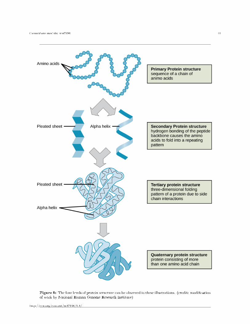
The four levels of protein structure (primary, secondary, tertiary, and quaternary) are illustrated inFigure 9.
http://cnx.org/content/m47196/1.4/
Connexions module: m47196 11
Figure 9: The four levels of protein structure can be observed in these illustrations. (credit: modi�cationof work by National Human Genome Research Institute)
http://cnx.org/content/m47196/1.4/

Connexions module: m47196 12
4 Denaturation and Protein Folding
Each protein has its own unique sequence and shape that are held together by chemical interactions. Asnoted by Mirsky and Pauling in the epigraph above, a denatured protein is one that has lost that uniqueshape and con�guration. If the protein is subject to changes in temperature, pH, or exposure to chemicals,the protein structure may change, losing its shape without losing its primary sequence in what is knownas denaturation. Denaturation is often reversible because the primary structure of the polypeptide isconserved in the process if the denaturing agent is removed, allowing the protein to resume its function.Sometimes denaturation is irreversible, leading to loss of function. One example of irreversible proteindenaturation is when an egg is fried. The albumin protein in the liquid egg white is denatured when placedin a hot pan. Not all proteins are denatured at high temperatures; for instance, bacteria that survive inhot springs have proteins that function at temperatures close to boiling. The stomach is also very acidic,has a low pH, and denatures proteins as part of the digestion process; however, the digestive enzymes of thestomach retain their activity under these conditions.
Protein folding is critical to its function. It was originally thought that the proteins themselves wereresponsible for the folding process. Only recently was it found that often they receive assistance in thefolding process from protein helpers known as chaperones (or chaperonins) that associate with the targetprotein during the folding process. They act by preventing aggregation of polypeptides that make up thecomplete protein structure, and they disassociate from the protein once the target protein is folded.
http://cnx.org/content/m47196/1.4/