Embed Size (px)
Citation preview
Proteins
Proteins are polypeptides
Biochemistry Lectures
Amino Acids, Proteins, Enzymes
DR. M. Sasvári
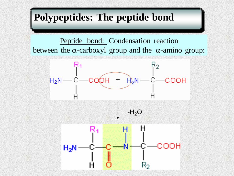
Polypeptides: The peptide bond
Peptide bond: Condensation reaction
between the -carboxyl group and the -amino group:
-H2O

How to write a dipeptide?
+H3N
C
C
N
C
COO-
R
R
O
H
N terminal C-terminal
H
H

-glutamyl-cystein
Glu -Cys
+ H2N-Gly
A biologically important tripeptide: Glutathion
+ Glu-COOH H2N-Cys
-glutamyl-cystein synthetase
-glutamyl-cysteinyl-glycine
glutathion
Glu -Cys-Gly
glutathion synthetase
Glu -Cys
ATP
ATP
N
C
H
O
C
CH2SH
HN
C
H
O
C
O
CC
CH2SH
HH
N
H
C
H
COO-
H
N
H
C
H
COO-
H
+ H 3 N
H2C
CH 2
C
O
C - COO
-
H
+ H 3 N CH 2
C
O
C
O O
C - COO
-
H H

Function of Glutathion
2 G-SH + H2O2 G-S-S-G + 2H2O
reduced glutathion (thiol group) oxidized glutathion (disulphide bridge)
Importance: Maintaining of cellular redox state
Reductive power against oxidative stress
Red blood cells: high O2 concentration
oxidative effect
free radical formation
hydrogen peroxide formation
lipid peroxidation

NutraSweet® (aspartame), an artificial sweetener
Dipeptide of Asp and Phe (carboxyl terminal is esterified)
+H3N
C
C
N
C
CO-O- CH3
CH2COO-
O
H
N terminal C-terminal
H
H
CH2
methylester

Calculation of Ip:
Polypeptides: Isoelectric point
Sequence of a tetrapeptide: Glu-His-Arg-Gly
Charges at pH 7:
Isoelectric form:
+ + + -
Ip:basic Ip: (6.0 + 9.7)/2
+
Arg (guanidino) (pKa = 12.5)
- -carboxyl group (pKa = 2.3)
-amino group (pKa = 9.7) + +
His (imidasol) (pKa = 6.0)
- Glu -carboxyl group) (pKa = 4.3)
Example:
+ + -
-
-
Lost Next

Other biologically important peptides
Peptide hormones:
thyrotropin releasing factor (3 amino acid residues)
oxytocin (9) – uterin contraction
bradykinin (9) – inhibits inflammation
enkephalins (CNS)
small proteins – large peptides
insuline (30+21)
glucagon (29)

Levels of protein
structure

Primary structure
Quaternary structure
Main molecular forces
- Amino acid sequence - covalent bonds
(peptide bond and disulphide bridge)
Secondary structure -H-bonds between the atoms of the peptide group
Tertiary structure - Secondary bonds between the side chains of the same
polypeptide
- Secondary bonds between the side chains of different
polypeptides (subunits)
CONFORMATION
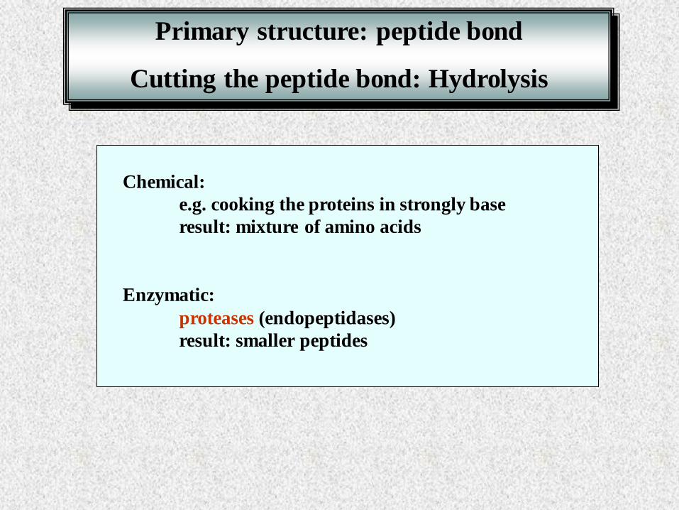
Chemical:
e.g. cooking the proteins in strongly base
result: mixture of amino acids
Enzymatic:
proteases (endopeptidases)
result: smaller peptides
Primary structure: peptide bond
Cutting the peptide bond: Hydrolysis

The peptide group is rigid and planar
RESONANCE INTERACTION
Consequences:
1. Planarity
2. Trans conformation
C-N bond will have some double bond character
C
O
N
H
.. C
C
carbonyl group
p bond
lone electron pair
of N
Six atoms of peptide group: C1 – CO-NH-C2
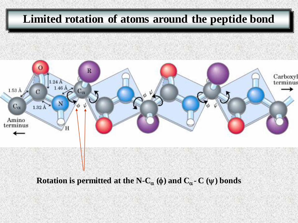
Rotation is permitted at the N-C (f) and C- C () bonds
Limited rotation of atoms around the peptide bond

Ramachandran plot: Prediction of secondary structure
possible angles of free rotation
C f N
H
O
C N
H
O
C H
R
b strands
helix

Molecular forces:
H-bond between the atoms of the peptide groups
O
H
C N
O
H
C N
Backbone structure
Side chains has only small influence
Secondary structure
Conformation of the polypeptide backbone
Examples:
-helix
parallel and antiparallel b-sheets
collagen helix (see later)
The conformational relationship of neighboring amino acids

Right handed -helix
R groups protrude outward from the helical backbone
Space-filling
model
Ball-and-stick model
backbone
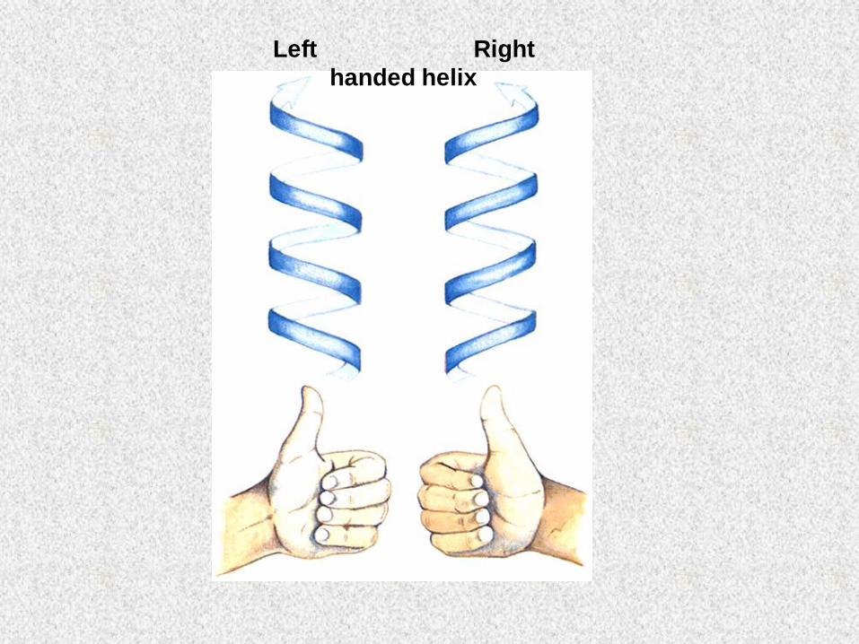
Left Right
handed helix

Stability of an -helix is decrease by
1. electrostatic repulsion (or attraction)
of adjacent positive (or negative) side chains
2. bulkiness of adjacent side chains
3. (ionic) interaction between amino acid side chains
spaced tree (four) residues apart
4. positively charged amino acid at the C-terminal, or
negatively charged amino acids at the N terminal
5. presence of Proline
Stability of the -helix and b-sheet is based on
- the minimized steric repulsion between the side chains
- the maximized H-bonds between the peptide groups
Stability of the -helix

Antiparallel chains of b-conformation
b-sheets: extended, zizag structure
Alternating pattern of R groups
H-bond

b-turn
Other secondary structures
A connection between
the ends of antiparallel b-chains
• a tight connection
• involves 4 amino acid residues
• H bond between the peptide groups
of the 1st and 4th amino acids
• Gly and Pro occur frequently
• often found near to the surface of the protein
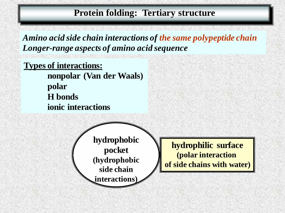
Protein folding: Tertiary structure
Amino acid side chain interactions of the same polypeptide chain
Longer-range aspects of amino acid sequence
Types of interactions:
nonpolar (Van der Waals)
polar
H bonds
ionic interactions
hydrophobic
pocket (hydrophobic
side chain
interactions)
hydrophilic surface (polar interaction
of side chains with water)

Conformations: many helices
Conformations: b sheets
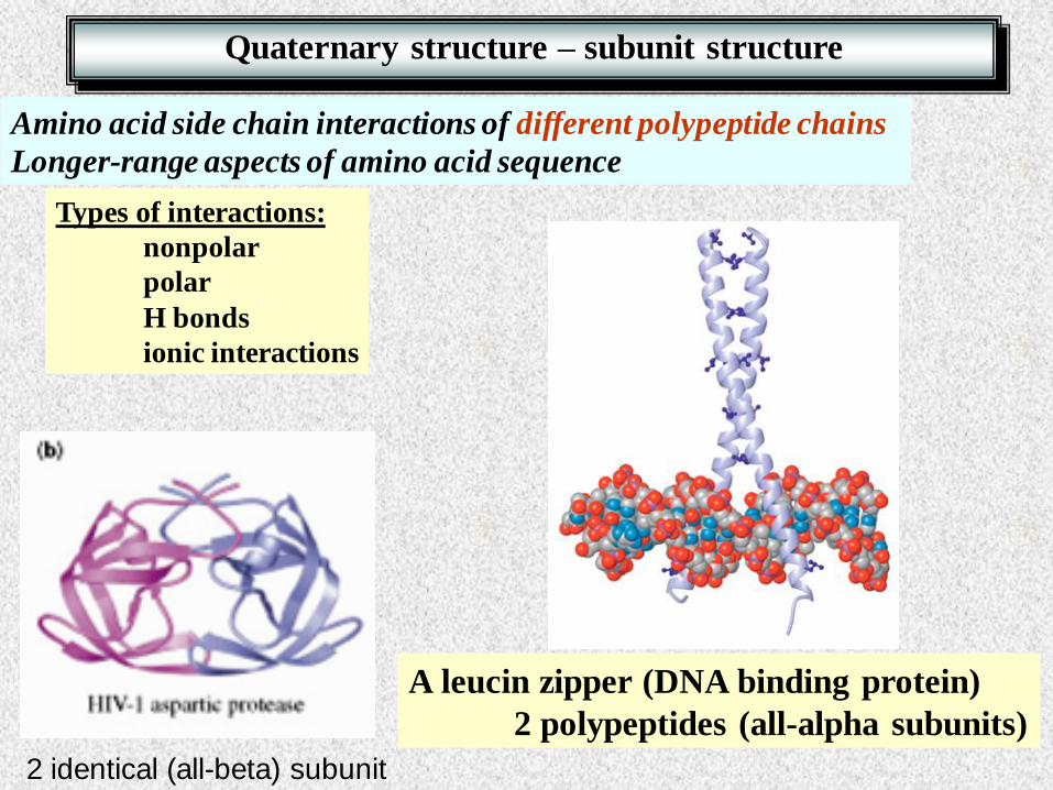
Quaternary structure – subunit structure
Amino acid side chain interactions of different polypeptide chains
Longer-range aspects of amino acid sequence
Types of interactions:
nonpolar
polar
H bonds
ionic interactions
A leucin zipper (DNA binding protein)
2 polypeptides (all-alpha subunits)
2 identical (all-beta) subunit

Enzymes are proteins
Cofactors
Prostetic groups
Metal ions
Coenzymes
Coordinative bond
Covalent bond
Secondary interactions

Optimal
Conditions:
Ionic strength
Temperature
Solvent
pH
Native conformation
Maximal catalytic activity

Temperature dependence of enzyme activity
T
Enzyme
activity
Heat denaturation
changes in conformation
Heat sensitivity is different
Heat-shock proteins
Q10 temperature coefficient
Optimal temperature

pH
Enzyme
activity
5 6 4
pH profiles
pH optimum Isoelectric pont =

pH dependence of enzyme activity
Active center:
Glu COOH pKa = 5.9
- Lysozyme
Asp bCOOH pKa = 4.5
Optimal pH?
Example:
pH
4 5 6 protonated deprotonated
4.5 5.9
Asp - Glu
optimal pH
Analysis and separation
of proteins
Appendix

Gel electrophoresis (practice)
SDS-PAGE
isoelectric focusing
two-dimensional electrophoresis
Gel filtration (practice)
Dialysis (desalting)
Affinity chromatography
Ion exchange chromatography

Analysis of protein mixtures – SDS-PAGE
1. SDS-PAGE (see: practice book) – separation of protein mixtures
Denaturation of proteins, negative charges

Crude extract (1st lane)
Proteins after the purification steps (others)
four subunits (after denaturation)
Purified protein
SDS-PAGE: Cotrolling the purification procedure

SDS-PAGE: Determination of molecular weight of proteins

Analysis of protein mixtures: Isoelectric focusing

Analysis of protein mixtures: Two dimensional electrophoresis
1st dimension:
Isoelectric focusing
2nd dimension:
SDS-PAGE

Identification of more than 1000 proteins from E. Coli
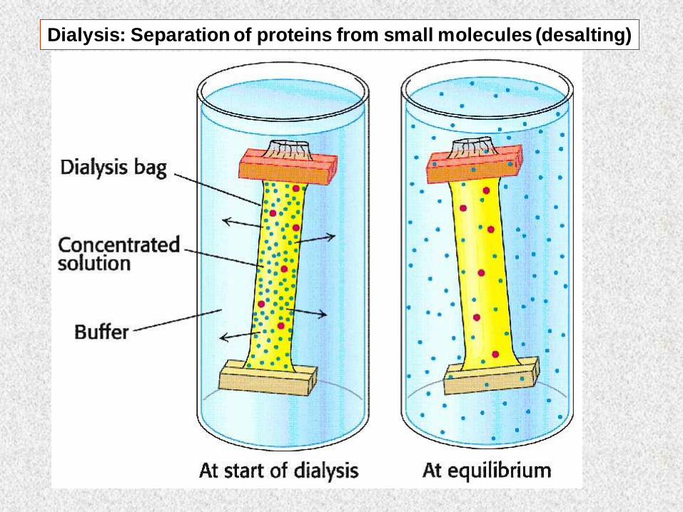
Dialysis: Separation of proteins from small molecules (desalting)

Gel filtration: Separation of proteins from small molecules (desalting)

Protein purification: Affinity chromatography
Separation is based on the
specific binding
between the LIGAND and the protein
The ligand is cross-linked to the beads
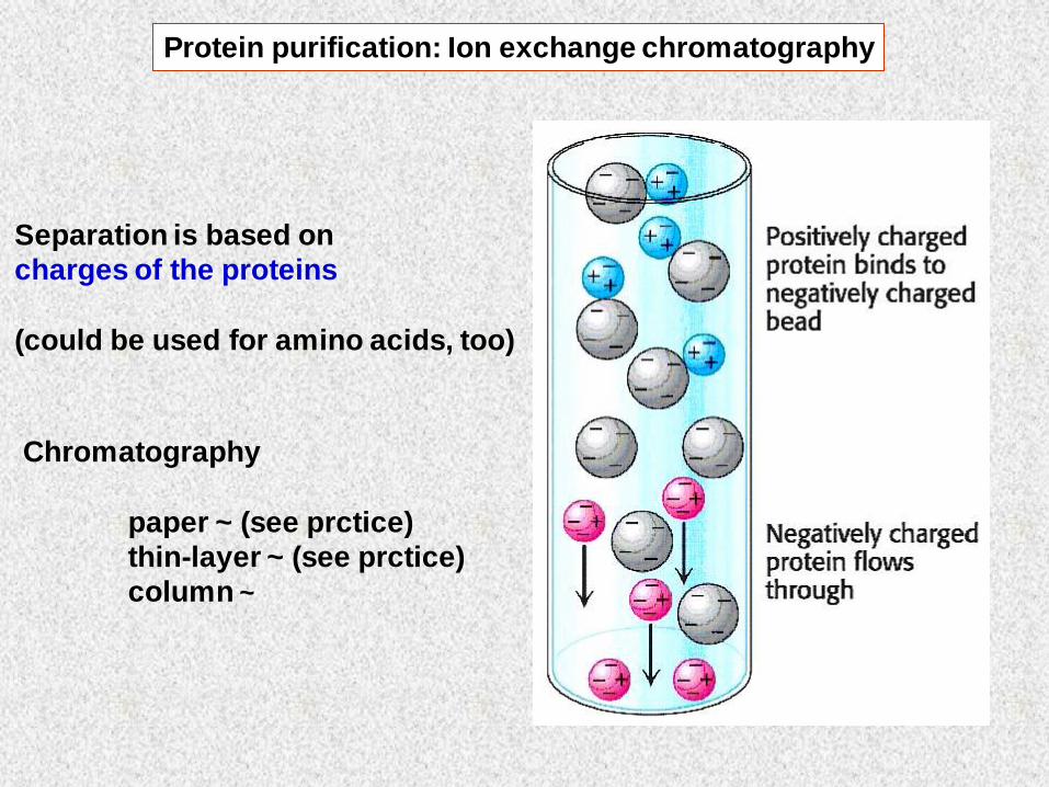
Protein purification: Ion exchange chromatography
Separation is based on
charges of the proteins
(could be used for amino acids, too)
Chromatography
paper ~ (see prctice)
thin-layer ~ (see prctice)
column ~