Embed Size (px)
Citation preview

OPEN
Proteolytic degradation and potential role ofonconeural protein cdr2 in neurodegeneration
J-Y Hwang1,2,8, J Lee1,8, C-K Oh1, HW Kang1, I-Y Hwang1, JW Um1, HC Park3, S Kim3, J-H Shin4, W-Y Park4, RB Darnell5, H-D Um6,KC Chung1, K Kim*,7 and YJ Oh*,1
Cerebellar degeneration-related protein 2 (cdr2) is expressed in the central nervous system, and its ectopic expression in tumorcells of patients with gynecological malignancies elicits immune responses by cdr2-specific autoantibodies and T lymphocytes,leading to neurological symptoms. However, little is known about the regulation and function of cdr2 in neurodegenerativediseases. Because we found that cdr2 is highly expressed in the midbrain, we investigated the role of cdr2 in experimental modelsof Parkinson’s disease (PD). We found that cdr2 levels were significantly reduced after stereotaxic injection of 1-methyl-4-phenylpyridinium (MPP+) into the striatum. cdr2 levels were also decreased in the brains of post-mortem PD patients. Usingprimary cultures of mesencephalic neurons and MN9D cells, we confirmed that MPP+ reduces cdr2 in tyrosine hydroxylase-positive dopaminergic neuronal cells. The MPP+-induced decrease of cdr2 was primarily caused by calpain- and ubiquitinproteasome system-mediated degradation, and cotreatment with pharmacological inhibitors of these enzymes or overexpressionof calcium-binding protein rendered cells less vulnerable to MPP+-mediated cytotoxicity. Consequently, overexpression of cdr2rescued cells from MPP+-induced cytotoxicity, whereas knockdown of cdr2 accelerated toxicity. Collectively, our findings provideinsights into the novel regulatory mechanism and potentially protective role of onconeural protein during dopaminergicneurodegeneration.Cell Death and Disease (2016) 7, e2240; doi:10.1038/cddis.2016.151; published online 2 June 2016
Cerebellar degeneration-related protein 2 (cdr2), an onco-neural protein, is associated with paraneoplastic cerebellardegeneration (PCD).1–3 Under physiological conditions, cdr2expression is restricted to cerebellar Purkinje neurons, brainstem neurons, and testes.4,5 However, cdr2 is ectopicallyexpressed in breast or ovarian tumors of PCD patients,resulting in the generation of autoantibodies6–8 that areassociated with neurodegeneration of Purkinje neurons.9–12
Although the regulation of cdr2 is not well understood, anearly study suggests that cdr2 is phosphorylated by PKN,13
and a more recent study shows that cdr2 is ubiquitinatedby anaphase-promoting complex/cyclosome (APC/C) anddegraded by proteasomes during the exit from mitosis.14
Despite these advances, the regulatory mechanisms andpotential role of cdr2 in neurodegenerative disorders have notbeen explored.Parkinson’s disease (PD) is a neurodegenerative disorder
characterized by a selective loss of dopaminergic neurons in
the substantia nigra (SN) pars compacta that is associatedwith both motor defects and nonmotor symptoms.15 Mitochon-drial dysfunction, oxidative stress, and inflammation areproposed to underlie the pathogenesis of familial and sporadicforms of PD.16,17 Accumulating evidence indicates thatprotease activation plays a critical role in the progression ofneurodegeneration in PD.18–27 In our previous studies, weobserved the activation of caspase and calpain in neurotoxin-induced dopaminergic neurodegeneration28,29 and found thatdegradation of endogenous substrates by activated proteasesleads to neurodegeneration.30,31 Therefore, in the presentstudy, we investigated the expression and protease-mediatedregulation of cdr2 in experimental models of PD. We foundthat cdr2 is downregulated by calpain and the ubiquitinproteasome system and that the restoration of cdr2 levelsrenders dopaminergic neurons less vulnerable to 1-methyl-4-phenylpyridinium (MPP+)-mediated cytotoxicity. To our knowl-edge, it is the first report providing evidence that cdr2 is
1Department of Systems Biology, Yonsei University College of Life Science and Biotechnology, Seoul 120-749, Korea; 2Dominick P. Purpura Department of Neuroscience,Albert Einstein College of Medicine, New York, NY 10461, USA; 3Graduate School of Medicine, Korea University, Ansan 425-707, Gyeonggi-do, Korea; 4Division ofPharmacology, Department of Molecular Cell Biology, Sungkyunkwan University School of Medicine, Suwon 440-746, Gyeonggi-do, Korea; 5Laboratory of MolecularNeuro-Oncology, Howard Hughes Medical Institute, The Rockefeller University, New York, NY 10065, USA; 6Division of Radiation Cancer Biology, Korean Institute ofRadiological & Medical Sciences, Seoul 01812, Korea and 7Department of Brain and Cognitive Sciences, Daegu Gyeongbuk Institute of Science and Technology (DGIST),Daegu 711-873, Korea*Corresponding author: K Kim, Department of Brain and Cognitive Sciences, DGIST, 333 Techno Jungang-daero, Hyeonoung-myeon, Dalseong-gun, Daegu 42988, Korea.Tel: +82 53 785 6144; Fax: +82 53 785 1219; E-mail: [email protected] YJ Oh, Department of Systems Biology, Yonsei University College of Life Science and Biotechnology, 134 Shinchon-Dong, Seodaemoon-Gu, Seoul 120-749, Korea.Tel: +82 2 2123 2662; Fax: +82 2 312 5657; E-mail: [email protected] two authors contributed equally to this work.
Received 25.2.16; revised 21.4.16; accepted 05.5.16; Edited by A Verkhratsky
Abbreviations: PD, Parkinson’s disease; cdr2, cerebellar degeneration-related protein 2; APC/C, anaphase-promoting complex/cyclosome; N2, N-2 supplement; MPP+,1-methyl-4-phenylpyridinium; MTT, 3-(4,5-dimethylthiazol-2-yl)-2,5-diphenyltetrazolium bromide; PCD, paraneoplastic cerebellar degeneration; TH, tyrosine hydroxylase;GABA, γ-aminobutyric acid; Ub, ubiquitin; SN, substantia nigra; CSF, cerebrospinal fluid; Z-VAD-fmk, N-benzyloxycarbonyl-Val-Ala-Asp-fluoromethylketone; β-lactone,clasto-lactacystin β-lactone
Citation: Cell Death and Disease (2016) 7, e2240; doi:10.1038/cddis.2016.151& 2016 Macmillan Publishers Limited All rights reserved 2041-4889/16
www.nature.com/cddis

proteolytically regulated andmay play a neuroprotective role indrug-induced model of neurodegeneration.
Results
cdr2 is highly expressed in the midbrain of normal adultrats. Previous studies show that cdr2 is normally expressedin cerebellar Purkinje neurons but is ectopically expressedin breast and ovarian tumors of PCD patients.4,5,32 To furthercharacterize the normal expression pattern of cdr2, lysatesfrom various tissues from adult rats were immunoprobed withanti-cdr2 antibody. We found that cdr2 was highly expressed
in the brain and kidney, whereas the heart and lung showedlower cdr2 expression (Figure 1a). This distinct spatial patternof cdr2 expression prompted us to investigate cdr2 levels inmore specific regions of the brain. We found that the medullaand midbrain showed the highest expression of cdr2,whereas the cerebellum, where Purkinje neurons reside,showed relatively lower cdr2 expression (Figure 1b). Doubleimmunofluorescent localization of tyrosine hydroxylase (TH)and cdr2 revealed that both TH-positive and -negative cellshighly expressed cdr2 in the midbrain including ventraltegmental area, SN pars compacta, and SN pars reticulata(Supplementary Figure S1). Varying levels of cdr2 wereexpressed in other brain regions including hippocampus,
Figure 1 cdr2 expression in rat tissues. (a) cdr2 protein levels in tissue lysates (50 μg) from adult rats (8 weeks old, male) were determined by immunoblotting with anti-cdr2 antibody.Lysates from MN9D dopaminergic neuronal cells transfected with or without T7-tagged mouse cdr2 were used as a positive control for the cdr2 band. Mouse monoclonal anti-GAPDHantibody was used as a loading control. (b) Immunoblot analysis comparing cdr2 levels across various brain regions. (c) Age-dependent expression of cdr2 in the cortex (E, embryonicday; P, postnatal day; W, postnatal week). Both short-term-exposed (S.E.) and long-term-exposed (L.E.) blots were presented. After normalization to GAPDH level, cdr2 levels in each lanewere calculated relative to cdr2 levels in the medulla (b) or the E16 cortex (c). Data are shown as the mean±S.D. of three independent experiments. **Po0.01; ***Po0.001
Regulation of cdr2 in neurodegenerationJ-Y Hwang et al
2
Cell Death and Disease

cortex, striatum, and hypothalamus (Figure 1b). In a pre-liminary study, quite equivalent levels of cdr2 were detected inthe spinal cord and olfactory bulb (data not shown). We alsofound abundant cdr2 expression in the cerebral cortex ofprenatal and early postnatal rats and a dramatic down-regulation in adult rats (Figure 1c), suggesting the temporalregulation of cdr2 expression in the brain. Invariably, weobserved more than one band of cdr2. The in vitro phos-phatase assay showed that the upper bands represent thephosphorylated forms of cdr2 (data not shown). Although wedid not pursue this observation further, it is worthy to note thata serine/threonine kinase PKN (also known as protein kinaseC-related kinase 1), which is involved in the pathogenesis ofAlzheimer’s disease and amyotrophic lateral sclerosis,33,34
interacts with and phosphorylates cdr2.13
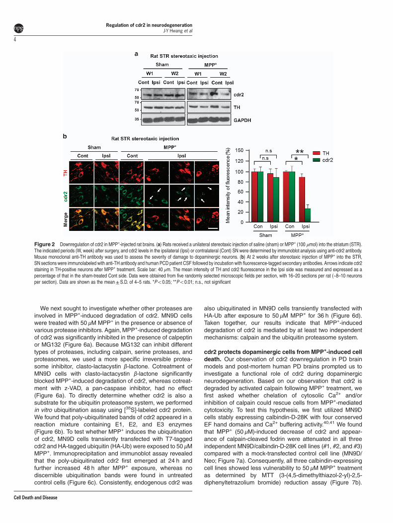
cdr2 protein is decreased in the SN of rodent models ofPD and post-mortem PD patients. PD is characterized by aselective loss of dopaminergic neurons in the SN andsubsequent dopamine deprivation in the striatum. Our findingthat cdr2 is highly expressed in the midbrain led us toexamine the regulation of cdr2 in PD pathogenesis. First,we measured levels of cdr2 protein using rodent models ofPD established by stereotaxic unilateral injection of MPP+
(100 μmol) into the striatum. In the ipsilateral side of the SNpars compacta injected with MPP+ for 2 weeks, the totalnumber of TH-positive dopaminergic neurons was reducedby ~ 29% over the sham control group or the contralateralside of MPP+ injection, as determined by unbiased stereo-logical cell counts (Supplementary Figure S2a). At 1 or2 weeks after MPP+ injection into the striatum, cdr2 levelswere reduced in the ipsilateral side of the SN pars compacta(Figure 2a and Supplementary Figure S2b). No discernibledecrease of cdr2 was detected in the contralateral side of themidbrain or in sham controls. Any obvious decrease of cdr2was not observed in the SN pars reticulata (SupplementaryFigure S2b). Similar pattern of cdr2 reduction in the ipsilateralside of the midbrain was also observed in rats that receivedstereotaxic injection of MPP+ into the middle forebrainbundle, another rodent model of PD (data not shown). ThisMPP+-induced decrease in cdr2 levels was associated with areduction of TH protein, indicating that decrease in cdr2levels may be ascribed to a loss of TH-positive dopaminergicneurons or lower expression levels of cdr2 in dying dopa-minergic neurons or both. Therefore, we next determinedwhether cdr2 levels are decreased in individual TH-positivedying neurons in the SN. At 2 weeks after MPP+ injection,cdr2 levels were well preserved in the cytosol of TH-positiveneurons in sham controls or the contralateral side of injection(Figure 2b). However, TH-positive neurons in the ipsilateralside showed morphology typical of retracted neurites, andtheir cdr2 levels were markedly decreased (Figure 2b,arrows). Quantitative analysis of fluorescence intensityshowed a significant decrease in cdr2 levels in ipsilateralindividual TH-positive neurons (Figure 2b, right). When wemeasured cdr2 levels in the SN of post-mortem PD patientsand age-matched controls (Figure 3a), we interestinglyobserved a significant decrease in cdr2 levels in all four PDbrains, whereas levels of HSP70, a molecular chaperone,were not altered (Figure 3b).
MPP+-induced reduction of cdr2 occurs in the TH-positivedopaminergic neurons. To further determine whether theMPP+-induced decrease of cdr2 occurs in TH-positive dyingdopaminergic cells, we used primary cultures of dopaminergicneurons derived from the rat embryonic mesencephalon or theMN9D dopaminergic neuronal cell line. Exposure of primarycultures of mesencephalic neurons to 3 μM MPP+ for 36 hinduced morphological changes such as neurite retractionand fragmentation (Figure 4a). In this condition, quantitativeanalysis indicated that cdr2 levels were significantly reducedin TH-positive dying neurons following MPP+ treatment(Figure 4a, right panel). In contrast, there were no discerniblechanges of cdr2 levels in γ-aminobutyric acid (GABA)-positiveneurons in the same culture. These data along with our findingthat cdr2 is preserved in neurons of SN pars reticulata suggestthat the MPP+-induced decrease in cdr2 levels is cell-typespecific. The MN9D cell line, a fusion product of mesence-phalic dopaminergic neurons and N18TG neuroblastoma, waspreviously demonstrated to synthesize, release, and take updopamine.35,36 As determined by immunocytochemistry, expo-sure of MN9D dopaminergic cells to 50 μM MPP+ for 36 h orlonger caused a significant reduction in cdr2 levels, whereasnumbers of cells in culture remained the same regardless ofdrug treatment (Figure 4b). Immunoblot analysis indicated thatMPP+ causes a time-dependent reduction of cdr2 (Figure 4c),Taken together, our data indicated that decrease in cdr2occurs in dying TH-positive neurons and is not simply due toMPP+-induced loss of dopaminergic neurons.
Activated calpain and the ubiquitin proteasome systemare responsible for MPP+-induced decrease of cdr2.Having established that cdr2 is downregulated in the brainsof experimental models of PD as well as post-mortem PDpatients, we next investigated the mechanism underlyingMPP+-induced reduction of cdr2. MPP+-induced cell death isaccompanied by or results from a burst of intracellular freeCa2+, leading to activation of calpain, a Ca2+-dependentcysteine protease in MN9D cells as previously demonstrated byus.30,37,38 Therefore, we determined whether Ca2+-mediatedcalpain activation is responsible for MPP+-induced reduction ofcdr2. As shown in Figure 5a, many MN9D cells stained positivefor fluo-3, a Ca2+-sensitive fluorescent dye, 36 h after 50 μMMPP+ treatment. Quantitative analysis revealed an approxi-mately fourfold increase in the intensity of fluo-3-stainedMN9D cells relative to untreated control cells 36 h afterMPP+ treatment (data not shown). Moreover, fodrin, a generalcalpain substrate, was cleaved in a time-dependent manner(Figure 5b), indicating that a rise of intracellular free Ca2+ leadsto calpain activation in MPP+-treated MN9D cells. We nextspecifically inquired whether cdr2 is a substrate of calpain byincubating lysates of MN9D cells in calpain-activating condi-tions. We found that the addition of either m- or μ-calpaincaused complete degradation of endogenous cdr2 (Figure 5c),and this event was blocked in the presence of calpeptin,a cell-permeable calpain inhibitor. To demonstrate that cdr2 is adirect substrate of activated calpain, we performed a calpaincleavage assay using in vitro translated [35S]-labeled cdr2.Both m- and μ-calpain led to complete degradation of cdr2(Figure 5d) that was inhibited in the presence of calpeptin orMG132, another calpain inhibitor.39
Regulation of cdr2 in neurodegenerationJ-Y Hwang et al
3
Cell Death and Disease
We next sought to investigate whether other proteases areinvolved in MPP+-induced degradation of cdr2. MN9D cellswere treated with 50 μM MPP+ in the presence or absence ofvarious protease inhibitors. Again, MPP+-induced degradationof cdr2 was significantly inhibited in the presence of calpeptinor MG132 (Figure 6a). Because MG132 can inhibit differenttypes of proteases, including calpain, serine proteases, andproteasomes, we used a more specific irreversible protea-some inhibitor, clasto-lactacystin β-lactone. Cotreatment ofMN9D cells with clasto-lactacystin β-lactone significantlyblocked MPP+-induced degradation of cdr2, whereas cotreat-ment with z-VAD, a pan-caspase inhibitor, had no effect(Figure 6a). To directly determine whether cdr2 is also asubstrate for the ubiquitin proteasome system, we performedin vitro ubiquitination assay using [35S]-labeled cdr2 protein.We found that poly-ubiquitinated bands of cdr2 appeared in areaction mixture containing E1, E2, and E3 enzymes(Figure 6b). To test whether MPP+ induces the ubiquitinationof cdr2, MN9D cells transiently transfected with T7-taggedcdr2 and HA-tagged ubiquitin (HA-Ub) were exposed to 50 μMMPP+. Immunoprecipitation and immunoblot assay revealedthat the poly-ubiquitinated cdr2 first emerged at 24 h andfurther increased 48 h after MPP+ exposure, whereas nodiscernible ubiquitination bands were found in untreatedcontrol cells (Figure 6c). Consistently, endogenous cdr2 was
also ubiquitinated in MN9D cells transiently transfected withHA-Ub after exposure to 50 μM MPP+ for 36 h (Figure 6d).Taken together, our results indicate that MPP+-induceddegradation of cdr2 is mediated by at least two independentmechanisms: calpain and the ubiquitin proteasome system.
cdr2 protects dopaminergic cells from MPP+-induced celldeath. Our observation of cdr2 downregulation in PD brainmodels and post-mortem human PD brains prompted us toinvestigate a functional role of cdr2 during dopaminergicneurodegeneration. Based on our observation that cdr2 isdegraded by activated calpain following MPP+ treatment, wefirst asked whether chelation of cytosolic Ca2+ and/orinhibition of calpain could rescue cells from MPP+-mediatedcytotoxicity. To test this hypothesis, we first utilized MN9Dcells stably expressing calbindin-D-28K with four conservedEF hand domains and Ca2+ buffering activity.40,41 We foundthat MPP+ (50 μM)-induced decrease of cdr2 and appear-ance of calpain-cleaved fodrin were attenuated in all threeindependent MN9D/calbindin-D-28K cell lines (#1, #2, and #3)compared with a mock-transfected control cell line (MN9D/Neo; Figure 7a). Consequently, all three calbindin-expressingcell lines showed less vulnerability to 50 μM MPP+ treatmentas determined by MTT (3-(4,5-dimethylthiazol-2-yl)-2,5-diphenyltetrazolium bromide) reduction assay (Figure 7b).
Figure 2 Downregulation of cdr2 in MPP+-injected rat brains. (a) Rats received a unilateral stereotaxic injection of saline (sham) or MPP+ (100 μmol) into the striatum (STR).The indicated periods (W, week) after surgery, and cdr2 levels in the ipsilateral (Ipsi) or contralateral (Cont) SN were determined by immunoblot analysis using anti-cdr2 antibody.Mouse monoclonal anti-TH antibody was used to assess the severity of damage to dopaminergic neurons. (b) At 2 weeks after stereotaxic injection of MPP+ into the STR,SN sections were immunolabeled with anti-TH antibody and human PCD patient CSF followed by incubation with fluorescence-tagged secondary antibodies. Arrows indicate cdr2staining in TH-positive neurons after MPP+ treatment. Scale bar: 40 μm. The mean intensity of TH and cdr2 fluorescence in the Ipsi side was measured and expressed as apercentage of that in the sham-treated Cont side. Data were obtained from five randomly selected microscopic fields per section, with 16–20 sections per rat (~8–10 neuronsper section). Data are shown as the mean±S.D. of 4–5 rats. *Po0.05; **Po0.01; n.s., not significant
Regulation of cdr2 in neurodegenerationJ-Y Hwang et al
4
Cell Death and Disease

Similarly, pharmacological inhibition of calpain by calpeptin orMG132, which partially preserved cdr2 levels, attenuatedMPP+-induced MN9D cell death (Figures 7c–f), suggestingthat the reduced vulnerability of calbindin-expressing cells orcalpeptin/MG132-treated MN9D cells may be in part attrib-uted to preserved cdr2 levels. To directly examine whetherthe level of cdr2 itself affects cell survival upon exposure to50 μM MPP+, we established three MN9D cell lines stablyoverexpressing T7-tagged cdr2 and two MN9D cell lines inwhich cdr2 was stably silenced by transfection with shorthairpin RNA (shRNA; Figures 8a and c). MTTreduction assayusing two highly cdr2-expressing cell lines (MN9D/cdr2 #2and #3) indicated that overexpression of cdr2 protects cellsfrom MPP+-induced cytotoxicity (Figure 8b). In contrast,MPP+-induced cell death was accelerated in cdr2-silencedMN9D cells (Figure 8d). Quite similar pattern was observedwhen stable MN9D cells were exposed to 50 μM MPP+ forvarying time periods (Supplementary Figure S3). Interest-ingly, we observed that cdr2 is also decreased during6-hydroxydopamine (6-OHDA)-induced neuronal death(Supplementary Figures S4a and b). As we previouslydemonstrated,28,29 6-OHDA led to reactive oxygen species-dependent MN9D cell death. As a consequence, 6-OHDA-mediated decrease in cdr2 was largely inhibited in thepresence of N-acetyl-L-cysteine but not calpeptin. These datasuggest that an additional calpain-independent degradationpathway may be involved in 6-OHDA-mediated decrease ofcdr2. We also observed that 6-OHDA-induced cell death wassignificantly blocked in cdr2-overexpressing MN9D cells
(Supplementary Figure S4c). Collectively, our results suggestthat cdr2 may play a neuroprotective role in drug-induceddopaminergic neurodegeneration.
Discussion
Although cdr2 mRNA is widely expressed, cdr2 proteinexpression is restricted to immune-privileged sites includingthe testis, brain stem, and cerebellum, suggesting that cdr2expression is regulated by a post-transcriptional mechanism.4,5
Here, we examined cdr2 expression patterns in several brainregions and found that the second most prominent site ofexpression was the midbrain. We also observed high levelsof cdr2 expression in the striatum, a target of dopaminergicprojections from the SN pars compacta. Therefore, weinvestigated whether cdr2 levels are altered in dopaminergicneurodegeneration. Indeed, we found lower cdr2 protein levelsin the brains of post-mortemPD patients and animal PDmodelsestablished by stereotaxic injection of MPP+ into the striatum.Using both cultured MN9D dopaminergic cells and primarycultures of mesencephalic neurons challenged with MPP+, wefound decreased levels of cdr2 in TH-positive dying dopami-nergic neurons but not in GABAergic neurons. This cell-typespecificity can be addressed by the fact that GABAergicneurons is more resistant to MPP+ or rotenone, anotherPD-related drug targeting mitochondria complex I.42
We showed that cdr2 degradation was mediated primarilyby calpain and the ubiquitin proteasome system. Conse-quently, pharmacological inhibition of these enzymessuccessfully blocked MPP+-induced degradation of cdr2and subsequent dopaminergic neurodegeneration. Previousstudies, including ones from our laboratory, report that MPP+-induced calpain activation via increased intracellular Ca2+
leads to cleavage of numerous cellular substrates andcontributes to neuronal cell death,28,30,43,44 supporting thenotion that degradation of critical cellular proteins by activatedcalpain may be linked to dopaminergic neurodegeneration. Anincrease in intracellular free Ca2+ is also known to occur inacute neurodegenerative conditions such as ischemic strokeand spinal cord injury.45–48 In preliminary studies, we foundthat cdr2 levels were decreased in a rodent ischemic strokemodel established by middle cerebral artery occlusion and acontusion spinal cord injury model (data not shown), suggest-ing that cdr2 may be degraded by activated calpainduring both acute and chronic neurodegeneration. Previously,it has been demonstrated that cdr2 undergoes APC/C-mediated poly-ubiquitination during the exit from mitosis.14
Although we did not determine the relationship betweenMPP+-induced ubiquitination of cdr2 and APC/C activity inMN9D cells, the results of our in vitro and cell-basedubiquitination assays raise the possibility that cdr2 is poly-ubiquitinated and subject to proteasome-mediated degrada-tion after MPP+ treatment. In a separate study, we found thatboth Parkin and SIAH bind to cdr2 in HEK293 cells and N2(N-2 supplement) neuroblastoma (data not shown). However,we do not yet have clear evidence of cdr2 poly-ubiquitinationby either of these two E3 ligases. Therefore, further attemptsare necessary to identify the specific E3 ligase for cdr2ubiquitination.
Figure 3 Downregulation of cdr2 in brains from post-mortem PD patients.(a) Characteristics of PD patients and age-matched controls, including diagnosis,age, sex, race, and post-mortem delay (PMD; h). (b) Tissue lysates from the SN ofpost-mortem humans were subjected to immunoblot analysis using anti-cdr2 antibodyand anti-HSP70 antibody. After normalization against GAPDH, relative intensity ofcdr2 in brains from post-mortem PD patients or age-matched controls. Data areshown as mean± S.D. **Po0.01
Regulation of cdr2 in neurodegenerationJ-Y Hwang et al
5
Cell Death and Disease

Our previous study showed that calpain-mediated cleavageof optineurin, peripherin, or arsenical pump-driving ATPaseoccurs in MPP+-treated MN9D dopaminergic cells, and anytreatments that restore their protein levels or overexpression ofone of these substrates rescue MN9D cells from MPP+-mediated cytotoxicity.30 In accordance with the results ofpharmacological inhibition of calpain, MN9D cells overexpres-sing calbindin-D-28K showed preservation of cdr2, resulting inmore resistance to MPP+-induced cytotoxicity. Similarly, wedemonstrated that MN9D cells overexpressing cdr2 are lessvulnerable to MPP+-induced cytotoxic damage. Conversely,MPP+-induced cell death was accelerated in MN9D cellssubjected to shRNA-mediated silencing of cdr2, suggestingthat cdr2 protein levels may be positively correlated with therate of neuroprotection in MN9D cells after MPP+ treatment.
Therefore, we are tempting to suggest that cdr2 exerts acertain neuroprotective function and thus its degradation maybe associated with drug-induced neurodegeneration. Intrigu-ingly, we also found that shRNA-mediated knockdown of cdr2in cultured hippocampal neurons enhanced spontaneousapoptotic cell death, raising the possibility that cdr2 mayhave a neuroprotective role during early development(Supplementary Figure S5). Furthermore, cdr2 morpholino-injected zebrafish embryos showed massive death of neuralprogenitor cells and post-mitotic differentiated neuronal cellsin the spinal cord (Supplementary Figure S6). Consideringthat developing neurons undergo programmed cell death tocontrol the number of neural progenitor cells and optimizeneural connections between differentiating neurons and theirtargets in both vertebrates and invertebrates,49 our
Figure 4 Downregulation of cdr2 in MPP+-treated dopaminergic neurons. (a) Primary cultures of rat mesencephalic dopaminergic neurons at DIV 5 or 6 were treated with orwithout 3 μMMPP+ for 36 h. Cells were subjected to double immunofluorescent staining using human PCD patient CSF and anti-TH antibody or anti-GABA antibody. TH-positiveneurons having retracted or fragmented neurites are indicated by white arrows. Scale bars: 20 μm. Mean intensity of TH/GABA or cdr2 fluorescence was measured andexpressed as a percentage of that in sham-treated control cultures. Data were obtained from 300–400 neurons from 5 to 10 randomly selected microscopic fields. Data are shownas the mean± S.D. of three independent experiments. ***Po0.001; n.s., not significant. (b) MN9D dopaminergic neuronal cells exposed to 50 μM MPP+ for 36 h wereimmunostained using human PCD patient CSF. Phase-contrast and fluorescent images were taken using an Axiovert 100. Scale bar: 50 μm. (c) Lysates (50 μg) from MN9D cellsexposed to 50 μM MPP+ for the indicated time period were subjected to immunoblot analysis using anti-cdr2 antibody. Anti-actin antibody was used as a loading control. Afternormalization against actin, levels of cdr2 were expressed as a percentage of that in untreated control cells. Data are shown as the mean± S.D. of three independentexperiments. *Po0.05; **Po0.01; n.s., not significant
Regulation of cdr2 in neurodegenerationJ-Y Hwang et al
6
Cell Death and Disease

preliminary findings suggest that cdr2 may be a key regulatorand not merely a bystander of neuronal cell survival, althoughit is highly speculative at present.Previous studies by others indicate that cdr2 is primarily
present in the cytoplasm and has a leucine zippermotif.6,13,50,51 Therefore, cdr2 could bind to other proteinswith a leucine zipper motif and exert transcriptional transacti-vation activity via binding to DNA. For example, cdr2 capturesc-myc in the cytoplasm that prevents the transactivation ofpro-apoptotic genes by nuclear c-myc activity in cerebellarPurkinje neurons,10 indicating that cdr2 can block c-myc-mediated apoptosis. As another example, cdr2 interacts with anuclear helix-loop-helix leucine zipper protein, MRG X,52 andcoexpression of cdr2 and MRG X prevents MRG X-inducedglioblastoma cell death. The same laboratory also reports thatcdr2 binds to the cell cycle-related protein MRG15 and thatoverexpression of cdr2 inhibits the derepression of B-mybtranscriptional activity by MRG15.53 The B-myb transcriptionfactor is induced in response to apoptotic stimuli, and itsknockdown in neurons is protective against nerve growthfactor deprivation or drug-induced cell death accompanyingDNA damage.54 Therefore, cdr2 may serve a neuroprotectiverole by repressing B-myb promoter activity. Although we didnot attempt to determine whether these scenarios hold true inMN9D cells, we observed that MPP+ treatment decreased
levels of c-myc in the nucleus regardless of whether cellsoverexpressed cdr2 (data not shown). Therefore, it seems thata neuroprotective role of cdr2 cannot be ascribed to itsknown regulation of the c-myc-mediated cell death pathway,at least in MN9D cells. We also found that cdr2 has achromosome segregation ATPase domain as determined by adatabase search and can bind to ATPas determined by in vitroATP binding assay (data not shown). Similarly, a colocalizationstudy performed in our laboratory indicates that cdr2 may bindtomicrotubules inMN9D cells (data not shown). At present, wedo not know whether and how these activities of cdr2 arerelated to its neuroprotective role in neuronal cells. Therefore,further studies delineating the biological and neuroprotectiveactivity of cdr2 in the nervous system are required.In summary, we characterized the cdr2 expression profile in
various regions of the brain, and examined its regulation byactivated proteases and its neuroprotective function duringneurotoxin-induced dopaminergic neurodegeneration. Ourfindings indicate that the negative regulation of cdr2 levelsby activated calpain and the ubiquitin proteasome systemmaycontribute to MPP+-induced neuronal cell death. Unveiling thedetail mechanisms involved in regulation of cdr2would expandour understanding of the potential role of cdr2 in pathologicalneurodegeneration.
Figure 5 Degradation of cdr2 by calpain. (a) MN9D cells were treated with 50 μM MPP+ for 36 h. Cells were then loaded with Fluo-3/AM dye and examined under afluorescence microscope to detect levels of intracellular free Ca2+. Scale bar: 100 μm. (b) Lysates (50 μg) from MN9D cells treated with or without 50 μM MPP+ for the indicatedtime period were subject to immunoblot analysis using monoclonal anti-fodrin antibody recognizing calpain-cleaved band. (c) Lysates from MN9D cells (50 μg) were incubatedwith recombinant m-calpain (0.343 units) or μ-calpain (0.134 units) in the presence of 1 mM CaCl2. Calpeptin (50 μM) was added to the lysates to block calpain activity. After thereaction, immunoblot analysis using anti-cdr2 antibody was performed to detect remaining cdr2. (d) For in vitro cleavage assay, [35S]-labeled cdr2 was incubated with m-calpain(0.343 units) or μ-calpain (0.134 units) in the presence of 1 mM CaCl2. Calpeptin (50 μM) or MG132 (10 μM) was added to the reaction mixtures. After incubation, all sampleswere separated by SDS-PAGE and subjected to autoradiography
Regulation of cdr2 in neurodegenerationJ-Y Hwang et al
7
Cell Death and Disease

Materials and MethodsAnimals, stereotaxic surgery, and post-mortem human brains.All experimental animal procedures were in accordance with the National Institutesof Health Guide for the Care and Use of Laboratory Animals and approved by theInstitutional Animal Care and Use Committees of Yonsei University. To measuretissue-specific expression of cdr2 protein, various body parts including subregions
of the brain were surgically removed from Sprague-Dawley (SD) rats (Orientbio,Seongnam, Korea) at different ages. For other groups of rats, stereotaxic surgerywas performed as previously described with modifications.55 Briefly, female SD rats(250–280 g; Daehan Biolink, Deajon, Korea) were anesthetized using anintraperitoneal injection of chloral hydrate (360 mg/kg) and placed in a stereotaxicapparatus (Kopf Instrument, Tujunga, CA, USA). Each rat received a unilateral
Regulation of cdr2 in neurodegenerationJ-Y Hwang et al
8
Cell Death and Disease

injection of MPP+ (Sigma, St. Louis, MO, USA) or sterilized saline into the rightstriatum (100 nmol; +1 mm anteroposterior, − 2.5 mm mediolateral, and -4.5 mmdorsoventral relative to bregma) according to Paxinos and Watson (1998).56
Injections were performed using a Hamilton syringe equipped with a 26S-gaugebeveled needle driven by a syringe pump (K.D. Scientific, Holliston, MA, USA). Theneedle was slowly retracted 10 min after the injection. On the predetermined day
Figure 6 Degradation of cdr2 by the ubiquitin proteasome system. (a) MN9D cells were treated with 50 μM MPP+ for 36 h in the presence or absence of calpeptin (50 μM),MG132 (2.5 μM), clasto-lactacystin β-lactone (2.5 μM), or Z-VAD-fmk (100 μM). Levels of cdr2 were measured by immunoblot analysis using anti-cdr2 antibody. Afternormalization against GAPDH, levels of cdr2 were expressed as a percentage of that in untreated controls. Data are shown as the mean± S.D. of three independent experiments.*Po0.05; **Po0.01; ***Po0.001; n.s., not significant. (b) [35S]-labeled cdr2 was incubated with or without a mixture of conjugation enzymes (E1, E2, and E3) plus ubiquitin andubiquitin-aldehyde. Reaction mixtures were separated by SDS-PAGE and subjected to autoradiography. (c) MN9D cells transiently transfected with T7-tagged cdr2 plus HA-Ubwere exposed to 50 μMMPP+ for the indicated time period. To prevent proteasome-mediated degradation of cdr2, MG132 (2.5 μM) was added to each culture 6 h before harvest.Cell lysates were processed for immunoprecipitation with mouse monoclonal anti-T7 and immunoblot analysis using mouse monoclonal anti-HA to detect poly-ubiquitinated cdr2.(d) To detect endogenous cdr2 ubiquitination, MN9D cells transiently transfected with HA-Ub were treated with or without 50 μM MPP+ for 36 h in the presence of MG132(2.5 μM). Cell lysates were subjected to immunoprecipitation with anti-cdr2 antibody and followed by immunoblot analysis using mouse monoclonal anti-HA
Figure 7 Preservation of cdr2 levels is linked to less vulnerability to MPP+-induced toxicity. (a) MN9D cells stably transfected with calbindin-D-28K (MN9D/calbindin-D-28K#1, #2, or #3) or control vector (MN9D/Neo) were exposed to 50 μM MPP+ for 36 h. Cell lysates from the indicated clones were processed for immunoblot analysis using theindicated antibodies. Mouse monoclonal anti-calbindin-D-28K antibody was used to detect its expression in stably established clones. (b) Cell viability was measured by MTTreduction assay. Data are shown as the mean±S.D. of three independent experiments. ***Po0.001. (c–f) MN9D/Neo cells were treated with 50 μM MPP+ for 36 h in thepresence or absence of the indicated concentration of calpeptin (c and d) or MG132 (e and f). (c and e) Immunoblot analysis was performed using the indicated antibodies.(b, d, and f) Cell viability was expressed as a percentage of that for untreated control cells. Data are shown as the mean± S.D. of three independent experiments. ***Po0.001
Regulation of cdr2 in neurodegenerationJ-Y Hwang et al
9
Cell Death and Disease

after surgery, 3–5 rats in each condition were killed by CO2, and the SN was rapidlydissected out for immunological analysis. The SNs from four post-mortem patientswith PD and four age-matched control individuals were provided by the Departmentof Pathology at Johns Hopkins University. Each brain underwent comprehensiveneuropathological analysis.57
Primary neuronal cultures. To prepare primary cultures of dopaminergicneurons, the ventral mesencephalon was removed from SD rats (Orientbio) onembryonic day (E)14 as previously described.28 Briefly, dopaminergic neuronal cultureswere plated at 1.0 × 10⁵ cells per 1 cm2 ACLAR embedding film (Electron MicroscopySciences, Fort Washington, PA, USA) precoated with 100 μg/ml poly-D-lysine (Sigma)and 4 μg/ml laminin (Invitrogen, San Diego, CA, USA) and maintained at 37 °C in ahumidified 5% CO2 atmosphere in modified Eagle’s medium (MEM; Gibco, GrandIsland, NY, USA) supplemented with 10% fetal bovine serum (FBS; Lonza, Walkersville,MD, USA), 2 mM L-glutamine (Sigma), and 6 g/l glucose (Sigma). At 5 or 6 days in vitro(DIV), cultures were washed with MEM and treated with 3 μM MPP+ for 36 h.
MN9D cell culture, drug treatment, and cell viability. MN9D dopa-minergic neuronal cultures were established from embryonic mesencephalicdopaminergic neurons by somatic fusion.35 As previously described by us,44 MN9Dcells were plated on 25 μg/ml poly-D-lysine precoated culture dishes or plates(Costar, Corning, NY, USA), maintained in DMEM (Gibco) supplemented with10% FBS in an incubator with 10% CO2 at 37 °C, and switched to serum-free N2medium58 containing various experimental reagents, including MPP+, calpeptin(Calbiochem, San Diego, CA, USA), MG132 (Calbiochem), N-benzyloxycarbonyl-Val-Ala-Asp-fluoromethylketone (Z-VAD-fmk, Enzyme Systems Products, Livermore,
CA, USA), and clasto-lactacystin β-lactone (Calbiochem). To assess the rate of cellsurvival after drug treatment, MTT reduction assay was performed as previouslydescribed.59 Briefly, after the indicated incubation period, MTT solution was addedto the culture at a final concentration of 1 mg/ml. Cells were then incubated for 1 hat 37 °C followed by lysis in 20% SDS in 50% aqueous dimethylformamide for 24 h.The optical density of dissolved formazan grains was measured at 540 nm usinga microplate reader (Molecular Devices, Sunnyvale, CA, USA). Values for eachtreatment group were calculated as a percentage relative to the untreated controlgroup (defined as 100% survival).
Immunoblot analysis. The SN of post-mortem human brains was processedfor immunoblot analysis as previously described by us.31 To measure cdr2expression in rats, various body parts including brain regions were dissected andsubjected to lysis. Briefly, dissected tissues were minced with blades, rinsed withice-cold phosphate-buffered saline (PBS), and briefly microcentrifuged. Theresulting pellets were lysed with RIPA buffer (50 mM Tris-HCl, pH 7.4, 1% NP-40,0.25% sodium deoxycholate, 150 mM NaCl, 1 mM EDTA) containing 0.1% SDS andprotease inhibitor cocktail (Roche, Mannheim, Germany) and then furtherhomogenized with a 1-ml syringe. SN obtained from rats that received stereotaxicinjection of MPP+ was similarly processed. For MN9D cells, cells were washed withice-cold PBS, lysed with RIPA buffer containing protease inhibitor cocktail, and thenhomogenized using a 1-ml syringe. Tissue or cell lysates were microcentrifuged at15 000 × g for 20 min at 4 °C. Protein content of the supernatants was measuredusing a Bio-Rad protein assay reagent (Hercules, CA, USA). Proteins from eachsample were separated on 10–12.5% SDS-PAGE, blotted onto prewetted PVDFnitrocellulose filters (Bio-Rad), and blocked with TBST including 5% skim milk for
Figure 8 Protective role for cdr2 in MPP+-induced cell death. (a) MN9D cells were stably transfected with T7-tagged mouse cdr2 (MN9D/cdr2 #1, #2, or #3) or empty vector(MN9D/Neo). Expression levels were validated by immunoblot analysis using mouse monoclonal anti-T7 antibody. (b) MN9D/Neo and two highly expressing MN9D/Cdr2 cell lineswere treated with 50 μMMPP+ for 36 h. Cell viability was measured using MTTreduction assay and expressed as a percentage of that for untreated control cells. Data are shownas the mean±S.D. of three independent experiments. *Po0.05. (c) MN9D cells were stably transfected with cdr2 shRNA- or control shRNA-expressing vectors. Extent of cdr2knockdown was determined by immunoblot analysis using anti-cdr2 antibody. (d) After treatment with 50 μM MPP+ for 36 h, MTT reduction assay was performed. Cell viabilitywas expressed as a percentage of that for untreated control cells. Data are shown as the mean± S.D. from three independent experiments. *Po0.05
Regulation of cdr2 in neurodegenerationJ-Y Hwang et al
10
Cell Death and Disease

1 h. The blots were immunoprobed with human PCD patient serum that reacts withcdr2 (1 : 10 000, generously provided by Dr Darnell at Rockefeller University,New York, NY, USA) or rabbit polyclonal anti-cdr2 (1 : 1000; Sigma), mousemonoclonal anti-tyrosine hydroxylase (TH, a rate-limiting enzyme of dopaminebiosynthesis: 1 : 1000; Pel-Freez, Rogers, AR, USA), mouse monoclonal anti-α-fodrin (1 : 4000; Enzo Life Science, Farmingdale, NY, USA), mouse monoclonalanti-hemagglutinin (HA; 1 : 4000; Santa Cruz Biotechnology, Dallas, TX, USA),mouse monoclonal anti-T7 (1 : 1000; Novagen, Madison, WI, USA), mousemonoclonal anti-HSP 70 (1 : 1000; Santa Cruz), or mouse monoclonal anti-calbindin-D-28K (1 : 2000; Swant, Fribourg, Switzerland). Rabbit polyclonal anti-actin (1 : 4000; Sigma) and mouse monoclonal anti-glyceraldehyde-3 phosphatedehydrogenase (GAPDH, 1 : 4000; Merck Millipore, Billerica, MA, USA) were usedas loading controls.
Immunofluorescent staining. For immunohistochemistry, rats were per-fused transcardially with saline solution containing 0.5% sodium nitrate and heparin(1000 units/ml, Sigma) before fixation. Brains were fixed at 4 °C overnight with 4%paraformaldehyde in 0.1 M phosphate buffer and incubated in 30% sucrose solutionfor 48–72 h at 4 °C until they sank. Brains were then cut into 30-μm-thick coronalsections using a sliding microtome. Sections were processed for immunohisto-chemical staining for TH and cdr2. For immunocytochemistry, primary cultures ofmesencephalic cells and MN9D dopaminergic cells were fixed, blocked, andincubated with primary antibodies as previously described.28 Primary antibodieswere mouse monoclonal anti-TH (1 : 7500, Pel-Freez), rabbit polyclonal GABAantibody (1 : 200; Sigma), and human PCD patient cerebrospinal fluid (CSF, 1 : 20;generously provided by Dr Darnell at Rockefeller University) that reacts with cdr2.After extensive washes with PBS, sections or cultures were incubated at RT for 1 hwith Alexa 546-conjugated goat anti-mouse antibody or Alexa 546-conjugated goatanti-rabbit antibody in combination with Alexa 488-conjugated goat anti-humanantibody (1 : 200, Molecular Probes, Eugene, OR, USA). After extensive washes,sections or cultures were then mounted with Vectashield mounting medium (VectorLaboratories, Burlingame, CA, USA) and examined under a Axiovert 100 micro-scope equipped with an epifluorescence and digital image analyzer (Carl Zeiss,Zena, Germany) or an LSM 510 Meta Laser Scanning Microscope (Carl Zeiss).Fluorescence intensity was measured and analyzed using ImageJ software (NIH,Bethesda, MD, USA).
Measurement of cytosolic free Ca2+ by Fluo-3. A method forvisualization of intracellular free Ca2+ levels was applied to MN9D cells usingFluo-3 calcium indicator (Molecular Probes). Briefly, MN9D cells treated with orwithout 50 μM MPP+ for the indicated time were loaded with 4 μM Fluo-3 AM andincubated at 37 °C for 30 min. After incubation, cells were washed twice withN2-supplemented medium and examined under an Axiovert 100 microscopeequipped with an epifluorescence and digital image analyzer (Carl Zeiss) at anexcitation wavelength of 488 nm.
In vitro and cell-based calpain cleavage assay. For in vitro calpaincleavage assays, the vector encoding T7-tagged mouse cdr2 in pcDNA3 wastranscribed and translated in the presence of [35S]-methionine (Perkin Elmer,Boston, MA, USA) using a TnT Quick coupled transcription/translation system(Promega, Madison, WI, USA) according to the manufacturer’s recommendations.For cell-based cleavage assay, MN9D cells were lysed in buffer containing 50 mMTris-HCl, pH 8.0, 2 mM EDTA, and 1% Triton X-100 buffer without protease inhibitorcocktail. [35S]-cdr2 or cell lysates (50 μg) were incubated for 1 h at 30 °C in acalpain activation buffer containing 1 mM CaCl2 in the presence or absence ofpurified m-calpain (0.343 units) or μ-calpain (0.134 units; both from Calbiochem) asrecommended by the manufacturer. If necessary, calpeptin (50 μM) or MG132(2.5 μM) was added to the reaction mixtures. Reactions were terminated by theaddition of 5 × protein sample buffer followed by boiling for 5 min. The resultingproducts were separated on 10% SDS-PAGE gel and processed for autoradio-graphy or immunoblot analysis.
In vitro and cell-based ubiquitination assay. By using an ubiquitinprotein conjugating kit (Calbiochem), in vitro ubiquitination of [35S]-cdr2 wasperformed as recommended by the manufacturer. Briefly, all components mixed at atotal volume of 25 μl were incubated for 3 h at 37 °C. The reaction was terminatedby adding 5 × protein sample buffer and boiling for 5 min. The reaction mixtureswere separated on 8% SDS-PAGE gel and processed for autoradiography. Toassess the ubiquitination pattern of exogenous and endogenous cdr2, MN9D cells
were transiently transfected with or without T7-tagged cdr2 in combination withHA-Ub and exposed to 50 μM MPP+ for the indicated period of time. To inhibitproteasome-mediated degradation of the ubiquitinated proteins, 2.5 μM MG132 wasadded to the culture medium. Cells were then harvested and subjected to lysis inRIPA buffer containing protease inhibitor cocktail. For immunoprecipitation, lysates(500 μg) were precleared with protein A agarose (Upstate Biotechnology, LakePlacid, NY, USA) for 2 h and further incubated with either mouse monoclonal anti-T7 antibody or rabbit polyclonal anti-cdr2 antibody (Sigma) with gentle rotationovernight at 4 °C. Immunocomplexes were collected by incubation with protein Aagarose for 2 h at 4 °C and subjected to centrifugation at 3000 × g at 4 °C for 2 min.After washing the beads three times, proteins were eluted by boiling with 1 × proteinsample buffer and then separated on SDS-PAGE gel and subjected to immunoblotanalysis using mouse monoclonal anti-ubiquitin antibody (Santa Cruz).
Plasmids and transfection. T7-tagged full-length mouse cdr2 (GenBankAccession Number U88588) in pcDNA3 eukaryotic expression vector was generouslyprovided from Dr Darnell at Rockefeller University. For constructs expressing cdr2-specific shRNA, the sequences 5′-GGATCCCGTTGATGCAACTAAATATCTCCTTGATATCCGGGAGATATTTAGTTGCATCAATTTTTTCCAAAAGCTT-3′ (#1) or 5′-GGATCCCGTTTCGCATGCTGCTCATTCATTTGATATCCGATGAATGAGCAGCATGCGAAATTTTTTCCAAAAGCTT-3′ (#2) were chosen based on recommendations by Genscript’sshRNA design center (http://www.genscript.com/design_center.html) and ligated intopRNAT-U6.1/Neo vectors. GFP signal under control of the CMV promoter in the vectorswas used to track transfection efficiency. MN9D cells were transfected with vectorcontaining T7-tagged cdr2 or cdr2 shRNAs using Lipofectamine 2000 (Invitrogen) asrecommended by the manufacturer. The transfected cells were maintained in the presenceof 500 μg/ml G418 (AG Scientific Inc., San Diego, CA, USA) for an additional 2 weeksand expanded in culture medium containing 250 μg/ml G418 for further experiments.
Statistical analyses. Data are shown as mean± S.D. Group differences wereanalyzed using Student’s t-tests (unpaired, two tailed) or one-way analysis ofvariance (ANOVA) followed by Tukey’s post hoc tests using GraphPad Prism 6software (La Jolla, CA, USA). Statistical significance was set at Po0.05.
Conflict of InterestThe authors declare no conflict of interest.
Acknowledgements. This research was supported by the Mid-CareerResearch Program through the National Research Foundation (NRF) funded bythe Ministry of Education, Science, and Technology (to YJO), and by the BrainResearch Program through NRF funded by the Ministry of Science, ICT & FuturePlanning: 2014M3C7A1064545 to KCC and 2012M2A2A7010422 to H-DU. JL is arecipient of the Brain Research Program through the NRF funded by the Ministry ofScience, ICT, and Future Planning (2013M3C7A1056731).
1. Albert ML, Darnell RB. Paraneoplastic neurological degenerations: keys to tumour immunity.Nat Rev Cancer 2004; 4: 36–44.
2. Darnell RB, Posner JB. Paraneoplastic syndromes affecting the nervous system. SeminOncol 2006; 33: 270–298.
3. Storstein A, Vedeler CA. Paraneoplastic neurological syndromes and onconeural antibodies:clinical and immunological aspects. Adv Clin Chem 2007; 44: 143–185.
4. Corradi JP, Yang C, Darnell JC, Dalmau J, Darnell RB. A post-transcriptional regulatorymechanism restricts expression of the paraneoplastic cerebellar degeneration antigen cdr2to immune privileged tissues. J Neurosci 1997; 17: 1406–1415.
5. Roberts WK, Darnell RB. Neuroimmunology of the paraneoplastic neurological degenera-tions. Curr Opin Immunol 2004; 16: 616–622.
6. Greenlee JE, Brashear HR. Antibodies to cerebellar Purkinje cells in patientswith paraneoplasticcerebellar degeneration and ovarian carcinoma. Ann Neurol 1983; 14: 609–613.
7. Cunningham J, Graus F, Anderson N, Posner JB. Partial characterization of the Purkinje cellantigens in paraneoplastic cerebellar degeneration. Neurology 1986; 36: 1163–1168.
8. Peterson K, Rosenblum MK, Kotanides H, Posner JB. Paraneoplastic cerebellardegeneration. I. A clinical analysis of 55 anti-Yo antibody-positive patients. Neurology1992; 42: 1931–1937.
9. Darnell RB. Onconeural antigens and the paraneoplastic neurologic disorders: at theintersection of cancer, immunity, and the brain. Proc Natl Acad Sci USA 1996; 93: 4529–4536.
10. Okano HJ, Park WY, Corradi JP, Darnell RB. The cytoplasmic Purkinje onconeural antigencdr2 down-regulates c-Myc function: implications for neuronal and tumor cell survival. GenesDev 1999; 13: 2087–2097.
Regulation of cdr2 in neurodegenerationJ-Y Hwang et al
11
Cell Death and Disease

11. Darnell RB, Posner JB. Paraneoplastic syndromes involving the nervous system. N Eng JMed 2003; 349: 1543–1554.
12. Greenlee JE, ClawsonSA,Hill KE,WoodBL, Tsunoda I, CarlsonNG.Purkinje cell death after uptakeof anti-Yo antibodies in cerebellar slice cultures. J Neuropathol Exp Neurol 2010; 69: 997–1007.
13. Takanaga H, Mukai H, Shibata H, Toshimori M, Ono Y. PKN interacts with a paraneoplasticcerebellar degeneration-associated antigen, which is a potential transcription factor. Exp CellRes 1998; 241: 363–372.
14. O'Donovan KJ, Diedler J, Couture GC, Fak JJ, Darnell RB. The onconeural antigen cdr2 is anovel APC/C target that acts in mitosis to regulate c-myc target genes in mammaliantumor cells. PloS One 2010; 5: e10045.
15. Jankovic J. Parkinson's disease: clinical features and diagnosis. J Neurol NeurosurgPsychiatry 2008; 79: 368–376.
16. Dauer W, Przedborski S. Parkinson's disease: mechanisms and models. Neuron 2003; 39:889–909.
17. Moore DJ, West AB, Dawson VL, Dawson TM. Molecular pathophysiology of Parkinson'sdisease. Annu Rev Neurosci 2005; 28: 57–87.
18. Hartmann A, Hunot S, Michel PP, Muriel MP, Vyas S, Faucheux BA et al. Caspase-3:a vulnerability factor and final effector in apoptotic death of dopaminergic neurons inParkinson's disease. Proc Natl Acad Sci USA 2000; 97: 2875–2880.
19. Viswanath V, Wu Y, Boonplueang R, Chen S, Stevenson FF, Yantirin F et al. Caspase-9activation results in downstream caspase-8 activation and bid cleavage in 1-methyl-4-phenyl-1,2,3,6-tetrahydropyridine-induced Parkinson's disease. J Neurosci 2001; 21: 9519–9528.
20. Bilsland J, Roy S, Xanthoudakis S, Nicholson DW, Han Y, Grimm E et al. Caspase inhibitorsattenuate 1-methyl-4-phenylpyridinium toxicity in primary cultures of mesencephalicdopaminergic neurons. J Neurosci 2002; 22: 2637–2649.
21. Mishizen-Eberz AJ, Guttmann RP, Giasson BI, Day GA 3rd, Hodara R, Ischiropoulos H et al.Distinct cleavage patterns of normal and pathologic forms of alpha-synuclein by calpain Iin vitro. J Neurochem 2003; 86: 836–847.
22. Smith PD, Mount MP, Shree R, Callaghan S, Slack RS, Anisman H et al. Calpain-regulatedp35/cdk5 plays a central role in dopaminergic neuron death through modulation of thetranscription factor myocyte enhancer factor 2. J Neurosci 2006; 26: 440–447.
23. Samantaray S, Ray SK, Banik NL. Calpain as a potential therapeutic target in Parkinson'sdisease. CNS Neurol Disord Drug Targets 2008; 7: 305–312.
24. Camins A, Crespo-Biel N, Junyent F, Verdaguer E, Canudas AM, Pallas M. Calpains as atarget for therapy of neurodegenerative diseases: putative role of lithium. Curr Drug Metab2009; 10: 433–447.
25. Harbison RA, Ryan KR, Wilkins HM, Schroeder EK, Loucks FA, Bouchard RJ et al.Calpain plays a central role in 1-methyl-4-phenylpyridinium (MPP+)-induced neurotoxicity incerebellar granule neurons. Neurotox Res 2011; 19: 374–388.
26. Diepenbroek M, Casadei N, Esmer H, Saido TC, Takano J, Kahle PJ et al. Overexpression ofthe calpain-specific inhibitor calpastatin reduces human alpha-Synuclein processing,aggregation and synaptic impairment in [A30P]alphaSyn transgenic mice. Hum Mol Genet2014; 23: 3975–3989.
27. Samantaray S, Knaryan VH, Shields DC, Cox A, Haque A, Banik NL. Inhibition of calpainactivation protects MPTP-induced nigral and spinal cord neurodegeneration, reducesinflammation, and improves gait dynamics in mice. Mol Neurobiol 2015; 52: 1054–1066.
28. Han BS, Hong HS, Choi WS, Markelonis GJ, Oh TH, Oh YJ. Caspase-dependent and-independent cell death pathways in primary cultures of mesencephalic dopaminergicneurons after neurotoxin treatment. J Neurosci 2003; 23: 5069–5078.
29. Choi WS, Eom DS, Han BS, Kim WK, Han BH, Choi EJ et al. Phosphorylation of p38 MAPKinduced by oxidative stress is linked to activation of both caspase-8- and -9-mediatedapoptotic pathways in dopaminergic neurons. J Biol Chem 2004; 279: 20451–20460.
30. Kim C, Yun N, Lee YM, Jeong JY, Baek JY, Song H et al. Gel-based protease proteomics foridentifying the novel calpain substrates in dopaminergic neuronal cell. J Biol Chem 2013;288: 36717–36732.
31. Yun N, Lee YM, Kim C, Shibayama H, Tanimura A, Hamanaka Y et al. Anamorsin, a novelcaspase-3 substrate in neurodegeneration. J Biol Chem 2014; 289: 22183–22195.
32. Darnell JC, Albert ML, Darnell RB. Cdr2, a target antigen of naturally occuring human tumorimmunity, is widely expressed in gynecological tumors. Cancer Res 2000; 60: 2136–2139.
33. Kawamata T, Taniguchi T, Mukai H, Kitagawa M, Hashimoto T, Maeda K et al. A proteinkinase, PKN, accumulates in Alzheimer neurofibrillary tangles and associated endoplasmicreticulum-derived vesicles and phosphorylates tau protein. J Neurosci 1998; 18: 7402–7410.
34. Manser C, Stevenson A, Banner S, Davies J, Tudor EL, Ono Y et al.Deregulation of PKN1 activitydisrupts neurofilament organisation and axonal transport. FEBS Lett 2008; 582: 2303–2308.
35. Choi HK,Won LA, Kontur PJ, HammondDN, Fox AP,Wainer BH et al. Immortalization of embryonicmesencephalic dopaminergic neurons by somatic cell fusion. Brain Res 1991; 552: 67–76.
36. Tang L, Todd RD, Heller A, O'Malley KL. Pharmacological and functional characterization ofD2, D3 and D4 dopamine receptors in fibroblast and dopaminergic cell lines. J PharmacolExp Ther 1994; 268: 495–502.
37. Choi WS, Lee E, Lim J, Oh YJ. Calbindin-D28K prevents drug-induced dopaminergicneuronal death by inhibiting caspase and calpain activity. Biochem Biophys Res Commun2008; 371: 127–131.
38. Lim J, Lee Y, Jung S, Youdim MB, Oh YJ. Impaired autophagic flux is critically involved indrug-induced dopaminergic neuronal death. Parkinsonism Relat Disord 2014; 20(Suppl 1):S162–S166.
39. Tsubuki S, Saito Y, Tomioka M, Ito H, Kawashima S. Differential inhibition of calpain andproteasome activities by peptidyl aldehydes of di-leucine and tri-leucine. J Biol Chem 1996;119: 572–576.
40. Persechini A, Moncrief ND, Kretsinger RH. The EF-hand family of calcium-modulatedproteins. Trends Neurosci 1989; 12: 462–467.
41. Baimbridge KG, Celio MR, Rogers JH. Calcium-binding proteins in the nervous system.Trends Neurosci 1992; 15: 303–308.
42. Choi WS, Kruse SE, Palmiter RD, Xia Z. Mitochondrial complex I inhibition is not required fordopaminergic neuron death induced by rotenone, MPP+, or paraquat. Proc Natl Acad SciUSA 2008; 105: 15136–15141.
43. Kubbutat MH, Vousden KH. Proteolytic cleavage of human p53 by calpain: a potentialregulator of protein stability. Mol Cell Biol 1997; 17: 460–468.
44. Choi WS, Lee EH, Chung CW, Jung YK, Jin BK, Kim SU et al. Cleavage of Bax is mediatedby caspase-dependent or -independent calpain activation in dopaminergic neuronal cells:protective role of Bcl-2. J Neurochem 2001; 77: 1531–1541.
45. Banik NL, Shields DC, Ray S, Davis B, Matzelle D, Wilford G et al. Role of calpain in spinalcord injury: effects of calpain and free radical inhibitors. Ann NY Acad Sci 1998; 844:131–137.
46. Kristian T, Siesjo BK. Calcium in ischemic cell death. Stroke 1998; 29,:705–718.47. Ray SK, Hogan EL, Banik NL. Calpain in the pathophysiology of spinal
cord injury: neuroprotection with calpain inhibitors. Brain Res Brain Res Rev 2003; 42:169–185.
48. Peng S, Kuang Z, Zhang Y, Xu H, Cheng Q. The protective effects and potential mechanismof Calpain inhibitor Calpeptin against focal cerebral ischemia-reperfusion injury in rats. MolBiol Rep 2011; 38: 905–912.
49. Dekkers MP, Nikoletopoulou V, Barde YA. Cell biology in neuroscience: death ofdeveloping neurons: new insights and implications for connectivity. J Cell Biol 2013; 203:385–393.
50. Fathallah-Shaykh H, Wolf S, Wong E, Posner JB, Furneaux HM. Cloning of a leucine-zipperprotein recognized by the sera of patients with antibody-associated paraneoplastic cerebellardegeneration. Proc Natl Acad Sci USA 1991; 88: 3451–3454.
51. Hida C, Tsukamoto T, Awano H, Yamamoto T. Ultrastructural localization of anti-Purkinje cellantibody-binding sites in paraneoplastic cerebellar degeneration. Arch Neurol 1994; 51:555–558.
52. Sakai K, Shirakawa T, Li Y, Kitagawa Y, Hirose G. Interaction of a paraneoplastic cerebellardegeneration-associated neuronal protein with the nuclear helix-loop-helix leucine zipperprotein MRG X. Mol Cell Neurosci 2002; 19: 477–484.
53. Sakai K, Kitagawa Y, Saiki S, Saiki M, Hirose G. Effect of a paraneoplastic cerebellardegeneration-associated neural protein on B-myb promoter activity. Neurobiol Dis 2004; 15:529–533.
54. Liu DX, Biswas SC, Greene LA. B-myb and C-myb play required roles in neuronal apoptosisevoked by nerve growth factor deprivation and DNA damage. J Neurosci 2004; 24:8720–8725.
55. Park ES, Kim SR, Jin BK. Transient receptor potential vanilloid subtype 1 contributes tomesencephalic dopaminergic neuronal survival by inhibiting microglia-originatedoxidative stress. Brain Res Bull 2012; 89: 92–96.
56. Paxinos G, Watson C (eds). The Rat Brain: In stereotaxic coordinates, 4th edn. Elsevier/Academic Press: NY, USA, 1998.
57. Shin JH, Ko HS, Kang H, Lee Y, Lee YI, Pletinkova O et al. PARIS (ZNF746) repression ofPGC-1alpha contributes to neurodegeneration in Parkinson's disease. Cell 2011; 144:689–702.
58. Bottenstein JE, Sato GH. Growth of a rat neuroblastoma cell line in serum-freesupplemented medium. Proc Natl Acad Sci USA 1979; 76: 514–517.
59. Shearman MS, Ragan CI, Iversen LL. Inhibition of PC12 cell redox activity is a specific, earlyindicator of the mechanism of beta-amyloid-mediated cell death. Proc Natl Acad Sci USA1994; 91: 1470–1474.
Cell Death and Disease is an open-access journalpublished by Nature Publishing Group. This work is
licensed under a Creative Commons Attribution 4.0 InternationalLicense. The images or other third party material in this article areincluded in the article’s Creative Commons license, unless indicatedotherwise in the credit line; if the material is not included under theCreative Commons license, users will need to obtain permission fromthe license holder to reproduce the material. To view a copy of thislicense, visit http://creativecommons.org/licenses/by/4.0/
Supplementary Information accompanies this paper on Cell Death and Disease website (http://www.nature.com/cddis)
Regulation of cdr2 in neurodegenerationJ-Y Hwang et al
12
Cell Death and Disease