Embed Size (px)
Citation preview
i
STRUCTURED REPORTING
PROTOCOL FOR EXCISIONS
AND COLPOSCOPIC BIOPSIES PERFORMED FOR THE
DIAGNOSIS AND TREATMENT
OF PRE-INVASIVE CERVICAL
NEOPLASIA
(1st edition 2017)
ii
ISBN: 978-1-76000-576-4 (online)
Publications number (SHPN): (CI) 160552
Online copyright
© RCPA 2017
This work (Protocol) is copyright. You may download, display, print and
reproduce the Protocol for your personal, non-commercial use or use within your
organisation subject to the following terms and conditions:
1. The Protocol may not be copied, reproduced, communicated or displayed, in
whole or in part, for profit or commercial gain.
2. Any copy, reproduction or communication must include this RCPA copyright
notice in full.
3. With the exception of Chapter 6 - the checklist, no changes may be made
to the wording of the Protocol including any Standards, Guidelines,
commentary, tables or diagrams. Excerpts from the Protocol may be used
in support of the checklist. References and acknowledgments must be
maintained in any reproduction or copy in full or part of the Protocol.
4. In regard to Chapter 6 of the Protocol - the checklist:
o The wording of the Standards may not be altered in any way and must be
included as part of the checklist.
o Guidelines are optional and those which are deemed not applicable may be
removed.
o Numbering of Standards and Guidelines must be retained in the checklist,
but can be reduced in size, moved to the end of the checklist item or
greyed out or other means to minimise the visual impact.
o Additional items for local use may be added but must not be numbered as
a Standard or Guideline, in order to avoid confusion with the RCPA
checklist items.
o Formatting changes in regard to font, spacing, tabulation and sequencing
may be made.
o Commentary from the Protocol may be added or hyperlinked to the
relevant checklist item.
Apart from any use as permitted under the Copyright Act 1968 or as set out
above, all other rights are reserved. Requests and inquiries concerning
reproduction and rights should be addressed to RCPA, 207 Albion St, Surry Hills,
NSW 2010, Australia.
First published: March 2017, 1st Edition (version 1.0)
iii
Disclaimer
The Royal College of Pathologists of Australasia ("College") has developed these
protocols as an educational tool to assist pathologists in reporting of relevant
information for specific cancers. The use of these standards and guidelines is
subject to the clinician’s judgement in each individual case.
The College makes all reasonable efforts to ensure the quality and accuracy of the
protocols and to update the protocols regularly. However subject to any
warranties, terms or conditions which may be implied by law and which cannot be
excluded, the protocols are provided on an "as is" basis. The College does not
warrant or represent that the protocols are complete, accurate, error-free, or up
to date. The protocols do not constitute medical or professional advice. Users
should obtain appropriate medical or professional advice, or where appropriately
qualified, exercise their own professional judgement relevant to their own
particular circumstances. Users are responsible for evaluating the suitability,
accuracy, currency, completeness and fitness for purpose of the protocols.
Except as set out in this paragraph, the College excludes: (i) all warranties, terms
and conditions relating in any way to; and (ii) all liability (including for
negligence) in respect of any loss or damage (including direct, special, indirect or
consequential loss or damage, loss of revenue, loss of expectation, unavailability
of systems, loss of data, personal injury or property damage) arising in any way
from or in connection with; the protocols or any use thereof. Where any statute
implies any term, condition or warranty in connection with the provision or use of
the protocols, and that statute prohibits the exclusion of that term, condition or
warranty, then such term, condition or warranty is not excluded. To the extent
permitted by law, the College's liability under or for breach of any such term,
condition or warranty is limited to the resupply or replacement of services or
goods.
iv
Contents
Scope ................................................................................................................ vi
Abbreviations ................................................................................................... vii
Definitions ......................................................................................................... ix
Introduction ..................................................................................................... 12
Authority and development .............................................................................. 16
1 Pre-analytical ......................................................................................... 19
2 Specimen handling and macroscopic findings ........................................ 21
3 Microscopic findings ............................................................................... 24
4 Ancillary studies findings ....................................................................... 32
5 Synthesis and overview ......................................................................... 33
6 Structured checklist ............................................................................... 36
7 Formatting of pathology reports ............................................................ 50
Appendix 1 Pathology request information .......................................... 51
Appendix 2 Guidelines for formatting of a pathology report ................ 54
Appendix 3 Examples of pathology reports .......................................... 55
Appendix 4 WHO Classification of Cervical Tumours ............................. 76
Appendix 5 Cervical Biopsies and Excision Specimens .......................... 78
Appendix 6 Macroscopic Cut-up Procedures ......................................... 80
Appendix 7 Transformation zone (TZ) and cervical excision types ....... 82
Appendix 8 Technical Aspects ............................................................... 83
Appendix 9 Clinico-pathological correlation .......................................... 84
Appendix 10 Factors contributing to specimen adequacy ....................... 85
v
Appendix 11 Diagnostic categories ......................................................... 87
Appendix 12 Margin status ..................................................................... 89
Appendix 13 Tumour types ..................................................................... 91
Appendix 14 Tumour grade..................................................................... 93
Appendix 15 Tumour measurements ...................................................... 96
Appendix 16 Ancillary studies ............................................................... 104
Appendix 17 Lymphovascular invasion ................................................. 107
References ..................................................................................................... 108

vi
Scope
Included in this protocol are standards and guidelines for the structured
pathology reporting of diagnostic biopsies such as targeted punch biopsies and
surgical cervical excisions (for example electrosurgical excisions, cold knife, and
laser).
This protocol covers squamous intraepithelial lesions and adenocarcinoma in situ,
as well as the reporting of the, albeit rare, carcinomas in these specimens. In the
majority of cases the pathologist reporting these diagnostic and therapeutic
specimens will be reporting elements related to pre-invasive lesions. The
dataset in this protocol does however specifically include elements required in the
unusual setting of malignancy (specifically carcinoma) being present in these
specimens.
Guidelines for excision specimens performed for the treatment of carcinoma are
provided in the RCPA cervical cancer structured reporting document. Whilst
diagnostic biopsies taken for confirmation of clinically suspected cervical cancer
are not specifically addressed by this protocol, the principles covered here may be
applied.
Separately labelled cervical specimens submitted concurrently, may be reported
individually, however in some cases an overarching comment synthesising the
findings may be beneficial.
vii
Abbreviations
ACA Adenocarcinoma
AIS Adenocarcinoma in situ
AJCC American Joint Committee on Cancer
CIN Cervical intraepithelial neoplasia
EEC Endometrial endometrioid adenocarcinoma
FIGO Federation Internationale de Gynecologie et d’Obstetrique
(International Federation of Obstetricians and Gynecologists)
GOG Gynecology Oncology Group
HPV Human papillomavirus
HSIL High-grade squamous intraepithelial lesion
IFCPC International Federation for Cervical Pathology and Colposcopy
IHC Immunohistochemical tests on formalin fixed tissues
LBC Liquid based cytology
LEEP Loop electrosurgical excision procedure
LLETZ Large loop excision of the transformation zone
LSIL low-grade squamous intraepithelial lesion
LVI Lymphovascular invasion by neoplastic cells
MDT multidisciplinary team
MGS multifactor grading systems
NCSP National cervical screening program
NEC Neuroendocrine carcinoma
Pap test Papanicolaou stained cervical smear
PBS Pharmaceutical benefits scheme
SI The International System of Units
SISCCA Superficially invasive squamous cell carcinoma
SCC Squamous cell carcinoma
SIL Squamous intraepithelial lesion
viii
SGO American Society of Gynecologic Oncology
SMILE Stratified mucin-producing intra-epithelial lesion
TNM Tumour–node–metastasis
TZ Transformation zone
UICC Union Internationale Contre le Cancer (International Union Against
Cancer)
WHO World Health Organization
ix
Definitions
The table below provides definitions for general or technical terms used in this
protocol. Readers should take particular note of the definitions for ‘standard’,
‘guideline’ and ‘commentary’, because these form the basis of the protocol.
Ancillary
study An ancillary study is any pathology investigation that may form part
of a cancer pathology report but is not part of routine histological
assessment.
Clinical
information Patient information required to inform pathological assessment,
usually provided with the specimen request form, also referred to as
“pre-test information”.
Commentary Commentary is text, diagrams or photographs that clarify the
standards (see below) and guidelines (see below), provide examples
and help with interpretation, where necessary (not every standard or
guideline has commentary).
Commentary is used to:
define the way an item should be reported, to foster
reproducibility
explain why an item is included (e.g. how does the item assist
with clinical management or prognosis of the specific cancer).
cite published evidence in support of the standard or guideline
state any exceptions to a standard or guideline.
In this document, commentary is prefixed with ‘CS’ (for commentary
on a standard) or ‘CG’ (for commentary on a guideline), numbered to
be consistent with the relevant standard or guideline, and with
sequential alphabetic lettering within each set of commentaries (eg
CS1.01a, CG2.05b).
General
commentary General commentary is text that is not associated with a specific
standard or guideline. It is used:
to provide a brief introduction to a chapter, if necessary
for items that are not standards or guidelines but are included in
the protocol as items of potential importance, for which there is
currently insufficient evidence to recommend their inclusion.
(Note: in future reviews of protocols, such items may be
reclassified as either standards or guidelines, in line with
diagnostic and prognostic advances, following evidentiary review).
x
Guideline Guidelines are recommendations; they are not mandatory, as
indicated by the use of the word ‘should’. Guidelines cover items that
are unanimously agreed should be included in the dataset but are not
supported by NHMRC level III-2 evidence.2 These elements may be
clinically important and recommended as good practice but are not yet validated or regularly used in patient management.
Guidelines include key information other than that which is essential
for clinical management, staging or prognosis of the cancer such as
macroscopic observations and interpretation, which are fundamental
to the histological diagnosis and conclusion eg macroscopic tumour
details, block identification key, may be included as either required or
recommended elements by consensus of the expert committee. Such
findings are essential from a clinical governance perspective, because
they provide a clear, evidentiary decision-making trail.
Guidelines are not used for research items.
In this document, guidelines are prefixed with ‘G’ and numbered
consecutively within each chapter (eg G1.10).
Macroscopic
findings Measurements, or assessment of a biopsy specimen, made by the
unaided eye.
Microscopic
findings In this document, the term ‘microscopic findings’ refers to histo-
morphological assessment.
Predictive
factor
A predictive factor is a measurement that is associated with response
or lack of response to a particular therapy.
Prognostic
factor
A prognostic factor is a measurement that is associated with clinical
outcome in the absence of therapy or with the application of a
standard therapy. It can be thought of as a measure of the natural
history of the disease.
Standard Standards are mandatory, as indicated by the use of the term ‘must’.
Standards are essential for the clinical management, staging or
prognosis of the cancer. These elements will either have evidentiary
support at Level III-2 or above (based on prognostic factors in the
NHMRC levels of evidence2 document). In rare circumstances, where
level III-2 evidence is not available an element may be made a
Standard where there is unanimous agreement in the expert
committee. An appropriate staging system eg Pathological TNM
staging would normally be included as a required element.These
elements must be recorded and at the discretion of the pathologist
included in the pathology report according to the needs of the
recipient of the report.
The summation of all standards represents the minimum dataset for
the cancer.
In this document, standards are prefixed with ‘S’ and numbered
consecutively within each chapter (eg S1.02).
Structured
report A report format which utilises standard headings, definitions and
nomenclature with required information.
xi
Synoptic
report A structured report in condensed form (as a synopsis or precis).
Synthesis Synthesis is the process in which two or more pre-existing elements
are combined, resulting in the formation of something new.
The Oxford dictionary defines synthesis as “the combination of
components or elements to form a connected whole”.
In the context of structured pathology reporting, synthesis represents
the integration and interpretation of information from two or more
modalities to derive new information.
12
Introduction
This protocol has been developed to support consistency of reporting and
adequate data capture for histology specimens related to the diagnosis and
treatment of pre-invasive cervical neoplasia, and is directly aligned with the
terminology and data required for the current National Cervical Screening
Program (NCSP)3, introduced in 2017. Presentation in a structured format assists
clinicopathological correlation and ensures required items are included in
pathology reports.
Cervical carcinoma remains a significant cause of morbidity and mortality world-
wide. In Australia in 2012, 869 women were diagnosed with cervical cancer and
there were 224 deaths from cervical cancer in 2013.4 The highly effective NCSP
has been in place in Australia since 1991 and mortality from cervical cancer in
Australia is now amongst the lowest in the world, with the majority of women
with cervical cancer either having never been screened or not having regular
screening tests.
Nearly all cases of cervical cancer are associated with human papillomavirus
(HPV) infection.5,6 In 2007, the National HPV Vaccination Program was introduced
for girls, with a quadrivalent vaccine (4vHPV), initially providing a vaccination
catch up program for all females aged 12-26, followed by the ongoing program
vaccinating girls in the first year of high school (age 12-13). In 2013 boys aged
12-13 years also commenced routine vaccination. In 2017, the NCSP changed
from a cytology based screening program with a Pap test every 2 years to an HPV
screening test every 5 years for women aged 25 to 74 years, for both HPV
vaccinated and unvaccinated. The NCSP involves a primary screening test for HPV
DNA with partial genotyping (to distinguish HPV types 16 and 18 from other
oncogenic or high risk [HR] types). All women with a positive HR HPV test result
will have (reflex) Liquid Based Cytology (LBC) testing on the residual sample.
Based on these results, referral for colposcopy is recommended for patients with
a positive HR HPV (type 16 or 18). Women with a positive HR HPV test (not
16/18) who have a positive reflex LBC result (possible high grade squamous
intraepithelial lesion or higher, or a glandular abnormality) will also be referred
for colposcopy.
Vaccine coverage in Australia is high, with over half of all eligible women fully
vaccinated during the catch up program. Vaccinated cohorts are therefore
approaching, or at the new starting age of 25 for primary HPV cervical screening
(with some already 35 years of age). With the transition to a 5 yearly primary
HPV based screening program the numbers of women requiring pre-cancer
treatment will fluctuate and there will be an associated fluctuation in the number
of diagnostic and therapeutic specimens. Colposcopic assessments and
therapeutic procedures will initially increase, partly due to the increased
sensitivity of the screening test. The fluctuation and overall levels are however
expected to decrease over time due to vaccine impact.7 Whilst there will remain
an unvaccinated cohort, the effect of herd protection will also increase over
time.7-9
In the diagnosis and treatment of pre-invasive cervical neoplasia, multiple types
of histological specimens are encountered. Targeted biopsies performed at
colposcopy are considered diagnostic specimens (small diagnostic biopsies).
Cervical excisions are both diagnostic and therapeutic procedures. Colposcopic
assessment of the transformation zone will guide the type of excision performed
(see appendix 7).
13
Documentation of specific variables assists in providing the treating clinician(s)
with degrees of certainty regarding diagnosis and adequacy of treatment. To
encourage consistency of reporting and facilitate data collection, this document
introduces the reporting of diagnostic categories for the squamous and glandular
components of both diagnostic biopsies and excisions.
Assessment of adequacy involves multiple factors and is dependent on several
variables. The pathological variables contributing to adequacy are addressed in
this document. This feedback allows the clinician to make an overall assessment
of adequacy. In excision specimens these include specimen
number/fragmentation, size and degree of tissue artefact and epithelial loss. The
surgical specimen received is influenced by clinical factors such as access, lesional
size, the type of transformation zone and the pre-treatment diagnosis.
In 2012, the Lower Anogenital Squamous Terminology Standardization Project for
HPV-Associated lesions (abbreviated to LAST) was published,1 unifying
terminology for HPV-associated lesions occurring at all sites within the lower
anogenital tract of females and males. This two-tier system has been widely
adopted, having greater histological reproducibility than the three-tier CIN
classification, and reflecting modern understanding of HPV biology and
pathogenesis. Low-grade lesions result from transient virion replication, whilst
high-grade lesions are precancerous, reflecting viral oncogene overexpression
driving clonal cell proliferation.
Importance of histopathological reporting
The information contained within a pathology report includes prognostic
information for the patient and treating clinical team. The content will assist in
subsequent management, this may be surveillance, further surgery, radiotherapy
or chemotherapy, or a combination of these modalities.
Benefits of structured reporting
The use of structured reporting checklists by pathologists ensures that all key
elements are included in the report specifically those which have clinical
management, staging or prognostic implications. Consequently minimum or
comprehensive datasets for the reporting of cancer have been developed10,11
around the world. Both the United Kingdom,12 and United States13 have produced
standardised cancer reporting protocols or “datasets” for national use for many
years.
The use of reporting checklists improves completeness and quality of reporting
and thereby ensures an improved outcome for the patient. This has long term
cost implications for public health, by ensuring the most effective and timely
treatment based on accurate and complete information.
The use of a structured reporting format also facilitates easy extraction of the
necessary information by secondary users of the information ie screening
services.
International Collaboration on Cancer Reporting
The International Collaboration on Cancer Reporting (ICCR), founded in 2011 by
the Australasian (RCPA), US (CAP) and UK (RCPath) Colleges of Pathology and
14
the Canadian Association of Pathology (Cap-ACP) in association with the Canadian
Partnership Against Cancer (CPAC), was established to explore the possibilities of
a collaborative approach to the development of common, internationally
standardised and evidence-based cancer reporting protocols for surgical
pathology specimens.
The ICCR, recognising that standardised cancer datasets have been shown to
provide significant benefits for patients and efficiencies for organisations through
the ease and completeness of data capture14-17 undertook to use the best
international approaches and the knowledge and experience of expert
pathologists, and produce cancer datasets which would ensure that cancer reports
across the world will be of the same high quality – ensuring completeness,
consistency, clarity, conciseness and above all, clinical utility.
Representatives from the four countries participating in the initial collaboration
undertook a pilot project in 2011 to develop four cancer datasets - Lung,
Melanoma, Prostate (Radical Prostatectomy), and Endometrium. Following on
from the success of this pilot project, the ICCR was joined by the European
Society of Pathology (ESP) in 2013 and in 2014 incorporated a not-for-profit
organisation focussed on the development of internationally agreed evidence-
based datasets developed by world leading experts. The ICCR Datasets are made
freely available from its website www.ICCR-Cancer.org
Design of this protocol
This protocol includes ICCR cancer dataset elements where applicable, as well as
additional information, elements and commentary as agreed by the RCPA expert
committee. This structured reporting protocol provides a complete framework for
the assessment and documentation of all the pathological features of small
biopsies taken at colposcopy, and cervical excisions performed for diagnosis and
treatment of pre-invasive cervical neoplasia.
Applicable ICCR dataset elements are included verbatim. ICCR Required
elements are mandatory and therefore represented as standards in this
document. ICCR Recommended elements, that is, those which are not
mandatory but are recommended, may be included as guidelines or upgraded to
a standard based on the consensus opinion of the local expert committee.
The ICCR elements are identified in each chapter with the ICCR logo placed
before the Standard or Guideline number or bullet and the ICCR element
description and commentary bordered by a grey box as shown below:
G3.03 The histological tumour grade should be recorded.
Additional commentary by the RCPA expert committee may be added to an ICCR
element, but is not included in the grey bordered area eg
G2.03 If present, the laterality of the lymph nodes submitted may be
recorded as left, right or bilateral.
CS2.03a If present, record site and number. All lymph node
tissue should be submitted for histological examination.
15
Further information on the ICCR is available at www.iccr-cancer.org
Checklist
Consistency and speed of reporting is improved by the use of discrete data
elements recorded from the checklist. Items suited to tick boxes are distinguished
from more complex elements requiring free text or narrative. A structured or
discrete approach to responses is favoured: however the pathologist is
encouraged to include free text or narrative where necessary to document any
other relevant issues, to give reasons for coming to a particular opinion and to
explain any points of uncertainty.
Report format
The structure provided by the following chapters, headings and subheadings
describes the elements of information and their groupings, but does not
necessarily represent the format of either a pathology report (Chapter 7) or
checklist (Chapter 6). These, and the structured pathology request form
(Appendix 1) are templates that represent information from this protocol,
organised and formatted differently to suit different purposes.
Key documentation
National Cervical Screening Program: Guidelines for the Management of
Screen-Detected Abnormalities, Screening in Specific Populations and
Investigation of Abnormal Vaginal Bleeding 3
Guidelines for Authors of Structured Cancer Pathology Reporting Protocols,
Royal College of Pathologists of Australasia, 200918
The Pathology Request-Test-Report Cycle — Guidelines for Requesters and
Pathology Provider19
World Health Organization Classification of Tumours of the female
reproductive organs 20146
Changes since the last edition
Not applicable
16
Authority and development
This section provides details of the committee involved in developing this protocol
and the process by which it was developed.
Protocol developers
This protocol was developed by an expert committee, with assistance from
relevant stakeholders.
Expert group
Dr Kerryn Ireland-Jenkin, pathologist (Co-Lead Author)
Dr Marsali Newman, pathologist (Co-Lead Author)
A/Prof Lyndal Anderson, Pathologist
A/Prof Jane Armes, Pathologist and chair of the RCPA Gynaecology Cancer Expert
Committee
A/Prof Marion Saville, Cytopathologist
Prof Suzanne Garland, Clinical Microbiologist
Mr David Wrede, Gynaecological Surgeon
A/Prof Sam Saidi, Gynaecological Oncologist
Acknowledgements
The Committee wishes to thank all the pathologists and clinicians who contributed
to the discussion around this document.
17
Stakeholders
ACT Health
ACT Cancer Registry
Anatomical Pathology Advisory Committee (APAC)
Australian Pathology
Australian Cancer Network
Australian Commission on Safety and Quality in Health Care
Australian Digital Health Agency
Australian Institute of Health and Welfare
Australian Society of Colposcopy and Cervical pathology (ASCCP)
Australian Society of Cytology (ASC)
Australian Society of Gynaecologic Oncologists (ASGO)
Cancer Australia
Cancer Council ACT
Cancer Council Queensland
Cancer Council Victoria
Cancer Council Western Australia
Cancer Institute NSW
Cancer Services Advisory Committee (CanSAC)
Cancer Voices NSW
Clinical Oncology Society of Australia (COSA)
Department of Health
Health Informatics Society of Australia (HISA)
Independent Review Group of Pathologists
International Federation of Obstetricians and Gynecologists (FIGO)
International Gynecological Cancer Society (IGCS)
Medical Software Industry Association (MSIA)
National Pathology Accreditation Advisory Council (NPAAC)
New Zealand Cancer Registry
Northern Territory Cancer Registry Public Pathology Australia
Queensland Cooperative Oncology Group (QCOG)
Representatives from laboratories specialising in anatomical pathology across
Australia
Royal Australasian College of Physicians (RACP)
South Australia Cancer Registry
Standards Australia
Tasmanian Cancer Registry
The Medical Oncology Group of Australia
18
The Royal Australasian College of Surgeons (RACS)
The Royal Australian and New Zealand College of Obstetricians & Gynaecologists
(RANZCOG)
The Royal Australian and New Zealand College of Radiologists (RANZCR)
The Royal Australian College of General Practitioners (RACGP)
The Royal College of Pathologists of Australasia (RCPA)
Western Australia Clinical Oncology Group (WACOG)
Secretariat
Meagan Judge, Royal College of Pathologists of Australasia.
Development process
This protocol has been developed following the seven-step process set out in
Guidelines for Authors of Structured Cancer Pathology Reporting Protocols.20
Where no reference is provided, the authority is the consensus of the expert
group.
19
1 Pre-analytical
This chapter relates to information that should be recorded on receipt of the
specimen in the laboratory.
The pathologist is reliant on the quality of information received from the clinicians
or requestor. Some of this information may be received in generic pathology
request forms: however, the additional information required by the pathologist
specifically for the reporting of colposcopic cervical biopsies and excision
specimens is outlined in Appendix 1. Appendix 1 also includes a standardised
request information sheet that may be useful in obtaining all relevant information
from the requestor.
Surgical handling procedures affect the quality of the specimen and
recommendations for appropriate surgical handling are included in Appendix 1.
S1.01 All demographic information provided on the request form and
with the specimen must be recorded.
CS1.01a The Royal College of Pathologists of Australasia (RCPA) The
Pathology Request-Test-Report Cycle — Guidelines for
Requesters and Pathology Providers must be adhered to.21
This document specifies the minimum information to be
provided by the requesting clinician for any pathology test.
CS1.01b Whether or not the woman identifies as Aboriginal and/ or
Torres Strait Islander. This is in support of a government
initiative to monitor the health of indigenous Australians
particularly in relation to cancer.
CS1.01c The patient’s health identifiers may include the patient’s
Medical Record Number as well as a national health number
such as a patient’s Medicare number (Australia), Individual
Healthcare Identifier (IHI) (Australia) or the National
Healthcare Identifier (New Zealand).
S1.02 All clinical information as documented on the request form must
be recorded.
CS1.02a The request information may be recorded as a single text
(narrative) field or it may be recorded in a structured
format.
CS1.02b In most cases all clinical information should be transcribed:
however in a small number of cases the pathologist may
exercise discretion regarding the inclusion of provided
clinical information, for instance, possibly erroneous
information or information that may impact on patient
privacy. In such case reference should be made as to the
location of the complete clinical information eg “Further
clinical information is available from the scanned request
form.”
S1.03 The pathology accession number of the specimen must be
recorded.
S1.04 The principal clinician involved in the patient’s care and
20
responsible for investigating the patient must be recorded.
CS1.04a It is important that the reporting pathologist should be able
to communicate with the managing clinician.
CS1.04b The Australian Healthcare identifiers i.e. Healthcare
Provider Identifier - Individual (HPI-I) and Healthcare
Provider Identifier - Organisation (HPI-O) should be
included, where possible, to identify the principal clinician
involved in the patient's care.
G1.01 Any clinical information received in other communications from the
requestor or other clinician should be recorded together with the source
of that information.
21
2 Specimen handling and macroscopic findings
This chapter relates to the procedures required after the information has been
handed over from the requesting clinician and the specimen has been received in
the laboratory.
Specimen handling
Detailed fixation and specimen handling instructions are available from
the RCPA online Cut-up Manual:
www.rcpa.edu.au/Library/Practising-Pathology/Macroscopic-Cut-Up
Macroscopic findings
S2.01 All measurements are in SI units, unless explicitly stated.
S2.02 The labelling of the specimen(s) must be clearly recorded.
G2.01 The type of procedure should be recorded if it differs from the specimen
labelling or contributes additional information.
CG2.01a This may include:
Cervical biopsy
Cervical excision type
o Type 1
o Type 2
o Type 3
Cervical excision modality
o Electrosurgical excision eg Loop excision such
as LLETZ, LEEP
o Cold-knife cone biopsy
o Laser cone biopsy
Endocervical curettage
Other (specify)
(Note more than one may apply).
The modality of excisional procedure should be specified.
Examples include electrosurgical procedures, cold knife cone
excisions and laser cone excisions. In addition, the type of
22
cervical excision that relates to the length of canal excised,
(Type 1, 2 or 3) should be specified. See appendix 7).
S2.03 The presence or absence of any orientating markers must be
recorded for cervical excisions.
CS2.03a Cold knife cone excisions are usually orientated, typically at
12 o’clock.
Ideally all excision specimens should be orientated. If not
orientated, the pathologist is often able to partially orient
according to the shape of the os, such that the parous os is
often slit like in the coronal plane (3 to 9 o’clock).
CS2.03b It may be useful to ink the various excision margins with
different colours to assist precise microscopic margin
recognition.
S2.04 Specimen measurements must be recorded.
CS2.04a If more than 1 piece is received, the number of pieces
submitted must be recorded.
CS2.04b For small diagnostic biopsies, the maximum dimension of
each piece submitted must be recorded.
CS2.04c For intact conical-shaped excision specimens measurements
should include:
length of the specimen (refer to Fig 2)
length of the canal* (from external os to the apex,
refer to Fig 2)
diameter of the ectocervix in two planes (if orientated
specify the 3-9 o’clock plane and the 6-12 o’clock
plane).
Refer to Figure 2 in Appendix 6.
*Length of canal is the measurement that the colposcopist
will use to correlate with the excision type. (See Appendix
7).
Suggested macroscopic cut-up procedures for loop/laser
excisions and cold-knife cones are included in Appendix 6.
CS2.04d For non-conical excision specimens when no canal is
identified, 3 dimensions of each piece should be recorded.
G2.02 A general description of the specimen may be recorded.
CG2.02a For example, the presence of significant macroscopic
artefact or the presence of significant irregularity/distortion
should be described.
S2.05 The presence of macroscopically evident lesions must be
recorded for excision specimens.
23
CS2.05a This includes the number of visible lesions.
G2.03 A descriptive or narrative field should be provided to allow
documentation of any relevant additional macroscopic information that is
not recorded in the above standards and guidelines.
CG2.03a The traditional macroscopic narrative recorded at the time of
specimen dissection is often reported separately from the
structured cancer dataset. Although this remains an option,
it is recommended that macroscopic information be recorded
within the overall structure of this protocol.
CG2.03b Much of the information recorded in a traditional
macroscopic narrative is covered in the standards and
guidelines above and in many cases, no further description
is required.
CG2.03c A traditional macroscopic description may be required when
the Laboratory Information System (LIS) does not allow a
structured approach.
CG2.03d Where the LIS offers an electronic interface for structured
data entry the need for narrative can be significantly
reduced to describe only information not otherwise
captured.
G2.04 A block identification key listing the nature and origin of all tissue blocks
should be recorded.
CG2.04a The origin/designation of all tissue blocks should be
recorded. This information should be documented in the final
pathology report and is particularly important should the
need for internal or external review arise. The reviewer
needs to be clear about the origin of each block in order to
provide an informed specialist opinion.
Recording the origin/designation of tissue blocks also
facilitates retrieval of blocks for further
immunohistochemical or molecular analysis, research
studies or clinical trials.
24
3 Microscopic findings
Microscopic findings relates to purely histological (morphological) assessment.
Information derived from multiple investigational modalities, or from two or more
chapters of this protocol, are described in Chapter 5.
S3.01 The tissue types present must be recorded.
CS3.01a Documentation of the tissue types present may assist in
adequacy assessment. An adequate cervical specimen
would be expected to include cervical mucosa. For
example a specimen comprising blood or mucus only
would not contribute to a diagnosis. However, normal
cervical mucosa is not required for assessment; for
example, a strip of HSIL epithelium would be an adequate
specimen. Documentation of tissue other than that
expected for the sampled site may provide adequacy
information. For example benign squamous mucosa only
in an endocervical curettage.
The presence of both squamous and endocervical
epithelial components may suggest appropriate sampling.
It should however be noted that sampling of the cervical
squamo-columnar junction in a small diagnostic biopsy is
not required for adequacy as the clinician is targeting the
colposcopic abnormality. Pathology reporting of
terminology relating to the presence or absence of the
transformation zone is not recommended, as this relates
to colposcopic assessment (refer to Appendix 7 on
transformation zone).
In summary, documentation of the tissues present
facilitates clinico-pathological correlation. For example, a
biopsy consisting of endocervical glands only will limit a
diagnosis of HSIL. A specific statement on adequacy by
the pathologist is not required, as adequacy requires
clinical correlation.
G3.01 Any tissue artefact should be recorded.
CG3.01a Tissue artefact may relate to the surgical procedure,
fixation, or to processing in the laboratory.
Thermal artefact in electrosurgical/laser excisions is a
normal finding related to the procedure. The extent to
which artefact hinders histological assessment of the
specimen is however variable and relates to loop
thickness and size, time, electrical energy and the
conductivity of the tissue.22
Assessing the degree of thermal artefact with the
following semi-quantitative categories is suggested (see
appendix 10).
minimal – thin rim of thermal artefact only with no
25
significant interference with histological
assessment
moderate – thermal artefact focal or partially
hinders histological assessment
extensive – thermal artefact significantly
interferes with histological assessment
Specify whether thermal artefact interferes with:
o margin assessment: if known the specific
margin(s) affected should be documented.
Whilst caution is advised in interpretation in
tissue affected by artefact, when there is
significant thermal artefact at a tissue margin,
p16 immunohistochemistry may assist in
clarifying margin assessment.
o diagnostic assessment – for example grading
of SIL, inability to exclude invasion, diagnostic
certainty of AIS.
o see example report 2.
G3.02 The degree of epithelial loss should be documented.
CG3.02a Loss of surface epithelium may hinder diagnosis or
assessment of margins. Epithelial loss may occur at the
time of the procedure or specimen handling in the
laboratory.23 Fixation prior to handling in the laboratory
may assist in minimising epithelial loss. The following
assessment of epithelial loss is suggested (refer also to
appendix 10):
minimal – there is no significant epithelial loss
(<10%)
moderate – focal or partial epithelial loss that may
hinder histological assessment (10-30%)
extensive – significant epithelial loss that is likely
to hinder histological assessment (>30%)
Specify as to whether epithelial loss influences:
o margin assessment: if known, specific
margin(s) affected should be documented
o diagnostic assessment – if there is a specific
known diagnostic issue this should be
documented, however generally significant
epithelial loss results in the possibility of a
missed diagnosis that is unable to be further
qualified.
o see example report 7.
26
ELEMENTS TO REPORT ONLY IF CERVICAL CARCINOMA
PRESENT This protocol is developed primarily for the reporting histology specimens taken
for the diagnosis and treatment of pre-invasive cervical neoplasia. The following
section relates to the finding of cervical carcinoma in these specimens. Guidelines
for excision specimens performed for the treatment of carcinoma are provided in
the RCPA cervical cancer structured reporting document.24 Whilst small
diagnostic biopsies taken for confirmation of clinically suspected cervical cancer
are not specifically addressed by this protocol, the principles covered here may be
applied.
S3.02 The presence of a cervical carcinoma and the specific type
must be recorded (refer to Appendix 4).
S3.03 The presence of multiple tumours must be recorded.
CS3.03a Multiple tumours are uncommon except in stage 1A
disease.
G3.03 The histological tumour grade should be recorded (refer to
Appendix 14).
CG3.03a The suggested grading system is:
G1: Well differentiated
G2: Moderately differentiated
G3: Poorly differentiated
GX: Cannot be graded
Not graded/applicable
S3.04 Tumour dimensions must be recorded (refer to Appendix 15).
CS3.04a This includes both the depth of invasion and horizontal
extent of the carcinoma.
CS3.04b The term microinvasive carcinoma should be avoided.
The term “microinvasive carcinoma” does not appear in
the FIGO staging system for cervical cancer.25
Furthermore, use of the term “microinvasive carcinoma”
has different connotations in different geographical
areas. (Refer to Appendix 15).
CS3.04c The term superficially invasive squamous cell carcinoma
(SISCCA) has been proposed for HPV associated
minimally invasive squamous cell carcinoma of the lower
anogenital tract that has been completely excised and is
potentially amenable to conservative surgical therapy.1
The term SISCCA should only be used if the carcinoma
is completely excised and fulfils the measurement and
clinical criteria for this diagnosis. Complete clinical data
(whether the carcinoma is grossly visible) may not be
27
available to the pathologist at the time of reporting.
Use of the term SISCCA does not replace the
requirement to provide adequate raw data
(measurements of depth of invasion and horizontal
extent of tumour).
In the cervix lymphovascular invasion is not part of the
definition of SISCCA. A small diagnostic biopsy is
usually inadequate to permit the diagnosis of SISCCA
except retrospectively, for example when a further
surgical procedure such as loop excision shows a biopsy
site with no residual lesion.
S3.05 The presence of lymphovascular invasion must be recorded
(refer to Appendix 17).
CS3.05a The significance of LVI in cervical carcinoma remains
somewhat controversial. Although studies conflict, there
is general agreement that LVI is an independent
predictor of adverse outcomes.26-36
CS3.05b The presence of LVI is more reliable when identified
beyond the tumour front.1
In cases of SISCCA, the presence of LVI guides
management (NCSP).3
CS3.05c The presence of true LVI in a biopsy without stromal
invasion identified in the biopsy (for example surface
HSIL and LVI) should be reported as evidence of a
carcinoma. The degree of suspicion will be dependent
on the degree of certainty regarding the presence of
LVI.
S3.06 Margin status for specimens with invasive carcinoma must
be recorded.
CS3.06a For cervical excision specimens, the status of all surgical
excision margins must be recorded (ectocervical,
endocervical, radial/deep stromal).
Refer to Appendix 12.
CS3.06b For small diagnostic biopsies, there are no true margins:
however, in the presence of malignancy, extension of
carcinoma to the tissue edges should be documented as
this may provide additional information to the clinician
regarding minimum tumour size (refer to Tumour
Dimensions and example report 14).
G3.04 If a margin is not involved by invasive carcinoma, the distance to
the surgical margin should be documented.
CG3.04a When invasive carcinoma is close to a surgical margin,
documentation of the distance to the margin is
recommended if less than 10mm. Margin measurements
of over 10mm may be designated as such. In
28
occasional cases where tumour involvement of the
margin cannot be determined for various reasons
(processing artefact, multiple pieces or poor tissue
orientation), it should be specified as “indeterminate”
and the reason explained.
REPORT THE FOLLOWING FOR ALL SPECIMENS
S3.07 The presence of High-grade Squamous Intraepithelial Lesion
(HSIL) must be recorded.
CS3.07a HSIL is a true squamous dysplasia and untreated can
progress to squamous cell carcinoma.
G3.05 The subcategorisation of HSIL with intraepithelial neoplasia (-IN)
terminology should be recorded.
CG3.05a HSIL encompasses CIN2 and CIN3. When a diagnosis of
CIN2 is suspected, p16 immunohistochemistry is
recommended to assist in confirming or refuting the
presence of HSIL (see chapter 4 ancillary studies).
The rationale for currently keeping the CIN 2
subcategory in parentheses in the diagnostic field
following the overarching HSIL categorisation is the
concern for overtreatment of CIN2. Conservative
management for this ‘intermediate’ category in women
of reproductive age may be appropriate.1 Retaining the
CIN 2 subcategory allows for additional analysis of the
outcomes of this group.37
It is however noted that CIN 2 has not been a
reproducible category amongst pathologists. More
reproducible diagnosis may result in more valid outcome
data. It is anticipated that the use of ancillary studies
with p16 immunohistochemistry that this category will
become better defined (see chapter 4 ancillary studies).
G3.06 The extent of HSIL should be recorded in excision specimens,
particularly in cases when the lesion is very small or is extensive.
CG3.06a Whilst designations such as focal or extensive are
descriptive and likely subjective, some clinicians are
interested in this information which may assist in
correlation with the colposcopic findings.
CG3.06b Extensive involvement of endocervical glands by HSIL
with an expansile growth pattern may have a higher
probability of association with or rapid progression to
invasive disease.38
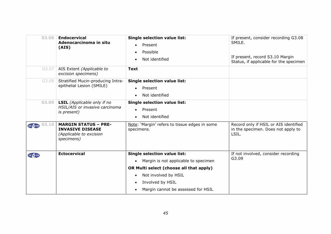
S3.08 The presence of Endocervical Adenocarcinoma In Situ (AIS)
must be recorded.
29
CS3.08a AIS is an HPV associated precursor lesion of
endocervical adenocarcinoma.
CS3.08b The precursor lesions of non-HPV-related cervical
adenocarcinomas are not well defined but lobular
endocervical glandular hyperplasia (LEGH), atypical
lobular endocervical glandular hyperplasia (ALEGH)
and adenocarcinoma in situ of gastric type have been
proposed as likely precursor lesions of gastric type
adenocarcinoma of the cervix.39
G3.07 The extent of AIS should be recorded in excision specimens,
particularly in cases when the lesion is very small or is extensive.
CG3.07a Whilst designations such as focal or extensive are
descriptive and likely subjective, some clinicians are
interested in this information which may assist in
correlation with the colposcopic findings.
CS3.07b Multifocal disease has been reported in 13–17% of
cases of AIS.
G3.08 The presence of Stratified Mucin-producing Intra-epithelial Lesion
(SMILE) should be recorded.
CG3.08a Stratified mucin producing intraepithelial lesion (SMILE)
is a premalignant lesion with morphological overlap
between SIL and AIS. In WHO 2014, it is regarded as a
variant of AIS6, but others consider it a form of high
grade reserve cell dysplasia and report it separately.40,41
In this protocol for the purposes of data capture and
treatment, the presence of SMILE is specified as a
subtype of AIS.
S3.09 The presence of a Low-grade Squamous Intraepithelial
Lesion (LSIL) must be recorded if no higher grade lesion is
present.
CS3.09a LSIL is the morphology associated with an HPV-
associated infection with a high spontaneous resolution
rate. LSIL, encompasses the HPV viral cytopathic effect
and changes, previously called ‘CIN1’.
If an HSIL lesion is present for example it is not
mandatory to record the coexistence of a LSIL.
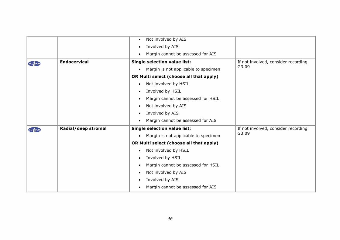
S3.10 Margin status must be recorded for excision specimens with
HSIL and/or AIS.
CS3.10a For excision specimens the status of all surgical excision
margins must be recorded (ectocervical, endocervical
and radial/deep stromal).
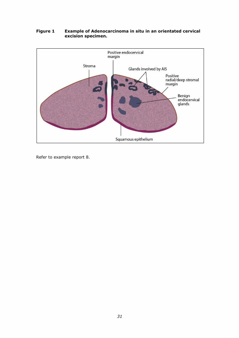
For each margin, the status of HSIL, AIS (including
SMILE) must be recorded. Refer to Appendix 12 and
30
Figure 1.
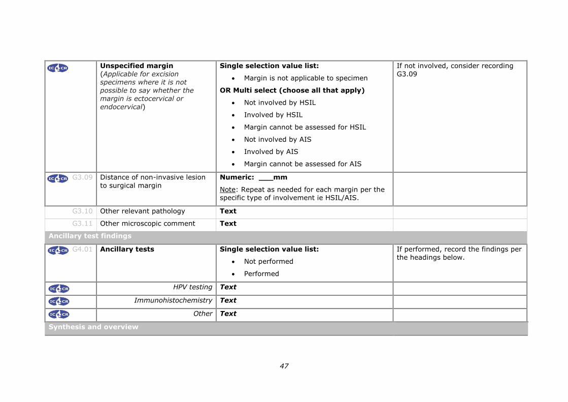
G3.09 If a margin is not involved by HSIL/AIS/SMILE, the distance to the
surgical margin should be documented.
CG3.09a In occasional cases where tumour involvement of the
margin cannot be determined for various reasons
(processing artefact, multiple pieces or poor tissue
orientation), it should be specified as “indeterminate”
and the reason explained.
CG3.09b The distance to the excision margin should be
documented if less than 10 mm.42,43
Information regarding the margin for AIS influences
management (NCSP 2017 guidelines)3. Close
surveillance is indicated if the margin for AIS is close
but apparently excised (less than 5 mm).
Women with positive margins for HSIL do not
necessarily require re-excision.3
G3.10 The presence of any other relevant pathology in the same or
accompanying specimens should be recorded.
CG3.10a For example,
endometrial carcinoma (refer to example report 9).
endometriosis
vaginal or vulval pre-neoplasia
G3.11 A descriptive or narrative field should be provided to record any
microscopic information that is not recorded in the above standards
and guidelines.
31
Figure 1 Example of Adenocarcinoma in situ in an orientated cervical
excision specimen.
Refer to example report 8.
32
4 Ancillary studies findings
HPV testing
G4.01 Results of molecular studies for HPV typing should be recorded if
performed (see appendix 16).
CG4.01a Human papillomavirus (HPV) is universally accepted
to play an aetiological role in cervical carcinogenesis
and HPVs are detectable in over 95% of pre-invasive
and invasive cervical carcinomas, with HPV 16 and 18
being the most frequent types.44
Molecular testing for HPV may occasionally be useful
in a diagnostic scenario. For example, this may be
useful in primary diagnosis when the differential
includes an HPV-related cervical cancer and a non
HPV-related neoplasm or in confirmation of a
metastatic HPV-related cervical neoplasm.
CG4.01b Although not usually required for individual
management, at an epidemiological level, genotyping
for cervical carcinoma and pre-neoplasia may be
important to assess the impact of the National HPV
Vaccination Program and the renewed NCSP.
Immunohistochemistry
G4.02 The results of any immunohistochemical testing should be
recorded if performed (refer to Appendix 16).
CG4.02a p16 immunohistochemistry may be beneficial:
Where there is diagnostic uncertainty for an
HSIL (eg cervicitis, difference in opinion
between pathologists.1).
In cases where the pathologist believes that
the morphology is CIN2, p16 should be
performed and only those cases that are p16
positive should be reported as HSIL. When
p16 negative, such cases should be reported
as LSIL. See example reports 12 and 13.
In the case of tissue artefact hindering
accurate assessment of specimen margins.
P16 may assist in clarifying margin status by
highlighting an area of HSIL or AIS. Caution
is however advised in interpretation of
immunohistochemistry in tissue affected by
artefact.
33
5 Synthesis and overview
Information that is synthesised from multiple modalities and therefore cannot
reside solely in any one of the preceding chapters is described here.
By definition, synthetic elements are inferential rather than observational, often
representing high-level information that is likely to form part of the report
‘Summary’ or ‘Diagnosis’ section in the final formatted report.
Overarching case comment is synthesis in narrative format. Although it may not
necessarily be required in any given report, the provision of the facility for
overarching commentary in a report is essential.
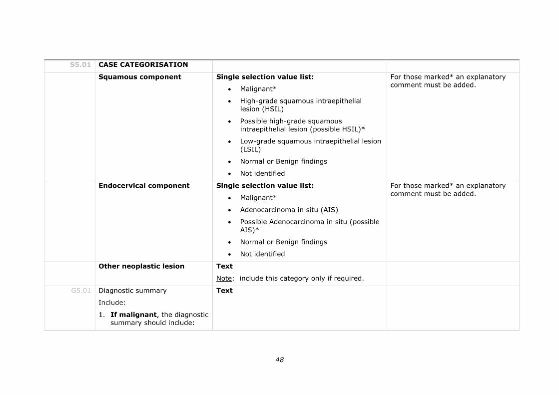
S5.01 A diagnostic category must be recorded.
CS5.01a This reflects assignment of the highest grade of abnormality for
each component.
Choose from:
1. Squamous component
Malignant*
High-grade squamous intraepithelial lesion (HSIL)
Possible high-grade squamous intraepithelial lesion
(possible HSIL)*#
Low-grade squamous intraepithelial lesion (LSIL)
Normal or Benign findings^
Not identified
2. Endocervical (glandular) component
Malignant*
Adenocarcinoma in situ (AIS)
Possible Adenocarcinoma in situ (possible AIS)*#
Normal or Benign findings^
Not identified
3. Other neoplastic lesion*
*These categories require further clarification and an
explanatory comment must be included.
#These categories have been introduced to allow for the rare
instance of diagnostic uncertainty.
34
^ The pathologist may select either normal or benign findings
at their discretion and as appropriate for the individual case.
For example, a case in which endometriosis is found would be
best classified as benign.
Refer to Appendix 11.
G5.01 A diagnostic summary should be included on the report.
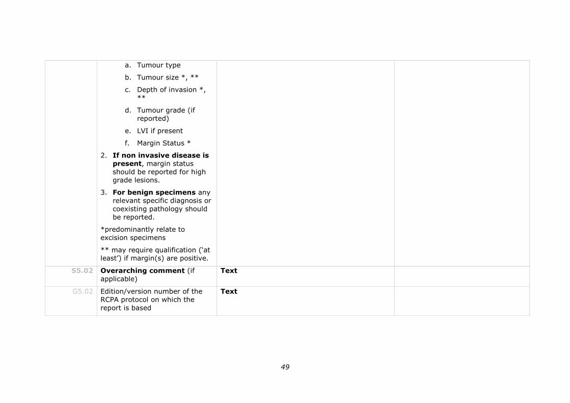
CG5.01a 1. If malignant, the diagnostic summary should include:
a. Tumour type
b. Tumour size *, **
c. Depth of invasion *, **
d. Tumour grade (if reported)
e. LVI if present
f. Margin status *
2. If non invasive disease is present, margin status, and
extent of disease should be reported for high grade lesions.
3. For benign specimens any relevant specific diagnosis or
coexisting pathology should be reported.
*predominantly relate to excision specimens
** may require qualification (‘at least’) if margin(s) are
positive.
CG5.01b Cervical carcinoma is clinically staged: however in lesions that
are not clinically visible (stage 1A) histological variables
contribute to staging and this may be documented following
clinico-pathological correlation.
S5.02 The reporting system must provide a field for free text or narrative
in which the reporting pathologist can give overarching case
comment.
CS5.02a This field may be used, for example, to:
highlight specific issues related to specimen artefact
issues relating to correlation with cervical cytology or
previous cervical histology
list any relevant ancillary tests
document any noteworthy adverse gross and/or histological
features
express any diagnostic subtlety or nuance that is beyond
35
synoptic capture
document further consultation or results still pending.
CS5.02a Use of this field is at the discretion of the reporting pathologist.
G5.02 The edition/version number of the RCPA protocol on which the report is
based should be included on the final report.
CG5.02a For example, the pathology report may include the following
wording at the end of the report: “the data fields within this
formatted report are aligned with the criteria as set out in the
RCPA document “ XXXXXXXXXX” XXXX Edition dated
XXXXXXX”.
36
6 Structured checklist
The following checklist includes the standards and guidelines for this protocol
which must be considered when reporting, in the simplest possible form. For
emphasis, standards (mandatory elements) are formatted in bold font.
S6.01 The structured checklist provided below may be modified as
required but with the following restrictions:
a. All standards and their respective naming conventions,
definitions and value lists must be adhered to.
b. Guidelines are not mandatory but are recommendations and
where used, must follow the naming conventions, definitions
and value lists given in the protocol.
G6.01 The order of information and design of the checklist may be varied
according to the laboratory information system (LIS) capabilities and as
described in Functional Requirements for Structured Pathology
Reporting of Cancer Protocols.18
CG6.01a Where the LIS allows dissociation between data entry and
report format, the structured checklist is usually best
formatted to follow pathologist workflow. In this situation,
the elements of synthesis or conclusions are necessarily at
the end. The report format is then optimised independently
by the LIS.
CG6.01b Where the LIS does not allow dissociation between data
entry and report format, (for example where only a single
text field is provided for the report), pathologists may elect
to create a checklist in the format of the final report. In this
situation, communication with the clinician takes precedence
and the checklist design is according to principles given in
Chapter 7.
G6.02 Where the checklist is used as a report template (see G6.01), the
principles in Chapter 7 and Appendix 2 apply.
CG6.02a All extraneous information, tick boxes and unused values
should be deleted.
G6.03 Additional comment may be added to an individual response where
necessary to describe any uncertainty or nuance in the selection of a
prescribed response in the checklist. Additional comment is not required
where the prescribed response is adequate.
37
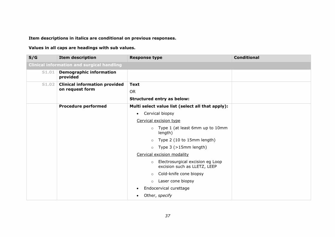
Item descriptions in italics are conditional on previous responses.
Values in all caps are headings with sub values.
S/G Item description Response type Conditional
Clinical information and surgical handling
S1.01 Demographic information
provided
S1.02 Clinical information provided
on request form
Text
OR
Structured entry as below:
Procedure performed Multi select value list (select all that apply):
Cervical biopsy
Cervical excision type
o Type 1 (at least 6mm up to 10mm
length)
o Type 2 (10 to 15mm length)
o Type 3 (>15mm length)
Cervical excision modality
o Electrosurgical excision eg Loop
excision such as LLETZ, LEEP
o Cold-knife cone biopsy
o Laser cone biopsy
Endocervical curettage
Other, specify
38
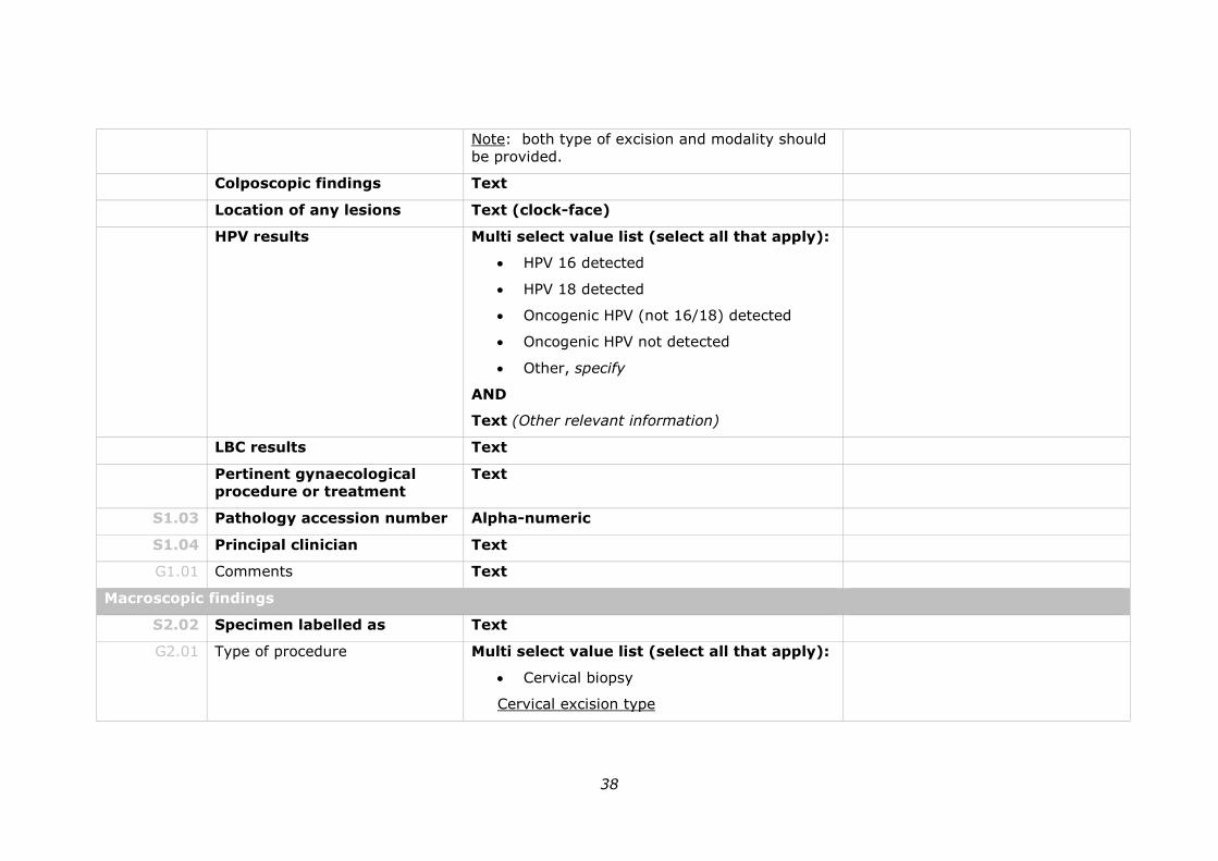
Note: both type of excision and modality should
be provided.
Colposcopic findings Text
Location of any lesions Text (clock-face)
HPV results Multi select value list (select all that apply):
HPV 16 detected
HPV 18 detected
Oncogenic HPV (not 16/18) detected
Oncogenic HPV not detected
Other, specify
AND
Text (Other relevant information)
LBC results Text
Pertinent gynaecological
procedure or treatment
Text
S1.03 Pathology accession number Alpha-numeric
S1.04 Principal clinician Text
G1.01 Comments Text
Macroscopic findings
S2.02 Specimen labelled as Text
G2.01 Type of procedure Multi select value list (select all that apply):
Cervical biopsy
Cervical excision type
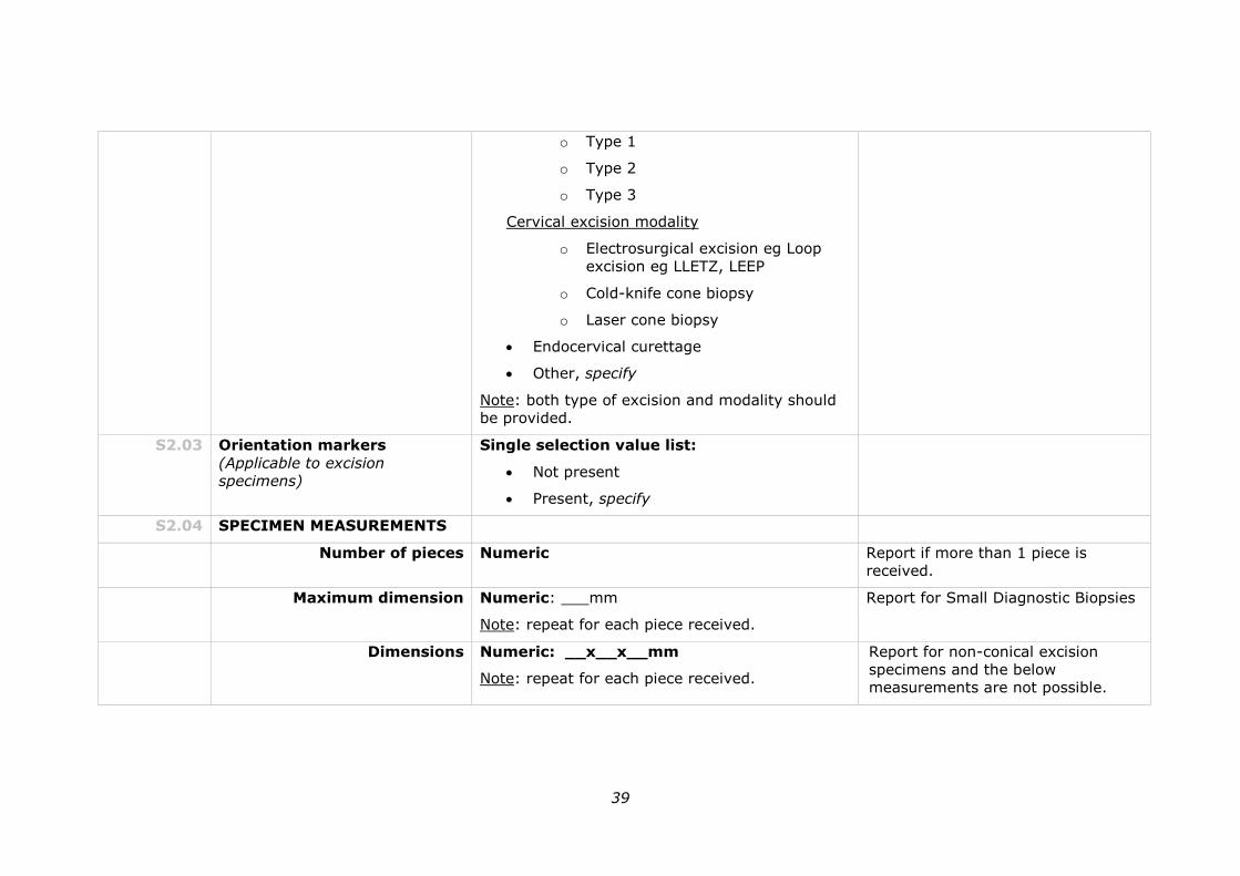
39
o Type 1
o Type 2
o Type 3
Cervical excision modality
o Electrosurgical excision eg Loop
excision eg LLETZ, LEEP
o Cold-knife cone biopsy
o Laser cone biopsy
Endocervical curettage
Other, specify
Note: both type of excision and modality should
be provided.
S2.03 Orientation markers
(Applicable to excision
specimens)
Single selection value list:
Not present
Present, specify
S2.04 SPECIMEN MEASUREMENTS
Number of pieces Numeric Report if more than 1 piece is
received.
Maximum dimension
Numeric: ___mm
Note: repeat for each piece received.
Report for Small Diagnostic Biopsies
Dimensions Numeric: __x__x__mm
Note: repeat for each piece received.
Report for non-conical excision
specimens and the below
measurements are not possible.
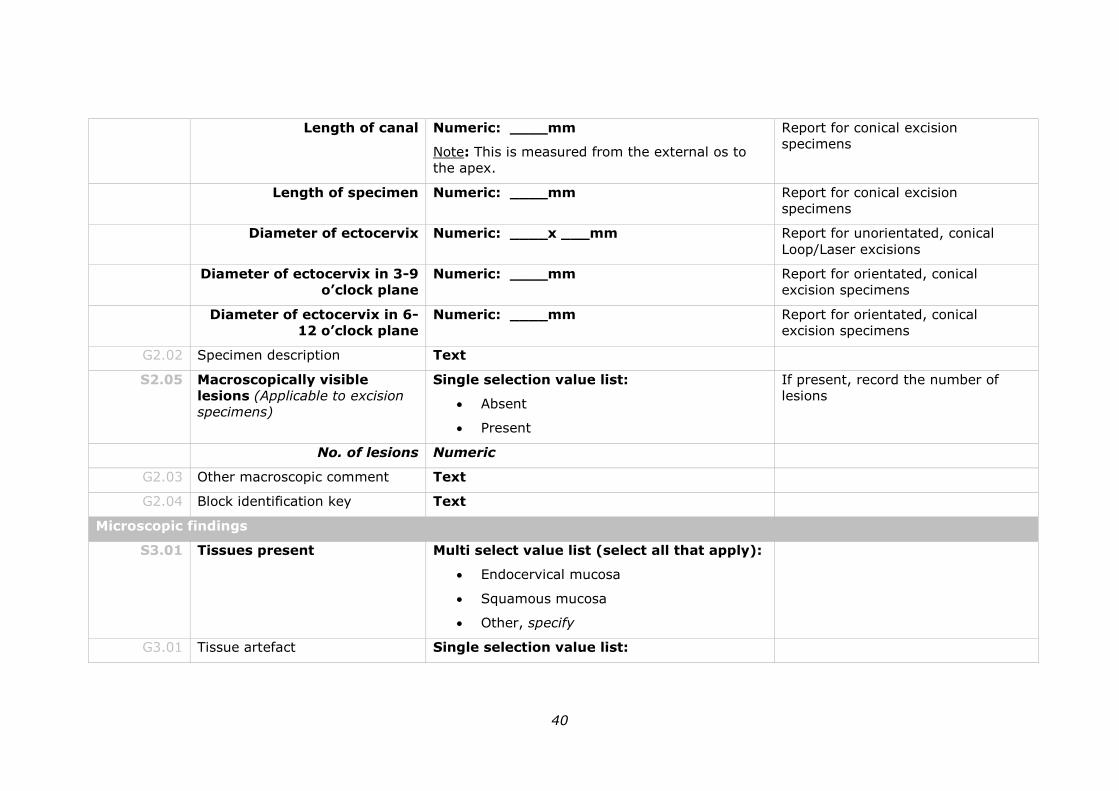
40
Length of canal Numeric: ____mm
Note: This is measured from the external os to
the apex.
Report for conical excision
specimens
Length of specimen Numeric: ____mm Report for conical excision
specimens
Diameter of ectocervix Numeric: ____x ___mm Report for unorientated, conical
Loop/Laser excisions
Diameter of ectocervix in 3-9
o’clock plane
Numeric: ____mm Report for orientated, conical
excision specimens
Diameter of ectocervix in 6-
12 o’clock plane
Numeric: ____mm Report for orientated, conical
excision specimens
G2.02 Specimen description Text
S2.05 Macroscopically visible
lesions (Applicable to excision
specimens)
Single selection value list:
Absent
Present
If present, record the number of
lesions
No. of lesions Numeric
G2.03 Other macroscopic comment Text
G2.04 Block identification key Text
Microscopic findings
S3.01 Tissues present Multi select value list (select all that apply):
Endocervical mucosa
Squamous mucosa
Other, specify
G3.01 Tissue artefact Single selection value list:
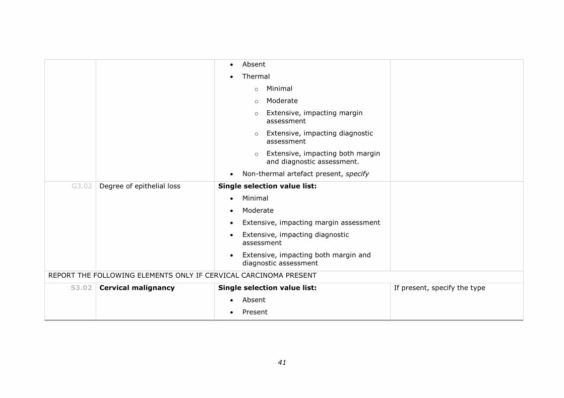
41
Absent
Thermal
o Minimal
o Moderate
o Extensive, impacting margin
assessment
o Extensive, impacting diagnostic
assessment
o Extensive, impacting both margin
and diagnostic assessment.
Non-thermal artefact present, specify
G3.02 Degree of epithelial loss Single selection value list:
Minimal
Moderate
Extensive, impacting margin assessment
Extensive, impacting diagnostic
assessment
Extensive, impacting both margin and
diagnostic assessment
REPORT THE FOLLOWING ELEMENTS ONLY IF CERVICAL CARCINOMA PRESENT
S3.02 Cervical malignancy Single selection value list:
Absent
Present
If present, specify the type
42
Tumour type Text
Note: Use values from the WHO Classification of
Tumours 2014 Appendix 4
S3.03 Multiple tumours Single selection value list:
Absent
Present
If present, record the number of
tumours
No. of tumours Numeric
G3.03 Tumour grade Single selection value list :
G1: Well differentiated
G2: Moderately differentiated
G3: Poorly differentiated
GX: Cannot be graded
Not graded/applicable
S3.04 TUMOUR DIMENSIONS Tumour dimensions cannot be determined
OR complete the following
Note: Repeat for each tumour identified.
Horizontal extent Numeric: __x__mm (at least)**
Note: ** It is advisable to include “at least” for
the tumour measurements when tumour is
present at an excision margin/s. If not
applicable, delete “at least”.
43
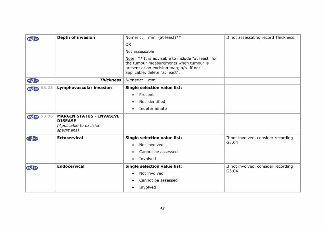
Depth of invasion Numeric:__mm (at least)**
OR
Not assessable
Note: ** It is advisable to include “at least” for
the tumour measurements when tumour is
present at an excision margin/s. If not
applicable, delete “at least”.
If not assessable, record Thickness.
Thickness Numeric:__mm
S3.05 Lymphovascular invasion Single selection value list:
Present
Not identified
Indeterminate
S3.06 MARGIN STATUS - INVASIVE
DISEASE
(Applicable to excision
specimens)
Ectocervical Single selection value list:
Not involved
Cannot be assessed
Involved
If not involved, consider recording
G3.04
Endocervical Single selection value list:
Not involved
Cannot be assessed
Involved
If not involved, consider recording
G3.04
44
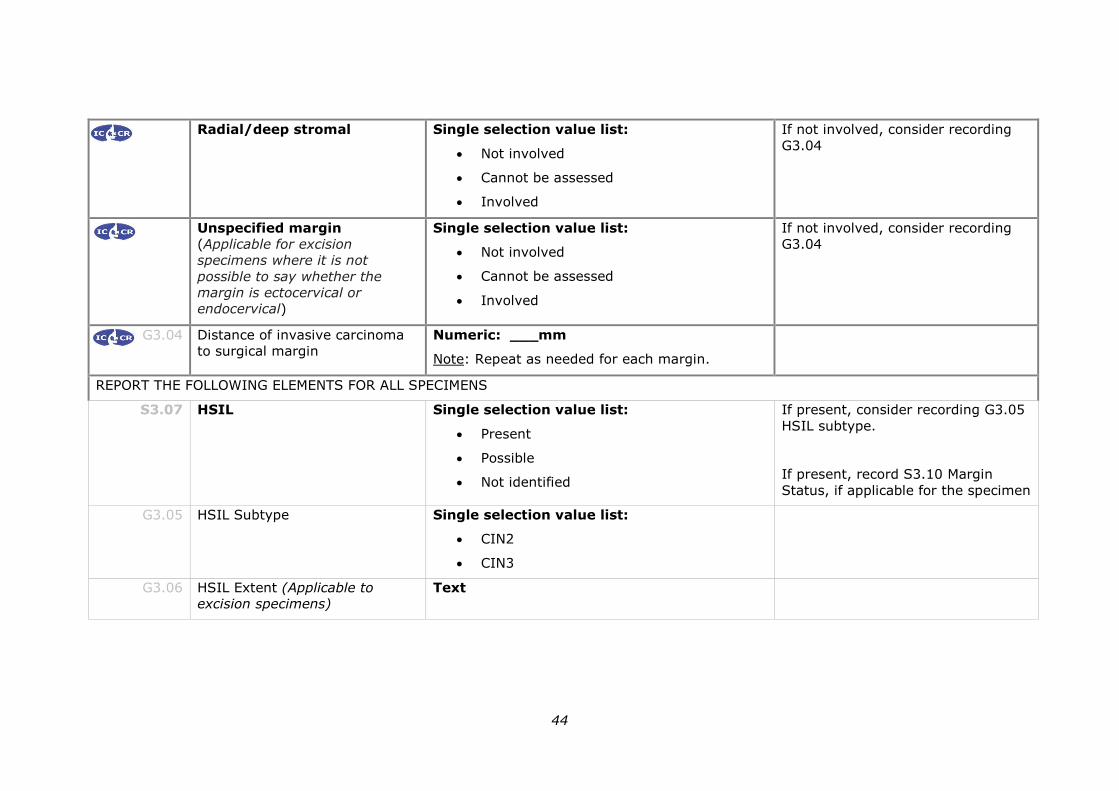
Radial/deep stromal Single selection value list:
Not involved
Cannot be assessed
Involved
If not involved, consider recording
G3.04
Unspecified margin
(Applicable for excision
specimens where it is not
possible to say whether the
margin is ectocervical or
endocervical)
Single selection value list:
Not involved
Cannot be assessed
Involved
If not involved, consider recording
G3.04
G3.04 Distance of invasive carcinoma
to surgical margin
Numeric: ___mm
Note: Repeat as needed for each margin.
REPORT THE FOLLOWING ELEMENTS FOR ALL SPECIMENS
S3.07 HSIL Single selection value list:
Present
Possible
Not identified
If present, consider recording G3.05
HSIL subtype.
If present, record S3.10 Margin
Status, if applicable for the specimen
G3.05 HSIL Subtype Single selection value list:
CIN2
CIN3
G3.06 HSIL Extent (Applicable to
excision specimens)
Text
45
S3.08 Endocervical
Adenocarcinoma in situ
(AIS)
Single selection value list:
Present
Possible
Not identified
If present, consider recording G3.08
SMILE.
If present, record S3.10 Margin
Status, if applicable for the specimen
G3.07 AIS Extent (Applicable to
excision specimens)
Text
G3.08 Stratified Mucin-producing Intra-
epithelial Lesion (SMILE)
Single selection value list:
Present
Not identified
S3.09 LSIL (Applicable only if no
HSIL/AIS or invasive carcinoma
is present)
Single selection value list:
Present
Not identified
S3.10 MARGIN STATUS – PRE-
INVASIVE DISEASE
(Applicable to excision
specimens)
Note: ‘Margin’ refers to tissue edges in some
specimens.
Record only if HSIL or AIS identified
in the specimen. Does not apply to
LSIL.
Ectocervical Single selection value list:
Margin is not applicable to specimen
OR Multi select (choose all that apply)
Not involved by HSIL
Involved by HSIL
Margin cannot be assessed for HSIL
If not involved, consider recording
G3.09
46
Not involved by AIS
Involved by AIS
Margin cannot be assessed for AIS
Endocervical Single selection value list:
Margin is not applicable to specimen
OR Multi select (choose all that apply)
Not involved by HSIL
Involved by HSIL
Margin cannot be assessed for HSIL
Not involved by AIS
Involved by AIS
Margin cannot be assessed for AIS
If not involved, consider recording
G3.09
Radial/deep stromal Single selection value list:
Margin is not applicable to specimen
OR Multi select (choose all that apply)
Not involved by HSIL
Involved by HSIL
Margin cannot be assessed for HSIL
Not involved by AIS
Involved by AIS
Margin cannot be assessed for AIS
If not involved, consider recording
G3.09
47
Unspecified margin
(Applicable for excision
specimens where it is not
possible to say whether the
margin is ectocervical or
endocervical)
Single selection value list:
Margin is not applicable to specimen
OR Multi select (choose all that apply)
Not involved by HSIL
Involved by HSIL
Margin cannot be assessed for HSIL
Not involved by AIS
Involved by AIS
Margin cannot be assessed for AIS
If not involved, consider recording
G3.09
G3.09 Distance of non-invasive lesion
to surgical margin
Numeric: ___mm
Note: Repeat as needed for each margin per the
specific type of involvement ie HSIL/AIS.
G3.10 Other relevant pathology Text
G3.11 Other microscopic comment Text
Ancillary test findings
G4.01 Ancillary tests Single selection value list:
Not performed
Performed
If performed, record the findings per
the headings below.
HPV testing Text
Immunohistochemistry Text
Other Text
Synthesis and overview
48
S5.01 CASE CATEGORISATION
Squamous component Single selection value list:
Malignant*
High-grade squamous intraepithelial
lesion (HSIL)
Possible high-grade squamous
intraepithelial lesion (possible HSIL)*
Low-grade squamous intraepithelial lesion
(LSIL)
Normal or Benign findings
Not identified
For those marked* an explanatory
comment must be added.
Endocervical component Single selection value list:
Malignant*
Adenocarcinoma in situ (AIS)
Possible Adenocarcinoma in situ (possible
AIS)*
Normal or Benign findings
Not identified
For those marked* an explanatory
comment must be added.
Other neoplastic lesion Text
Note: include this category only if required.
G5.01 Diagnostic summary
Include:
1. If malignant, the diagnostic
summary should include:
Text
49
a. Tumour type
b. Tumour size *, **
c. Depth of invasion *,
**
d. Tumour grade (if
reported)
e. LVI if present
f. Margin Status *
2. If non invasive disease is
present, margin status
should be reported for high
grade lesions.
3. For benign specimens any
relevant specific diagnosis or
coexisting pathology should
be reported.
*predominantly relate to
excision specimens
** may require qualification (‘at
least’) if margin(s) are positive.
S5.02 Overarching comment (if
applicable)
Text
G5.02 Edition/version number of the
RCPA protocol on which the
report is based
Text
50
7 Formatting of pathology reports
Good formatting of the pathology report is essential for optimising communication
with the clinician, and will be an important contributor to the success of cancer
reporting protocols. The report should be formatted to provide information clearly
and unambiguously to the treating doctors, and should be organised with their
use of the report in mind. In this sense, the report differs from the structured
checklist, which is organised with the pathologists’ workflow as a priority. For
example pathology reports, please refer to Appendix 3.
Uniformity in the format as well as in the data items of cancer reports between
laboratories makes it easier for treating doctors to understand the reports; it is
therefore seen as an important element of the systematic reporting of cancer. For
guidance on formatting pathology reports, please refer to Appendix 2.
51
Appendix 1 Pathology request information
This appendix describes the information that should be collected before the
pathology test. Some of this information can be provided on generic pathology
request forms; any additional information required specifically for the reporting of
small cervical specimens may be provided by the clinician on a separate request
information sheet. An example request information sheet is included below.
Elements which are in bold text are those which pathologists consider to be
required information. Those in non-bold text are recommended.
Also included in this appendix are the procedures that are recommended before
handover of specimens to the laboratory.
Patient information
Adequate demographic and request information should be
provided with the specimen.
Items relevant to cancer reporting protocols include:
patient name
date of birth
sex
identification and contact details of requesting doctor
date of request
Whether or not the woman identifies as Aboriginal and/ or Torres
Strait Islander. This is in support of a government initiative to
monitor the health of indigenous Australians particularly in
relation to cancer.
The patient’s health identifiers should be provided.
The patient’s health identifiers may include the patient’s Medical
Record Number as well as a national health number such as a
patient’s Medicare number (Australia), Individual Healthcare
Identifier (IHI) (Australia) or the National Healthcare Identifier
(New Zealand).
The Australian Healthcare identifiers i.e. Healthcare Provider Identifier -
Individual (HPI-I) and Healthcare Provider Identifier - Organisation (HPI-
O) should be use, where possible, to identify the requesting doctor.
52
Clinical Information
The specific procedure performed should be described on the
request form.
The colposcopic findings should be included.
The location of any lesion should be noted.
Ideally the location of the lesion should be indicated by clock
position.
The patient’s previous HPV test results should be included.
The dates and results of previous HPV testing, including partial
genotyping information (HPV (16/18) detected or oncogenic HPV
(not 16/18) detected, oncogenic HPV not detected), should be
included in the request.
Information about LBC tests should be included if performed.
The request should indicate the results of any LBC tests
performed before colposcopy. LBC results are important to
correlate with the colposcopy and histopathology results.
The request should indicate if an LBC has been performed at the
time of colposcopy.
Any pertinent gynaecological procedure or treatment should be
included.
For example,
previous cancer, in particular the type and management
(including radiotherapy) of any previous pelvic malignancy
previous diagnostic procedure or treatment for HSIL or AIS
Any other relevant information should be included.
This should include information such as pregnancy and
immunodeficiency.
Surgical handling
The specimen should be capable of orientation if the status of specific
margins is critical in determining the need for, or extent of, further
surgery.
Where there are no anatomical landmarks, specimen orientation
may be indicated with marking sutures or other techniques. If a
specimen is orientated, the orientation should be indicated on the
specimen request form (this may be facilitated by the use of a
diagram).
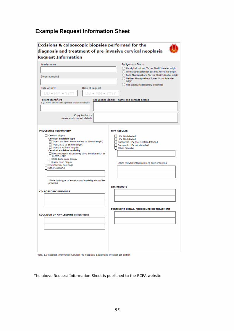
53
Example Request Information Sheet
The above Request Information Sheet is published to the RCPA website
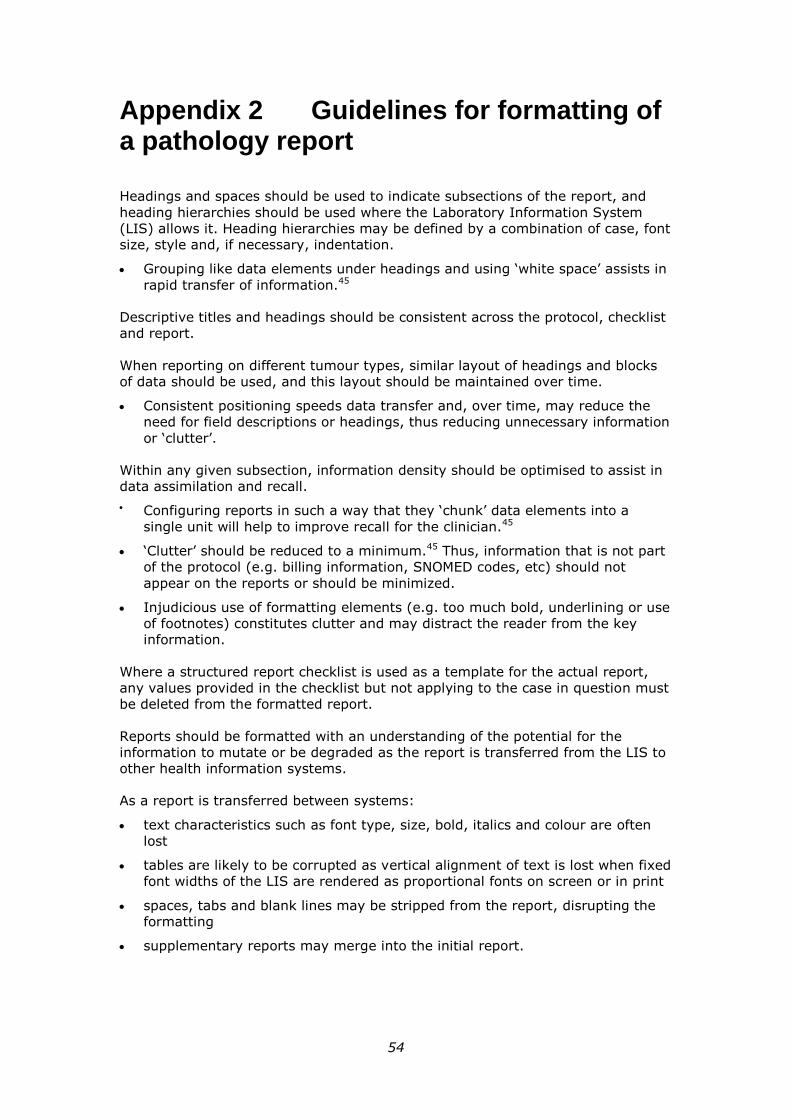
54
Appendix 2 Guidelines for formatting of a pathology report
Headings and spaces should be used to indicate subsections of the report, and
heading hierarchies should be used where the Laboratory Information System
(LIS) allows it. Heading hierarchies may be defined by a combination of case, font
size, style and, if necessary, indentation.
Grouping like data elements under headings and using ‘white space’ assists in
rapid transfer of information.45
Descriptive titles and headings should be consistent across the protocol, checklist
and report.
When reporting on different tumour types, similar layout of headings and blocks
of data should be used, and this layout should be maintained over time.
Consistent positioning speeds data transfer and, over time, may reduce the
need for field descriptions or headings, thus reducing unnecessary information
or ‘clutter’.
Within any given subsection, information density should be optimised to assist in
data assimilation and recall.
Configuring reports in such a way that they ‘chunk’ data elements into a
single unit will help to improve recall for the clinician.45
‘Clutter’ should be reduced to a minimum.45 Thus, information that is not part
of the protocol (e.g. billing information, SNOMED codes, etc) should not
appear on the reports or should be minimized.
Injudicious use of formatting elements (e.g. too much bold, underlining or use
of footnotes) constitutes clutter and may distract the reader from the key
information.
Where a structured report checklist is used as a template for the actual report,
any values provided in the checklist but not applying to the case in question must
be deleted from the formatted report.
Reports should be formatted with an understanding of the potential for the
information to mutate or be degraded as the report is transferred from the LIS to
other health information systems.
As a report is transferred between systems:
text characteristics such as font type, size, bold, italics and colour are often
lost
tables are likely to be corrupted as vertical alignment of text is lost when fixed
font widths of the LIS are rendered as proportional fonts on screen or in print
spaces, tabs and blank lines may be stripped from the report, disrupting the
formatting
supplementary reports may merge into the initial report.
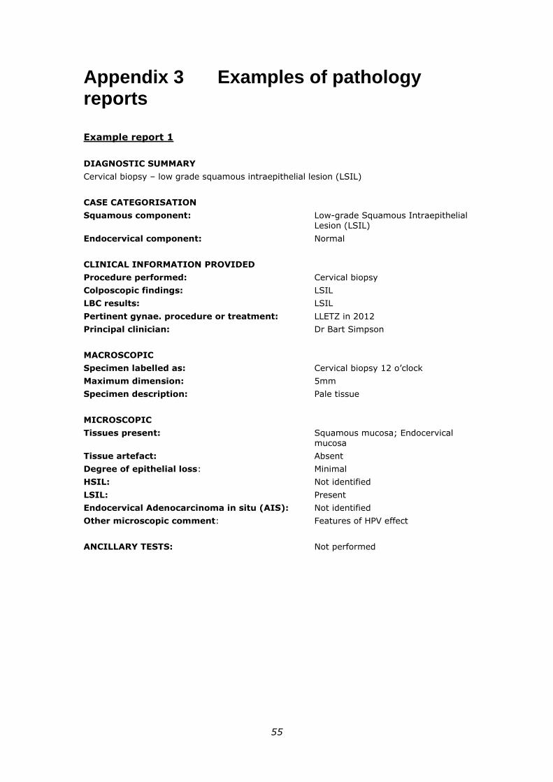
55
Appendix 3 Examples of pathology reports
Example report 1
DIAGNOSTIC SUMMARY
Cervical biopsy – low grade squamous intraepithelial lesion (LSIL)
CASE CATEGORISATION
Squamous component: Low-grade Squamous Intraepithelial Lesion (LSIL)
Endocervical component: Normal
CLINICAL INFORMATION PROVIDED
Procedure performed: Cervical biopsy
Colposcopic findings: LSIL
LBC results: LSIL
Pertinent gynae. procedure or treatment: LLETZ in 2012
Principal clinician: Dr Bart Simpson
MACROSCOPIC
Specimen labelled as: Cervical biopsy 12 o’clock
Maximum dimension: 5mm
Specimen description: Pale tissue
MICROSCOPIC
Tissues present: Squamous mucosa; Endocervical mucosa
Tissue artefact: Absent
Degree of epithelial loss: Minimal
HSIL: Not identified
LSIL: Present
Endocervical Adenocarcinoma in situ (AIS): Not identified
Other microscopic comment: Features of HPV effect
ANCILLARY TESTS: Not performed
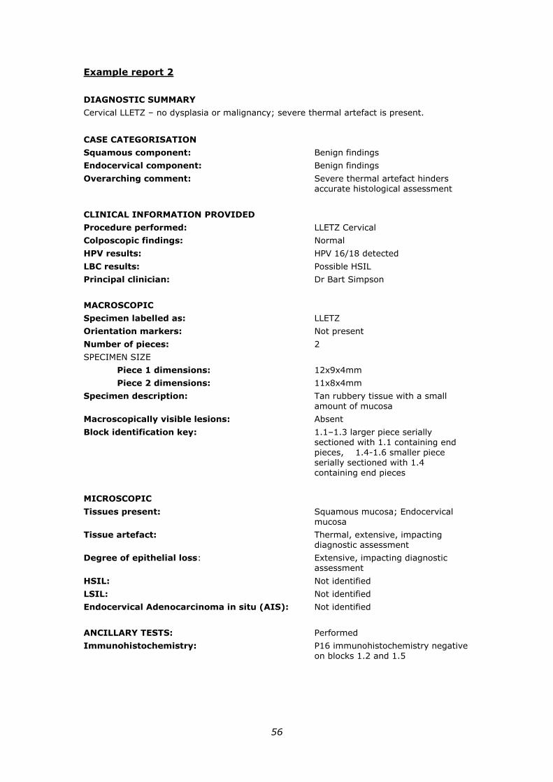
56
Example report 2
DIAGNOSTIC SUMMARY
Cervical LLETZ – no dysplasia or malignancy; severe thermal artefact is present.
CASE CATEGORISATION
Squamous component: Benign findings
Endocervical component: Benign findings
Overarching comment: Severe thermal artefact hinders accurate histological assessment
CLINICAL INFORMATION PROVIDED
Procedure performed: LLETZ Cervical
Colposcopic findings: Normal
HPV results: HPV 16/18 detected
LBC results: Possible HSIL
Principal clinician: Dr Bart Simpson
MACROSCOPIC
Specimen labelled as: LLETZ
Orientation markers: Not present
Number of pieces: 2
SPECIMEN SIZE
Piece 1 dimensions: 12x9x4mm
Piece 2 dimensions: 11x8x4mm
Specimen description: Tan rubbery tissue with a small amount of mucosa
Macroscopically visible lesions: Absent
Block identification key: 1.1–1.3 larger piece serially sectioned with 1.1 containing end pieces, 1.4-1.6 smaller piece serially sectioned with 1.4 containing end pieces
MICROSCOPIC
Tissues present: Squamous mucosa; Endocervical mucosa
Tissue artefact: Thermal, extensive, impacting diagnostic assessment
Degree of epithelial loss: Extensive, impacting diagnostic assessment
HSIL: Not identified
LSIL: Not identified
Endocervical Adenocarcinoma in situ (AIS): Not identified
ANCILLARY TESTS: Performed
Immunohistochemistry: P16 immunohistochemistry negative on blocks 1.2 and 1.5
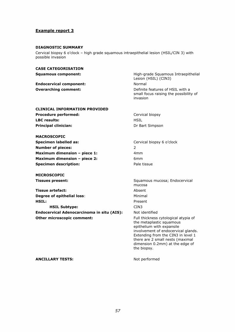
57
Example report 3
DIAGNOSTIC SUMMARY
Cervical biopsy 6 o’clock – high grade squamous intraepithelial lesion (HSIL/CIN 3) with possible invasion
CASE CATEGORISATION
Squamous component: High-grade Squamous Intraepithelial
Lesion (HSIL) (CIN3)
Endocervical component: Normal
Overarching comment: Definite features of HSIL with a small focus raising the possibility of invasion
CLINICAL INFORMATION PROVIDED
Procedure performed: Cervical biopsy
LBC results: HSIL
Principal clinician: Dr Bart Simpson
MACROSCOPIC
Specimen labelled as: Cervical biopsy 6 o’clock
Number of pieces: 2
Maximum dimension – piece 1: 4mm
Maximum dimension – piece 2: 6mm
Specimen description: Pale tissue
MICROSCOPIC
Tissues present: Squamous mucosa; Endocervical
mucosa
Tissue artefact: Absent
Degree of epithelial loss: Minimal
HSIL: Present
HSIL Subtype: CIN3
Endocervical Adenocarcinoma in situ (AIS): Not identified
Other microscopic comment: Full thickness cytological atypia of the metaplastic squamous epithelium with expansile involvement of endocervical glands. Extending from the CIN3 in level 1 there are 2 small nests (maximal dimension 0.2mm) at the edge of
the biopsy.
ANCILLARY TESTS: Not performed
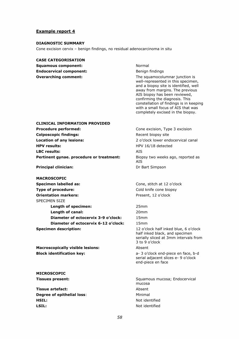
58
Example report 4
DIAGNOSTIC SUMMARY
Cone excision cervix – benign findings, no residual adenocarcinoma in situ
CASE CATEGORISATION
Squamous component: Normal
Endocervical component: Benign findings
Overarching comment: The squamocolumnar junction is
well-represented in this specimen, and a biopsy site is identified, well away from margins. The previous AIS biopsy has been reviewed, confirming the diagnosis. This constellation of findings is in keeping
with a small focus of AIS that was completely excised in the biopsy.
CLINICAL INFORMATION PROVIDED
Procedure performed: Cone excision, Type 3 excision
Colposcopic findings: Recent biopsy site
Location of any lesions: 2 o’clock lower endocervical canal
HPV results: HPV 16/18 detected
LBC results: AIS
Pertinent gynae. procedure or treatment: Biopsy two weeks ago, reported as AIS
Principal clinician: Dr Bart Simpson
MACROSCOPIC
Specimen labelled as: Cone, stitch at 12 o’clock
Type of procedure: Cold knife cone biopsy
Orientation markers: Present, 12 o’clock
SPECIMEN SIZE
Length of specimen: 25mm
Length of canal: 20mm
Diameter of ectocervix 3-9 o’clock: 15mm
Diameter of ectocervix 6-12 o’clock: 15mm
Specimen description: 12 o’clock half inked blue, 6 o’clock half inked black, and specimen serially sliced at 3mm intervals from 3 to 9 o’clock
Macroscopically visible lesions: Absent
Block identification key: a- 3 o’clock end-piece en face, b-d serial adjacent slices e- 9 o’clock end-piece en face
MICROSCOPIC
Tissues present: Squamous mucosa; Endocervical mucosa
Tissue artefact: Absent
Degree of epithelial loss: Minimal
HSIL: Not identified
LSIL: Not identified
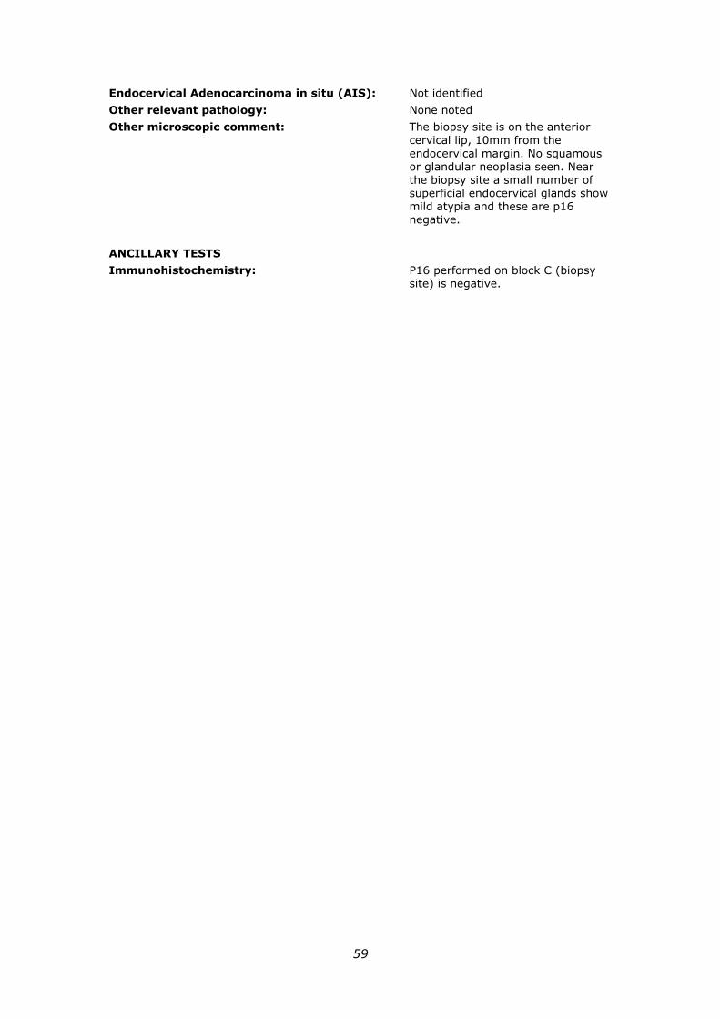
59
Endocervical Adenocarcinoma in situ (AIS): Not identified
Other relevant pathology: None noted
Other microscopic comment: The biopsy site is on the anterior cervical lip, 10mm from the
endocervical margin. No squamous or glandular neoplasia seen. Near the biopsy site a small number of superficial endocervical glands show mild atypia and these are p16 negative.
ANCILLARY TESTS
Immunohistochemistry: P16 performed on block C (biopsy site) is negative.
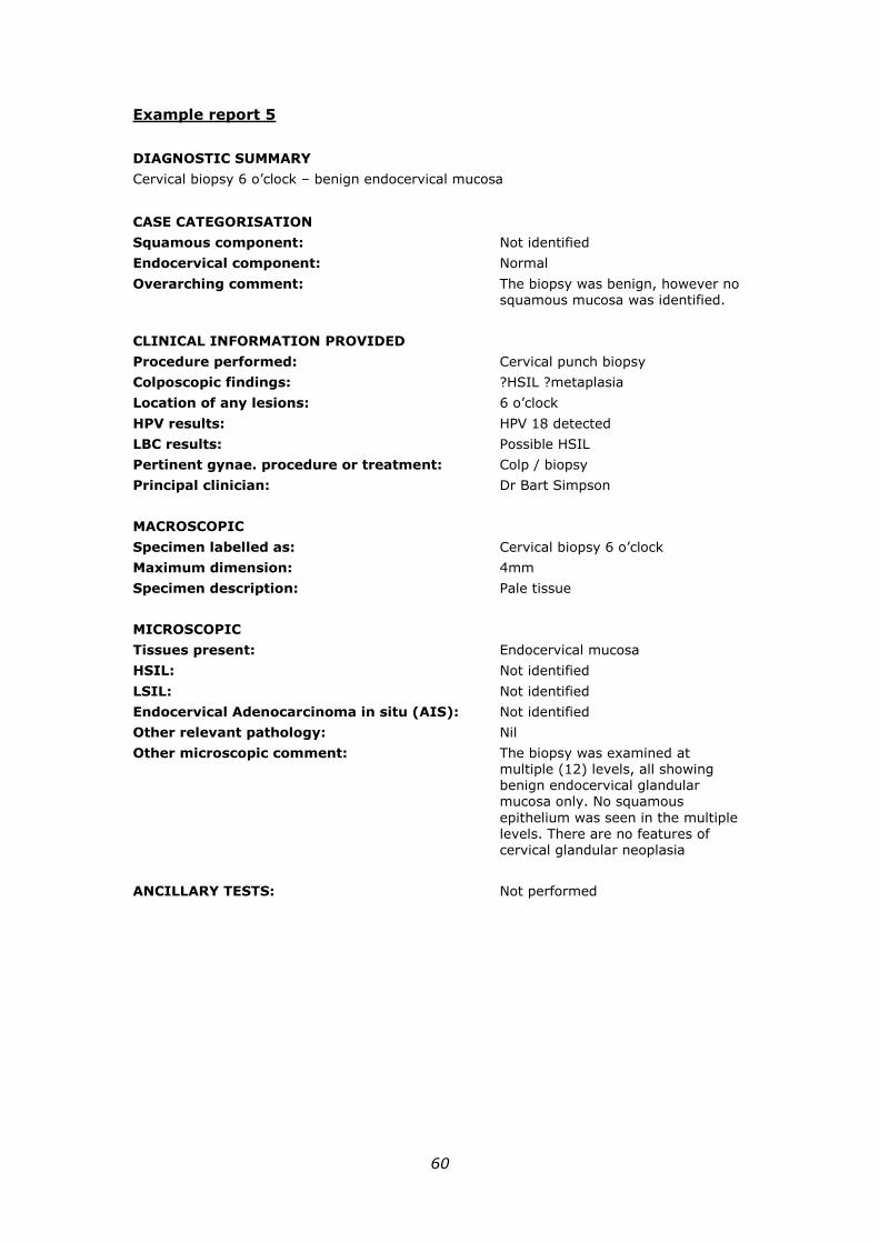
60
Example report 5
DIAGNOSTIC SUMMARY
Cervical biopsy 6 o’clock – benign endocervical mucosa
CASE CATEGORISATION
Squamous component: Not identified
Endocervical component: Normal
Overarching comment: The biopsy was benign, however no squamous mucosa was identified.
CLINICAL INFORMATION PROVIDED
Procedure performed: Cervical punch biopsy
Colposcopic findings: ?HSIL ?metaplasia
Location of any lesions: 6 o’clock
HPV results: HPV 18 detected
LBC results: Possible HSIL
Pertinent gynae. procedure or treatment: Colp / biopsy
Principal clinician: Dr Bart Simpson
MACROSCOPIC
Specimen labelled as: Cervical biopsy 6 o’clock
Maximum dimension: 4mm
Specimen description: Pale tissue
MICROSCOPIC
Tissues present: Endocervical mucosa
HSIL: Not identified
LSIL: Not identified
Endocervical Adenocarcinoma in situ (AIS): Not identified
Other relevant pathology: Nil
Other microscopic comment: The biopsy was examined at multiple (12) levels, all showing benign endocervical glandular mucosa only. No squamous
epithelium was seen in the multiple levels. There are no features of cervical glandular neoplasia
ANCILLARY TESTS: Not performed
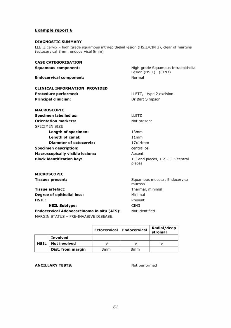
61
Example report 6
DIAGNOSTIC SUMMARY
LLETZ cervix – high grade squamous intraepithelial lesion (HSIL/CIN 3), clear of margins (ectocervical 3mm, endocervical 8mm)
CASE CATEGORISATION
Squamous component: High-grade Squamous Intraepithelial Lesion (HSIL) (CIN3)
Endocervical component: Normal
CLINICAL INFORMATION PROVIDED
Procedure performed: LLETZ, type 2 excision
Principal clinician: Dr Bart Simpson
MACROSCOPIC
Specimen labelled as: LLETZ
Orientation markers: Not present
SPECIMEN SIZE
Length of specimen: 13mm
Length of canal: 11mm
Diameter of ectocervix: 17x14mm
Specimen description: central os
Macroscopically visible lesions: Absent
Block identification key: 1.1 end pieces, 1.2 – 1.5 central pieces
MICROSCOPIC
Tissues present: Squamous mucosa; Endocervical
mucosa
Tissue artefact: Thermal, minimal
Degree of epithelial loss: Minimal
HSIL: Present
HSIL Subtype: CIN3
Endocervical Adenocarcinoma in situ (AIS): Not identified
MARGIN STATUS – PRE-INVASIVE DISEASE:
Ectocervical Endocervical Radial/deep stromal
HSIL
Involved
Not involved √ √ √
Dist. from margin 3mm 8mm
ANCILLARY TESTS: Not performed
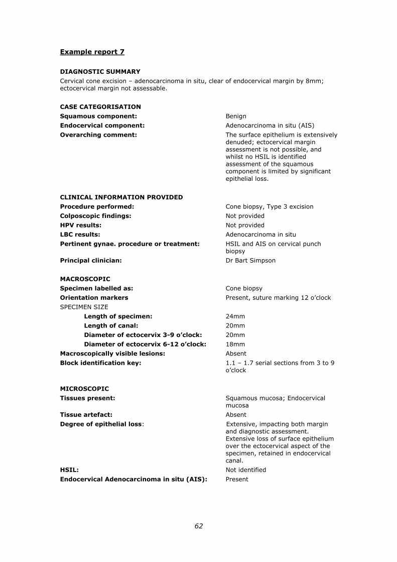
62
Example report 7
DIAGNOSTIC SUMMARY
Cervical cone excision – adenocarcinoma in situ, clear of endocervical margin by 8mm; ectocervical margin not assessable.
CASE CATEGORISATION
Squamous component: Benign
Endocervical component: Adenocarcinoma in situ (AIS)
Overarching comment: The surface epithelium is extensively denuded; ectocervical margin assessment is not possible, and whilst no HSIL is identified assessment of the squamous component is limited by significant
epithelial loss.
CLINICAL INFORMATION PROVIDED
Procedure performed: Cone biopsy, Type 3 excision
Colposcopic findings: Not provided
HPV results: Not provided
LBC results: Adenocarcinoma in situ
Pertinent gynae. procedure or treatment: HSIL and AIS on cervical punch biopsy
Principal clinician: Dr Bart Simpson
MACROSCOPIC
Specimen labelled as: Cone biopsy
Orientation markers Present, suture marking 12 o’clock
SPECIMEN SIZE
Length of specimen: 24mm
Length of canal: 20mm
Diameter of ectocervix 3-9 o’clock: 20mm
Diameter of ectocervix 6-12 o’clock: 18mm
Macroscopically visible lesions: Absent
Block identification key: 1.1 – 1.7 serial sections from 3 to 9 o’clock
MICROSCOPIC
Tissues present: Squamous mucosa; Endocervical mucosa
Tissue artefact: Absent
Degree of epithelial loss: Extensive, impacting both margin and diagnostic assessment. Extensive loss of surface epithelium over the ectocervical aspect of the specimen, retained in endocervical canal.
HSIL: Not identified
Endocervical Adenocarcinoma in situ (AIS): Present

63
MARGIN STATUS – PRE-INVASIVE DISEASE
ANCILLARY TESTS: Not Performed
Margin
AIS
Involved Cannot be assessed
Not involved
Dist. from margin (mm)
Ectocervical √
Endocervical √ 8mm
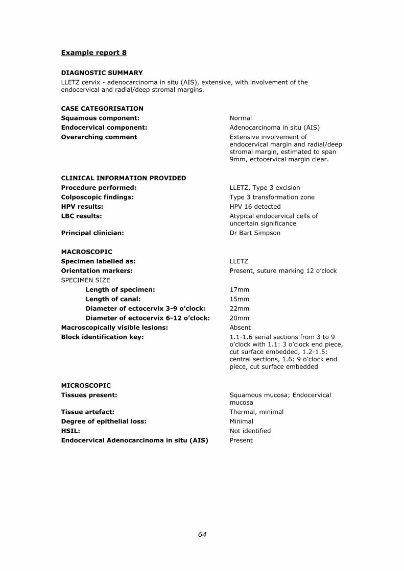
64
Example report 8
DIAGNOSTIC SUMMARY
LLETZ cervix - adenocarcinoma in situ (AIS), extensive, with involvement of the endocervical and radial/deep stromal margins.
CASE CATEGORISATION
Squamous component: Normal
Endocervical component: Adenocarcinoma in situ (AIS)
Overarching comment Extensive involvement of endocervical margin and radial/deep stromal margin, estimated to span 9mm, ectocervical margin clear.
CLINICAL INFORMATION PROVIDED
Procedure performed: LLETZ, Type 3 excision
Colposcopic findings: Type 3 transformation zone
HPV results: HPV 16 detected
LBC results: Atypical endocervical cells of uncertain significance
Principal clinician: Dr Bart Simpson
MACROSCOPIC
Specimen labelled as: LLETZ
Orientation markers: Present, suture marking 12 o’clock
SPECIMEN SIZE
Length of specimen: 17mm
Length of canal: 15mm
Diameter of ectocervix 3-9 o’clock: 22mm
Diameter of ectocervix 6-12 o’clock: 20mm
Macroscopically visible lesions: Absent
Block identification key: 1.1-1.6 serial sections from 3 to 9 o’clock with 1.1: 3 o’clock end piece, cut surface embedded, 1.2-1.5: central sections, 1.6: 9 o’clock end
piece, cut surface embedded
MICROSCOPIC
Tissues present: Squamous mucosa; Endocervical mucosa
Tissue artefact: Thermal, minimal
Degree of epithelial loss: Minimal
HSIL: Not identified
Endocervical Adenocarcinoma in situ (AIS) Present
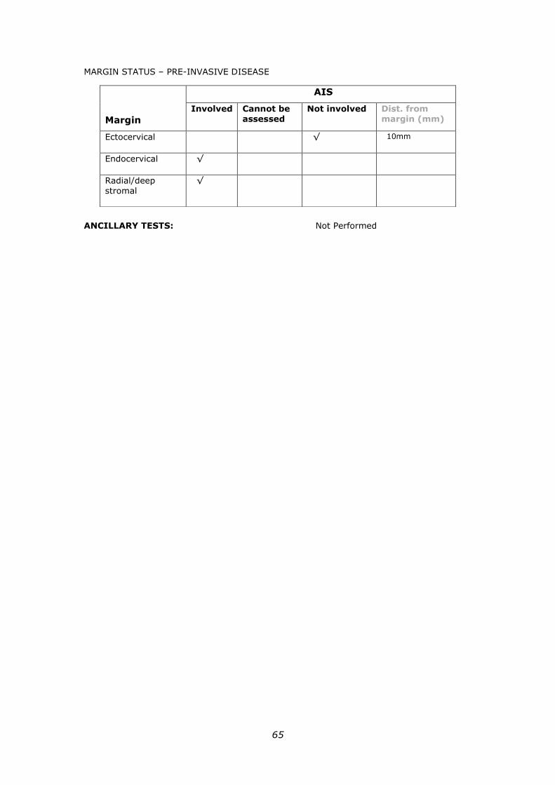
65
MARGIN STATUS – PRE-INVASIVE DISEASE
ANCILLARY TESTS: Not Performed
Margin
AIS
Involved Cannot be assessed
Not involved Dist. from margin (mm)
Ectocervical √ 10mm
Endocervical √
Radial/deep stromal
√
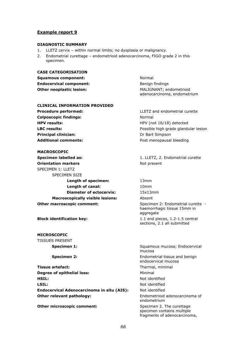
66
Example report 9
DIAGNOSTIC SUMMARY
1. LLETZ cervix – within normal limits; no dysplasia or malignancy.
2. Endometrial curettage – endometrioid adenocarcinoma, FIGO grade 2 in this specimen.
CASE CATEGORISATION
Squamous component: Normal
Endocervical component: Benign findings
Other neoplastic lesion: MALIGNANT; endometrioid adenocarcinoma, endometrium
CLINICAL INFORMATION PROVIDED
Procedure performed: LLETZ and endometrial curette
Colposcopic findings: Normal
HPV results: HPV (not 16/18) detected
LBC results: Possible high grade glandular lesion
Principal clinician: Dr Bart Simpson
Additional comments: Post menopausal bleeding
MACROSCOPIC
Specimen labelled as: 1. LLETZ, 2. Endometrial curette
Orientation markers Not present
SPECIMEN 1: LLETZ
SPECIMEN SIZE
Length of specimen: 13mm
Length of canal: 10mm
Diameter of ectocervix: 15x13mm
Macroscopically visible lesions: Absent
Other macroscopic comment: Specimen 2: Endometrial curette - haemorrhagic tissue 15mm in aggregate
Block identification key: 1.1 end pieces, 1.2-1.5 central sections, 2.1 all submitted
MICROSCOPIC
TISSUES PRESENT
Specimen 1: Squamous mucosa; Endocervical mucosa
Specimen 2: Endometrial tissue and benign
endocervical mucosa
Tissue artefact: Thermal, minimal
Degree of epithelial loss: Minimal
HSIL: Not identified
LSIL: Not identified
Endocervical Adenocarcinoma in situ (AIS): Not identified
Other relevant pathology: Endometrioid adenocarcinoma of endometrium
Other microscopic comment: Specimen 2. The curettage specimen contains multiple fragments of adenocarcinoma,

67
comprising cribriform glands with
focal squamous differentiation and small areas of solid growth (10%). There is background atypical
hyperplasia.
ANCILLARY TESTS: Not Performed
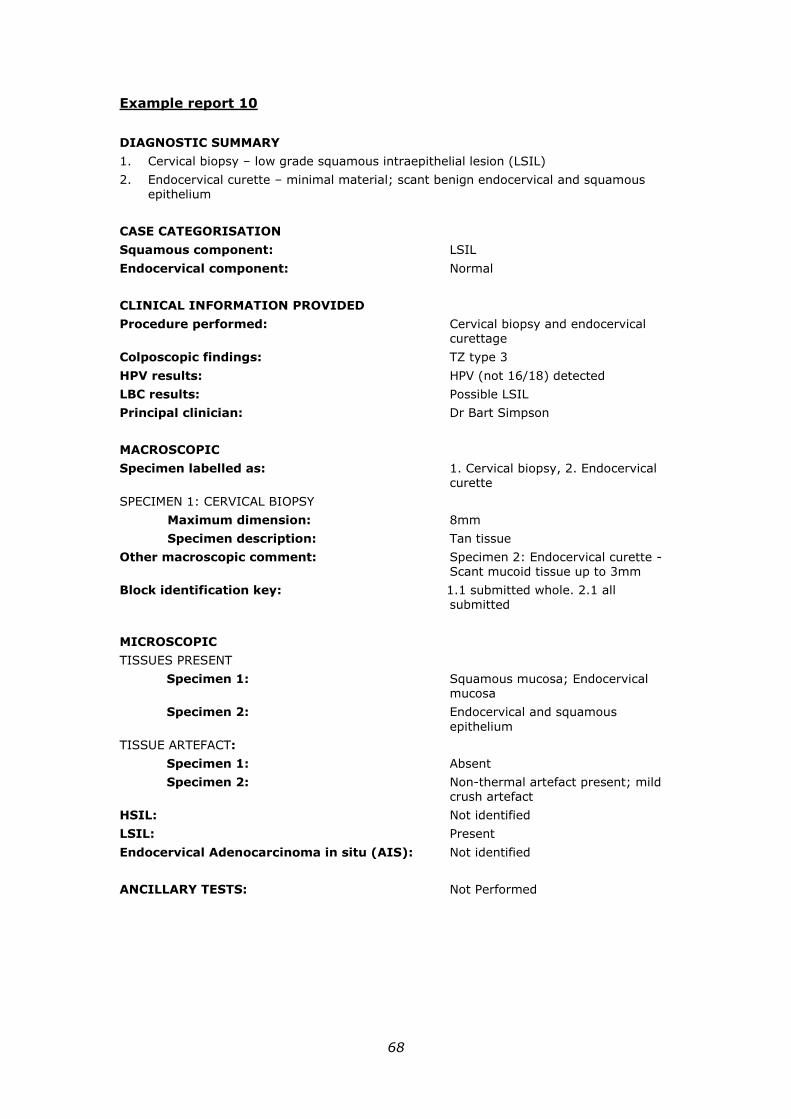
68
Example report 10
DIAGNOSTIC SUMMARY
1. Cervical biopsy – low grade squamous intraepithelial lesion (LSIL)
2. Endocervical curette – minimal material; scant benign endocervical and squamous epithelium
CASE CATEGORISATION
Squamous component: LSIL
Endocervical component: Normal
CLINICAL INFORMATION PROVIDED
Procedure performed: Cervical biopsy and endocervical curettage
Colposcopic findings: TZ type 3
HPV results: HPV (not 16/18) detected
LBC results: Possible LSIL
Principal clinician: Dr Bart Simpson
MACROSCOPIC
Specimen labelled as: 1. Cervical biopsy, 2. Endocervical curette
SPECIMEN 1: CERVICAL BIOPSY
Maximum dimension: 8mm
Specimen description: Tan tissue
Other macroscopic comment: Specimen 2: Endocervical curette - Scant mucoid tissue up to 3mm
Block identification key: 1.1 submitted whole. 2.1 all
submitted
MICROSCOPIC
TISSUES PRESENT
Specimen 1: Squamous mucosa; Endocervical mucosa
Specimen 2: Endocervical and squamous
epithelium
TISSUE ARTEFACT:
Specimen 1: Absent
Specimen 2: Non-thermal artefact present; mild crush artefact
HSIL: Not identified
LSIL: Present
Endocervical Adenocarcinoma in situ (AIS): Not identified
ANCILLARY TESTS: Not Performed
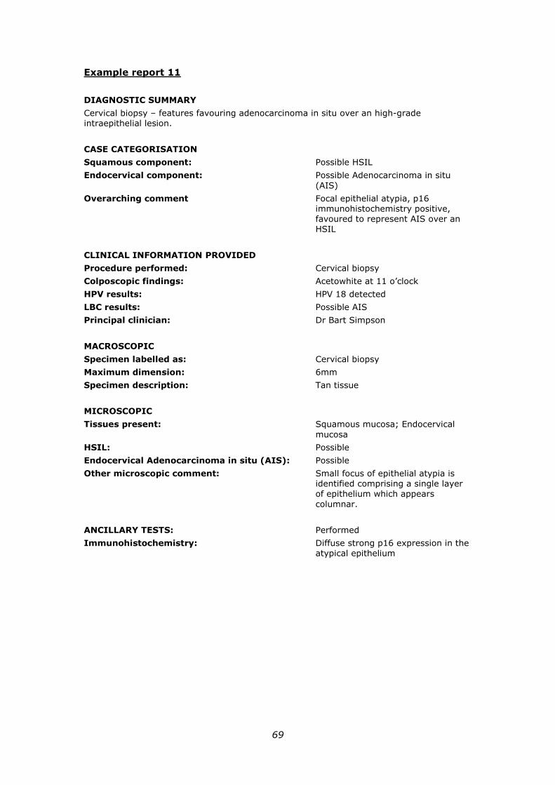
69
Example report 11
DIAGNOSTIC SUMMARY
Cervical biopsy – features favouring adenocarcinoma in situ over an high-grade intraepithelial lesion.
CASE CATEGORISATION
Squamous component: Possible HSIL
Endocervical component: Possible Adenocarcinoma in situ
(AIS)
Overarching comment Focal epithelial atypia, p16 immunohistochemistry positive, favoured to represent AIS over an HSIL
CLINICAL INFORMATION PROVIDED
Procedure performed: Cervical biopsy
Colposcopic findings: Acetowhite at 11 o’clock
HPV results: HPV 18 detected
LBC results: Possible AIS
Principal clinician: Dr Bart Simpson
MACROSCOPIC
Specimen labelled as: Cervical biopsy
Maximum dimension: 6mm
Specimen description: Tan tissue
MICROSCOPIC
Tissues present: Squamous mucosa; Endocervical
mucosa
HSIL: Possible
Endocervical Adenocarcinoma in situ (AIS): Possible
Other microscopic comment: Small focus of epithelial atypia is identified comprising a single layer of epithelium which appears
columnar.
ANCILLARY TESTS: Performed
Immunohistochemistry: Diffuse strong p16 expression in the atypical epithelium
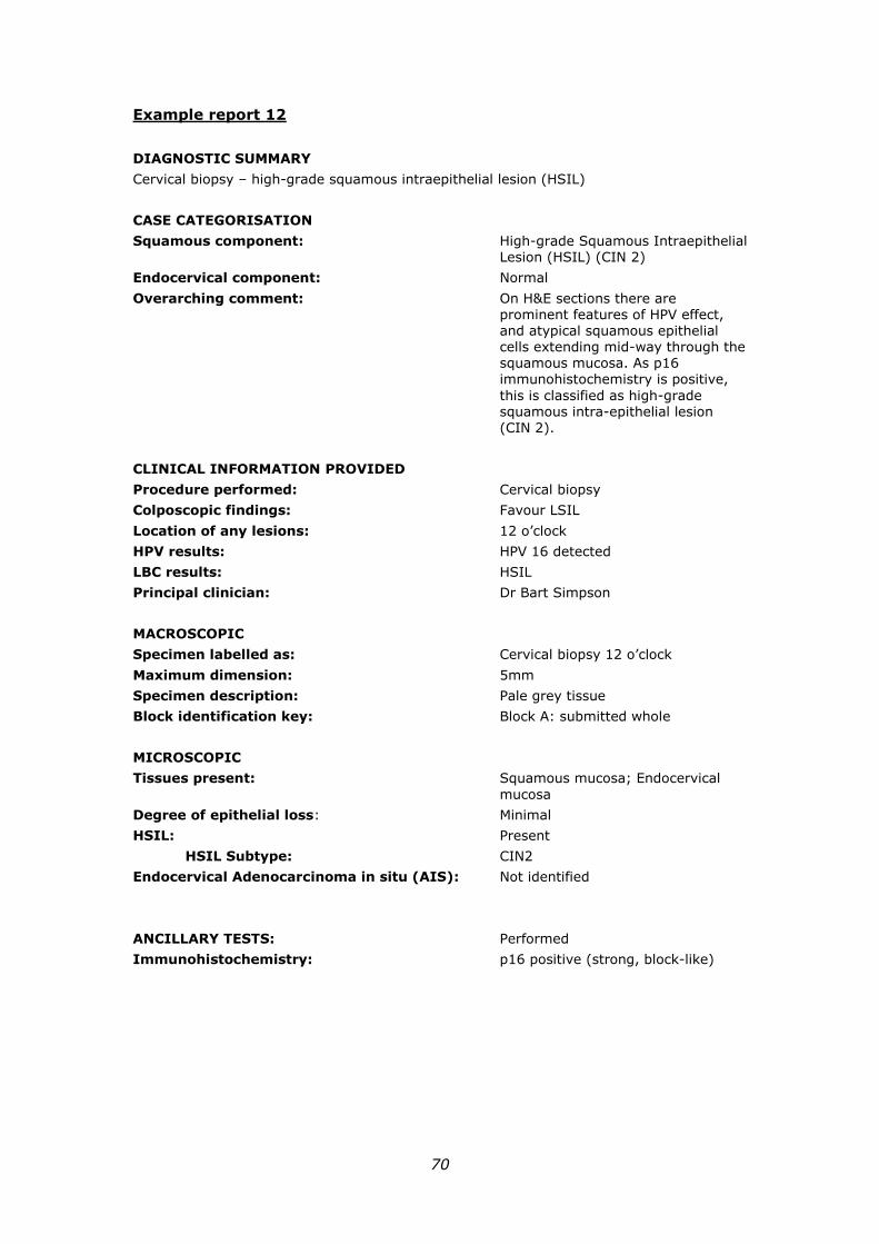
70
Example report 12
DIAGNOSTIC SUMMARY
Cervical biopsy – high-grade squamous intraepithelial lesion (HSIL)
CASE CATEGORISATION
Squamous component: High-grade Squamous Intraepithelial Lesion (HSIL) (CIN 2)
Endocervical component: Normal
Overarching comment: On H&E sections there are prominent features of HPV effect, and atypical squamous epithelial cells extending mid-way through the squamous mucosa. As p16 immunohistochemistry is positive,
this is classified as high-grade squamous intra-epithelial lesion (CIN 2).
CLINICAL INFORMATION PROVIDED
Procedure performed: Cervical biopsy
Colposcopic findings: Favour LSIL
Location of any lesions: 12 o’clock
HPV results: HPV 16 detected
LBC results: HSIL
Principal clinician: Dr Bart Simpson
MACROSCOPIC
Specimen labelled as: Cervical biopsy 12 o’clock
Maximum dimension: 5mm
Specimen description: Pale grey tissue
Block identification key: Block A: submitted whole
MICROSCOPIC
Tissues present: Squamous mucosa; Endocervical
mucosa
Degree of epithelial loss: Minimal
HSIL: Present
HSIL Subtype: CIN2
Endocervical Adenocarcinoma in situ (AIS): Not identified
ANCILLARY TESTS: Performed
Immunohistochemistry: p16 positive (strong, block-like)
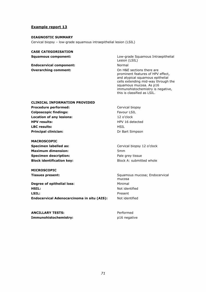
71
Example report 13
DIAGNOSTIC SUMMARY
Cervical biopsy – low-grade squamous intraepithelial lesion (LSIL)
CASE CATEGORISATION
Squamous component: Low-grade Squamous Intraepithelial Lesion (LSIL)
Endocervical component: Normal
Overarching comment: On H&E sections there are prominent features of HPV effect, and atypical squamous epithelial cells extending mid-way through the squamous mucosa. As p16 immunohistochemistry is negative,
this is classified as LSIL.
CLINICAL INFORMATION PROVIDED
Procedure performed: Cervical biopsy
Colposcopic findings: Favour LSIL
Location of any lesions: 12 o’clock
HPV results: HPV 16 detected
LBC results: HSIL
Principal clinician: Dr Bart Simpson
MACROSCOPIC
Specimen labelled as: Cervical biopsy 12 o’clock
Maximum dimension: 5mm
Specimen description: Pale grey tissue
Block identification key: Block A: submitted whole
MICROSCOPIC
Tissues present: Squamous mucosa; Endocervical mucosa
Degree of epithelial loss: Minimal
HSIL: Not identified
LSIL: Present
Endocervical Adenocarcinoma in situ (AIS): Not identified
ANCILLARY TESTS: Performed
Immunohistochemistry: p16 negative
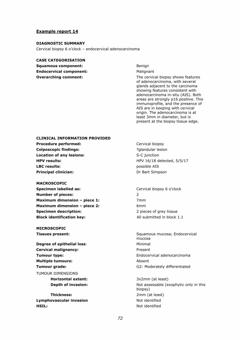
72
Example report 14
DIAGNOSTIC SUMMARY
Cervical biopsy 6 o’clock – endocervical adenocarcinoma
CASE CATEGORISATION
Squamous component: Benign
Endocervical component: Malignant
Overarching comment: The cervical biopsy shows features
of adenocarcinoma, with several glands adjacent to the carcinoma showing features consistent with adenocarcinoma in-situ (AIS). Both areas are strongly p16 positive. This immunoprofile, and the presence of
AIS are in keeping with cervical origin. The adenocarcinoma is at least 3mm in diameter, but is present at the biopsy tissue edge.
CLINICAL INFORMATION PROVIDED
Procedure performed: Cervical biopsy
Colposcopic findings: ?glandular lesion
Location of any lesions: S-C junction
HPV results: HPV 16/18 detected, 5/5/17
LBC results: possible AIS
Principal clinician: Dr Bart Simpson
MACROSCOPIC
Specimen labelled as: Cervical biopsy 6 o’clock
Number of pieces: 2
Maximum dimension – piece 1: 7mm
Maximum dimension – piece 2: 6mm
Specimen description: 2 pieces of grey tissue
Block identification key: All submitted in block 1.1
MICROSCOPIC
Tissues present: Squamous mucosa; Endocervical mucosa
Degree of epithelial loss: Minimal
Cervical malignancy: Present
Tumour type: Endocervical adenocarcinoma
Multiple tumours: Absent
Tumour grade: G2: Moderately differentiated
TUMOUR DIMENSIONS
Horizontal extent: 3x2mm (at least)
Depth of invasion: Not assessable (exophytic only in this biopsy)
Thickness: 2mm (at least)
Lymphovascular invasion Not identified
HSIL: Not identified

73
Adenocarcinoma in situ (AIS) Present
Other microscopic comment: The adenocarcinoma shows a mixture of glandular and solid growth.
ANCILLARY TESTS: Performed
Immunohistochemistry: p16 positive
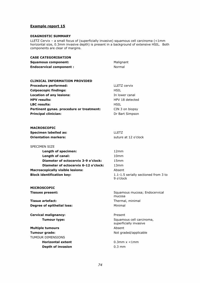
74
Example report 15
DIAGNOSTIC SUMMARY
LLETZ Cervix – a small focus of (superficially invasive) squamous cell carcinoma (<1mm horizontal size, 0.3mm invasive depth) is present in a background of extensive HSIL. Both components are clear of margins.
CASE CATEGORISATION
Squamous component: Malignant
Endocervical component : Normal
CLINICAL INFORMATION PROVIDED
Procedure performed: LLETZ cervix
Colposcopic findings: HSIL
Location of any lesions: In lower canal
HPV results: HPV 18 detected
LBC results: HSIL
Pertinent gynae. procedure or treatment: CIN 3 on biopsy
Principal clinician: Dr Bart Simpson
MACROSCOPIC
Specimen labelled as: LLETZ
Orientation markers: suture at 12 o’clock
SPECIMEN SIZE
Length of specimen: 12mm
Length of canal: 10mm
Diameter of ectocervix 3-9 o’clock: 15mm
Diameter of ectocervix 6-12 o’clock: 13mm
Macroscopically visible lesions: Absent
Block identification key: 1.1-1.5 serially sectioned from 3 to 9 o’clock
MICROSCOPIC
Tissues present: Squamous mucosa; Endocervical mucosa
Tissue artefact: Thermal, minimal
Degree of epithelial loss: Minimal
Cervical malignancy: Present
Tumour type: Squamous cell carcinoma, superficially invasive
Multiple tumours Absent
Tumour grade: Not graded/applicable
TUMOUR DIMENSIONS
Horizontal extent 0.3mm x <1mm
Depth of invasion 0.3 mm
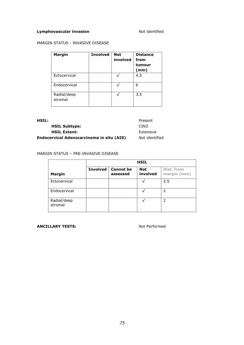
75
Lymphovascular invasion Not identified
MARGIN STATUS - INVASIVE DISEASE
Margin Involved Not
involved
Distance
from
tumour
(mm)
Ectocervical √ 4.5
Endocervical √ 6
Radial/deep
stromal
√ 3.5
HSIL: Present
HSIL Subtype: CIN3
HSIL Extent: Extensive
Endocervical Adenocarcinoma in situ (AIS) Not identified
MARGIN STATUS – PRE-INVASIVE DISEASE
ANCILLARY TESTS: Not Performed
Margin
HSIL
Involved Cannot be
assessed
Not
involved
Dist. from
margin (mm)
Ectocervical √ 2.5
Endocervical √ 2
Radial/deep stromal
√ 2
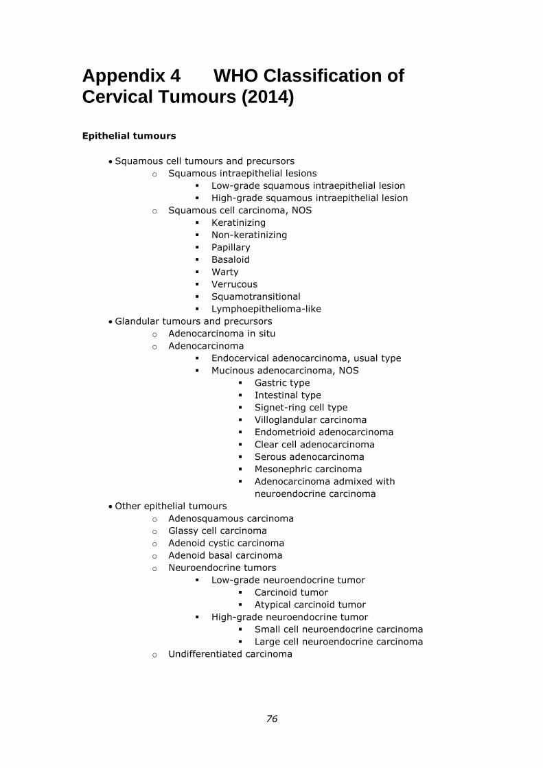
76
Appendix 4 WHO Classification of Cervical Tumours (2014)
Epithelial tumours
Squamous cell tumours and precursors
o Squamous intraepithelial lesions
Low-grade squamous intraepithelial lesion
High-grade squamous intraepithelial lesion
o Squamous cell carcinoma, NOS
Keratinizing
Non-keratinizing
Papillary
Basaloid
Warty
Verrucous
Squamotransitional
Lymphoepithelioma-like
Glandular tumours and precursors
o Adenocarcinoma in situ
o Adenocarcinoma
Endocervical adenocarcinoma, usual type
Mucinous adenocarcinoma, NOS
Gastric type
Intestinal type
Signet-ring cell type
Villoglandular carcinoma
Endometrioid adenocarcinoma
Clear cell adenocarcinoma
Serous adenocarcinoma
Mesonephric carcinoma
Adenocarcinoma admixed with
neuroendocrine carcinoma
Other epithelial tumours
o Adenosquamous carcinoma
o Glassy cell carcinoma
o Adenoid cystic carcinoma
o Adenoid basal carcinoma
o Neuroendocrine tumors
Low-grade neuroendocrine tumor
Carcinoid tumor
Atypical carcinoid tumor
High-grade neuroendocrine tumor
Small cell neuroendocrine carcinoma
Large cell neuroendocrine carcinoma
o Undifferentiated carcinoma
77
Mixed epithelial and mesenchymal tumours
Carcinosarcoma (malignant mullerian mixed tumour)
78
Appendix 5 Cervical Biopsies and Excision Specimens
Cold-knife cone biopsy
Cold-knife cone biopsy is a cervical excision specimen performed using a surgical
blade without electrocautery, and is performed in an operating theatre with general
anaesthesia. Cold-knife cone biopsies have the highest rates of single specimens and
type 3 excisions, and there is no thermal damage. Cold–knife cone biopsies have
been used in the treatment of invasive disease and suspected or known glandular
disease. Disadvantages of the procedure include higher rates of primary
haemorrhage and pre-term labour compared to other excisional procedures.46,47,48-51
Electrosurgical excisions
Electrosurgical procedures such as LEEP/ LLETZ or straight wire, are used for the
diagnosis and treatment of predominantly squamous intraepithelial neoplasia,3 and
are used as a type 2/3 excision for adenocarcinoma in situ in some units.52 They are
the most commonly used therapy for HSIL in resource-rich countries.
These specimens may show variable degrees of specimen fragmentation and thermal
artefact which appear to be related to technique.22,53-57 Thermal artefact should
ideally be limited to 0.2mm in the histological specimen.
The NCSP Guidelines3 provides for further information regarding the types of
procedures and clinical guidelines.
Laser Cone Biopsy
Laser Cone Biopsy was introduced to obtain a histologically assessable specimen,
following concern that laser ablation on its own might result in the under-diagnosis of
invasive disease.
Laser cone biopsies result in thermal artefact. They are now an uncommon
specimen. The duration of the laser cone procedure is longer than LLETZ/LEEP and is
considered to require greater expertise.3
Small diagnostic biopsies
Targeted punch biopsy is a small diagnostic biopsy performed at the time of
colposcopy directed at the area of colposcopic abnormality.
The NSCP Guidelines3 consensus-based recommendations state that the cervix should
be biopsied when the LBC prediction is pHSIL or HSIL and the colposcopic appearance
shows major change (as per International Federation for Cervical Pathology and
Colposcopy (IFCPC) definition) and the abnormal TZ is visible (Type 1 or Type 2 TZ).
Biopsy is also required prior to ablative treatment.
Practice points (based on expert opinion and formulated by a consensus process)
from NCSP3 relating to other lesions include:
Biopsy visible lesion if suspicious for invasion when T3 TZ colposcopy
79
In some situations, when there is a visible high-grade lesion on the ectocervix but
there is a T3 TZ (lesion extends into canal out of visual range), it may be
reasonable to take a cervical biopsy of the visible lesion if there is any suspicion
of superficially invasive or invasive carcinoma.
Biopsy of low-grade lesions is encouraged but not always necessary.
Women with a LBC prediction of pLSIL or LSIL and a colposcopic impression of
low-grade disease or less may not always require a biopsy. However, biopsy is
accepted practice for confirmation of the colposcopic impression and exclusion of
high-grade disease, and should be encouraged, especially for less experienced
colposcopists.
Endocervical curettage
Endocervical curettage is an outpatient procedure previously uncommonly practiced
in Australia. A negative endocervical curettage specimen may contribute to support
an observational approach with early repeat HPV testing in the setting of type 3
transformation zone and liquid based cytology and histology <HSIL. (Refer to
example report 10).
Practice point (based on expert opinion and formulated by a consensus process) from
NCSP3 regarding the role of ECC in Type 3 TZ colposcopy following LBC prediction of
pLSIL/LSIL:
Despite a lack of evidence, endocervical curettage can be considered for
women who have a positive oncogenic HPV test result (any type) with a LBC
report of persistent pLSIL/LSIL and colposcopy reported as Type 3 TZ. A
negative ECC may provide additional reassurance for a conservative
(observational) approach.
Ablative procedures
Ablative procedures are treatment procedures with no excisional component. In the
setting of a known diagnosis of HSIL for example, ablation of the mucosa is
performed without a surgical specimen.
The NCSP guidelines3 consensus recommendations state that ablative therapy should
be reserved for women intending to have children, and when the following conditions
have all been met:
TZ is completely visible (Type 1 or Type 2)
There is no evidence of invasive or glandular disease
A biopsy has been performed prior to treatment
HSIL (CIN2/3) has been histologically confirmed
There is no significant discordance between the histopathology and referral
cytology results.
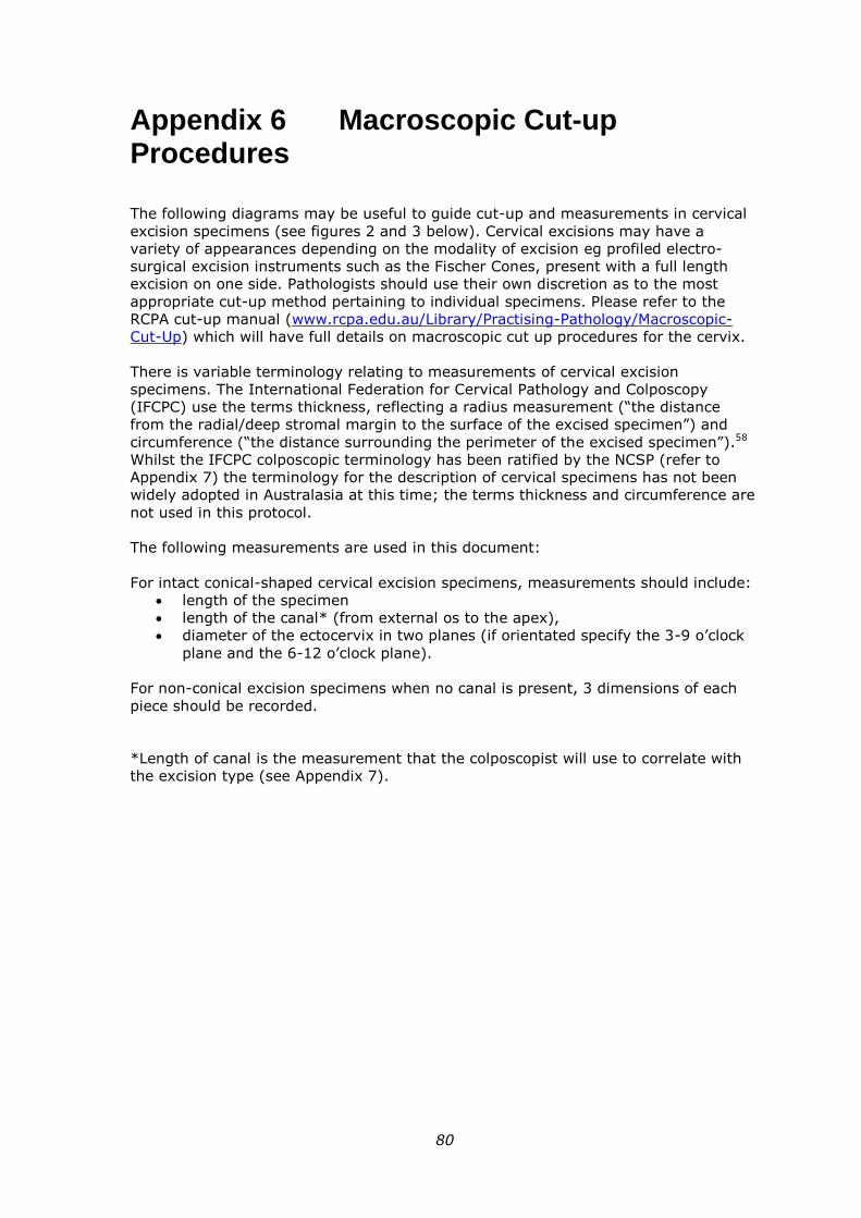
80
Appendix 6 Macroscopic Cut-up Procedures
The following diagrams may be useful to guide cut-up and measurements in cervical
excision specimens (see figures 2 and 3 below). Cervical excisions may have a
variety of appearances depending on the modality of excision eg profiled electro-
surgical excision instruments such as the Fischer Cones, present with a full length
excision on one side. Pathologists should use their own discretion as to the most
appropriate cut-up method pertaining to individual specimens. Please refer to the
RCPA cut-up manual (www.rcpa.edu.au/Library/Practising-Pathology/Macroscopic-
Cut-Up) which will have full details on macroscopic cut up procedures for the cervix.
There is variable terminology relating to measurements of cervical excision
specimens. The International Federation for Cervical Pathology and Colposcopy
(IFCPC) use the terms thickness, reflecting a radius measurement (“the distance
from the radial/deep stromal margin to the surface of the excised specimen”) and
circumference (“the distance surrounding the perimeter of the excised specimen”).58
Whilst the IFCPC colposcopic terminology has been ratified by the NCSP (refer to
Appendix 7) the terminology for the description of cervical specimens has not been
widely adopted in Australasia at this time; the terms thickness and circumference are
not used in this protocol.
The following measurements are used in this document:
For intact conical-shaped cervical excision specimens, measurements should include:
length of the specimen
length of the canal* (from external os to the apex),
diameter of the ectocervix in two planes (if orientated specify the 3-9 o’clock
plane and the 6-12 o’clock plane).
For non-conical excision specimens when no canal is present, 3 dimensions of each
piece should be recorded.
*Length of canal is the measurement that the colposcopist will use to correlate with
the excision type (see Appendix 7).
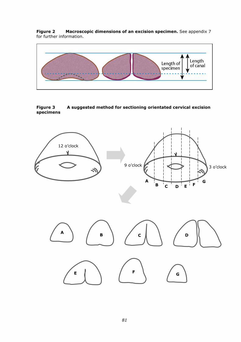
81
Figure 2 Macroscopic dimensions of an excision specimen. See appendix 7
for further information.
Figure 3 A suggested method for sectioning orientated cervical excision
specimens
12 o’clock
3 o’clock 9 o’clock
A B C D E F
G
A B C D
E F G
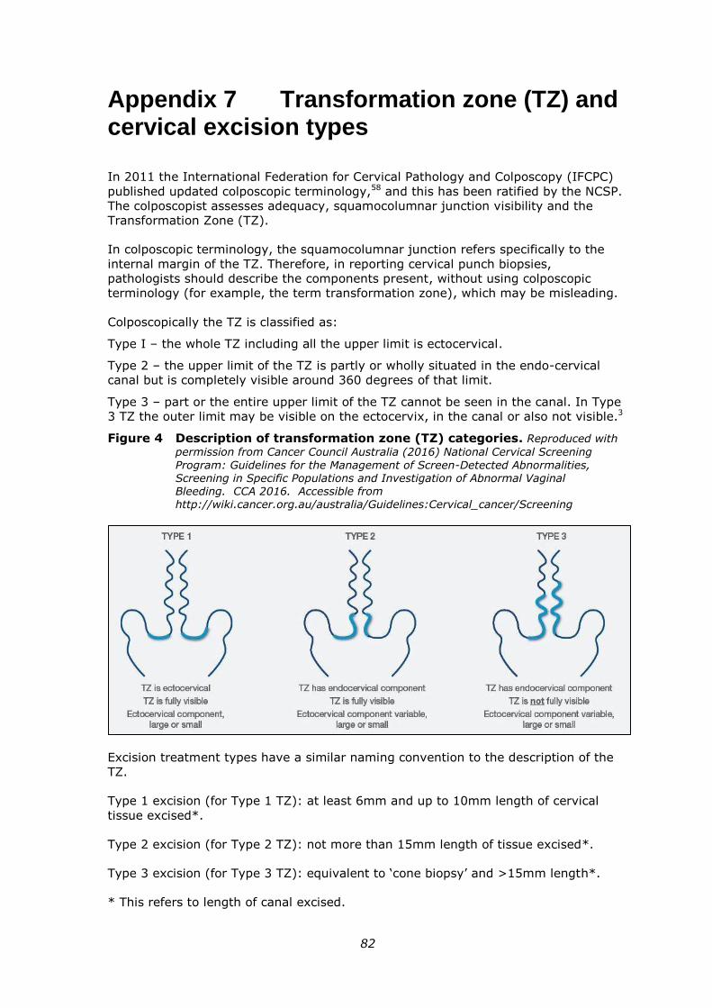
82
Appendix 7 Transformation zone (TZ) and cervical excision types
In 2011 the International Federation for Cervical Pathology and Colposcopy (IFCPC)
published updated colposcopic terminology,58 and this has been ratified by the NCSP.
The colposcopist assesses adequacy, squamocolumnar junction visibility and the
Transformation Zone (TZ).
In colposcopic terminology, the squamocolumnar junction refers specifically to the
internal margin of the TZ. Therefore, in reporting cervical punch biopsies,
pathologists should describe the components present, without using colposcopic
terminology (for example, the term transformation zone), which may be misleading.
Colposcopically the TZ is classified as:
Type I – the whole TZ including all the upper limit is ectocervical.
Type 2 – the upper limit of the TZ is partly or wholly situated in the endo-cervical
canal but is completely visible around 360 degrees of that limit.
Type 3 – part or the entire upper limit of the TZ cannot be seen in the canal. In Type
3 TZ the outer limit may be visible on the ectocervix, in the canal or also not visible.3
Figure 4 Description of transformation zone (TZ) categories. Reproduced with
permission from Cancer Council Australia (2016) National Cervical Screening
Program: Guidelines for the Management of Screen-Detected Abnormalities, Screening in Specific Populations and Investigation of Abnormal Vaginal Bleeding. CCA 2016. Accessible from http://wiki.cancer.org.au/australia/Guidelines:Cervical_cancer/Screening
Excision treatment types have a similar naming convention to the description of the
TZ.
Type 1 excision (for Type 1 TZ): at least 6mm and up to 10mm length of cervical
tissue excised*.
Type 2 excision (for Type 2 TZ): not more than 15mm length of tissue excised*.
Type 3 excision (for Type 3 TZ): equivalent to ‘cone biopsy’ and >15mm length*.
* This refers to length of canal excised.
83
Appendix 8 Technical Aspects
Small diagnostic biopsies and cervical excision specimens should be handled to
optimise tissue integrity and fixation. Small diagnostic biopsies should be placed
immediately into formalin. Excision specimens are also ideally handled using a
technique that does not denude the mucosa, and fixation intact prior to cutup is
recommended.23
Appropriate orientation of the specimen at embedding is necessary for optimal
assessment of the squamous and endocervical mucosa and in the setting of
malignancy, in assessment of variables such as depth of invasion. This may not be
optimised in some smaller diagnostic specimens. In pre-invasive lesions one study
found that orientation was moderate or good in 86% of cervical punch biopsies and
orientation was not a factor in altering the predictability of the final grade of
squamous intraepithelial lesion on subsequent LLETZ.59
Small diagnostic biopsies are optimally examined at multiple levels. Six levels are
recommended. The depth at which these levels are performed depends on the
thickness of the tissue in the block. For a 3-5mm punch biopsy, this may approximate
100µ, however caution is required, and an individual approach to each case should be
adopted in the laboratory. Importantly, initially sectioning the entire tissue is not
acceptable prior to histologic evaluation, particularly as p16 immunohistochemistry
may be required to refine the diagnosis. Further levels may then be required, for
example to resolve any discrepancy between the histological findings and the
colposcopic and cytological assessment.
Due to a combination of the tissue density and specimen heat effect, electrosurgical
and laser excisions may at times present challenges at microtomy. Avoidance of
overcrowding the tissue cassettes will facilitate both the processing and microtomy
stages of laboratory handling, and avoid processed tissue ‘popping out’ of the paraffin
during microtomy. These specimens are also optimally examined at multiple levels;
200µ is a guide, however as with small biopsies, selecting the depth of levels must be
on an individualised basis. Three levels are typically examined at the initial
assessment, with additional levels performed in cases that are discordant with the
previous small diagnostic biopsy or cytology findings and/or to refine the diagnosis.
The presence of a high grade lesion in end pieces may require levelling through the
block to assess these margins. This is may be a safer method than re-embedding the
block.
84
Appendix 9 Clinico-pathological correlation
Review of a patient’s cytology and histology is required in the setting of discordant
test results. The review is optimally performed in a multidisciplinary meeting and
reviewing pathologist(s) and the gynaecologist(s) are expected to have expertise in
this area. Multidisciplinary review may not always be possible at all centres, however
the essential point is that all relevant specimens are reviewed together to synthesise
the findings in a clinico-pathological forum.
The NCSP guidelines3 recommendations for pathology review include:
“REC7.12: Pathology review of discordant test results
For women who have had a colposcopy with significant discordance between the
histopathology and the referral cytology, both specimens should be reviewed by a
pathologist from at least one of the reporting laboratories who should then convey
the results of the review to the colposcopist in order to inform the management
plan.” a
“REC9.6: Cytology review essential when test results are discordant
For women who have a positive oncogenic HPV (any type) test result with a
histologically confirmed LSIL after LBC prediction of pHSIL/HSIL, both the cytology
and the histopathology should be reviewed by a pathologist from at least one of the
reporting laboratories who should then convey the results of the review to the
colposcopist in order to inform the management plan.” a
In the case of invasive disease, review of pathology results typically occurs in a
gynaecological-oncology multidisciplinary meeting. Cervical carcinoma is clinically
staged, however for lesions that are not clinically visible (stage 1A) histological
variables contribute to staging and finalising staging is optimally performed following
clinico-pathological correlation.
Refer to example reports 4 and 11.
a Reproduced with permission from Cancer Council Australia (2016) National Cervical Screening Program: Guidelines for the Management of Screen-Detected Abnormalities, Screening in Specific Populations and Investigation of Abnormal Vaginal Bleeding. CCA 2016. Accessible from http://wiki.cancer.org.au/australia/Guidelines:Cervical_cancer/Screening
85
Appendix 10 Factors contributing to specimen adequacy
Documentation of specific specimen variables assists in providing treating clinician(s)
with degrees of certainty regarding diagnosis and adequacy of treatment. The
surgical specimen received is influenced by clinical factors such as access, the size of
the lesion, the type of transformation zone and the pre-treatment diagnosis.
The NCSP guidelines suggest that of all punch biopsies and excisional biopsies taken,
more than 90% should be of suitable for quality histological examination.3,60
Adequacy assessment requires integration of multiple variables, many of which are
typically more relevant to excision specimens than small diagnostic biopsies. A study
investigating adequacy in cervical punch biopsies found that the number of fragments
present, the biopsy size and the number of tissue types sampled did not influence
whether the biopsy was predictive of the subsequent grade of squamous
intraepithelial lesion on subsequent LLETZ.59
When the pathologist considers histological interpretation is hindered a comment
should be made indicating this, with reference to the specific variable(s). The
histological variables that may contribute to adequacy in this document include:
1. Number of fragments received (S2.04). In loop/laser excisions this relates to
number of passes performed. The NCSP guidelines indicate specimens are
optimally removed in one piece, as multiple pieces can hinder histological
assessment. In particular in the interpretation of margins, completeness of
excision and the evaluation of invasive disease. This is important in cases
confirmed or suspected AIS. Due to anatomical variation it may be necessary
to excise the TZ in more than 1 piece (for example when the TZ is very large).
In this instance accurate orientation is required to optimise histological
evaluation.3
2. Specimen size (S2.04). Macroscopic documentation of specimen size may
provide information regarding adequacy, however this requires correlation
with the microscopic findings, for example a specimen comprising abundant
blood clot or mucus may not provide useful diagnostic information despite
being of significant size. Microscopic specimen size measurement is not
required; it is generally not practical and may be inaccurate in the case of a
fragmented specimen.
3. Ability to orientate the specimen (S2.03). In the case of excision specimens
the orientation of the specimen allows for assessment of specific margins.
4. Assessment of thermal or other artefact (G3.01). In electrosurgical/laser
excisions semi-quantitative documentation of thermal artefact is suggested to
indicate the degree to which this interferes with diagnostic assessment (for
example grading of SIL) and/or margin assessment.
The following assessment of thermal artefact is suggested:
minimal – thin rim of thermal artefact only with no significant interference
with histological assessment
moderate – thermal artefact focal or partially hinders histological
assessment
86
extensive – thermal artefact significantly interferes with histological
assessment
Specifying whether thermal artefact interferes with:
o margin assessment: if known the specific margin(s) affected should be
documented. Whilst caution is advised in interpretation in tissue affected
by artefact, when there is significant thermal artefact at a tissue margin,
p16 immunohistochemistry may assist in clarifying margin assessment.
o diagnostic assessment – for example grading of SIL, inability to exclude
invasion, diagnostic certainty of AIS.
5. Assessment of epithelial loss (G3.02): Loss of surface epithelium may hinder
diagnosis or assessment of margins. Epithelial loss may occur at the time of
the procedure or specimen handling in the laboratory.23 Fixation prior to
handling in the laboratory may assist in minimising epithelial loss. Grading of
epithelial loss may be relevant in small diagnostic biopsies as well as
excisional specimens. A semi-quantitative assessment is suggested, with
percentages provided as a guide.
The following assessment of epithelial loss is suggested
minimal – there is no significant epithelial loss (<10%)
moderate – focal or partially epithelial loss that may hinder histological
assessment (10-30%)
extensive – significant epithelial loss that is likely to hinder histological
assessment (>30%)
Specifying as to whether epithelial loss influences:
o margin assessment: if known, specific margin(s) affected should be
documented
o diagnostic assessment – if there is a specific known diagnostic issue this
should be documented, however generally significant epithelial loss results
in the possibility of a missed diagnosis that is unable to be further
qualified.
87
Appendix 11 Diagnostic categories
Diagnostic categories have been introduced to encourage consistency and clarity of
reporting and to assist with data collection. Squamous and endocervical glandular
components are required to be categorised, with the provision for the categorisation
of other tissue types if present. The categories are:
1. Squamous component
Malignant*
High-grade squamous intraepithelial lesion (HSIL)
Possible high-grade squamous intraepithelial lesion (possible HSIL)*#
Low-grade squamous intraepithelial lesion (LSIL)
Normal or Benign findings^
Not identified
2. Endocervical (glandular) component
Malignant*
Adenocarcinoma in situ (AIS)
Possible Adenocarcinoma in situ (possible AIS)*#
Normal or Benign findings^
Not identified
3. Other neoplastic lesion*
*These categories require further clarification and an explanatory comment must be
included.
#These categories have been introduced to allow for the rare instance of diagnostic
uncertainty.
^ The pathologist may select either normal or benign findings at their discretion and
as appropriate for the individual case. For example, a case in which endometriosis is
found would be best classified as benign.
It is acknowledged that a very small number of small diagnostic biopsies and
electrosurgical/laser specimens may have findings that do not readily fit into the
endocervical or squamous diagnostic categories eg endometrial or rectal carcinoma
and these should be included in the ‘Other neoplastic lesion’ category.
Stratified mucin producing intraepithelial lesion (SMILE) is a premalignant lesion with
morphological overlap between SIL and AIS. In WHO 2014, this is regarded as a
variant of AIS6, others consider it a form of high grade reserve cell dysplasia,
however for the purposes of this categorisation system, SMILE should be included
under the endocervical category.
88
Possible HSIL and possible AIS categories
It is recognized that there will be cases of diagnostic uncertainty and this is reflected
in the inclusion of, Possible HSIL and Possible AIS categories. Categories with
‘possible’ prefix should be used judiciously, reserved for those unusual cases in which
a definitive diagnosis is not possible. Before assigning this category every effort
should be made to refine the diagnosis, and this may include examining additional
levels of the specimen, ancillary studies and obtaining a second opinion. An
explanatory comment must be made describing the diagnostic difficulties
encountered to help guide subsequent management. As an example, a focus
suspicious for HSIL in the first levels of the specimen, however the lesion is not
present in the deeper levels so that confirmatory p16 immunohistochemistry is
unable to be performed. The use of these categories would be very uncommon in
resection specimens, mainly reserved for examples of extensive artefact.
Refer to example report 11.
Recommendation for HSIL and AIS with possible invasion
Where there are features of a definite high grade lesion (HSIL or AIS) but additional
features that are suspicious for, but not diagnostic of, an invasive lesion, the
appropriate high grade diagnostic category should be recorded, with information in
the overarching comment to address the additional features that fall short of invasive
malignancy.
Refer to example report 3.
Malignant
Whilst HSIL or AIS categories are used in cases where there is definite evidence of a
high grade lesion, but diagnostic uncertainty with regard to invasion (see above), the
malignant category should be used when invasion is definitive. It should be noted
that some cases show very limited invasion and while categorised as malignant the
diagnostic summary must describe the findings to allow for appropriate
management.1,61-64 (Refer to the section on SISCCA in Appendix 15).
Refer to example report 15.
89
Appendix 12 Margin status
Invasive
Margin Involved Cannot
be
assessed
Not
involved
G3.04
Distance
from tumour
(mm)
Ectocervical
Endocervical
Radial/deep
stromal
Unspecified **
90
Pre-invasive
Margin
HSIL AIS# Margin is not
applicable to specimen
Involved Cannot be assessed^
Not involved
G3.09 Dist. from margin (mm)
Involved Cannot be assessed^
Not involved
G3.09 Dist. from margin (mm)
Ectocervical
Endocervical
Radial /deep stromal
Unspecified**
**Use for excisions where it is not possible to say whether the margin is ectocervical or endocervical
#Use for cases with SMILE
^In occasional cases where tumour involvement of the margin cannot be determined for various reasons (processing artefact, multiple pieces or poor tissue orientation), the margin status should be specified as “cannot be assessed” and the reason explained.
91
Appendix 13 Tumour types
All cervical carcinomas should be typed according to the 2014 WHO classification.6
Carcinosarcoma is also included since, although it is included in the category of mixed
epithelial and mesenchymal tumours, it is essentially a carcinoma which has
undergone sarcomatous differentiation/metaplasia. The major subtypes of cervical
carcinoma are squamous cell carcinoma (SCC), adenocarcinoma (with various
subtypes), adenosquamous carcinoma and neuroendocrine tumours. While it is
beyond the remit of this document to detail the morphological appearances of the
different tumour types in detail, a few points should be noted.
SCCs are nearly all caused by high-risk human papillomavirus (HPV) with rare
exception65,66 and are subclassified by the WHO based on their histological growth
pattern and the presence of keratinization. However, the subclassification of SCC
seems to have little or no bearing on clinical behaviour and so it is not considered
necessary to specify the subtype (keratinizing, papillary, basaloid, warty, verrucous
etc). However, it may be useful to record unusual subtypes, for example
lymphoepithelioma-like, since the behaviour of these is not well established.
There are several subtypes of cervical adenocarcinoma, the most common being the
usual type which, in the majority of cases, is associated with high-risk HPV. The
other, less common subtypes (gastric type, mesonephric, clear cell and others) are
generally unassociated with HPV infection and have different and distinct histologic
appearances. While there is limited information regarding the clinical behaviour of the
adenocarcinoma subtypes, it is now well established that gastric type
adenocarcinomas of the cervix (adenoma malignum or mucinous variant of minimal
deviation adenocarcinoma is the morphologically well differentiated end of the
spectrum of gastric type adenocarcinoma) have a particularly aggressive behaviour
with poor prognosis, even in early stage disease.67-69 Therefore, it is extremely
important from both a prognostic stance as well as an aetiologic and epidemiologic
perspective (in light of widespread HPV vaccination programs) to correctly identify
these tumour subtypes. The ubiquitous use of and reliance on p16
immunohistochemistry to diagnose cervical adenocarcinoma may cause diagnostic
problems for HPV negative tumours, since these do not exhibit the diffuse block-type
immunoreactivity characteristic of HPV-related tumours (see section on ANCILLARY
STUDIES).70,71 In addition, in the era of molecular characterization and targeted
therapy, correct identification of the tumour subtypes will be even more crucial for
understanding tumour biology and discovery of potential therapeutic targets.
Adenosquamous carcinomas (defined in WHO 2014 blue book as a malignant
epithelial tumour comprising both adenocarcinoma and squamous carcinoma6) are
usually related to high-risk HPV. To make a diagnosis of adenosquamous carcinoma,
malignant squamous and glandular components should be identifiable on routine
haematoxylin and eosin stained sections. The demonstration of foci of
intracytoplasmic mucin by mucin stains in an otherwise typical squamous carcinoma
should not result in diagnosis of an adenosquamous carcinoma. Carcinomas which
lack evidence of squamous differentiation (intercellular bridges, keratinisation) but
have abundant mucin-producing cells should be diagnosed as poorly-differentiated
adenocarcinomas. Adenosquamous carcinoma should also be distinguished from a
spatially separate squamous carcinoma and adenocarcinoma, which occasionally
occurs. While some studies have indicated a worse outcome than pure squamous or
adenocarcinomas, there is not robust evidence to confirm these findings.72,73
92
Primary serous carcinoma of the cervix is exceedingly rare and some doubt its
existence, although it is included in the 2014 WHO Classification. Most cases reported
as primary cervical serous carcinoma are likely to represent a metastasis from the
corpus or extrauterine sites or a usual HPV-related adenocarcinoma with marked
nuclear atypia. Metastasis should be excluded before diagnosing a primary cervical
serous carcinoma. Usual type cervical adenocarcinomas can have a papillary growth
pattern and may show high-grade nuclear atypia, which can mimic serous carcinoma.
Whether true p53 mutation-associated serous carcinoma of the cervix exists is
unresolved at this time.
While endometrioid type adenocarcinoma of the cervix is a subtype listed in the 2014
WHO classification, in the past this has been an over-used diagnostic category and
some even doubt its existence as a primary cervical neoplasm. Most
adenocarcinomas classified as primary cervical endometrioid adenocarcinomas in the
literature represent usual type cervical adenocarcinomas with mucin depletion. These
are different from true endometrioid type adenocarcinomas of the uterine corpus or
adnexa which are driven by hormones and not HPV-associated. If endometrioid
adenocarcinoma occurs as a primary neoplasm in the cervix, it is most likely in the
setting of endometriosis and has the same histologic and immunohistochemical
profiles as endometrioid adenocarcinomas of the uterine corpus or ovary. As with
serous carcinoma, extreme caution should be exercised before diagnosing a primary
cervical endometrioid adenocarcinoma.
Neuroendocrine carcinomas (NECs) (small cell and large cell neuroendocrine
carcinoma) are uncommon but well described in the cervix and can occur in pure
form or associated with another tumour type, typically adenocarcinoma, squamous
carcinoma or adenosquamous carcinoma. These are referred to in the WHO 2014 blue
book as high-grade neuroendocrine carcinomas. The term ‘small cell neuroendocrine
carcinoma’ is preferred to ‘small cell carcinoma’ since a small cell variant of
squamous carcinoma occurs and if the term “neuroendocrine” is not applied, this may
result in confusion. When mixed with another tumour type, the percentage of the
neuroendocrine component should be given. Regardless of the percentage of NEC, it
is recommended that the tumour be reported as mixed since all tumours containing a
component of NEC have a very poor prognosis and the NEC component may be
underestimated in a limited sample.74 Several studies of small cell neuroendocrine
carcinomas of the cervix have shown that adjuvant chemotherapy after surgery for
early stage disease provides significant clinical benefit compared to surgery alone and
therefore, it is extremely important to correctly diagnose any component of NEC.
Additionally, in many institutions surgical resection is not undertaken for a NEC even
if early stage but instead chemotherapy treatment is given. Diagnosing NEC or a
component of NEC can be difficult, especially in small samples, but a combination of
synaptophysin, chromogranin, CD56, TTF1 and p63 has been shown to be helpful in
making the distinction between NEC and poorly-differentiated non-NEC (see section
on ANCILLARY STUDIES).75,76
93
Appendix 14 Tumour grade
Grading of cervical carcinoma
Tumour grade is regularly included in histopathology reports of cervical squamous cell
carcinoma (SCC) and adenocarcinoma (ACA). However, at present no particular
grading system(s) has achieved universal acceptance and grading of these tumours
remains of uncertain clinical value.30,77,78 For example, grade is not amongst the
factors considered in determining the Gynecology Oncology Group (GOG) score which
is used to assess the need for adjuvant therapy following surgery for low-stage
cervical carcinomas.79 Not uncommonly, studies that assess grade as a potential
prognostic variable provide no details of the grading system employed, and this is
also true of large multicentre investigations such as SEER analyses.80,81 For these
and other reasons (discussed below), tumour grading is not listed as a required but
rather a recommended element. Furthermore, no particular grading system for
squamous carcinoma or adenocarcinoma is recommended.
General considerations
1. As with tumours arising in other anatomical sites, grading of cervical
carcinomas has a considerable subjective component and this probably
explains, at least in part, the variable proportion of well, moderately, and
poorly-differentiated tumours reported in different studies. However, some
investigators have demonstrated reasonable intra- and inter-observer
agreement using more complex multifactor grading schemes in SCC
(discussed below).
2. Almost all cervical SCCs are HPV-associated and given that HPV-associated
SCCs very commonly have a “basaloid” morphology with minimal
keratinisation, they are very commonly poorly-differentiated.
3. Most clinically advanced cervical carcinomas are treated with primary
chemoradiation rather than surgery and histological sampling may be limited
to a small diagnostic biopsy. This may not be fully representative due to
tumour heterogeneity and could be potentially misleading as regards tumour
differentiation or grade.77 This may be particularly relevant since less
differentiated appearing tumour elements may be located more deeply
towards the invasive margin.30
4. There is an implicit correlation between tumour subtype and grade in certain
cervical carcinomas and therefore a separate grade may not be applicable. For
example, pure villoglandular ACA of the cervix is by definition a low-grade
neoplasm while serous and clear cell carcinoma, as in the endometrium, are
considered high-grade by default. Similarly, ‘gastric-type’ cervical ACAs and
NECs are clinically aggressive regardless of their histological pattern and
therefore are best considered high-grade automatically.67,68 There is no
published grading system for cervical mesonephric ACAs. Several variants of
cervical SCC are also recognised, although most do not differ from
conventional SCC in terms of prognosis or therapy.82
5. It is uncertain whether a truly ‘undifferentiated’ cervical carcinoma should be
regarded as a separate tumour subtype analogous, for example, to similar
tumours arising in the endometrium.
6. Grading of very small superficially (‘early’) invasive carcinomas of either
squamous or glandular type is probably not possible or relevant.
94
Grading of Cervical SCC
Historically, cervical SCCs were graded using Broder’s system or modifications thereof
based upon the degree of keratinisation, cytological atypia and mitotic activity. In
some schemes, the pattern of invasion (pushing versus infiltrating) has also been
taken into account. Traditionally, SCCs have also been subclassified into large cell
keratinising, large cell non-keratinising and small cell non-keratinising categories,
with these sometimes being regarded as approximately equivalent to well,
moderately and poorly-differentiated, respectively. As noted above, this raises the
issue whether such categorisation represents a tumour subtype (arguably not further
graded), or a grade within a spectrum of a single type of tumour. It should be noted
that some studies have found that the keratinising variant of large cell SCC actually
has a poorer prognosis than the non-keratinising variant, an apparently paradoxical
finding if keratinisation is deemed to be evidence of better differentiation. It is also
uncertain what proportion of “small cell SCCs” reported in the older literature would
now be classified as high-grade NECs (small cell NEC), and this could potentially bias
the supposedly poor outcome of this tumour category.
More complex multifactor grading systems (MGS) that include both tumour and
host/stromal parameters have been assessed in cervical carcinomas, mainly SCC.83-87
For example, the system employed by Stendahl et al,83 based upon that used in head
and neck SCC, comprised eight features, 4 of which were tumour-related (growth
pattern, differentiation, pleomorphism and mitoses) and four of which were stromal-
related (pattern of invasion, stage/depth of invasion, vascular invasion and
inflammatory reaction). Each factor was scored from 1 to 3 and thus the potential
total MGS score ranged from 8-24 points. Simplified modifications to the MGS have
also been described including systems that selectively focus upon the invasive
tumour border or the patterns of tumour invasion.88-91 However, the “cut-off value”
for tumour grade has varied in different studies and not all have demonstrated a
correlation with prognosis.30,92,93 At present, none of these grading systems has been
widely adopted in routine diagnostic practice.
Grading of Cervical ACA
As with SCC, it is controversial whether grading has independent prognostic value in
cervical ACA. Whilst a correlation between higher grade and adverse outcomes has
been reported,94-98 at least for poorly differentiated tumours, this has not been a
universal finding.99,100 It should also be noted that some studies have included a
variable proportion of less common histological subtypes such as adenosquamous
carcinoma, mesonephric, gastric-type and clear cell carcinoma94,97,98 and often
tumour details are not provided. Therefore, it is not clear whether the reported
grading data are applicable to usual-type cervical ACA or have been biased by the
inclusion of other more aggressive tumour subtypes (for example, gastric-type ACA).
Most grading systems for cervical ACA have been based upon the relative proportion
of glandular differentiation, typically following the FIGO system for endometrial
endometrioid adenocarcinoma (EEC). However, the maximum permitted extent of
solid growth for a grade 1 cervical ACA has been variably specified to be 5%101,102 or
10%.98,103 As with EEC, an upward grade adjustment has been suggested for those
tumours exhibiting more marked cytological atypia. However, it is pertinent that
usual-type cervical ACAs typically demonstrate more marked nuclear atypia, mitotic
and apoptotic activity than architecturally similar EECs. 104 There are no separate
grading systems for the various non-HPV related cervical ACAs.
Recently, a system of assessing cervical ACAs based upon their invasive growth
pattern has been developed, and this has been shown to be reproducible amongst
pathologists and to correlate with the risk of lymph node metastasis and patient
95
outcomes.105-108 If these findings are confirmed by additional studies it may be
argued whether this system could be considered a complement to, or even an
alternative to, conventional grading. The latter has traditionally been based upon the
cytoarchitectural pattern of the neoplasm itself but as noted above, tumour-stromal
relationships including the pattern of stromal invasion have been included in earlier
grading schemes of cervical SCC.
Grading of Cervical Adenosquamous Carcinoma
Although it has been suggested that adenosquamous carcinomas are graded on the
basis of the degree of differentiation of both the glandular and squamous
components, there is no well-established grading system for these neoplasms which
has been shown to be of prognostic significance.
96
Appendix 15 Tumour measurements
If there are separate tumours the dimensions for each tumour must be
specified.
It is advisable to include “at least” for the tumour measurements in loop/laser
excisions or cone biopsies when tumour is present at a resection margin (refer to
MARGIN STATUS (S3.06)). In poorly orientated small biopsies care should be
taken not to overestimate depth of invasion.
Reasons for accurate tumour measurement
Measurement of tumour dimensions in cervical carcinomas is important for accurate
FIGO staging of early cervical cancers, patient management and patient
prognostication. Tumours should be measured in mm in three dimensions, namely
two measurements of horizontal extent and the depth of invasion (Figure A). There
are multiple problems with regard to measuring cervical tumours and these are
discussed in detail in this section. In addition, it may not be possible to provide
accurate tumour dimensions in fragmented or thermally damaged specimens. In
situations where the tumour extends to resection margins, the tumour dimensions
should be qualified by use of the term ‘at least’ to indicate that the measurements
may not indicate the true/final tumour size.
In most datasets, separate gross and microscopic measurements are mandated but
this may result in confusion if different measurements are given. Some tumours
(especially larger ones) are more accurately measured grossly while others
(especially smaller tumours and some larger tumours with a diffusely infiltrative
pattern or with marked tumour associated fibrosis) are best measured (or can only
be measured) microscopically. In this dataset, separate gross and microscopic
measurements are not included but rather one set of measurements is required
which is based on a correlation of the gross and microscopic features with gross
examination being more important in some cases and microscopic examination in
others. A few other points are emphasised:-
1. In providing the final tumour dimensions, the measurements in any prior
specimens, for example loop/cone excisions, will need to be taken into
account. Although it may overestimate the maximum horizontal extent, it
is recommended to add together the maximum horizontal measurement in
different specimens when calculating the final horizontal extent. The depth
of invasion can be taken as the maximum depth of invasion in the two
different specimens. Similar comments pertain if loop/cone excisions are
received in more than one piece and where multifocal tumour can be
excluded.
2. Many cervical carcinomas of large size or advanced stage are treated by
chemoradiation, without surgical resection, once the diagnosis has been
confirmed on a small biopsy specimen. In such cases, the tumour
dimensions will be derived from clinical examination and the radiological
appearances. As indicated previously, this dataset applies only to
excision/resection specimens and not to small biopsy specimens.
3. Occasionally resections are undertaken following chemoradiation for
cervical carcinoma. In such cases, there may be no residual tumour or only
small microscopic foci making it impossible to assess the tumour

97
dimensions. In such cases, the pre-treatment clinical or radiological
tumour dimensions should be used for staging.
Specific situations where tumour measurements are important
These include:-
1. Small carcinomas where accurate measurement is paramount in
distinguishing between FIGO stage IA1, IA2 and IB1 neoplasms.25 As well
as providing an accurate stage, this may also be critical in dictating patient
management. For example, FIGO IA1 neoplasms are often treated by local
excision ensuring that the margins are clear of pre-invasive and invasive
disease while IA2 and IB1 neoplasms are usually treated by radical surgery
(radical hysterectomy or trachelectomy).
2. In patients with FIGO stage IB tumours treated by radical hysterectomy,
the tumour size is often one of the parameters used (in conjunction with
tumour differentiation, presence or absence of lymphovascular invasion
and distance to margins) in assessing the need for adjuvant therapy.
3. The tumour measurements may be important in helping to determine
whether radical hysterectomy or trachelectomy is performed; sometimes a
cut-off size of 2 cm is used for performing a radical trachelectomy,
although some surgeons would still perform this procedure for larger size
lesions. Following radical trachelectomy, the recurrence rate is statistically
higher with tumour size greater than 2 cm and rates of adjuvant treatment
are higher.109,110 There is also a trend towards more conservative surgery
(simple as opposed to radical hysterectomy) in patients with tumours less
than 2 cm as the probability of parametrial infiltration is very low.
4. Several studies have shown that in FIGO stage IB1 cervical carcinomas, a
cut-off size of 2 cm may be of prognostic value.111,112
5. A cut-off of 4 cm is similarly of prognostic significance in distinguishing
between FIGO IB1 and IB2 neoplasms and between IIA1 and IIA2
neoplasms.25,113
Measurement of horizontal extent of tumour (Figures A and B)
The horizontal extent (two dimensions, i.e. both tumour length and width,
measurements ‘b’ and ‘c’ in Figure A) must be measured in all cases. As discussed
earlier, in large tumours, this may best be done grossly if large block processing is
not available, because in many cases these neoplasms will need to be submitted in
multiple cassettes and the maximum tumour dimension may not be represented on a
single slide. If a gross measurement is not performed in large circumferential
tumours, there is a risk of overestimating the maximum horizontal extent of the
tumour. This can occur when a circumferential tumour is “opened-up” and submitted
in several sequential cassettes. When the other horizontal dimension (the third
dimension) is calculated by adding up sequential slices in this situation (see below),
this may result in an artificially greater measurement than is accurate.
In smaller neoplasms, the horizontal extent is best determined histologically (Figure
B). One dimension is the measurement in a single slide in which the extent of
invasion is the greatest (measurement ‘e’, Figure B). If the invasive focus is only
represented in 1 block, then the other horizontal dimension is taken to be the
thickness of the block (usually 2.5-3 mm, or estimated as indicated below). In some
cases, the maximum horizontal extent may need to be calculated in the manner
below if this is not represented in one section but is spread over several adjacent
sections (measurement ‘c’, Figure A). If invasive carcinoma is present in several
adjacent sections of tissue and the invasive foci co-localise in the sections, the
horizontal extent of the carcinoma should be calculated by an estimate of the
98
thickness of the blocks, which is determined from the macroscopic dimensions of the
specimen and the number of blocks taken. However, pathologists should be mindful
that thickness of large or outsize blocks can vary from block to block, as compared
with standard-sized blocks. Whilst it is acknowledged that measurements from
calculating block thickness may be somewhat inaccurate, it will in some cases be the
only way to determine the maximum horizontal extent and this may affect staging,
especially in small tumours. A few points regarding measurement of the horizontal
extent of tumours are listed below:-
1. in a case where a single tongue of stromal invasion is seen in continuity
with the epithelium of origin (surface or glandular), the width of the single
focus of invasion is measured across the invasive tongue.
2. where clustered foci of stromal invasion arise close together from a single
crypt or from dysplastic surface epithelium as detached cell groups, the
maximum horizontal extent must encompass all the foci of invasion in the
immediate area and the horizontal extent should be measured from the
edge at which invasion is first seen to the most distant edge at which
invasion is detected.
3. where several foci of invasion arise in one single piece of cervical tissue as
separate foci of invasion, but in close proximity (see section below on
MEASUREMENT OF MULTIFOCAL CARCINOMAS), either as contiguous
tongues of invasion or detached epithelial groups, the maximum horizontal
extent is taken from the edge at which invasion is first seen to the most
distant edge at which invasion is detected. The small amount of
intervening tissue with no invasion (usually with in situ neoplasia) is
included in the measurement.
Measurement of depth of invasion (Figure B)
The maximum depth of invasion must be measured in all cases. This measurement is
taken from the base of the epithelium (surface or crypt) from which the carcinoma
arises to the deepest point of invasion, as specified in the FIGO classification.25 If the
deepest point of invasion involves the deep margin of the specimen, comment should
be made regarding the possibility of underestimation of the depth of invasion; this is
particularly applicable to loop/cone specimens. When the invasive focus is in
continuity with the dysplastic epithelium from which it originates, this measurement
is straightforward. If the invasive focus or foci are not in continuity with the
dysplastic epithelium, the depth of invasion should be measured from the tumour
base (deepest focus of tumour invasion) to the base of the nearest dysplastic crypt or
surface epithelium (Figure B, measurements ‘a’ and ‘c’). If there is no obvious
epithelial origin despite multiple levels of the tissue block, the depth is measured
from the tumour base (deepest focus of tumour invasion) to the base of the nearest
surface epithelium, regardless of whether it is dysplastic or not (Figure B,
measurement ‘d’)
There are some situations where it is impossible to measure the depth of invasion. In
such cases, the tumour thickness may be measured and this should be clearly stated
on the pathology report along with the reasons for providing the thickness rather
than the depth of invasion. In such cases, the pathologist and clinician should equate
the tumour thickness with depth of invasion for staging and management purposes.
Situations where it may be necessary to measure the tumour thickness rather than
the depth of invasion include:-
1. in some glandular lesions, it may be impossible to accurately assess where
adenocarcinoma in situ (AIS) ends and where invasive adenocarcinoma
begins. This is because, in general, identification of invasion in a glandular
99
lesion is more difficult than in a squamous lesion and this is an area where
a specialist opinion may be of value. In some cases where the thickness is
measured (from the epithelial surface to the deepest point of the tumour)
because the point of origin is impossible to establish, this may result in
overestimation of the depth of invasion.
2. in ulcerated tumours with no obvious origin from overlying epithelium, the
thickness may need to be measured. In this situation, measurement of
tumour thickness may result in an underestimate of the depth of invasion.
3. uncommonly, squamous carcinomas, adenocarcinomas and other
morphological subtypes are polypoid with an exclusive or predominant
exophytic growth pattern. In such cases, the carcinoma may project above
the surface with little or even no invasion of the underlying stroma. These
should not be regarded as in-situ lesions and the tumour thickness will
need to be measured in such cases (from the surface of the tumour to the
deepest point of invasion). Depth of invasion i.e. the extent of infiltration
below the level of the epithelial origin, should not be provided in these
cases as it may not be a true reflection of the biological potential of such
tumours.
Avoid the term microinvasive carcinoma
The term “microinvasive carcinoma” does not appear in the FIGO staging system for
cervical cancer.25 Furthermore, use of the term “microinvasive carcinoma” has
different connotations in different geographical areas. For example, in the United
Kingdom, microinvasive carcinoma was considered to be synonymous with FIGO
stage IA1 and IA2 disease in most, but not all, institutions (some used the term
“microinvasive carcinoma” to denote only FIGO stage IA1 tumours). In the United
States and Canada where the Lower Anogenital Squamous Terminology (LAST)1
recommendations have been adopted, the term superficially invasive squamous cell
carcinoma (SISCCA) is used to describe FIGO stage 1A1 tumours with negative
margins, and the term “microinvasive squamous cell carcinoma” is no longer in
routine use. Confusingly, however, the American Society of Gynecologic Oncology
(SGO) has its own definition of stage IA tumours, which is limited not only by the
depth of tumour invasion, but, in contrast to FIGO and TNM, also by the absence of
lymphovascular invasion.114 According to the SGO, cancers invading less than 3 mm
but with lymphovascular involvement are classified as FIGO stage IB1. Thus, in order
to avoid confusion, it is recommended to avoid using the term “microinvasive
carcinoma” for all morphological subtypes and to use the specific FIGO stage.
100
Measurement of multifocal carcinomas
Early invasive carcinomas of the cervix, especially squamous, are sometimes
multifocal comprising tumours that show multiple foci of invasion arising from
separate sites in the cervix and separated by uninvolved cervical tissue. Specifically,
multifocal tumours should be diagnosed if foci of invasion are:
separated by blocks of uninvolved cervical tissue (levels must be cut to
confirm this)
located on separate cervical lips with discontinuous tumour, not involving
the curvature of the canal
situated far apart from each other in the same section (see below).
The individual foci of stromal invasion may be attached to, or discontinuous from, the
epithelium from which they arise. Multifocal carcinomas should not be confused with
the scenario in which tongues or buds of invasion originate from more than one place
in a single zone of transformed epithelium and will, over time, coalesce to form a
single invasive tumour which represents unifocal disease (and should be measured as
indicated above, in three dimensions).
The frequency of multifocality in FIGO stage IA1 cervical squamous carcinomas has
been reported to be between 12 and 25% 64,115,116 although multifocality in larger,
advanced tumours is uncommon. There are few (and some rather dated) guidelines
regarding measurement of multifocal carcinomas. Although pre-invasive disease may
be present, when foci of stromal invasion arise from separate sites or are separated
by cervical tissue without invasion (after levels/deeper sections have been cut to
confirm this), the foci of invasion should be measured separately, in 3 dimensions, as
described above, and staged according to the dimensions of the larger/largest tumour
with a clear statement that the tumour is multifocal. However, in the last of the
scenarios mentioned above (foci of stromal invasion situated far apart from each
other in the same section) measurement of the multifocal disease is problematical.
The term superficially invasive squamous cell carcinoma (SISCCA) has been
proposed for minimally invasive squamous cell carcinoma of the lower anogenital
tract that has been completely excised and is potentially amenable to conservative
surgical therapy.1
In the cervix SISCCA is defined as an invasive squamous cell carcinoma that:
o Is not a grossly visible lesion, AND
o Has and invasive depth of ≤3mm from the basement membrane of the
point of origin, AND
o Has a horizontal spread of ≤7mm in maximal extent AND
o Has been completely excised.
In the cervix lymphovascular invasion is not part of the definition of SISCCA. A
small diagnostic biopsy is usually inadequate to permit the diagnosis of SISCCA
except retrospectively, for example when a further surgical procedure such as loop
excision shows a biopsy site with no residual lesion.
101
Options include measuring from the edge of one invasive focus to the edge of the
furthest invasive focus according to FIGO guidelines (irrespective of the distance
between foci of invasion), adding the maximum horizontal extent of each invasive
focus together (which clearly does not reflect the biological potential of the individual
invasive foci) or regarding widely separated foci as representing small independent
areas of invasion.64,115-118 For tumours with a shallow depth of invasion (up to 3mm),
the assessment and measurement of multifocal disease have implications for staging.
It is in the context of these early, shallow tumours in loop/cone excisions that
management may be significantly affected if the maximum horizontal extent is taken
from the edge of one invasive focus to the edge of the furthest invasive focus, when
the invasive foci are separate from each other. This may upstage a small superficially
invasive carcinoma to FIGO stage IB1, leading to radical surgery (radical
hysterectomy or trachelectomy) in patients who are often young and wish to retain
their fertility. An alternative view is that when widely separated, these foci of invasion
could be regarded as separate foci of IA1 disease, which can be treated by local
excision or simple hysterectomy.
The SHAPE trial119 sets out to address this problematic issue. However, two recent
studies have regarded such lesions as representing multiple foci of invasion
(multifocal FIGO IA1 carcinomas) if the foci of invasion are clearly separated.
However, the distance of separation is not defined and FIGO provides no guidance on
this matter. An arbitrary minimum distance of 2 mm between each separate focus of
invasion has been applied in the 2 studies.64,115 Follow-up of patients in these two
studies, which include a combined total of 46 cases of ‘‘multifocal IA1 cervical
squamous carcinomas’’ treated by local excisional methods (loop/cone excision) with
margins clear of premalignant and malignant disease, has shown no evidence of
recurrent premalignant or malignant disease with median follow-up periods of 45
months and 7 years respectively.64,115 Moreover, one of the studies also showed that
the prevalence of residual pre-invasive (20%) and invasive disease (5%) on repeat
excision were comparable to data available for unifocal FIGO stage IA1 cases.115
These studies included cases which would have been regarded as FIGO stage IB1 had
the horizontal extent been measured from the edge of one invasive focus to the edge
of the furthest invasive focus, as per FIGO guidelines. Although limited by a relatively
small number of cases and the selection of an arbitrary distance of separation of 2
mm, the findings support the hypothesis that with regard to tumour staging and
management, it may be appropriate to consider superficial, widely separated foci of
invasion as representing multifocal lesions, to measure each focus separately, and to
determine the FIGO stage on the basis of the invasive focus with the higher/highest
FIGO stage. Of course, the possibility that such lesions behave as FIGO stage IA1
tumours may reflect the shallow depth of invasion, which clinicians do not seem to
take account of when faced with a tumour whose maximum horizontal width is 7 mm
or more, and the spectre of a FIGO IB1 tumour is raised.
Although the ICCR does not have a mandate to implement an approach based only
on 2 studies involving 46 patients in total, the ICCR recommends that this approach
be considered and discussed at the Tumour Board/multidisciplinary team (MDT)
meetings to avoid unnecessary surgery in young patients who wish to preserve their
fertility in this specific clinical situation. This approach needs to be verified by
additional larger collaborative studies and trials. It is also stressed that in such cases,
the tissue blocks containing the invasive foci and those in between should be levelled
to confirm that the invasive foci are truly separate and ensure that there is no occult
stromal invasion in the intervening areas. If this approach is adopted, the pathology
report should clearly indicate how the measurements have been obtained to arrive at
a diagnosis of multifocal invasion, provide the dimensions of the separate foci of
invasion and indicate how the FIGO stage has been ascertained. Such cases may
need to be referred to Cancer Centres for review and, as indicated above, should be
discussed individually at the tumour board/MDT meeting. There have been no similar
102
studies for multifocal adenocarcinomas but anecdotally these are less common than
multifocal squamous carcinomas and until further evidence becomes available, a
similar approach is recommended.
Measurement of tumour volume
In most studies, tumour size is based on measurement of two dimensions but in a
few studies, tumour volume (based on the three measured tumour dimensions) has
been shown to predict prognosis more reliably than measurements in only one or two
dimensions. Some older studies have suggested tumour volume as a reliable
prognostic factor for early stage tumours: a volume of less than 420 mm3 has been
suggested to be associated with no lymph node metastasis.120-122 This is one of the
main reasons for recommending that three tumour dimensions (two of horizontal
extent and one of depth of invasion or tumour thickness) are provided. However, only
a few centres continue to routinely factor tumour volume into patient management.
Figure A: Measurement of cervical tumours in three dimensions
CIN3 with involvement of endocervical gland crypts is represented by the dark blue-
coloured areas, non-dysplastic squamous epithelium is pink, and grey areas indicate
foci of stromal invasion. The depth of invasion, (a), and horizontal tumour
dimension/width, (b) are measured in unifocal disease.
Third dimension: when stromal invasion is present in three or more consecutive
blocks of a loop or cone biopsy the third tumour dimension, (c), may exceed 7 mm,
i.e. the carcinoma may be more than FIGO stage IA2. This dimension is determined
by calculating the block thickness (usually 2.5 - 3.0 mm) from the macroscopic
specimen dimensions and multiplying this by the number of sequential blocks through
which the invasion extends.
103
Figure B: Measurement of width and depth of invasion in cervical tumours
The dark blue-coloured areas represent CIN3 with involvement of endocervical gland
crypts, non-dysplastic squamous epithelium is pink, and grey areas indicate foci of
stromal invasion.
Depth of invasion: when invasion originates from the surface epithelium, (a), or
gland crypts (b and c), the depth of invasion is taken from the base of the epithelium
from which the invasive carcinoma arises, to the deepest focus of invasion, as
specified in the FIGO classification. Measurements are taken in the same way,
regardless of whether the invasive foci remain attached to the gland crypt (b) or not
(c). Where invasion occurs and no obvious surface (or crypt) epithelial origin is seen,
the depth of invasion is measured from the deepest focus of tumour invasion, to the
base of the nearest non-neoplastic surface epithelium, (d).
Horizontal dimension/width in unifocal tumours, (e): this is measured in the
slice of tissue in which the width is greatest (from the edge at which invasion is first
seen, to the most distant edge at which invasion is identified), in sections where the
foci of invasion arise in close proximity to each other, even if those foci are separated
by short stretches of normal epithelium.
104
Appendix 16 Ancillary studies
IMMUNOHISTOCHEMISTRY
p16 Immunohistochemistry
Diffuse immunoreactivity (nuclear and cytoplasmic) for p16 is a surrogate marker for
malignant or high-grade, premalignant epithelial lesions associated with high-risk
HPV infections.123 In high-grade premalignant squamous lesions, this is referred to as
“block type” immunoreactivity. AIS and high-risk HPV-associated cervical cancers
also show strong diffuse p16 nuclear and cytoplasmic staining. However, it should be
remembered that other gynaecological malignancies, for example uterine serous
carcinoma and high-grade serous carcinoma of the ovary/fallopian tube typically
exhibit such strong diffuse immunoreactivity with p16. This should be distinguished
from focal/patchy (so-called “mosaic-type”) staining, which is not in keeping with a
high-risk HPV associated neoplasm.
In the diagnosis of pre-neoplastic cervical lesions p16 immunohistochemistry may be
beneficial:
Where there is diagnostic uncertainty for an HSIL (eg cervicitis, difference in
opinion between pathologists.1)
In cases where the pathologist believes that the morphology is CIN2, p16 should
be performed and only those cases that are p16 positive should be reported as
HSIL. When p16 negative, such cases should be reported as LSIL.1
In the case of tissue artefact hindering accurate assessment of specimen margins.
P16 may assist in clarifying margin status by highlighting an area of HSIL or AIS.
Caution is however advised in interpretation of immunohistochemistry in tissue
affected by artefact.
The LAST1 working party recommends against the use of p16 IHC as a routine
adjunct to histologic assessment of biopsy specimens with morphologic
interpretations of negative, -IN1, and -IN3.
Immunohistochemistry: Cervical versus Endometrial Adenocarcinoma
Immunohistochemistry can be helpful in the differential diagnosis between a cervical
and an endometrial adenocarcinoma.124 In the distinction between an endometrial
and a cervical origin for an adenocarcinoma, the panels of markers which are useful
will depend on the morphological subtype and not just the site of origin. In the
distinction between a high-risk HPV-related (usual type) cervical adenocarcinoma and
a low-grade endometrial endometrioid adenocarcinoma, the most useful
immunohistochemical markers are p16 and hormone receptors (oestrogen receptor
(ER) and progesterone receptor (PR)) with cervical adenocarcinomas exhibiting
diffuse immunoreactivity with p16 and usually being negative or only focally positive
with hormone receptors. In contrast, low-grade endometrial endometrioid
adenocarcinomas are usually diffusely positive with hormone receptors and exhibit
patchy “mosaic-type” staining with p16. Even when low-grade endometrial
endometrioid adenocarcinomas exhibit diffuse positivity with p16, this is still usually
patchy with alternating positive and negative areas. Vimentin (usually positive in low-
grade endometrial endometrioid adenocarcinoma and negative in cervical
adenocarcinomas) and CEA (usually positive in cervical adenocarcinomas and
negative in low-grade endometrial endometrioid adenocarcinomas) may also be of
105
value. However, it is stressed that there may be unexpected positive and negative
staining reactions with any of the markers. HPV studies will be of value in such cases.
In the distinction between a high-risk HPV-related (usual type) cervical
adenocarcinoma and a high-grade endometrial adenocarcinoma, p16 and hormone
receptors are often of limited value. p53 immunohistochemistry and HPV studies may
be of value in this scenario. Most uterine serous carcinomas and many other high-
grade endometrial carcinomas exhibit mutation-type p53 staining (“all or nothing”
staining) and are HPV negative. High-risk HPV-related cervical adenocarcinomas
rarely, if ever, exhibit “mutation-type” p53 expression.
Immunohistochemistry of Non-HPV Related Cervical Adenocarcinomas
Non-HPV related cervical adenocarcinomas have a different immunophenotype than
usual HPV related adenocarcinomas. They tend to be negative or only focally
positive with p16 and some, such as gastric type adenocarcinomas, may exhibit
mutation-type staining with p53.125 Gastric type adenocarcinomas are usually
positive with gastric markers such as MUC6 and HIK1083 and are flat negative with
hormone receptors.125 There is no specific immunohistochemical marker of
mesonephric adenocarcinomas but they tend to be flat negative with hormone
receptors and may stain with CD10 and GATA3.126,127 Clear cell carcinomas are
usually hormone receptor negative, exhibit wild-type staining with p53 and may be
positive with Napsin A and hepatocyte nuclear factor 1-beta.
Immunohistochemistry of Cervical Neuroendocrine Carcinomas
Cervical neuroendocrine carcinomas are variably positive with the neuroendocrine
markers chromogranin, CD56, synaptophysin and PGP9.5. Of these, CD56 and
synaptophysin are the most sensitive but CD56 lacks specificity. Chromogranin is the
most specific neuroendocrine marker but lacks sensitivity with only about 50% of
these neoplasms being positive.76 Chromogranin positivity is often very focal in small
cell neuroendocrine carcinomas with punctate cytoplasmic immunoreactivity which is
only visible on high-power magnification. A diagnosis of small cell neuroendocrine
carcinoma can be made in the absence of neuroendocrine marker positivity if the
morphological appearances are typical. Small cell neuroendocrine carcinoma may be
only focally positive (often punctate cytoplasmic staining) or even negative with
broad-spectrum cytokeratins. A diagnosis of large cell neuroendocrine carcinoma
requires neuroendocrine marker positivity and most of these neoplasms are diffusely
positive with broad-spectrum cytokeratins.
A high percentage of primary cervical neuroendocrine carcinomas are TTF1 positive,
including some with diffuse immunoreactivity, and this marker is of no value in
distinction from a pulmonary metastasis.76 Most cervical neuroendocrine carcinomas
are diffusely positive with p16 secondary to the presence of high-risk HPV.76 Diffuse
p63 nuclear positivity is useful in confirming a small cell variant of squamous
carcinoma rather than small cell neuroendocrine carcinoma. However, occasional
cervical neuroendocrine carcinomas exhibit p63 nuclear immunoreactivity.76
106
MOLECULAR STUDIES
Molecular testing for HPV may occasionally be useful in a diagnostic scenario. For
example, this may be useful in primary diagnosis when the differential includes an
HPV-related cervical cancer and a non HPV-related neoplasm or in confirmation of a
metastatic HPV-related cervical neoplasm.
Although not usually required for individual management, at an epidemiological level,
genotyping for cervical carcinoma and pre-neoplasia may be important to assess the
impact of the National HPV Vaccination Program and the renewed NCSP.
It is beyond the scope of this protocol to detail molecular methods for HPV testing,
however it should be noted that a control for DNA adequacy is essential. A DNA
target from a ubiquitous host gene with a single haplotype copy, such as human
beta-globin, should be amplified. The crossing point (Cp) value [cycle threshold (Ct),
quantitation cycle (Cq)] should be no more than 40 to indicate adequate cellularity,
successful nucleic acid extraction and a lack of amplification inhibition. The size of the
amplified target should be approximately 260 base pairs to detect non-degraded
cellular DNA. For mRNA assays this will be different, beta-actin mRNA (ACTB) is
suitable for an mRNA internal control as it is expressed ubiquitously.
107
Appendix 17 Lymphovascular invasion
Lymphovascular invasion (LVI) does not affect FIGO or TNM staging (for example if
there is LVI in tissues outside the cervix but the tumour itself is confined to the
cervix, this is still FIGO stage I) but should be clearly documented in the pathology
report. The significance of LVI in cervical carcinoma has been debated for predicting
overall survival (OS), disease free interval (DFI), recurrence free survival (RFS) and
regional lymph node metastasis for decades. Although studies conflict, there is
general agreement that LVI is an independent predictor of adverse outcome.26-36
Early studies indicated that LVI was an independent predictor of DFI with one study
reporting a 1.7 times higher rate of recurrence in patients with LVI compared to
those without LVI in low-stage cervical carcinoma.28 This has been confirmed in later
studies, particularly in low-stage (FIGO stage IB) cervical carcinoma.30 The
significance of LVI in superficially invasive squamous cell carcinoma (SISCCA) is
unclear, likely due to the rarity of adverse outcomes including lymph node metastasis
in SISCCA. Studies have shown that LVI does not predict lymph node metastasis in
cases of SISCCA with a depth of invasion of <3 mm.63,128-130,
Lack of standardised criteria and marked variability in recognition of LVI have
undoubtedly lead to conflicting outcomes in previous studies. Fixation retraction
around tumour cell groups is a well-recognized artifact which mimics LVI. Features
that may help in the recognition of LVI include a tumour nest within a space
associated with other vascular structures, the presence of an endothelial lining,
adherence of the tumour cell group to the side of the space, the contour of the
intravascular component matching the contour of the vessel and the presence of
adherent fibrin. Immunohistochemical demonstration of an endothelial cell lining may
assist but is not performed routinely. D2-40 (recognizing lymphatic endothelium) and
CD31 and CD34 (recognizing both lymphatic and blood vascular endothelium) may be
useful in confirming the presence of LVI.131-134
In rare situations when specimens are severely traumatised or diathermied, LVI may
be suspected but it may not be possible to reliably determine whether or not LVI is
present. In these circumstances ‘indeterminate’ should be recorded in the reporting
guide, although it is expected this will be a rare response.
Most studies which have examined the significance of LVI in cervical carcinoma have
not distinguished between lymphatic and blood vessel invasion and there is little
evidence to support separating out the type of invasion, especially since this is not
reliable in haematoxylin and eosin stained sections. Occasional studies have found
blood vessel invasion to have a worse prognosis than lymphatic invasion and to be a
predictor of ovarian involvement.135 However, there is insufficient evidence to warrant
inclusion of blood vessel and lymphatic invasion as separate data items.
108
References
1 Darragh TM, Colgan TJ, Cox JT, Heller DS, Henry MR, Luff RD, McCalmont T,
Nayar R, Palefsky JM, Stoler MH, Wilkinson EJ, Zaino RJ, Wilbur DC and
Members of LAST Project Work Groups (2012). The Lower Anogenital
Squamous Terminology Standardization Project for HPV-Associated Lesions:
background and consensus recommendations from the College of American
Pathologists and the American Society for Colposcopy and Cervical Pathology.
Arch Pathol Lab Med 136(10):1266-1297.
2 Merlin T, Weston A and Tooher R (2009). Extending an evidence hierarchy to
include topics other than treatment: revising the Australian 'levels of
evidence'. BMC Med Res Methodol 9:34.
3 Cancer Council Australia (2016). National Cervical Screening Program:
Guidelines for the Management of Screen-Detected Abnormalities, Screening
in Specific Populations and Investigation of Abnormal Vaginal Bleeding. CCA.
Accessible from
http://wiki.cancer.org.au/australia/Guidelines:Cervical_cancer/Screening
4 AIHW (Australian Institute of Health and Welfare) (2016). Australian Cancer
Incidence and Mortality (ACIM). Available at: http://www.aihw.gov.au/acim-
books/. (Accessed 17th Nov 2016).
5 An HJ, Kim KR, Kim IS, Kim DW, Park MH, Park IA, Suh KS, Seo EJ, Sung SH,
Sohn JH, Yoon HK, Chang ED, Cho HI, Han JY, Hong SR and Ahn GH (2005).
Prevalence of human papillomavirus DNA in various histological subtypes of
cervical adenocarcinoma: a population-based study. Mod Pathol 18(4):528-
534.
6 Kurman RJ, Carcangiu ML, Herrington CS and Young RH (2014). WHO
classification of tumours of the female reproductive organs. IARC press, Lyon.
7 Smith MA, Gertig D, Hall M, Simms K, Lew JB, Malloy M, Saville M and Canfell
K (2016). Transitioning from cytology-based screening to HPV-based screening
at longer intervals: implications for resource use. BMC Health Serv Res
16:147.
8 Tabrizi SN, Brotherton JM, Kaldor JM, Skinner SR, Liu B, Bateson D, McNamee
K, Garefalakis M, Phillips S, Cummins E, Malloy M and Garland SM (2014).
Assessment of herd immunity and cross-protection after a human
papillomavirus vaccination programme in Australia: a repeat cross-sectional
study. Lancet Infect Dis 14(10):958-966.
9 Naber SK, Matthijsse SM, Rozemeijer K, Penning C, de Kok IM and van
Ballegooijen M (2016). Cervical Cancer Screening in Partly HPV Vaccinated
Cohorts - A Cost-Effectiveness Analysis. PLoS ONE 11(1):e0145548.
109
10 Australian Cancer Network Colorectal Cancer Guidelines Revision Committee
(2005). Guidelines for the Prevention, Early Detection and Management of
Colorectal Cancer. The Cancer Council Australia and Australian Cancer
Network, Sydney.
11 Maughan NJ, Morris E, Forman D and Quirke P (2007). The validity of the
Royal College of Pathologists' colorectal cancer minimum dataset within a
population. British Journal of Cancer 97(10):1393–1398.
12 Royal College of Pathologists (2015). Cancer datasets and tissue pathways.
Available from: https://www.rcpath.org/profession/publications/cancer-
datasets.html. http://www.rcpath.org/index.asp?PageID=254 (Accessed 19th
Feb 2016).
13 College of American Pathologists (2015). Cancer protocol templates. Available
from: http://www.cap.org/web/home/resources/cancer-reporting-
tools/cancer-protocol-templates?_adf.ctrl-
state=10jd5draq2_17&_afrLoop=78742816534289#!%40%40%3F_afrLoop%
3D78742816534289%26_adf.ctrl-state%3D4596lsm96_4.
http://www.cap.org/apps/cap.portal?_nfpb=true&cntvwrPtlt_actionOverride=
%2Fportlets%2FcontentViewer%2Fshow&_windowLabel=cntvwrPtlt&cntvwrPtlt
%7BactionForm.contentReference%7D=committees%2Fcancer%2Fcancer_pro
tocols%2Fprotocols_index.html&_state=maximized&_pageLabel=cntvwr
(Accessed 19th Feb 2016).
14 Cross SS, Feeley KM and Angel CA (1998). The effect of four interventions on
the informational content of histopathology reports of resected colorectal
carcinomas. J Clin Oncol 51(6):481–482.
15 Mathers M, Shrimankar J, Scott D, Charlton F, Griffith C and Angus B (2001).
The use of a standard proforma in breast cancer reporting. J Clin Pathol
54(10):809–811.
16 Srigley JR, McGowan T, MacLean A, Raby M, Ross J, Kramer S and Sawka C
(2009). Standardized synoptic cancer pathology reporting: A population-based
approach. J Surg Oncol 99(8):517–524.
17 Gill AJ, Johns AL, Eckstein R, Samra JS, Kaufman A, Chang DK, Merrett ND,
Cosman PH, Smith RC, Biankin AV and Kench JG (2009). Synoptic reporting
improves histopathological assessment of pancreatic resection specimens.
Pathology 41(2):161–167.
18 Royal College of Pathologists of Australasia (2011). Functional Requirements
for Laboratory Information Systems to support Structured Pathology Reporting
of Cancer Protocols https://www.rcpa.edu.au/Library/Practising-
Pathology/Structured-Pathology-Reporting-of-Cancer/Implementation.
110
19 RCPA (Royal College of Pathologists of Australasia) (2004). The Pathology
Request-Test-Report Cycle — Guidelines for Requesters and Pathology
Providers, RCPA, Surry Hills, NSW.
20 RCPA (Royal College of Pathologists of Australasia (2009 ). Guidelines for
Authors of Structured Cancer Pathology Reporting Protocols. RCPA, Surry Hills
NSW.
21 RCPA (Royal College of Pathologists of Australasia) (2004). Chain of
Information Custody for the Pathology Request-Test-Report Cycle —
Guidelines for Requesters and Pathology Providers. RCPA, Surry Hills, NSW.
22 Messing MJ, Otken L, King LA and Gallup DG (1994). Large loop excision of the
transformation zone (LLETZ): a pathologic evaluation. Gynecol Oncol
52(2):207-211.
23 Golbang P, Scurry J, de Jong S, McKenzie D, Planner R, Pyman J and Davoren
R (1997). Investigation of 100 consecutive negative cone biopsies. Br J Obstet
Gynaecol 104(1):100-104.
24 Royal College of Pathologists of Australasia (2010- 2015). Cancer Protocols.
Available from: www.rcpa.edu.au/Library/Practising-Pathology/Structured-
Pathology-Reporting-of-Cancer/Cancer-Protocols. (Accessed 19th Feb 2016).
25 Pecorelli S, Zigliani L and Odicino F (May 2009). Revised FIGO staging for
carcinoma of the cervix. Int J Gynaecol Obstet. 105(2):107-108. Epub 2009
Apr 2001.
26 Benson WL and Norris HJ (1977). A critical review of the frequency of lymph
node metastasis and death from microinvasive carcinoma of the cervix. Obstet
Gynecol 49(5):632-638.
27 Maiman MA, Fruchter RG, DiMaio TM and Boyce JG (1988). Superficially
invasive squamous cell carcinoma of the cervix. Obstet Gynecol 72(3 Pt
1):399-403.
28 Delgado G, Bundy BN, Fowler WC, Jr., Stehman FB, Sevin B, Creasman WT,
Major F, DiSaia P and Zaino R (1989). A prospective surgical pathological
study of stage I squamous carcinoma of the cervix: a Gynecologic Oncology
Group Study. Gynecol Oncol 35(3):314-320.
29 Copeland LJ, Silva EG, Gershenson DM, Morris M, Young DC and Wharton JT
(1992). Superficially invasive squamous cell carcinoma of the cervix. Gynecol
Oncol 45(3):307-312.
111
30 Zaino R, Ward S, Delgado G, Bundy B, Gore H, Fetter G, Ganjei P and
Frauenhofer E (1992). Histopathologic Predictors of the Behavior of Surgically
Treated Stage IB Squamous Cell Carcinoma of the Cervix. A Gynecologic
Oncology Group Study. Cancer 69(7):1750-1758.
31 Buckley SL, Tritz DM, Van Le L, Higgins R, Sevin BU, Ueland FR, DePriest PD,
Gallion HH, Bailey CL, Kryscio RJ, Fowler W, Averette H and van Nagell JR, Jr.
(1996). Lymph node metastases and prognosis in patients with stage IA2
cervical cancer. Gynecol Oncol 63(1):4-9.
32 Obermair A, Wanner C, Bilgi S, Speiser P, Reisenberger K, Kaider A, Kainz C,
Leodolter S, Breitenecker G and Gitsch G (1998). The influence of vascular
space involvement on the prognosis of patients with stage IB cervical
carcinoma: correlation of results from hematoxylin and eosin staining with
results from immunostaining for factor VIII-related antigen. Cancer
82(4):689-696.
33 Morimura Y, Nishiyama H, Hashimoto T, Fujimori K, Yamada H, Yanagida K
and Sato A (1999). Re-assessment of stage I uterine cervical carcinoma
according to revised JSGO (1997) staging. Fukushima J Med Sci 45(2):109-
116.
34 Elliott P, Coppleson M, Russell P, Liouros P, Carter J, MacLeod C and Jones M
(2000). Early invasive (FIGO stage IA) carcinoma of the cervix: a clinico-
pathologic study of 476 cases. Int J Gynecol Cancer 10(1):42-52.
35 Morice P, Piovesan P, Rey A, Atallah D, Haie-Meder C, Pautier P, Sideris L,
Pomel C, Duvillard P and Castaigne D (2003). Prognostic value of
lymphovascular space invasion determined with hematoxylin-eosin staining in
early stage cervical carcinoma: results of a multivariate analysis. Ann Oncol
14(10):1511-1517.
36 Singh N and Arif S (2004). Histopathologic parameters of prognosis in cervical
cancer - a review. Int J Gynecol Cancer. 14(5):741-750.
37 Sykes P, Innes C, Harker D, Whitehead M, van der Griend R, Lawton B, Hibma
M, Fitzgerald P, Dudley N, Petrich S, Faherty J, Bergzoll C, Eva L, Parker C,
Sadler L and Simcock B (2016). Observational Management of CIN 2 in Young
Women: A Prospective Multicenter Trial. J Low Genit Tract Dis.
38 al-Nafussi AI and Hughes DE (1994). Histological features of CIN3 and their
value in predicting invasive microinvasive squamous carcinoma. J Clin Pathol
47(9):799-804.
39 Mikami Y and McCluggage WG (2013). Endocervical glandular lesions
exhibiting gastric differentiation: an emerging spectrum of benign,
premalignant, and malignant lesions. Adv Anat Pathol 20(4):227-237.
112
40 Park JJ, Sun D, Quade BJ, Flynn C, Sheets EE, Yang A, McKeon F and Crum CP
(2000). Stratified mucin-producing intraepithelial lesions of the cervix:
adenosquamous or columnar cell neoplasia? Am J Surg Pathol 24(10):1414-
1419.
41 Boyle DP and McCluggage WG (2015). Stratified mucin-producing
intraepithelial lesion (SMILE): report of a case series with associated
pathological findings. Histopathology 66(5):658-663.
42 Tierney KE, Lin PS, Amezcua C, Matsuo K, Ye W, Felix JC and Roman LD
(2014). Cervical conization of adenocarcinoma in situ: a predicting model of
residual disease. Am J Obstet Gynecol 210(4):366.e361-365.
43 Lea JS, Shin CH, Sheets EE, Coleman RL, Gehrig PA, Duska LR, Miller DS and
Schorge JO (2002). Endocervical curettage at conization to predict residual
cervical adenocarcinoma in situ. Gynecol Oncol 87(1):129-132.
44 Wheeler CM, Hunt WC, Joste NE, Key CR, Quint WG and Castle PE (2009).
Human papillomavirus genotype distributions: implications for vaccination and
cancer screening in the United States. J Natl Cancer Inst 101(7):475-487.
45 Valenstein PN (2008). Formatting pathology reports: applying four design
principles to improve communication and patient safety. Arch Path Lab Med.
132(1):84–94.
46 Kristensen J, Langhoff-Roos J and Kristensen FB (1993). Increased risk of
preterm birth in women with cervical conization. Obstet Gynecol 81(6):1005-
1008.
47 Sadler L, Saftlas A, Wang W, Exeter M, Whittaker J and McCowan L (2004).
Treatment for cervical intraepithelial neoplasia and risk of preterm delivery.
JAMA 291(17):2100-2106.
48 Kyrgiou M, Koliopoulos G, Martin-Hirsch P, Arbyn M, Prendiville W and
Paraskevaidis E (2006). Obstetric outcomes after conservative treatment for
intraepithelial or early invasive cervical lesions: systematic review and meta-
analysis. Lancet 367(9509):489-498.
49 Arbyn M, Kyrgiou M, Simoens C, Raifu AO, Koliopoulos G, Martin-Hirsch P,
Prendiville W and Paraskevaidis E (2008). Perinatal mortality and other severe
adverse pregnancy outcomes associated with treatment of cervical
intraepithelial neoplasia: meta-analysis. BMJ 337:a1284.
50 Mathevet P, Dargent D, Roy M and Beau G (1994). A randomized prospective
study comparing three techniques of conization: cold knife, laser, and LEEP.
Gynecol Oncol 54(2):175-179.
113
51 Giacalone PL, Laffargue F, Aligier N, Roger P, Combecal J and Daures JP
(1999). Randomized study comparing two techniques of conization: cold knife
versus loop excision. Gynecol Oncol 75(3):356-360.
52 Munro A, Leung Y, Spilsbury K, Stewart CJ, Semmens J, Codde J, Williams V,
O'Leary P, Steel N and Cohen P (2015). Comparison of cold knife cone biopsy
and loop electrosurgical excision procedure in the management of cervical
adenocarcinoma in situ: What is the gold standard? Gynecol Oncol
137(2):258-263.
53 Dalrymple C and Russell P (1999). Thermal artefact after diathermy loop
excision and laser excision cone biopsy. Int J Gynecol Cancer 9(3):238-242.
54 Jakus S, Edmonds P, Dunton C and King SA (2000). Margin status and
excision of cervical intraepithelial neoplasia: a review. Obstet Gynecol Surv
55(8):520-527.
55 Ioffe OB, Brooks SE, De Rezende RB and Silverberg SG (1999). Artifact in
cervical LLETZ specimens: correlation with follow-up. Int J Gynecol Pathol
18(2):115-121.
56 Garcia Ramos AM, Garcia Ramos ES, Dos Reis HL and de Rezende RB (2015).
Quality evaluation of cone biopsy specimens obtained by large loop excision of
the transformation zone. J Clin Med Res 7(4):220-224.
57 Nagar HA, Dobbs SP, McClelland HR, Price JH, McClean G and McCluggage WG
(2004). The large loop excision of the transformation zone cut or blend
thermal artefact study: a randomized controlled trial. Int J Gynecol Cancer
14(6):1108-1111.
58 Bornstein J, Bentley J, Bosze P, Girardi F, Haefner H, Menton M, Perrotta M,
Prendiville W, Russell P, Sideri M, Strander B, Tatti S, Torne A and Walker P
(2012). 2011 colposcopic terminology of the International Federation for
Cervical Pathology and Colposcopy. Obstet Gynecol 120(1):166-172.
59 Heatley MK (2010). An audit to establish the proportion of cervical punch
biopsies that meet criteria for adequacy. J Obstet Gynaecol 30(5):501-504.
60 Luesley D and Leeson S (eds) (2004). Colposcopy and Programme
Management Guidelines for the NHS Cervical Screening Programme. :
Publication No. 20, NHSCSP, Sheffield.
61 Östör AG (2000). Early Invasive Adenocarcinoma of the Uterine Cervix. Int J
Gynecol Pathol 19(1):29-38.
114
62 Lee SJ, Kim WY, Lee JW, Kim HS, Choi YL, Ahn GH, Lee JH, Kim BG and Bae
DS (2009). Conization using electrosurgical conization and cold coagulation for
international federation of gynecology and obstetrics stage IA1 squamous cell
carcinomas of the uterine cervix. Int J Gynecol Cancer 19(3):407-411.
63 Ostor AG and Rome RM (1994). Micro-invasive squamous cell carcinoma of the
cervix: a clinico-pathologic study of 200 cases with long-term follow-up. Int J
Gynecol Cancer 4(4):257-264.
64 McIlwaine P, Nagar H and McCluggage WG (2014). Multifocal FIGO stage 1A1
cervical squamous carcinomas have an extremely good prognosis equivalent
to unifocal lesions. Int J Gynecol Pathol 33(3):213-217.
65 Casey S, Harley I, Jamison J, Molijn A, van den Munckhof H and McCluggage
WG (2015). A rare case of HPV-negative cervical squamous cell carcinoma. Int
J Gynecol Pathol 34(2):208-212.
66 Rodriguez-Carunchio L, Soveral I, Steenbergen RD, Torne A, Martinez S, Fuste
P, Pahisa J, Marimon L, Ordi J and del Pino M (2015). HPV-negative carcinoma
of the uterine cervix: a distinct type of cervical cancer with poor prognosis.
Bjog 122(1):119-127.
67 Kojima A, Mikami Y, Sudo T, Yamaguchi S, Kusanagi Y, Ito M and Nishimura R
(2007). Gastric morphology and immunophenotype predict poor outcome in
mucinous adenocarcinoma of the uterine cervix. Am J Surg Pathol 31(5):664-
672.
68 Karamurzin YS, Kiyokawa T, Parkash V, Jotwani AR, Patel P, Pike MC, Soslow
RA and Park KJ (2015). Gastric-type Endocervical Adenocarcinoma: An
Aggressive Tumor With Unusual Metastatic Patterns and Poor Prognosis. Am J
Surg Pathol 39(11):1449-1457.
69 Park KJ, Kiyokawa T, Soslow RA, Lamb CA, Oliva E, Zivanovic O, Juretzka MM
and Pirog EC (2011). Unusual endocervical adenocarcinomas: an
immunohistochemical analysis with molecular detection of human
papillomavirus. Am J Surg Pathol 35(5):633-646.
70 Howitt BE, Herfs M, Brister K, Oliva E, Longtine J, Hecht JL and Nucci MR
(2013). Intestinal-type endocervical adenocarcinoma in situ: an
immunophenotypically distinct subset of AIS affecting older women. Am J Surg
Pathol 37(5):625-633.
71 Vang R, Gown AM, Farinola M, Barry TS, Wheeler DT, Yemelyanova A,
Seidman JD, Judson K and Ronnett BM (2007). p16 expression in primary
ovarian mucinous and endometrioid tumors and metastatic adenocarcinomas
in the ovary: utility for identification of metastatic HPV-related endocervical
adenocarcinomas. Am J Surg Pathol 31(5):653-663.
115
72 Look KY, Brunetto VL, Clarke-Pearson DL, Averette HE, Major FJ, Alvarez RD,
Homesley HD and Zaino RJ (1996). An analysis of cell type in patients with
surgically staged stage IB carcinoma of the cervix: a Gynecologic Oncology
Group study. Gynecol Oncol 63(3):304-311.
73 Lea JS, Coleman RL, Garner EO, Duska LR, Miller DS and Schorge JO (2003).
Adenosquamous histology predicts poor outcome in low-risk stage IB1 cervical
adenocarcinoma. Gynecol Oncol 91(3):558-562.
74 Horn LC, Hentschel B, Bilek K, Richter CE, Einenkel J and Leo C (2006). Mixed
small cell carcinomas of the uterine cervix: prognostic impact of focal
neuroendocrine differentiation but not of Ki-67 labeling index. Ann Diagn
Pathol 10(3):140-143.
75 Ganesan R, Hirschowitz L, Dawson P, Askew S, Pearmain P, Jones PW, Singh
K, Chan KK and Moss EL (2016). Neuroendocrine Carcinoma of the Cervix:
Review of a Series of Cases and Correlation With Outcome. Int J Surg Pathol
24(6):490-496.
76 McCluggage WG, Kennedy K and Busam KJ (2010). An immunohistochemical
study of cervical neuroendocrine carcinomas: Neoplasms that are commonly
TTF1 positive and which may express CK20 and P63. Am J Surg Pathol
34(4):525-532.
77 Benda JA (1996). Histopathologic prognostic factors in early stage cervical
carcinoma. J Natl Cancer Inst Monogr(21):27-34.
78 Tiltman AJ (2005). The pathology of cervical tumours. Best Pract Res Clin
Obstet Gynaecol 19(4):485-500.
79 Delgado G, Bundy B, Zaino R, Sevin BU, Creasman WT and Major F (1990).
Prospective surgical-pathological study of disease-free interval in patients with
stage IB squamous cell carcinoma of the cervix: a Gynecologic Oncology
Group study. Gynecol Oncol 38:352-357.
80 Vinh-Hung V, Bourgain C, Vlastos G, Cserni G, De Ridder M, Storme G and
Vlastos AT (2007). Prognostic value of histopathology and trends in cervical
cancer: a SEER population study. BMC Cancer 7:164.
81 Macdonald OK, Chen J, Dodson M, Lee CM and Gaffney DK (2009). Prognostic
significance of histology and positive lymph node involvement following radical
hysterectomy in carcinoma of the cervix. Am J Clin Oncol 32(4):411-416.
82 Stoler M, Bergeron C and Colgan TJ et al (2014). Squamous cell tumours and
precursors. In: WHO Classification of Tumours of Female Reproductive Organs,
116
4th ed., Kurman RJ, Carcangiu ML, Herrington CS and Young RH (eds), IARC,
Lyon, 172-182.
83 Stendahl U, Eklund G and Willen R (1983). Prognosis of invasive squamous
cell carcinoma of the uterine cervix: a comparative study of the predictive
values of clinical staging IB--III and a histopathologic malignancy grading
system. Int J Gynecol Pathol 2(1):42-54.
84 Bichel P and Jakobsen A (1985). Histopathologic grading and prognosis of
uterine cervical carcinoma. Am J Clin Oncol 8(3):247-254.
85 Kristensen GB, Abeler VM, Risberg B, Trop C and Bryne M (1999). Tumor size,
depth of invasion, and grading of the invasive tumor front are the main
prognostic factors in early squamous cell cervical carcinoma. Gynecol Oncol
74(2):245-251.
86 Lindahl B, Ranstam J and Willen R (2007). Prospective malignancy grading of
invasive squamous carcinoma of the uterine cervix. Prognostic significance in a
long-term follow-up. Anticancer Res 27(4c):2829-2832.
87 Eggen T, Arnes M, Moe B, Straume B and Orbo A (2007). Prognosis of early
cervical cancer (FIGO Stages IA2, IB, and IIA) in northern Norway predicted
by malignancy grading score and objective morphometric image analysis. Int J
Gynecol Pathol 26(4):447-456.
88 Stendahl U, Eklund G and Willen R (1981). Invasive squamous cell carcinoma
of the uterine cervix. IV. Analysis of a histopathologic malignancy grading
system and construction of a partial index. Acta Radiol Oncol 20(5):289-284.
89 Graflund M, Sorbe B, Hussein A, Bryne M and Karlsson M (2002). The
prognostic value of histopathologic grading parameters and microvessel
density in patients with early squamous cell carcinoma of the uterine cervix.
Int J Gynecol Cancer 12(1):32-41.
90 Horn LC, Fischer U, Raptis G, Bilek K, Hentschel B, Richter CE, Braumann UD
and Einenkel J (2006). Pattern of invasion is of prognostic value in surgically
treated cervical cancer patients. Gynecol Oncol 103(3):906-911.
91 Horn LC, Hentschel B and Braumann UD (2008). Malignancy grading, pattern
of invasion, and juxtatumoral stromal response (desmoplastic change) in
squamous cell carcinoma of the uterine cervix. Int J Gynecol Pathol
27(4):606-607.
92 Crissman JD, Budhraja M, Aron BS and Cummings G (1987). Histopathologic
prognostic factors in stage II and III squamous cell carcinoma of the uterine
cervix. An evaluation of 91 patients treated primarily with radiation therapy.
Int J Gynecol Pathol 6(2):97-103.
117
93 Samlal RA, van der Velden J, Schilthuis MS, Gonzalez Gonzalez D, Ten Kate FJ,
Hart AA and Lammes FB (1997). Identification of high-risk groups among
node-positive patients with stage IB and IIA cervical carcinoma. Gynecol Oncol
64(3):463-467.
94 McLellan R, Dillon MB, Woodruff JD, Heatley GJ, Fields AL and Rosenshein NB
(1994). Long-term follow-up of stage I cervical adenocarcinoma treated by
radical surgery. Gynecol Oncol 52(2):253-259.
95 Goodman HM, Buttlar CA, Niloff JM, Welch WR, Marck A, Feuer EJ, Lahman EA,
Jenison EL and Knapp RC (1989). Adenocarcinoma of the uterine cervix:
prognostic factors and patterns of recurrence. Gynecol Oncol 33(2):241-247.
96 Lea JS, Sheets EE, Wenham RM, Duska LR, Coleman RL, Miller DS and
Schorge JO (2002). Stage IIB-IVB cervical adenocarcinoma: prognostic factors
and survival. Gynecol Oncol 84(1):115-119.
97 Eifel PJ, Morris M, Oswald MJ, Wharton JT and Delclos L (1990).
Adenocarcinoma of the uterine cervix. Prognosis and patterns of failure in 367
cases. Cancer 65(11):2507-2514.
98 Baalbergen A, Ewing-Graham PC, Hop WC, Struijk P and Helmerhorst TJ
(2004). Prognostic factors in adenocarcinoma of the uterine cervix. Gynecol
Oncol 92(1):262-267.
99 Alfsen GC, Kristensen GB, Skovlund E, Pettersen EO and Abeler VM (2001).
Histologic Subtype Has Minor Importance for Overall Survival in Patients with
Adenocarcinoma of the Uterine Cervix. A Population-Based Study of Prognostic
Factors in 505 Patients with Nonsquamous Cell Carcinomas of the Cervix.
Cancer 92(9):2471–2483.
100 Leminen A, Paavonen J, Forss M, Wahlstrom T and Vesterinen E (1990).
Adenocarcinoma of the uterine cervix. Cancer 65(1):53-59.
101 Silverberg SG and Ioffe OB (2003). Pathology of cervical cancer. Cancer J
9(5):335-347.
102 D’Angelo E and Prat J (2014). Cervical glandular neoplasia. In: Pathology of
the Female Reproductive Tract, 3rd edition, Mutter GL and Prat J (eds),
Churchill Livingstone, 251-289.
103 Lawrence WD, Abdul-Karim FW, Crum C and Fu Y-S (2000). Recommendations
for the Reporting of Surgical Specimens Containing Uterine Cervical
Neoplasms. Mod Pathol 13(4):1029-1033.
118
104 Young RH and Clement PB (2002). Endocervical adenocarcinoma and its
variants: their morphology and differential diagnosis. Histopathology 41:185-
207.
105 Roma AA, Diaz De Vivar A, Park KJ, Alvarado-Cabrero I, Rasty G, Chanona-
Vilchis JG, Mikami Y, Hong SR, Teramoto N, Ali-Fehmi R, Rutgers JK, Barbuto
D and Silva EG (2015). Invasive endocervical adenocarcinoma: a new pattern-
based classification system with important clinical significance. Am J Surg
Pathol 39(5):667-672.
106 Diaz De Vivar A, Roma AA, Park KJ, Alvarado-Cabrero I, Rasty G, Chanona-
Vilchis JG, Mikami Y, Hong SR, Arville B, Teramoto N, Ali-Fehmi R, Rutgers JK,
Tabassum F, Barbuto D, Aguilera-Barrantes I, Shaye-Brown A, Daya D and
Silva EG (2013). Invasive endocervical adenocarcinoma: proposal for a new
pattern-based classification system with significant clinical implications: a
multi-institutional study. Int J Gynecol Pathol 32(6):592-601.
107 Roma AA, Mistretta TA, De Vivar AD, Park KJ, Alvarado-Cabrero I, Rasty G,
Chanona-Vilchis JG, Mikami Y, Hong SR, Teramoto N, Ali-Fehmi R, Barbuto D,
Rutgers JK and Silva EG (2016). New pattern-based personalized risk
stratification system for endocervical adenocarcinoma with important clinical
implications and surgical outcome. Gynecol Oncol 141(1):36-42.
108 Rutgers JK, Roma AA, Park KJ, Zaino RJ, Johnson A, Alvarado I, Daya D, Rasty
G, Longacre TA, Ronnett BM and Silva EG (2016). Pattern classification of
endocervical adenocarcinoma: reproducibility and review of criteria. Mod
Pathol 29(9):1083-1094.
109 Plante M, Gregoire J, Renaud MC and Roy M (2011). The vaginal radical
trachelectomy: an update of a series of 125 cases and 106 pregnancies.
Gynecol Oncol 121(2):290-297.
110 Park JY, Joo WD, Chang SJ, Kim DY, Kim JH, Kim YM, Kim YT and Nam JH
(2014). Long-term outcomes after fertility-sparing laparoscopic radical
trachelectomy in young women with early-stage cervical cancer: an Asan
Gynecologic Cancer Group (AGCG) study. J Surg Oncol 110(3):252-257.
111 Turan T, Yildirim BA, Tulunay G, Boran N and Kose MF (2010). Prognostic
effect of different cut-off values (20mm, 30mm and 40mm) for clinical tumor
size in FIGO stage IB cervical cancer. J Surg Oncol 19(2):106-113.
112 Horn LC, Bilek K, Fischer U, Einenkel J and Hentschel B (2014). A cut-off value
of 2 cm in tumor size is of prognostic value in surgically treated FIGO stage IB
cervical cancer. Gynecol Oncol 134(1):42-46.
113 Horn LC, Fischer U, Raptis G, Bilek K and Hentschel B (2007). Tumor size is of
prognostic value in surgically treated FIGO stage II cervical cancer. Gynecol
Oncol 107(2):310-315.
119
114 Girardi F, Burghardt E and Pickel H (1994). Small FIGO stage IB cervical
cancer. Gynecol Oncol 55(3 Pt 1):427-432.
115 Day E, Duffy S, Bryson G, Syed S, Shanbhag S, Burton K, Lindsay R, Siddiqui
N and Millan D (2016). Multifocal FIGO Stage IA1 Squamous Carcinoma of the
Cervix: Criteria for Identification, Staging, and its Good Clinical Outcome. Int J
Gynecol Pathol.
116 Reich O, Pickel H, Tamussino K and Winter R (2001). Microinvasive carcinoma
of the cervix: site of first focus of invasion. Obstet Gynecol 97(6):890-892.
117 Reich O and Pickel H (2002). Multifocal Stromal Invasion in Microinvasive
Squamous Cell Carcinoma of the Cervix: How to Measure and Stage these
Lesions. Int J Gynecol Pathol 21:416-417.
118 Hirschowitz L, Nucci M and Zaino RJ (2013). Problematic issues in the staging
of endometrial, cervical and vulval carcinomas. Histopathology 62(1):176-202.
119 Cancer Research UK, (2015). A trial looking at surgery for cervical cancer
(SHAPE). Available at: http://www.cancerresearchuk.org/about-cancer/find-
a-clinical-trial/a-trial-looking-at-surgery-for-cervical-cancer-shape. (Accessed
11th May 2016).
120 Burghardt E, Baltzer J, Tulusan AH and Haas J (1992). Results of surgical
treatment of 1028 cervical cancers studied with volumetry. Cancer 70(3):648-
655.
121 Trattner M, Graf AH, Lax S, Forstner R, Dandachi N, Haas J, Pickel H, Reich O,
Staudach A and Winter R (2001). Prognostic factors in surgically treated stage
ib-iib cervical carcinomas with special emphasis on the importance of tumor
volume. Gynecol Oncol 82(1):11-16.
122 Burghardt E and Holzer E (1977). Diagnosis and treatment of microinvasive
carcinoma of the cervix uteri. Obstet Gynecol 49(6):641-653.
123 Kalof AN and Cooper K (2006). p16INK4a immunoexpression: surrogate
marker of high-risk HPV and high-grade cervical intraepithelial neoplasia. Adv
Anat Pathol 13(4):190-194.
124 Castrillon DH, Lee KR and Nucci MR (2002). Distinction between endometrial
and endocervical adenocarcinoma: an immunohistochemical study. Int J
Gynecol Pathol 21:4-10.
125 Carleton C, Hoang L, Sah S, Kiyokawa T, Karamurzin YS, Talia KL, Park KJ and
McCluggage WG (2016). A Detailed Immunohistochemical Analysis of a Large
120
Series of Cervical and Vaginal Gastric-type Adenocarcinomas. Am J Surg
Pathol 40(5):636-644.
126 Howitt BE, Emori MM, Drapkin R, Gaspar C, Barletta JA, Nucci MR, McCluggage
WG, Oliva E and Hirsch MS (2015). GATA3 Is a Sensitive and Specific Marker
of Benign and Malignant Mesonephric Lesions in the Lower Female Genital
Tract. Am J Surg Pathol 39(10):1411-1419.
127 Kenny SL, McBride HA, Jamison J and McCluggage WG (2012). Mesonephric
adenocarcinomas of the uterine cervix and corpus: HPV-negative neoplasms
that are commonly PAX8, CA125, and HMGA2 positive and that may be
immunoreactive with TTF1 and hepatocyte nuclear factor 1-beta. Am J Surg
Pathol 36(6):799-807.
128 Benedet JL (1997). Cervical cancer staging systems: the endless debate.
Gynecol Oncol 65(1):6-7.
129 Lee SW, Kim YM, Son WS, You HJ, Kim DY, Kim JH, Kim YT and Nam JH
(2009). The efficacy of conservative management after conization in patients
with stage IA1 microinvasive cervical carcinoma. Acta Obstet Gynecol Scand
88(2):209-215.
130 Robert ME and Fu YS (1990). Squamous cell carcinoma of the uterine cervix--
a review with emphasis on prognostic factors and unusual variants. Semin
Diagn Pathol 7(3):173-189.
131 Birner P, Obermair A, Schindl M, Kowalski H, Breitenecker G and Oberhuber G
(2001). Selective immunohistochemical staining of blood and lymphatic
vessels reveals independent prognostic influence of blood and lymphatic vessel
invasion in early-stage cervical cancer. Clin Cancer Res 7(1):93-97.
132 Alexander-Sefre F, Singh N, Ayhan A, Salveson HB, Wilbanks G and Jacobs IJ
(2003). Detection of tumour lymphovascular space invasion using dual
cytokeratin and CD31 immunohistochemistry. J Clin Pathol 56(10):786-788.
133 Urabe A, Matsumoto T, Kimura M, Sonoue H and Kinoshita K (2006). Grading
system of lymphatic invasion according to D2-40 immunostaining is useful for
the prediction of nodal metastasis in squamous cell carcinoma of the uterine
cervix. Histopathology 49(5):493-497.
134 Lim CS, Alexander-Sefre F, Allam M, Singh N, Aleong JC, Al-Rawi H and Jacobs
IJ (2008). Clinical value of immunohistochemically detected lymphovascular
space invasion in early stage cervical carcinoma. Ann Surg Oncol 15(9):2581-
2588.
135 Sakuragi N, Takeda N, Hareyama H, Fujimoto T, Todo Y, Okamoto K, Takeda
M, Wada S, Yamamoto R and Fujimoto S (2000). A Multivariate Analysis of

121
Blood Vessel and Lymph Vessel Invasion as Predictors of Ovarian and Lymph
Node Metastases in Patients with Cervical Carcinoma. Cancer 88:2578-2583.


![Neoplasia intraepitelial cervical[1]](https://img.pdfslide.net/doc/110x75/556b39b4d8b42adc248b470c/neoplasia-intraepitelial-cervical1.jpg)