Embed Size (px)
Citation preview
8/12/2019 Protocol FISH Invitrogen
http://slidepdf.com/reader/full/protocol-fish-invitrogen 1/17
Revised: 10–March–2006 | MP 32956
FISH Tag™ RNA Multicolor Kit
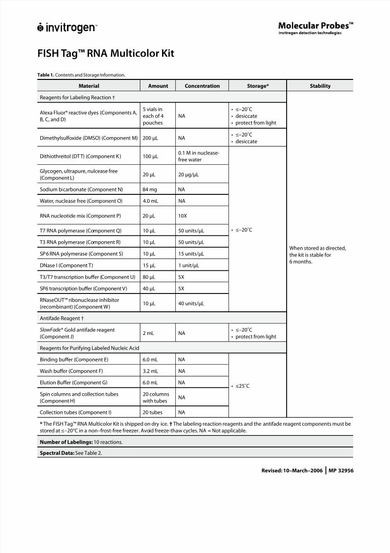
Table 1. Contents and Storage Information.
Material Amount Concentration Storage* Stability
Reagents for Labeling Reaction †
When stored as directed,
the kit is stable for
6 months.
Alexa Fluor® reactive dyes (Components A,
B, C, and D)
5 vials in
each of 4
pouches
NA
≤–20˚C
desiccate
protect from light
•
•
•
Dimethylsulfoxide (DMSO) (Component M) 200 µL NA≤–20˚C
desiccate
•
•
Dithiothreitol (DTT) (Component K) 100 µL0.1 M in nuclease-
free water
≤–20˚C•
Glycogen, ultrapure, nulcease free
(Component L)20 µL 20 µg/µL
Sodium bicarbonate (Component N) 84 mg NA
Water, nuclease free (Component O) 4.0 mL NA
RNA nucleotide mix (Component P) 20 µL 10X
T7 RNA polymerase (Component Q) 10 µL 50 units/µL
T3 RNA polymerase (Component R) 10 µL 50 units/µL
SP6 RNA polymerase (Component S) 10 µL 15 units/µL
DNase I (Component T) 15 µL 1 unit/µL
T3/T7 transcription buffer (Component U) 80 µL 5X
SP6 transcription buffer (Component V) 40 µL 5X
RNaseOUT™ ribonuclease inhibitor
(recombinant) (Component W)10 µL 40 units/µL
Antifade Reagent †
SlowFade® Gold antifade reagent
(Component J)2 mL NA
≤–20˚C
protect from light
•
•
Reagents for Purifying Labeled Nucleic Acid
Binding buffer (Component E) 6.0 mL NA
≤25˚C•
Wash buffer (Component F) 3.2 mL NA
Elution Buffer (Component G) 6.0 mL NA
Spin columns and collection tubes
(Component H)
20 columns
with tubesNA
Collection tubes (Component I) 20 tubes NA
* The FISH Tag™ RNA Multicolor Kit is shipped on dry ice. † The labeling reaction reagents and the antifade reagent components must be
stored at ≤–20°C in a non–frost-free freezer. Avoid freeze-thaw cycles. NA = Not applicable.
Number of Labelings: 10 reactions.
Spectral Data: See Table 2.
8/12/2019 Protocol FISH Invitrogen
http://slidepdf.com/reader/full/protocol-fish-invitrogen 2/17
FISH Tag™ RNA Multicolor Kit | 2
Contents
Introduction . . . . . . . . . . . . . . . . . . . . . . . . . . . . . . . . . . . . . . . . . . . . . . . . . . . . . . . . . . . . . . . . . . . . . . . . . . . . . . . . . . . . . . . 3
Fluorescence In Situ Hybridization . . . . . . . . . . . . . . . . . . . . . . . . . . . . . . . . . . . . . . . . . . . . . . . . . . . . . . . . . . . . . 3
FISH Tag™ Kits. . . . . . . . . . . . . . . . . . . . . . . . . . . . . . . . . . . . . . . . . . . . . . . . . . . . . . . . . . . . . . . . . . . . . . . . . . . . . . . . . 4
Before you Begin. . . . . . . . . . . . . . . . . . . . . . . . . . . . . . . . . . . . . . . . . . . . . . . . . . . . . . . . . . . . . . . . . . . . . . . . . . . . . . . . . . . 5
Materials Required but Not Supplied. . . . . . . . . . . . . . . . . . . . . . . . . . . . . . . . . . . . . . . . . . . . . . . . . . . . . . . . . . . 5
Handling of Amine-Reactive Fluorescent Dyes . . . . . . . . . . . . . . . . . . . . . . . . . . . . . . . . . . . . . . . . . . . . . . . . . 5
Storage of DMSO. . . . . . . . . . . . . . . . . . . . . . . . . . . . . . . . . . . . . . . . . . . . . . . . . . . . . . . . . . . . . . . . . . . . . . . . . . . . . . 5
Preparing Binding Buffer with Isopropanol . . . . . . . . . . . . . . . . . . . . . . . . . . . . . . . . . . . . . . . . . . . . . . . . . . . . . 6
Preparing Wash Buffer with Ethanol . . . . . . . . . . . . . . . . . . . . . . . . . . . . . . . . . . . . . . . . . . . . . . . . . . . . . . . . . . . . 6
Preparing the Sodium Bicarbonate Solution . . . . . . . . . . . . . . . . . . . . . . . . . . . . . . . . . . . . . . . . . . . . . . . . . . . . 6
Choosing the Appropriate DNA Template for In Vitro Transcription . . . . . . . . . . . . . . . . . . . . . . . . . . . . . . 6
Pre-Protocol Reading. . . . . . . . . . . . . . . . . . . . . . . . . . . . . . . . . . . . . . . . . . . . . . . . . . . . . . . . . . . . . . . . . . . . . . . . . . 6
Synthesis of Amine-Modified RNA . . . . . . . . . . . . . . . . . . . . . . . . . . . . . . . . . . . . . . . . . . . . . . . . . . . . . . . . . . . . . . . . . . 7
In Vitro Transcription . . . . . . . . . . . . . . . . . . . . . . . . . . . . . . . . . . . . . . . . . . . . . . . . . . . . . . . . . . . . . . . . . . . . . . . . . . 7
Purifying the Amine-Modified RNA . . . . . . . . . . . . . . . . . . . . . . . . . . . . . . . . . . . . . . . . . . . . . . . . . . . . . . . . . . . . 8
Ethanol Precipitation of the Amine-Modified RNA . . . . . . . . . . . . . . . . . . . . . . . . . . . . . . . . . . . . . . . . . . . . . . 8
Labeling the Amine-Modified RNA with Fluorescent Dye . . . . . . . . . . . . . . . . . . . . . . . . . . . . . . . . . . . . . . . . . . . . 9
Purifiying the Fluorescent Dye–Labeled RNA . . . . . . . . . . . . . . . . . . . . . . . . . . . . . . . . . . . . . . . . . . . . . . . . . . . 9
Ethanol Precipitation of th Fluorescent Dye–Labeled RNA . . . . . . . . . . . . . . . . . . . . . . . . . . . . . . . . . . . . .10
RNA Probe Fragmentation (Optional) . . . . . . . . . . . . . . . . . . . . . . . . . . . . . . . . . . . . . . . . . . . . . . . . . . . . . . . . . 11Suggested Hybridization Protocols . . . . . . . . . . . . . . . . . . . . . . . . . . . . . . . . . . . . . . . . . . . . . . . . . . . . . . . . . . . . . . . . 11
P r e - H y b r i d i z a t i o n . . . . . . . . . . . . . . . . . . . . . . . . . . . . . . . . . . . . . . . . . . . . . . . . . . . . . . . . . . . . . . . . . . . . . . . . . . . . 1 2
Hybridization . . . . . . . . . . . . . . . . . . . . . . . . . . . . . . . . . . . . . . . . . . . . . . . . . . . . . . . . . . . . . . . . . . . . . . . . . . . . . . . . 13
Post-Hybridization . . . . . . . . . . . . . . . . . . . . . . . . . . . . . . . . . . . . . . . . . . . . . . . . . . . . . . . . . . . . . . . . . . . . . . . . . . . 13
Tips for Success . . . . . . . . . . . . . . . . . . . . . . . . . . . . . . . . . . . . . . . . . . . . . . . . . . . . . . . . . . . . . . . . . . . . . . . . . . . . . . . . . . . 13
Sensitivity. . . . . . . . . . . . . . . . . . . . . . . . . . . . . . . . . . . . . . . . . . . . . . . . . . . . . . . . . . . . . . . . . . . . . . . . . . . . . . . . . . . . 13
Length of Probe . . . . . . . . . . . . . . . . . . . . . . . . . . . . . . . . . . . . . . . . . . . . . . . . . . . . . . . . . . . . . . . . . . . . . . . . . . . . . . 13
S p e c i m e n I n t e g r i t y . . . . . . . . . . . . . . . . . . . . . . . . . . . . . . . . . . . . . . . . . . . . . . . . . . . . . . . . . . . . . . . . . . . . . . . . . . . 1 4
Imaging . . . . . . . . . . . . . . . . . . . . . . . . . . . . . . . . . . . . . . . . . . . . . . . . . . . . . . . . . . . . . . . . . . . . . . . . . . . . . . . . . . . . . 14
Troubleshooting . . . . . . . . . . . . . . . . . . . . . . . . . . . . . . . . . . . . . . . . . . . . . . . . . . . . . . . . . . . . . . . . . . . . . . . . . . . . . . . . . . 14
Yield . . . . . . . . . . . . . . . . . . . . . . . . . . . . . . . . . . . . . . . . . . . . . . . . . . . . . . . . . . . . . . . . . . . . . . . . . . . . . . . . . . . . . . . . . 14
D e g r e e o f L a b e l i n g . . . . . . . . . . . . . . . . . . . . . . . . . . . . . . . . . . . . . . . . . . . . . . . . . . . . . . . . . . . . . . . . . . . . . . . . . . . 1 4
Hybridization . . . . . . . . . . . . . . . . . . . . . . . . . . . . . . . . . . . . . . . . . . . . . . . . . . . . . . . . . . . . . . . . . . . . . . . . . . . . . . . . 15
Calculating the Labeling Efficiency and Concentration of Nucleic Acid . . . . . . . . . . . . . . . . . . . . . . . . . . . . . . 15Measuring the Base:Dye Ratio . . . . . . . . . . . . . . . . . . . . . . . . . . . . . . . . . . . . . . . . . . . . . . . . . . . . . . . . . . . . . . . . 1 5
Measuring the Concentration of Nucleic Acid . . . . . . . . . . . . . . . . . . . . . . . . . . . . . . . . . . . . . . . . . . . . . . . . . 1 6
References. . . . . . . . . . . . . . . . . . . . . . . . . . . . . . . . . . . . . . . . . . . . . . . . . . . . . . . . . . . . . . . . . . . . . . . . . . . . . . . . . . . . . . . . 16
Product List . . . . . . . . . . . . . . . . . . . . . . . . . . . . . . . . . . . . . . . . . . . . . . . . . . . . . . . . . . . . . . . . . . . . . . . . . . . . . . . . . . . . . . 17
Additional Products . . . . . . . . . . . . . . . . . . . . . . . . . . . . . . . . . . . . . . . . . . . . . . . . . . . . . . . . . . . . . . . . . . . . . . . . . . . . . . . 17
Contact Information . . . . . . . . . . . . . . . . . . . . . . . . . . . . . . . . . . . . . . . . . . . . . . . . . . . . . . . . . . . . . . . . . . . . . . . . . . . . . . 17
8/12/2019 Protocol FISH Invitrogen
http://slidepdf.com/reader/full/protocol-fish-invitrogen 3/17
FISH Tag™ RNA Multicolor Kit | 3
Introduction
Fluorescence In Situ Hybridization Fluorescence in situ hybridization (FISH) technology permits detection of specific nucleic
acid targets within a biological specimen, or in situ meaning where it lies. RNA and DNAtargets such as mRNAs expressed in a tissue or genes present on a chromosome can belocalized using this technology. Detection of a nucleic acid target in situ is achieved throughhybridization of complementary sequence, fluorescent dye–labeled nucleic acid “probe” to
the specimen. Once the hybridization assay is complete, the specimen is viewed under afluorescence microscope to visualize the hybridized fluorescent probe. Fluorescent dyes, orfluorophores, having different excitation and emission spectra generate fluorescence of dif-ferent colors when viewed under a f luorescence microscope. Different fluorophores can beused to label different nucleic acid probes for detection of multiple targets simultaneously.Multiplex FISH (MFISH) refers to the simultaneous localization of multiple sequence-specificnucleic acid targets using spectrally distinct fluorescent dye labels.
The labeling technology provided in the FISH Tag™ RNA Kits uses a two step approach.1 Inthe first step, in vitro transcription is used to enzymatically incorporate an amine-modifiednucleotide into the probe template. The modified nucleotide is UTP having an NH
2 group
attached through a linker to the C5 position of the base. In the second step, dye labeling of thepurified amine-modified RNA is achieved by incubation with amine-reactive dyes. These
active ester compounds react with the primary amines incorporated into the probe template,covalently conjugating the dye to the modified nucleotide base. The purified probe is thenready for hybridization to the specimen.
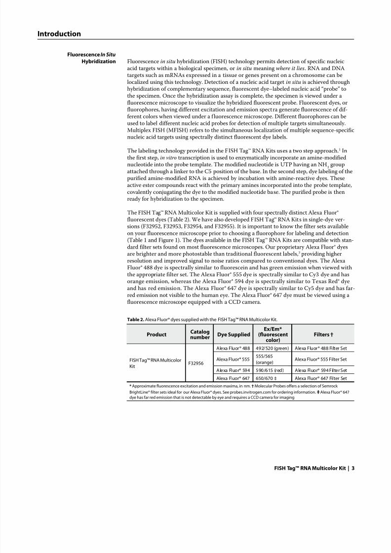
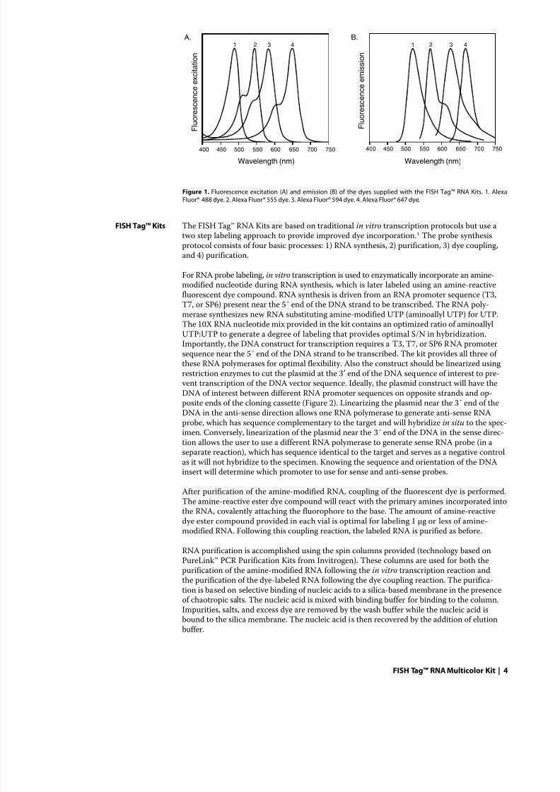
The FISH Tag™ RNA Multicolor Kit is supplied with four spectrally distinct Alexa Fluor®fluorescent dyes (Table 2). We have also developed FISH Tag™ RNA Kits in single-dye ver-sions (F32952, F32953, F32954, and F32955). It is important to know the filter sets availableon your fluorescence microscope prior to choosing a fluorophore for labeling and detection(Table 1 and Figure 1). The dyes available in the FISH Tag™ RNA Kits are compatible with stan-dard filter sets found on most fluorescence microscopes. Our proprietary Alexa Fluor® dyesare brighter and more photostable than traditional fluorescent labels,2 providing higherresolution and improved signal to noise ratios compared to conventional dyes. The AlexaFluor® 488 dye is spectrally similar to fluorescein and has green emission when viewed withthe appropriate filter set. The Alexa Fluor® 555 dye is spectrally similar to Cy3 dye and has
orange emission, whereas the Alexa Fluor® 594 dye is spectrally similar to Texas Red® dyeand has red emission. The Alexa Fluor® 647 dye is spectrally similar to Cy5 dye and has far-red emission not visible to the human eye. The Alexa Fluor® 647 dye must be viewed using afluorescence microscope equipped with a CCD camera.
Table 2. Alexa Fluor® dyes supplied with the FISH Tag™ RNA Multicolor Kit.
Product Catalognumber Dye Supplied
Ex/Em*(fluorescent
color)Filters †
FISH Tag™ RNA Multicolor
KitF32956
Alexa Fluor® 488 492/520 (green) Alexa Fluor® 488 Filter Set
Alexa Fluor® 555555/565
(orange)Alexa Fluor® 555 Filter Set
Alexa Fluor® 594 590/615 (red) Alexa Fluor® 594 Filter Set
Alexa Fluor® 647 650/670 ‡ Alexa Fluor® 647 Filter Set
* Approximate fluorescence excitation and emission maxima, in nm. † Molecular Probes offers a selection of Semrock
BrightLine® filter sets ideal for our Alexa Fluor® dyes. See probes.invitrogen.com for ordering information. ‡ Alexa Fluor® 647
dye has far red emission that is not detectable by eye and requires a CCD camera for imaging
8/12/2019 Protocol FISH Invitrogen
http://slidepdf.com/reader/full/protocol-fish-invitrogen 4/17
FISH Tag™ RNA Multicolor Kit | 4
FISH Tag™ Kits The FISH Tag™ RNA Kits are based on traditional in vitro transcription protocols but use atwo step labeling approach to provide improved dye incorporation.1 The probe synthesisprotocol consists of four basic processes: 1) RNA synthesis, 2) purification, 3) dye coupling,and 4) purification.
For RNA probe labeling, in vitro transcription is used to enzymatically incorporate an amine-modified nucleotide during RNA synthesis, which is later labeled using an amine-reactivefluorescent dye compound. RNA synthesis is driven from an RNA promoter sequence (T3,T7, or SP6) present near the 5´ end of the DNA strand to be transcribed. The RNA poly-merase synthesizes new RNA substituting amine-modified UTP (aminoallyl UTP) for UTP.The 10X RNA nucleotide mix provided in the kit contains an optimized ratio of aminoallylUTP:UTP to generate a degree of labeling that provides optimal S/N in hybridization.Importantly, the DNA construct for transcription requires a T3, T7, or SP6 RNA promotersequence near the 5´ end of the DNA strand to be transcribed. The kit provides all three ofthese RNA polymerases for optimal flexibility. Also the construct should be linearized usingrestriction enzymes to cut the plasmid at the 3′ end of the DNA sequence of interest to pre- vent transcription of the DNA vector sequence. Ideally, the plasmid construct will have theDNA of interest between different RNA promoter sequences on opposite strands and op-
posite ends of the cloning cassette (Figure 2). Linearizing the plasmid near the 3´ end of theDNA in the anti-sense direction allows one RNA polymerase to generate anti-sense RNAprobe, which has sequence complementary to the target and will hybridize in situ to the spec-imen. Conversely, linearization of the plasmid near the 3´ end of the DNA in the sense direc-tion allows the user to use a different RNA polymerase to generate sense RNA probe (in aseparate reaction), which has sequence identical to the target and serves as a negative controlas it will not hybridize to the specimen. Knowing the sequence and orientation of the DNAinsert will determine which promoter to use for sense and anti-sense probes.
After purification of the amine-modified RNA, coupling of the fluorescent dye is performed.The amine-reactive ester dye compound will react with the primary amines incorporated intothe RNA, covalently attaching the fluorophore to the base. The amount of amine-reactivedye ester compound provided in each vial is optimal for labeling 1 µg or less of amine-modified RNA. Following this coupling reaction, the labeled RNA is purified as before.
RNA purification is accomplished using the spin columns provided (technology based onPureLink™ PCR Purification Kits from Invitrogen). These columns are used for both thepurification of the amine-modified RNA following the in vitro transcription reaction andthe purification of the dye-labeled RNA following the dye coupling reaction. The purifica-tion is based on selective binding of nucleic acids to a silica-based membrane in the presenceof chaotropic salts. The nucleic acid is mixed with binding buffer for binding to the column.Impurities, salts, and excess dye are removed by the wash buffer while the nucleic acid isbound to the silica membrane. The nucleic acid is then recovered by the addition of elutionbuffer.
F l u o r e s c e n c e e m i s s i o n
F l u o r e s c e n c e e x c i t a t i o n
Wavelength (nm)Wavelength (nm)
400 450 500 550 600 650 700 750400 450 500 550 600 650 700 750
A. B.44 33 22 11
Figure 1. Fluorescence excitation (A) and emission (B) of the dyes supplied with the FISH Tag™ RNA Kits. 1. AlexaFluor® 488 dye. 2. Alexa Fluor® 555 dye. 3. Alexa Fluor® 594 dye. 4. Alexa Fluor® 647 dye.
8/12/2019 Protocol FISH Invitrogen
http://slidepdf.com/reader/full/protocol-fish-invitrogen 5/17
FISH Tag™ RNA Multicolor Kit | 5
The RNA hybridization protocol provided in this manual for in situ hybridization is basedon RNA hybridization to Drosophila (fruit fly) embryos3 and should be generally applicableto tissues. It is provided as an example. Depending on your model system or specimen require-ments, optimization of this protocol may be required.
Before you Begin
Materials Required but NotSupplied DNA template for transcription
100% isopropanol
100% ethanol
70% ethanol
3M sodium acetate, pH 5.2
incubator at 37°C
heat block at 65°C
microcentrifuge
Handling of Amine-ReactiveFluorescent Dyes Amine-reactive fluorescent dyes are sensitive to light and moisture. Ensure that the amine-
reactive fluorescent dyes remain desiccated. Minimize the exposure of the labeled probe(both during the labeling reaction and during your experiments) to light.
Storage of DMSO The DMSO used for dissolving the amine-reactive dye compounds (Component M) is hygro-scopic. Store at ≤–20°C or room temperature, tightly sealed.
Preparing Binding Buffer withIsopropanol 1.1 To the binding buffer concentrate supplied in the kit (6 mL, Component E) add 4 mL of
100% isopropanol to make a final volume of 10 mL of binding buffer.
•
•
•
•
•
•
•
•
RNA promoter 1
DNA insert
Plasmid Construct
sense strand
RNA promoter 2
antisense strand
RNA promoter 1
RNA promoter 2
5´
3´
3´
5´
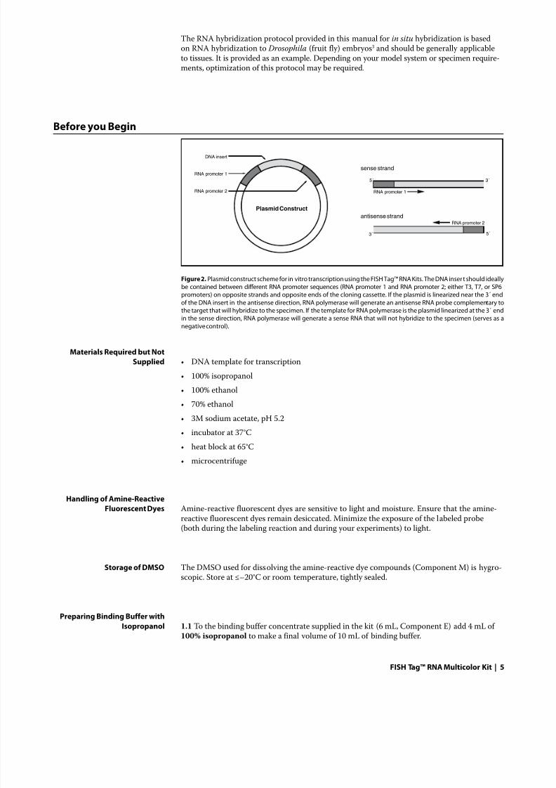
Figure 2. Plasmid construct scheme for in vitro transcription using the FISH Tag™ RNA Kits. The DNA insert should ideallybe contained between different RNA promoter sequences (RNA promoter 1 and RNA promoter 2; either T3, T7, or SP6promoters) on opposite strands and opposite ends of the cloning cassette. If the plasmid is linearized near the 3´ end
of the DNA insert in the antisense direction, RNA polymerase will generate an antisense RNA probe complementary tothe target that will hybridize to the specimen. If the template for RNA polymerase is the plasmid linearized at the 3´ endin the sense direction, RNA polymerase will generate a sense RNA that will not hybridize to the specimen (serves as a
negative control).
8/12/2019 Protocol FISH Invitrogen
http://slidepdf.com/reader/full/protocol-fish-invitrogen 6/17
FISH Tag™ RNA Multicolor Kit | 6
1.2 Mix well.
1.3 Mark the checkbox on the bottle label to indicate that isopropanol has been added. Theworking solution of binding buffer is stable for 6 months at room temperature.
Preparing Wash Buffer withEthanol 2.1 To the wash buffer concentrate supplied in the kit (3.2 mL, Component F) add 12.8 mL of
100% ethanol to make a final volume of 15 mL of wash buffer.
2.2 Mix well.
2.3 Mark the checkbox on the bottle label to indicate that ethanol has been added. The work-ing solution of wash buffer is stable for 6 months at room temperature.
Preparing the SodiumBicarbonate Solution 3.1 To the tube containing the sodium bicarbonate powder (Component N) add 1 mL of
nuclease-free water (Component O)
3.2 Vortex until solid material is no longer visible in the tube.
3.3 Store at ≤–20˚C when not in use. This solution of sodium bicarbonate will be stable for6 months.
Choosing the AppropriateDNA Template for In Vitro
Transcription The DNA template should be a plasmid construct that has T7, T3, or SP6 RNA polymerasepromoter sequences at opposite ends and on opposite strands of the DNA insert of interest.This type of construct allows one to generate sense (negative control) and anti-sense hybridiza-tion probes. The vector should be linearized by restriction digest at one end of the insert orthe other in order to utilize the respective RNA promoter sequence to initiate RNA synthesis.The nucleotide sequence of the DNA insert and proximity to the RNA polymerase promoter
will determine which polymerase to use for each linearized form of the plasmid (Figure 2).
Pre-Protocol Reading At the end of this instruction manual are two sections entitled Tips for Success and Trouble- shooting . It may be beneficial to read through these topics before you start your experiment,especially if you are relatively new to the preparation and use of FISH probes.
8/12/2019 Protocol FISH Invitrogen
http://slidepdf.com/reader/full/protocol-fish-invitrogen 7/17
FISH Tag™ RNA Multicolor Kit | 7
Synthesis of Amine-Modified RNA
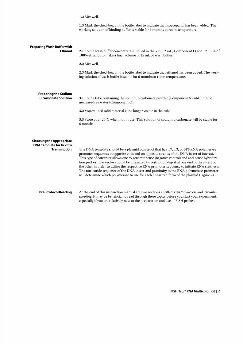
In Vitro Transcription 4.1 Remove the following components from the freezer, thaw to room temperature, and mixby vortexing:
water, nuclease free (Component O)
5X transcription buffer of choice (T3/T7 transcription buffer, Component U or SP6 tran-
scription buffer, Component V)0.1M DTT (Component K)
10X RNA nucleotide mix (Component P)
RNaseOUT™ ribonuclease inhibitor (Component W)
4.2 Remove the following components from the freezer, and place them on ice or in a –20°Cbench top cooler. Do not vortex.
RNA polymerase of choice (T7 RNA polymerase, Component Q or T3 RNA polymerse,Component R, or SP6 RNA polymerase, Component S)
Note: When in use, ensure these enzymes remain in the –20°C bench top cooler or on ice
and return them to the non–frost-free freezer as soon as possible after use.
4.3 If you plan to use SP6 RNA polymerase, first make a 0.01 M DTT solution by diluting1 µL of 0.1 M DTT (Component K) into 9 µL of nuclease-free water (Component O). Use thisdiluted solution of DTT if the in vitro transcription reaction (step 4.4) uses the SP6 RNApolymerase.
4.4 Prepare in vitro transcription reactions on ice as described below.
Component Volumewater, nuclease free to final 20 µL5X transcription buffer (either T3/T7 or SP6) 4 µLDTT 1–2 µL*
10X RNA nucleotide mix 2 µLlinearized DNA template (user supplied) 1 µgRNaseOUT™ inhibitor 1 µLRNA polymerase (T7, T3, or SP6) 1 µLFinal Volume 20 µL
* If the reaction uses T3/T7 RNA polymerase, add 1 µL of 0.1 M DTT (Component K) in thisreaction. If the reaction uses SP6 RNA polymerase, add 2 µL of 0.01 M DTT (prepared instep 4.3).
4.5 Mix gently by slowly pipetting the mixture up and down three times (do not vortex).
4.6 Incubate at 37°C for 1 hour.
4.7 Add 1 µl of DNase I (Component T) and mix gently by slowly pipetting the mixture upand down three times (do not vortex).
4.8 Incubate at 37°C for 15 minutes.
4.9 Add 79 µl of nuclease-free water to the sample (Component O) and vortex the reactionat maximum speed for 10 seconds. The vortexing is important to inactivate the DNase I.
4.10 Proceed immediately to Purifying the Amine-Modified RNA.
•
•
•
•
•
•
8/12/2019 Protocol FISH Invitrogen
http://slidepdf.com/reader/full/protocol-fish-invitrogen 8/17
FISH Tag™ RNA Multicolor Kit | 8
Purifying the Amine-ModifiedRNA 5.1 Add 400 µL of binding buffer with isopropanol (see Before You Begin, above) to the
synthesis reaction and mix well.
5.2 Add the entire volume (500 µL) to a spin column seated inside a collection tube(Component H).
5.3 Centrifuge the column at >10,000 × g for 1 minute. The RNA is bound to the column.Discard the flow-through.
5.4 Wash the column with 650 µL of wash buffer with ethanol (see Before You Begin, above).
5.5 Centrifuge the column at >10,000 × g for 1 minute. Discard the flow-through.
5.6 Centrifuge the column >10,000 × g for 1 minute to remove any residual wash buffer.
5.7 Place the spin column in a clean 1.7 mL collection tube (Component I).
5.8 Apply 55 µL of elution buffer (Component G) to the center of the column.
5.9 Allow the column to stand at room temperature for 1 minute.
5.10 Centrifuge the column > 10,000 × g for 1 minute.
5.11 The collection tube contains your purified amine-modified RNA. Discard the columnand proceed to Ethanol Precipitation of the Amine-Modified RNA.
Ethanol Precipitation of theAmine-Modified RNA 6.1 To the eluted RNA from step 5.11, add:
10 µL of 3M sodium acetate (pH 5.2)
1 µL of glycogen (Component L)
39 µL of nuclease-free water (Component O)
6.2 Add 300 µL of 100% ethanol.
6.3 Store sample at –20˚C for 30 minutes.
6.4 Centrifuge the sample at >10,000 × g for 10 minutes.
6.5 Remove the supernatant. Be careful not to lose the pellet.
6.6 Carefully rinse the pellet with 400 µL of 70% ethanol. Remove the supernatant andrepeat this rinse step.
Note: Free amines carried over with the RNA will inhibit the efficiency of the dye couplingreaction. These rinse steps with 70% ethanol are important to eliminate any trace amines.
6.7 With a pipet, remove as much of the residual 70% ethanol as possible without disturbingthe pellet and then allow the sample to air dry (about 5–10 minutes).
6.8 Add 5 µL of nuclease-free water (Component O) to the pellet (buffer should not be usedin order to avoid introduction of free amines).
6.9 Incubate the sample at 37°C for 5 minutes.
6.10 Vortex the sample to fully resuspend the RNA and place the sample on ice.
•
•
•
8/12/2019 Protocol FISH Invitrogen
http://slidepdf.com/reader/full/protocol-fish-invitrogen 9/17
FISH Tag™ RNA Multicolor Kit | 9
Note: The sample can be stored at this stage for up to 2 weeks.
6.11 Determine the concentration of the sample (see Calculating the Labeling Efficiency andConcentration of Nucleic Acid ).
Note: The in vitro transcription reaction often generates 1–4 µg of RNA depending on DNAtemplate. The dye conjugation reaction (below) is optimized for 1 µg of amine-modifiedRNA. Using more RNA per reaction will result in lowered labeling efficiency.
6.12 Adjust the concentration of the sample with water to a final concentration of 0.2 µg/µL.
6.13 Proceed to Labeling the Amine-Modified RNA with Fluorescent Dye.
Labeling the Amine-Modified RNA with Fluorescent Dye
7.1 Denature 1 µg (5 µL) of the RNA by incubating it at 65°C for 5 minutes.
7.2 Place the sample on ice for 3 minutes.
7.3 Centrifuge the sample at >10,000 × g for 3 minutes.
7.4 Add 3 µL of sodium bicarbonate solution to the sample (prepared in step 3.3).
Note: The thawed sodium bicarbonate solution may precipitate. Vortex thoroughly before using.
7.5 Remove the label from a vial of reactive dye (Components A, B, C, or D) in order tobetter see the dye pellet.
7.6. Resuspend the reactive dye in 2 µl of DMSO (Component M). Vortex well (10 seconds athigh speed) in order to fully resuspend the dye.
7.7 Transfer the 2 µl of reactive dye in DMSO to the RNA sample at room temperature.
7.8 Vortex the mixture at maximum speed for at least 15 seconds.
Note: Sufficient mixing of the labeling reaction is critical.
7.9 Centrifuge the sample briefly in order to collect the labeling reaction in the bottom ofthe tube.
7.10 Incubate the labeling reaction at room temperature in the dark for 1 hour.
7.11 Add 90 µL of water to the sample.
7.12 Proceed immediately to Purifying the Fluorescent Dye–Labeled RNA.
Purifiying the FluorescentDye–Labeled RNA 8.1 Add 400 µL of binding buffer with isopropanol (see Before You Begin, above) to the
labeling reaction and mix well.
8.2 Add the entire volume (500 µL) to a spin column seated inside a collection tube(Component H).
8/12/2019 Protocol FISH Invitrogen
http://slidepdf.com/reader/full/protocol-fish-invitrogen 10/17
FISH Tag™ RNA Multicolor Kit | 10
8.3 Centrifuge the column at >10,000 × g for 1 minute. The labeled RNA is bound to thecolumn. Discard the flow-through.
8.4 Wash the column with 650 µL of wash buffer with ethanol (see Before You Begin, above).
8.5 Centrifuge the column at >10,000 × g for 1 minute. Discard the flow-through.
8.6 Centrifuge the column >10,000 × g for 1 minute to remove any residual wash buffer.
8.7 Place the spin column in a clean 1.7 mL collection tube (Component I).
8.8 Apply 55 µL of elution buffer (Component G) to the center of the column.
8.9 Allow the column to stand at room temperature for 1 minute.
8.10 Centrifuge the column >10,000 × g for 1 minute.
8.11 The collection tube contains your purified fluorescent dye–labeled RNA. Discardthe column.
Ethanol Precipitation of th
Fluorescent Dye–Labeled RNA 9.1 To the eluted dye-labeled RNA from step 8.11 add:
10 µL of 3M sodium acetate (pH 5.2)
1 µL of glycogen (Component L)
39 µL of nuclease-free water (Component O)
9.2 Add 300 µL of 100% ethanol.
9.3 Store sample at –20˚C for 30 minutes.
9.4 Centrifuge the sample at > 10,000 × g for 10 minutes.
9.5 Remove the supernatant. Be careful not to lose the pellet.
9.6 Carefully rinse the pellet with 400 µL of 70% ethanol. Remove the supernatant and repeatthis rinse.
9.7 With a pipet, remove as much of the residual 70% ethanol as possible without disturbingthe pellet and then allow the sample to air dry (about 5–10 minutes).
9.8 Add 10 µL of nuclease-free water (Component L) to the pellet.
9.9 Incubate the sample at 37˚C for 5 minutes.
9.10 Vortex the sample to fully resuspend the dye-labeled RNA and store on ice.
9.11 Determine the concentration of the sample (see Calculating the Labeling Efficiency andConcentration of Nucleic Acid ).
9.12 The dye–labeled RNA is now ready for hybridization buffer. Alternatively, store the dye-labeled RNA at ≤–70˚C until ready for use. It is stable when protected from light for up to2 weeks when stored at ≤–70˚C.
•
•
•
8/12/2019 Protocol FISH Invitrogen
http://slidepdf.com/reader/full/protocol-fish-invitrogen 11/17
FISH Tag™ RNA Multicolor Kit | 11
RNA Probe Fragmentation(Optional) For RNA probes greater than 500 bases, the following optional fragmentation protocol can
be used to reduce the size of the labeled RNA probe to an average length of 500 bases. Werecommend the user test fragmented and non-fragmented probes in hybridization to deter-mine which gives the best signal to noise ratio.
10.1 Prepare 2X carbonate buffer
127.2 mg sodium carbonate
67.2 mg sodium bicarbonate
add water to 10 mL
pH to 10.2 with NaOH
store in small aliquots at ≤–20˚C.
10.2 Prepare stop solution
164.1 mg sodium acetate
add water to 10 mL
pH to 6.0 with acetic acid
store in small aliquots at ≤–20˚C.
10.3 Mix 10 µL of labeled RNA with 10 µl 2X carbonate buffer.
10.4 Incubate at 42˚C for 20–30 minutes.
10.5 Add 20 µL stop solution and precipitate with ethanol (as described above).
10.6 Verify the length by gel electrophoresis. Decrease or increase the time of fragmentationaccordingly.
Suggested Hybridization Protocols
The RNA hybridization protocol for in situ hybridization is based on RNA hybridization to Drosophila (fruit fly) embryos3 and should be generally applicable to tissues. It is providedas an example. Depending on your model system or specimen requirements, optimizationof this protocol may be required.
Useful protocols for various in situ hybridization applications can be found in In Situ Hy-bridization: A Practical Approach by D.G. Wilkinson (Ed.) Oxford University Press; 2nd edition (1999), In Situ Hybridization Protocols (Methods in Molecular Biology) byI. A. Darby (Ed.) Humana Press; 2nd edition (2000), Practical in Situ Hybridization byT. Schwarchzacher and P. Heslop-Harrison, BIOS Scientific Publishers (1999), and Intro-duction to Fluorescence In Situ Hybridization: Principles and Clinical Applications byM. Andreeff (Ed.) and D. Pinkel, Wiley-Liss; 1st edition (1999).
Note: perform these steps at room temperature unless specified otherwise and never let thespecimen become dry at any point as this will increase autofluorescence.
•
•
•
•
•
•
•
•
•
8/12/2019 Protocol FISH Invitrogen
http://slidepdf.com/reader/full/protocol-fish-invitrogen 12/17
FISH Tag™ RNA Multicolor Kit | 12
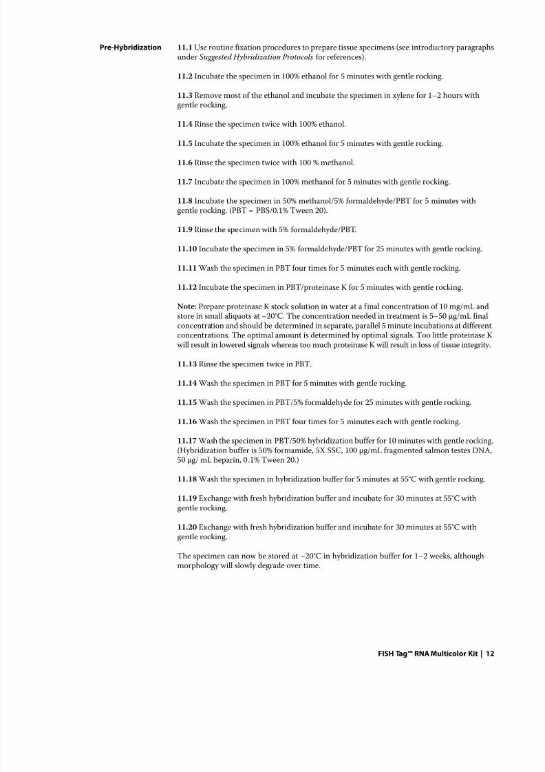
Pre-Hybridization 11.1 Use routine fixation procedures to prepare tissue specimens (see introductory paragraphsunder Suggested Hybridization Protocols for references).
11.2 Incubate the specimen in 100% ethanol for 5 minutes with gentle rocking.
11.3 Remove most of the ethanol and incubate the specimen in xylene for 1–2 hours withgentle rocking.
11.4 Rinse the specimen twice with 100% ethanol.
11.5 Incubate the specimen in 100% ethanol for 5 minutes with gentle rocking.
11.6 Rinse the specimen twice with 100 % methanol.
11.7 Incubate the specimen in 100% methanol for 5 minutes with gentle rocking.
11.8 Incubate the specimen in 50% methanol/5% formaldehyde/PBT for 5 minutes withgentle rocking. (PBT = PBS/0.1% Tween 20).
11.9 Rinse the specimen with 5% formaldehyde/PBT.
11.10 Incubate the specimen in 5% formaldehyde/PBT for 25 minutes with gentle rocking.
11.11 Wash the specimen in PBT four times for 5 minutes each with gentle rocking.
11.12 Incubate the specimen in PBT/proteinase K for 5 minutes with gentle rocking.
Note: Prepare proteinase K stock solution in water at a final concentration of 10 mg/mL andstore in small aliquots at –20°C. The concentration needed in treatment is 5–50 µg/mL finalconcentration and should be determined in separate, parallel 5 minute incubations at differentconcentrations. The optimal amount is determined by optimal signals. Too little proteinase Kwill result in lowered signals whereas too much proteinase K will result in loss of tissue integrity.
11.13 Rinse the specimen twice in PBT.
11.14 Wash the specimen in PBT for 5 minutes with gentle rocking.
11.15 Wash the specimen in PBT/5% formaldehyde for 25 minutes with gentle rocking.
11.16 Wash the specimen in PBT four times for 5 minutes each with gentle rocking.
11.17 Wash the specimen in PBT/50% hybridization buffer for 10 minutes with gentle rocking.(Hybridization buffer is 50% formamide, 5X SSC, 100 µg/mL fragmented salmon testes DNA,50 µg/ mL heparin, 0.1% Tween 20.)
11.18 Wash the specimen in hybridization buffer for 5 minutes at 55°C with gentle rocking.
11.19 Exchange with fresh hybridization buffer and incubate for 30 minutes at 55°C withgentle rocking.
11.20 Exchange with fresh hybridization buffer and incubate for 30 minutes at 55°C withgentle rocking.
The specimen can now be stored at –20°C in hybridization buffer for 1–2 weeks, althoughmorphology will slowly degrade over time.
8/12/2019 Protocol FISH Invitrogen
http://slidepdf.com/reader/full/protocol-fish-invitrogen 13/17
8/12/2019 Protocol FISH Invitrogen
http://slidepdf.com/reader/full/protocol-fish-invitrogen 14/17
FISH Tag™ RNA Multicolor Kit | 14
Specimen Integrity Proper fixation of the specimen is critical to successful hybridization of the probe to thetarget. Specimens for RNA hybridization should be treated to maintain the integrity ofthe target RNA and obviate RNA degradation by RNase activity. We recommendconsulting the in situ hybridization text books above for proper fixation technique.
Imaging Prior to imaging the labeled specimen, it is important to verify the correct filter sets tomatch the dye choice are available on the microscope and that they are in good condition.
They filter sets for each channel should accommodate accurately the spectral character-istics of the dye (see Figure 1 and Table 2). The filter sets should be inspected for wearthat might lead to excitation/emission beyond the filter window specifications. Multi-color experiments should be designed with the available filter sets in mind such that theemission windows accommodate separation of individual dye emissions cleanly withoutoverlap or bleed-through. All fluorescent dyes are subject to photobleaching, so labeledspecimens should be protected from light whenever possible. We provide SlowFade® Goldantifade reagent for mounting Alexa Fluor® dye–labeled specimens because it is optimizedfor high photostability of these dyes where other more traditional mounting media fail.The SlowFade® Gold antifade reagent is non-gelling. ProLong® Gold mounting mediaprovides the same level of photostability as SlowFade® Gold, but slowly gels over time.Both mounting media are available with DAPI counterstain added.
Troubleshooting
In troubleshooting your work, consider the following topics:
Yield The standard in vitro transcription protocol is optimized for use with 1 µg of linearizedtemplate DNA. Typical yield expected with SP6 RNA polymerase is 1–2 µg and with T3 orT7 RNA polymerase is 1–4 µg. RNase contamination can degrade RNA and reduce yield so
good molecular biology technique is important to success. It is important to evaluate the sizeof the RNA template by gel electrophoresis in order to be sure there is not RNase contamina-tion. The optional fragmentation protocol should be optimized to result in an average size of500 bases. The DNase I step in the in vitro transcription protocol is important to eliminatethe DNA template, which could otherwise lead to overestimation of RNA yield as well as bemisinterpreted as RNA in gel electrophoresis, especially if the RNA has been degraded in theprocess by RNase contamination.
Degree of Labeling DOL is a measure of the number of dyes per 100 bases of nucleic acid probe, as determinedfrom absorbance readings at 260 nm and at the dye maximal absorbance (Table 3). The cal-culation is provided below and is available on our website at probes.invitrogen.com. Accurateabsorbance readings require the entire sample in the smallest volume possible. Microcuvettesof 1 cm pathlength and 100 µL can be used. Other microscale spectrophotometers are available.It is important to blank the instrument with the diluent prior to measurement and not todilute the sample too greatly as to fall into the non-linear dynamic range of the instrument.Expected DOLs should be from 1–6 dyes per 100 bases, depending on the dye. It is importantto follow the guidelines in the instruction manual in detail in order to obviate the possibilityof free amine contamination that will result in low DOLs. We strictly recommend two, large volume 70% washes of the amine-modified nucleic acid pellet in order to eliminate free
8/12/2019 Protocol FISH Invitrogen
http://slidepdf.com/reader/full/protocol-fish-invitrogen 15/17
FISH Tag™ RNA Multicolor Kit | 15
amines. The amine-modified RNA should be fully resuspended prior to the coupling reactionby vortexing and using low heat (37˚C) if resuspension is problematic. The amine-reactivedye is extremely sensitive to moisture and thus, must be stored sealed tightly in its pouch bagwith desiccant to prevent loss of activity, which can result in low DOLs. Thorough mixing ofthe coupling reaction is important to optimal labeling and vortexing the reaction at full speedfor a full 15 seconds is highly recommended in order to avoid low DOL.
Hybridization The hybridization protocols suggested are provided as a general guideline to standard RNA
FISH and your model system will require some optimization. Consult published scientificliterature and the handbooks above for further details on general in situ hybridization tech-nique. By far the most important aspect of the experimental design is a reliable positive con-trol that will verify that the hybridization and detection protocols are working. Moderate tostrongly expressed marker genes work well as a positive control in RNA FISH. RNA FISH canfail for multiple reasons but it is important to be able to verify that the RNA in the specimenis intact and has not been degraded by RNase. A reliable positive control is crucial to success-fully troubleshooting your model system.
Calculating the Labeling Efficiency and Concentration of Nucleic Acid
The relative efficiency of a labeling reaction can be evaluated by calculating the approximateratio of bases to dye molecules.1 This ratio can be determined, as described below, by mea-suring the absorbance of the nucleic acid at 260 nm and the absorbance of the dye at itsabsorbance maximum (λ
max). The calculations are based on the Beer-Lambert law:
A = ε × path length (cm) × concentration (M),
where ε is the extinction coefficient in cm–1M–1. The absorbance measurements can also beused to determine the concentration of nucleic acid in the sample. Values needed for thesecalculations are found in Table 3. Alternatively, the ratio can be determined by using ourBase:Dye Ratio Calculator on our website (probes.invitrogen.com) in the Resources section.
Measuring the Base:Dye Ratio 14.1 Measure the absorbance of the nucleic acid–dye conjugate at 260 nm (A260
) and the λmax
for the dye (A
dye). Measure the background absorbance at 260 nm and λ
max, using buffer alone,
and subtract these numbers from the raw absorbance values for the sample. The λmax
valuesfor the fluorophores are given in Table 3.
To perform these measurements, the nucleic acid–dye conjugate should be at a concentra-tion of at least 5 μg/mL. Depending on the dye used and the degree of labeling, a higherconcentration may be required.
For most applications, it will be necessary to measure the absorbance of the entire sampleusing either a conventional spectrophotometer with a 100 μL cuvette or an absorbance
microplate reader with a microplate.Use a cuvette or microplate that does not block UV light and that is clean and nuclease-free.Note that most plastic disposable cuvettes and microplates have significant absorption inthe UV.
•
•
•
8/12/2019 Protocol FISH Invitrogen
http://slidepdf.com/reader/full/protocol-fish-invitrogen 16/17
FISH Tag™ RNA Multicolor Kit | 16
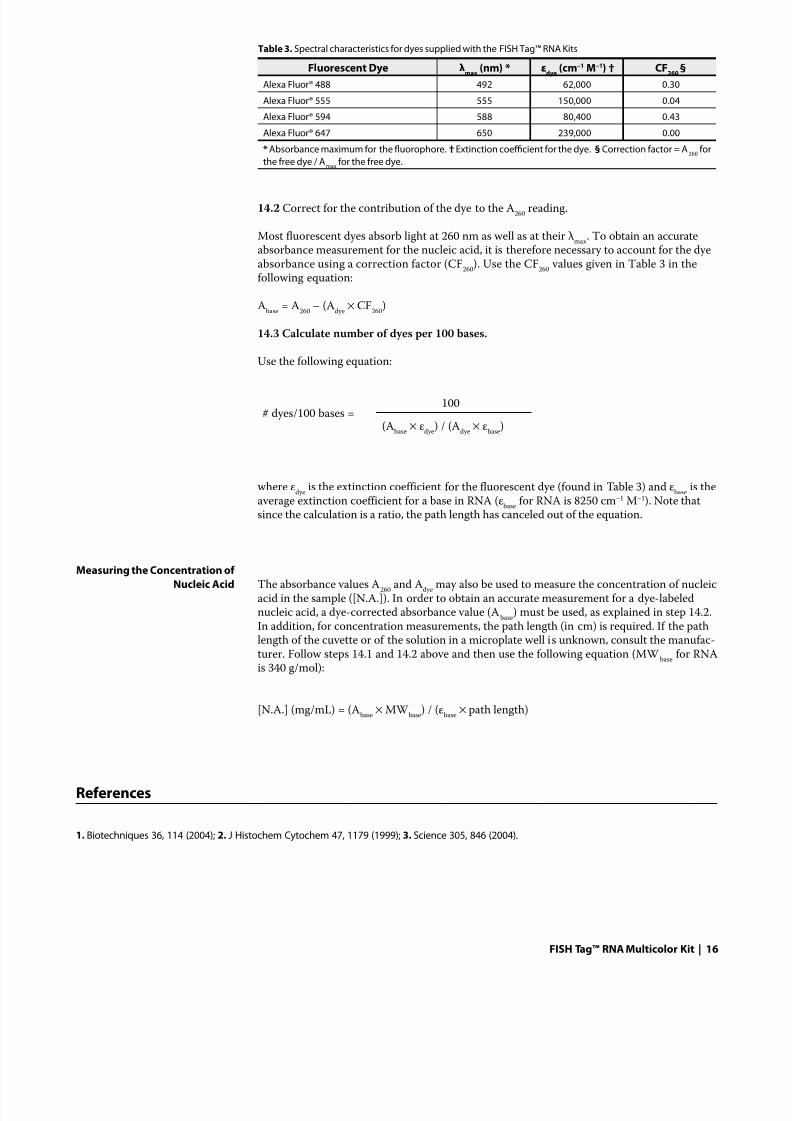
14.2 Correct for the contribution of the dye to the A260
reading.
Most fluorescent dyes absorb light at 260 nm as well as at their λmax
. To obtain an accurateabsorbance measurement for the nucleic acid, it is therefore necessary to account for the dyeabsorbance using a correction factor (CF
260). Use the CF
260 values given in Table 3 in the
following equation:
Abase
= A260
– (Adye
× CF260
)
14.3 Calculate number of dyes per 100 bases.
Use the following equation:
# dyes/100 bases = 100
(Abase
× εdye
) / (Adye
× εbase
)
where εdye
is the extinction coefficient for the fluorescent dye (found in Table 3) and εbase
is theaverage extinction coefficient for a base in RNA (ε
base for RNA is 8250 cm–1 M–1). Note that
since the calculation is a ratio, the path length has canceled out of the equation.
Measuring the Concentration ofNucleic Acid The absorbance values A
260 and A
dye may also be used to measure the concentration of nucleic
acid in the sample ([N.A.]). In order to obtain an accurate measurement for a dye-labelednucleic acid, a dye-corrected absorbance value (A
base) must be used, as explained in step 14.2.
In addition, for concentration measurements, the path length (in cm) is required. If the pathlength of the cuvette or of the solution in a microplate well is unknown, consult the manufac-turer. Follow steps 14.1 and 14.2 above and then use the following equation (MW
base for RNA
is 340 g/mol):
[N.A.] (mg/mL) = (Abase
× MW base
) / (εbase
× path length)
References
1. Biotechniques 36, 114 (2004); 2. J Histochem Cytochem 47, 1179 (1999); 3. Science 305, 846 (2004).
Table 3. Spectral characteristics for dyes supplied with the FISH Tag™ RNA Kits
Fluorescent Dye λmax
(nm) * εdye
(cm–1 M–1) † CF260
§
Alexa Fluor® 488 492 62,000 0.30
Alexa Fluor® 555 555 150,000 0.04
Alexa Fluor® 594 588 80,400 0.43
Alexa Fluor® 647 650 239,000 0.00
* Absorbance maximum for the fluorophore. † Extinction coefficient for the dye. § Correction factor = A260
for
the free dye / Amax
for the free dye.
8/12/2019 Protocol FISH Invitrogen
http://slidepdf.com/reader/full/protocol-fish-invitrogen 17/17
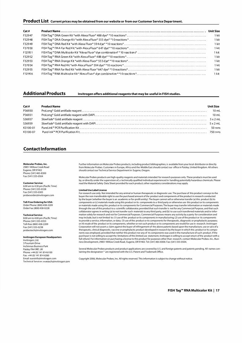
Contact Information
Molecular Probes, Inc.29851 Willow Creek Road
Eugene, OR 97402
Phone: (541) 465-8300Fax: (541) 335-0504
Customer Service: 6:00 am to 4:30 pm (Pacific Time)
Phone: (541) 335-0338
Fax: (541) 335-0305
Toll-Free Ordering for USA:Order Phone: (800) 438-2209
Order Fax: (800) 438-0228
Technical Service: 8:00 am to 4:00 pm (Pacific Time)
Phone: (541) 335-0353
Toll-Free (800) 438-2209
Fax: (541) 335-0238
Invitrogen European HeadquartersInvitrogen, Ltd.
3 Fountain Drive
Inchinnan Business Park
Paisley PA4 9RF, UK
Phone: +44 (0) 141 814 6100
Fax: +44 (0) 141 814 6260
Email: [email protected]
Technical Services: [email protected]
Further information on Molecular Probes products, including product bibliographies, is available from your local distributor or directly
from Molecular Probes. Customers in Europe, Africa and the Middle East should contact our office in Paisley, United Kingdom. All others
should contact our Technical Service Department in Eugene, Oregon.
Molecular Probes products are high-quality reagents and materials intended for research purposes only. These products must be used
by, or directly under the supervision of, a technically qualified individual experienced in handling potentially hazardous chemicals. Please
read the Material Safety Data Sheet provided for each product; other regulatory considerations may apply.
Limited Use Label License
For research use only. Not intended for any animal or human therapeutic or diagnostic use. The purchase of this product conveys to the
buyer the non-transferable right to use the purchased amount of the product and components of the product in research conducted
by the buyer (whether the buyer is an academic or for-profit entity). The buyer cannot sell or otherwise transfer (a) this product (b) its
components or (c) materials made using this product or its components to a third party or otherwise use this product or its components
or materials made using this product or its components for Commercial Purposes. The buyer may transfer information or materials made
through the use of this product to a scientific collaborator, provided that such transfer is not for any Commercial Purpose, and that such
collaborator agrees in writing (a) to not transfer such materials to any third party, and (b) to use such transferred materials and/or infor-
mation solely for research and not for Commercial Purposes. Commercial Purposes means any activity by a party for consideration and
may include, but is not limited to: (1) use of the product or its components in manufacturing; (2) use of the product or its components
to provide a service, information, or data; (3) use of the product or its components for therapeutic, diagnostic or prophylactic purposes;
or (4) resale of the product or its components, whether or not such product or its components are resold for use in research. Invitrogen
Corporation will not assert a claim against the buyer of infringement of the above patents based upon the manufacture, use or sal e of a
therapeutic, clinical diagnostic, vaccine or prophylactic product developed in research by the buyer in which this product or its compo-
nents was employed, provided that neither this product nor any of its components was used in the manufacture of such product. If thepurchaser is not willing to accept the limitations of this limited use statement, Invitrogen is willing to accept return of the product with a
full refund. For information on purchasing a license to this product for purposes other than research, contact Molecular Probes, Inc., Busi-
ness Development, 29851 Willow Creek Road, Eugene, OR 97402. Tel: (541) 465-8300. Fax: (541) 335-0504.
Several Molecular Probes products and product applications are covered by U.S. and foreign patents and patents pending. All names con-
taining the designation ® are registered with the U.S. Patent and Trademark Office.
Copyright 2006, Molecular Probes, Inc. All rights reserved. This information is subject to change without notice.
Product List Current prices may be obtained from our website or from our Customer Service Department.
Cat # Product Name . . . . . . . . . . . . . . . . . . . . . . . . . . . . . . . . . . . . . . . . . . . . . . . . . . . . . . . . . . . . . . . . . . . . . . . . . . . . . . . . . . . . . . . . . . . . . . . . . . . . . . . . . . Unit Size
F32947 FISH Tag™ DNA Green Kit *with Alexa Fluor® 488 dye* *10 reactions* . . . . . . . . . . . . . . . . . . . . . . . . . . . . . . . . . . . . . . . . . . . . . . . . . . . . . . . . . . . . . . 1 kit
F32948 FISH Tag™ DNA Orange Kit *with Alexa Fluor® 555 dye* *10 reactions* . . . . . . . . . . . . . . . . . . . . . . . . . . . . . . . . . . . . . . . . . . . . . . . . . . . . . . . . . . . . 1 kit
F32949 FISH Tag™ DNA Red Kit *with Alexa Fluor® 594 dye* *10 reac tions* . . . . . . . . . . . . . . . . . . . . . . . . . . . . . . . . . . . . . . . . . . . . . . . . . . . . . . . . . . . . . . . . 1 kit
F32950 FISH Tag™ DNA Far Red Kit *with Alexa Fluor® 647 dye* *10 reactions* . . . . . . . . . . . . . . . . . . . . . . . . . . . . . . . . . . . . . . . . . . . . . . . . . . . . . . . . . . . . 1 kit
F32951 FISH Tag™ DNA Multicolor Kit *Alexa Fluor® dye combination* *10 reactions* . . . . . . . . . . . . . . . . . . . . . . . . . . . . . . . . . . . . . . . . . . . . . . . . . . . . . 1 kit
F32952 FISH Tag™ RNA Green Kit *with Alexa Fluor® 488 dye* *10 reactions* . . . . . . . . . . . . . . . . . . . . . . . . . . . . . . . . . . . . . . . . . . . . . . . . . . . . . . . . . . . . . . 1 kitF32953 FISH Tag™ RNA Orange Kit *with Alexa Fluor® 555 dye* *10 reac tions*. . . . . . . . . . . . . . . . . . . . . . . . . . . . . . . . . . . . . . . . . . . . . . . . . . . . . . . . . . . . . 1 kit
F32954 FISH Tag™ RNA Red Kit *with Alexa Fluor® 594 dye* *10 reactions* . . . . . . . . . . . . . . . . . . . . . . . . . . . . . . . . . . . . . . . . . . . . . . . . . . . . . . . . . . . . . . . . 1 kit
F32955 FISH Tag™ RNA Far Red Kit *with Alexa Fluor® 647 dye* *10 reactions* . . . . . . . . . . . . . . . . . . . . . . . . . . . . . . . . . . . . . . . . . . . . . . . . . . . . . . . . . . . . . 1 kit
F32956 FISH Tag™ RNA Multicolor Kit *Alexa Fluor® dye combination* *10 reactions*. . . . . . . . . . . . . . . . . . . . . . . . . . . . . . . . . . . . . . . . . . . . . . . . . . . . . . 1 kit
Additional Products Invitrogen offers additional reagents that may be useful in FISH studies.
Cat # Product Name Unit Size
P36930 ProLong® Gold antifade reagent . . . . . . . . . . . . . . . . . . . . . . . . . . . . . . . . . . . . . . . . . . . . . . . . . . . . . . . . . . . . . . . . . . . . . . . . . . . . . . . . . . . . . . . . . . . . . . . . . 10 mL
P36931 ProLong® Gold antifade reagent with DAPI . . . . . . . . . . . . . . . . . . . . . . . . . . . . . . . . . . . . . . . . . . . . . . . . . . . . . . . . . . . . . . . . . . . . . . . . . . . . . . . . . . . . . . . 10 mLS36937 SlowFade® Gold antifade reagent . . . . . . . . . . . . . . . . . . . . . . . . . . . . . . . . . . . . . . . . . . . . . . . . . . . . . . . . . . . . . . . . . . . . . . . . . . . . . . . . . . . . . . . . . . . . . . . . 5 x 2 mL
S36939 SlowFade® Gold antifade reagent with DAPI . . . . . . . . . . . . . . . . . . . . . . . . . . . . . . . . . . . . . . . . . . . . . . . . . . . . . . . . . . . . . . . . . . . . . . . . . . . . . . . . . . . . . . 5 x 2 mL
K3100-01 PureLink™ PCR Purification Kit . . . . . . . . . . . . . . . . . . . . . . . . . . . . . . . . . . . . . . . . . . . . . . . . . . . . . . . . . . . . . . . . . . . . . . . . . . . . . . . . . . . . . . . . . . . . . . . . . . . 50 rxns
K3100-02 PureLink™ PCR Purification Kit. . . . . . . . . . . . . . . . . . . . . . . . . . . . . . . . . . . . . . . . . . . . . . . . . . . . . . . . . . . . . . . . . . . . . . . . . . . . . . . . . . . . . . . . . . . . . . . . . . . 250 rxns