Embed Size (px)
Citation preview
Proc. Natl Acad. Sci. USAVol. 80, pp. 160-164, January 1983Biophysics
Anionic lipid headgroups as a proton-conducting pathway alongthe surface of membranes: A hypothesis
(proton pumps/proton translocation/chemiosmosis/acid membranes/acid-anions)
THOMAS H. HAINESDepartment of Chemistry, City College of City University of New York, New York, New York 10031
Communicated by Albert L. Lehninger, June 30, 1982
ABSTRACT Evidence has been gathering from several lab-oratories that protons in proton-pumping membranes move alongor within the bilayer rather than exchange with the bulk phase.These experiments are typically conducted on the natural mem-brane in vivo or in vitro or on fragments of natural membrane.Anionic lipids are present in all proton-pumping membranes.Model studies on the protonation state of the fatty acids of lipo-somes containing entrapped water show that the bilayers alwayscontain mixtures of protonated and deprotonated carboxylates.Protonated fatty acids form stable acid-anion pairs with depro-tonatedfatty acids through unusually strong hydrogen bonds. Suchacid-anion dimers have a single negative charge, which is sharedby the four negative oxygens of both headgroups. The two pK val-ues of the resulting dimer will be significantly different from thepK of the monomeric species, so that the dimer will be stable overa wide pH range. It is proposed that anionic lipid headgroups inbiological membranes share protons as acid-anion dimers and thatanionic lipids thus trap and conduct protons along the headgroupdomain of bilayers that contain such anionic lipids-. Protonspumped from the other side ofthe membrane may enter and movewithin the headgroup sheet because the protonation rate of neg-atively charged proton acceptors is 5 orders of magnitude fasterthan that of water. Protons trapped in the acidic headgroup sheetneed not leave this region in order to be utilized by a responsiveproton-translocating pore (a transport protein using the protongradient). Experiments suggest the proton concentration in theheadgroup domain may vary widely and the anionic lipid head-group sheet may therefore function as a proton buffer. -Due to theGouy-Chapman-Stern layer at polyanionic surfaces, anionic lip-ids will also sequester protons from the bulk solution at low andmoderate ionic strengths. At high ionic strength metal cations mayreplace protons sequestered near the headgroups, but these cat-ions cannot substitute for protons in the "proton-conducting path-way," which is based on hydrogen bonding.
Recent experiments in several laboratories have suggested thatprotons that are translocated by proton-pumping membranes,such as those of mitochondria and halobacteria, need not enterthe external bulk water phase in order to be utilized by proton-transporting proteins ofthe membrane, such as ATP synthetase.Notable among these experiments are -those of Michel and Oes-terhelt (1), who made careful measurements of proton-depen-dent ATP synthesis and pH changes in and near illuminated,Halobacterium halobium cells. They found, that the pH is notlowered in the bulk aqueous phase as the protons pumped.bythe purple patches of bacteriorhodopsin are utilized by ATPsynthetase, which is not present in the purple patches but isfound at some distance in red patches of the same plasma mem-brane. They concluded that.the protons do not pass through thebulk aqueous phase but move very close to the membrane sur-
face. This system is unique because the organism is culturedin 6 M NaCl, which implies that the Gouy-Chapman-Sternlayer contains exclusively Na+ and is not rich in H30+.
Other reports (2-18) have described proton movements inrestricted domains or in isolated open membrane sheets notcapable of sustaining a transmembrane pH gradient (whichnevertheless can be energized, ostensibly by generation of anelectrochemical H+ gradient for ATP synthesis) or have dem-onstrated that the pH may not account for the activities attrib-uted to membrane "energization." Taken together, the obser-vations suggest that there may be specific proton-conductingpathways in or on such energy-transducing membranes thatallow them to carry out energy transduction without necessarilyinvolving a pH gradient across the membrane.
Considerations developed in this paper suggest that the an-ionic headgroups of the lipids of energy-transducing mem-branes have the capacity to bind and conduct protons along themembrane surface. It is proposed that anionic groups, such asphosphodiester anions, form acid-anion dimers, two anionssharing a single proton via a hydrogen bond. Evidence is re-viewed suggesting that such putative acid-anion dimers may beprotonated and deprotonated at a high rate, permitting themto constitute a proton-conducting sheet or continuum, rapidlymoving protons through the headgroups (along the surface) fromH+-pumping (e.g., electron transport chain protein) and H+-utilizing systems (ATP synthetases) anywhere in the membrane.Protons trapped in such a proton-conducting continuum can,eventually, equilibrate with the bulk aqueous phase. This mayaccount for the capacity of an imposed pH gradient to be usedexperimentally to generate ATP in proton-pumping membranevesicles.
These suggestions came from experimental studies designedto explain the structure and dynamics ofthe flagellar membraneof the phytoflagellate Ochromonas danica. We had focused onthe protonation state of the primary and secondary sulfates ofthe docosanediol 1, 14-disulfate and the series ofanalogues con-taining one to six chloro groups on an otherwise saturated chain(19-28). This unusual natural membrane is devoid of phospho-lipids. This type of membrane occurs in many freshwater algae(29, 30) and may represent a-diversion in the evolution of bio-logical membranes (31). Natural membranes made of anionicdetergents are surprising, more so when the detergent struc-ture suggests that their anionic groups may be buried in thehydrophobic domain of the bilayer. Elemental analyses of suchmembrane preparations (unpublished data) excluded all poten-tial counterions except for the proton. We therefore institutedstudies on.model bilayers containing alkyl sulfates or fatty acidsin an attempt to understand how protons may stabilize suchmembranes and participate in their function.
Fatty Acid Liposomes.. Unsaturated fatty acid liposomes thatentrap aqueous compartments were described by Gebicki andHicks 10years ago (32). Liposomes may also be made of satu-
160
The publication costs ofthis article were defrayed in part by page charge'payment. This article must therefore be hereby marked "advertise-ment" in accordance with 18 U. S. C. §1734 solely to indicate this fact.
Proc. Natl. Acad. Sci. USA 80 (1983) 161
I.-
0
-ax._
0
0--
4-'
0
3)
ZL
pH
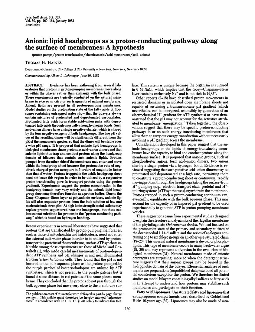
FIG. 1. Titration of a dispersion of 50 mM oleic acid and 10 mM [1-_3C]lauric acid from pH 12 to pH 6. 13C NMR experiments were performedon a Varian CFT 20 20-MHz spectrometer in pulse-Fourier transform mode. Most spectra were obtained after 30,000 transients. The chemical shiftof the labeled carbon is sensitive to the protonation state of the carboxylate. The percent of total chemical shift in ppm is directly proportional tothe percent dissociation of carboxylate. The percent total shift is plotted against pH (A). For comparison, a titration plotting the percent of totalHOl required for complete protonation vs. the pH is shown (v). This titration displays two inflection points, at pH 9.45 and 7.15. The higher inflectionpoint is associated with a transition from micelles to liposomes; the lower inflection point accompanies a phase change from liposomes to oil droplets.This titration is essentially that of Hargreaves and Deamer (33); they confirmed the presence of liposomes by interference microscopy. They andGebicki and Hicks (32) measured the trapped volume of these liposomes. The pK of the fatty acid titration as estimated by NMR is 8.6. Stableliposomes are found between pH 9.5 and 7.2 and are oligolamellar.
rated fatty acids ofshort chain length (C14) at room temperatureor of longer chain lengths (C18) at higher temperatures (33).Such membranes are stable only between pH 7.0 and 9.6. Incollaboration with Michael Heller, the stability of such lipo-somes has been studied as a function of pH and of the proton-ation state of the carboxylate group as measured by 13C NMR,using the chemical shift of the carboxyl carbon (Fig. 1). Twophase changes occur during a titration of fatty acids (50 mM) inwater (33). Above pH 10 the fatty acids are exclusively in themicellar form; belowpH 7 they form oil droplets. Between thesepH values vesicular liposomes are formed; only in this pH rangeare both protonated and nonprotonated species simultaneouslypresent. The liposomes are stable throughout a wide range ofrelative concentrations of each. The titration curve shows twoapparent inflection points, presumably identified with the twophase changes.
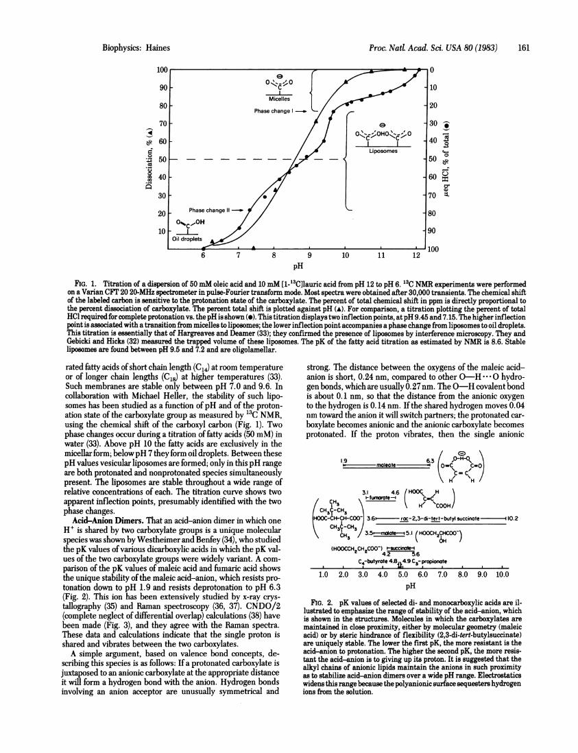
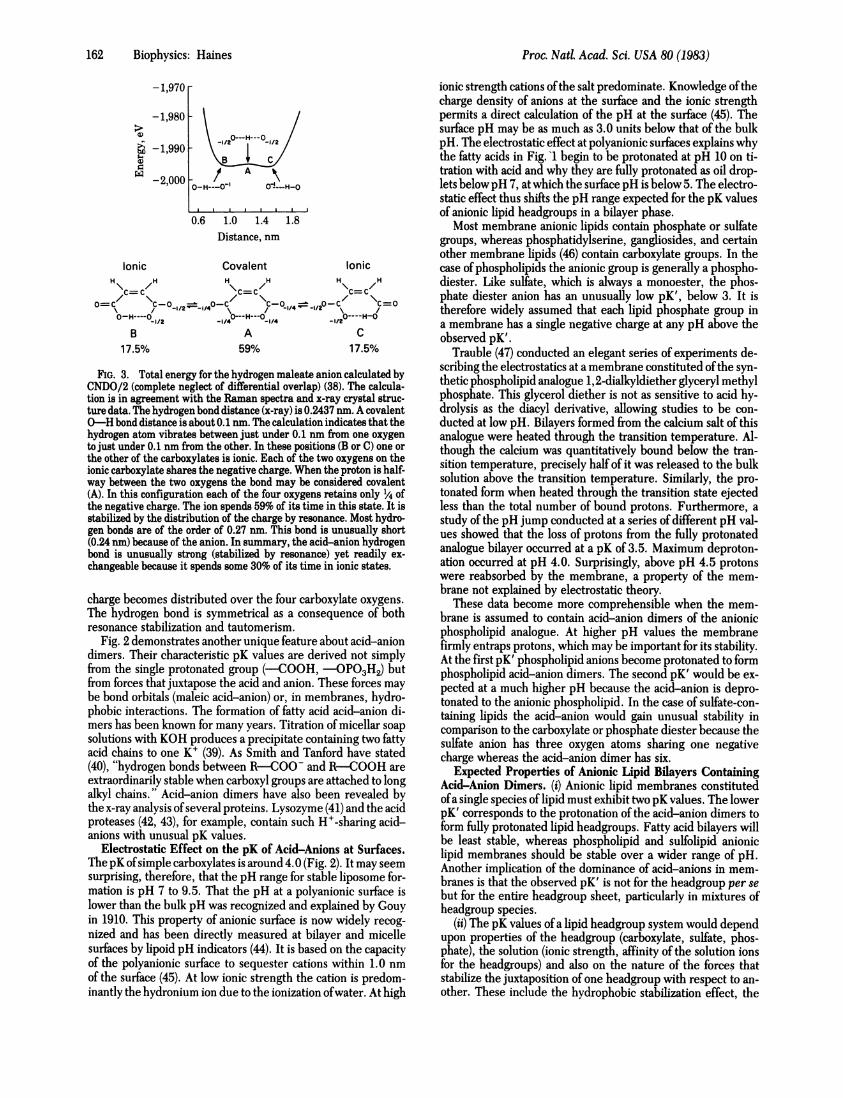
Acid-Anion Dimers. That an acid-anion dimer in which oneH' is shared by two carboxylate groups is a unique molecularspecies was shown by Westheimer and Benfey (34), who studiedthe pK values of various dicarboxylic acids in which the pK val-ues of the two carboxylate groups were widely variant. A com-parison of the pK values of maleic acid and fumaric acid showsthe unique stability of the maleic acid-anion, which resists pro-tonation down to pH 1.9 and resists deprotonation to pH 6.3(Fig. 2). This ion has been extensively studied by x-ray crys-tallography (35) and Raman spectroscopy (36, 37). CNDO/2(complete neglect of differential overlap) calculations (38) havebeen made (Fig. 3), and they agree with the Raman spectra.These data and calculations indicate that the single proton isshared and vibrates between the two carboxylates.A simple argument, based on valence bond concepts, de-
scribing this species is as follows: If a protonated carboxylate isjuxtaposed to an anionic carboxylate at the appropriate distanceit will form a hydrogen bond with the anion. Hydrogen bondsinvolving an anion acceptor are unusually symmetrical and
strong. The distance between the oxygens of the maleic acid-anion is short, 0.24 nm, compared to other O-H- --0 hydro-gen bonds, which are usually 0.27 nm. The O-H covalent bondis about 0.1 nm, so that the distance from the anionic oxygento the hydrogen is 0.14 nm. If the shared hydrogen moves 0.04nm toward the anion it will switch partners; the protonated car-boxylate becomes anionic and the anionic carboxylate becomesprotonated. If the proton vibrates, then the single anionic
1.9 ____ ~~6.3 frHQO \mleote (-o=C_ co
3.1 4.6 HOOC Hefumorote- C.\ ~~H/%COOH
-) 3.6 i- roc-2,3-di-tert-butyl succinote -10.2
3.5-rmale 5.1 (HOOCH2CHCOO-)OH1
(HOOCCH2CH2COO-) t-succinotef-4.2 5.6
C4-butyrote 4.8 49 C3-propionate. . .~ mI.. . . .1.0 2.0 3.0 4.0 5.0 6.0 7.0 8.0 9.0 10.0
pH
FIG. 2. pK values of selected di- and monocarboxylic acids are il-lustrated to emphasize the range of stability of the acid-anion, whichis shown in the structures. Molecules in which the carboxylates aremaintained in close proximity, either by molecular geometry (maleicacid) or by steric hindrance of flexibility (2,3-di-tert-butylsuccinate)are uniquely stable. The lower the first pK, the more resistant is theacid-anion to protonation. The higher the second pK, the more resis-tant the acid-anion is to giving up its proton. It is suggested that thealkyl chains of anionic lipids maintain the anions in such proximityas to stabilize acid-anion dimers over a wide pH range. Electrostaticswidens this range because the polyanionic surface sequesters hydrogenions from the solution.
Biophysics: Haines
Proc. Natd. Acad. Sci. USA 80 (1983)
-1,970
-1,980
6 -1,990a)
w-2,0001
-1/2--- H---0_1/2
/ A \
O-H---O| 0-1---H-0
0.6 1.0 1.4 1.8Distance, nm
Ionic Covalent IonicH\ H H /H H
\H
C=C/C=C/C=C
O~~c \C-O - O-C \~~~~~~C-O0 \12O-O-H----AC/4---H---0are 0----H-0
B A C17.5% 59% 17.5%
FIG. 3. Total energy for the hydrogen maleate anion calculated byCNDO/2 (complete neglect of differential overlap) (38). The calcula-tion is in agreement with the Raman spectra and x-ray crystal struc-ture data. The hydrogen bond distance (x-ray) is 0.2437 nm. A covalent0-H bond distance is about 0.1 rum. The calculation indicates that thehydrogen atom vibrates between just under 0.1 nm from one oxygento just under 0.1 nm from the other. In these positions (B or C) one orthe other of the carboxylates is ionic. Each of the two oxygens on theionic carboxylate shares the negative charge. When the proton is half-way between the two oxygens the bond may be considered covalent(A). In this configuration each of the four oxygens retains only 1X4 ofthe negative charge. The ion spends 59% of its time in this state. It isstabilized by the distribution of the charge by resonance. Most hydro-gen bonds are of the order of 0.27 nm. This bond is unusually short(0.24 nm) because of the anion. In summary, the acid-anion hydrogenbond is unusually strong (stabilized by resonance) yet readily ex-changeable because it spends some 30% of its time in ionic states.
charge becomes distributed over the four carboxylate oxygens.The hydrogen bond is symmetrical as a consequence of bothresonance stabilization and tautomerism.
Fig. 2 demonstrates another unique feature about acid-aniondimers. Their characteristic pK values are derived not simplyfrom the single protonated group (-COOH, -OP03H2) butfrom forces that juxtapose the acid and anion. These forces maybe bond orbitals (maleic acid-anion) or, in membranes, hydro-phobic interactions. The formation of fatty acid acid-anion di-mers has been known for many years. Titration of micellar soap
solutions with KOH produces a precipitate containing two fattyacid chains to one K+ (39). As Smith and Tanford have stated(40), "hydrogen bonds between R-COO- and R-COOH are
extraordinarily stable when carboxyl groups are attached to longalkyl chains." Acid-anion dimers have also been revealed bythe x-ray analysis of several proteins. Lysozyme (41) and the acidproteases (42, 43), for example, contain such H+-sharing acid-anions with unusual pK values.
Electrostatic Effect on the pK of Acid-Anions at Surfaces.The pK ofsimple carboxylates is around 4.0 (Fig. 2). It may seem
surprising, therefore, that the pH range for stable liposome for-mation is pH 7 to 9.5. That the pH at a polyanionic surface islower than the bulk pH was recognized and explained by Gouyin 1910. This property of anionic surface is now widely recog-
nized and has been directly measured at bilayer and micellesurfaces by lipoid pH indicators (44). It is based on the capacityof the polyanionic surface to sequester cations within 1.0 nmof the surface (45). At low ionic strength the cation is predom-inantly the hydronium ion due to the ionization ofwater. At high
ionic strength cations ofthe salt predominate. Knowledge ofthecharge density of anions at the surface and the ionic strengthpermits a direct calculation of the pH at the surface (45). Thesurface pH may be as much as 3.0 units below that of the bulkpH. The electrostatic effect at polyanionic surfaces explains whythe fatty acids in Fig. l begin to be protonated at pH 10 on ti-tration with acid and why they are fully protonated as oil drop-lets belowpH 7, at which the surface pH is below 5. The electro-static effect thus shifts the pH range expected for the pK valuesof anionic lipid headgroups in a bilayer phase.
Most membrane anionic lipids contain phosphate or sulfategroups, whereas phosphatidylserine, gangliosides, and certainother membrane lipids (46) contain carboxylate groups. In thecase of phospholipids the anionic group is generally a phospho-diester. Like sulfate, which is always a monoester, the phos-phate diester anion has an unusually low pK', below 3. It istherefore widely assumed that each lipid phosphate group ina membrane has a single negative charge at any pH above theobserved pK'.
Trauble (47) conducted an elegant series of experiments de-scribing the electrostatics at a membrane constituted ofthe syn-thetic phospholipid analogue 1,2-dialkyldiether glyceryl methylphosphate. This glycerol diether is not as sensitive to acid hy-drolysis as the diacyl derivative, allowing studies to be con-ducted at low pH. Bilayers formed from the calcium salt of thisanalogue were heated through the transition temperature. Al-though the calcium was quantitatively bound below the tran-sition temperature, precisely half of it was released to the bulksolution above the transition temperature. Similarly, the pro-tonated form when heated through the transition state ejectedless than the total number of bound protons. Furthermore, astudy of the pH jump conducted at a series of different pH val-ues showed that the loss of protons from the fully protonatedanalogue bilayer occurred at a pK of 3.5. Maximum deproton-ation occurred at pH 4.0. Surprisingly, above pH 4.5 protonswere reabsorbed by the membrane, a property of the mem-brane not explained by electrostatic theory.
These data become more comprehensible when the mem-brane is assumed to contain acid-anion dimers of the anionicphospholipid analogue. At higher pH values the membranefirmly entraps protons, which may be important for its stability.At the first pK' phospholipid anions become protonated to formphospholipid acid-anion dimers. The second pK' would be ex-pected at a much higher pH because the acid-anion is depro-tonated to the anionic phospholipid. In the case of sulfate-con-taining lipids the acid-anion would gain unusual stability incomparison to the carboxylate or phosphate diester because thesulfate anion has three oxygen atoms sharing one negativecharge whereas the acid-anion dimer has six.
Expected Properties of Anionic Lipid Bilayers ContainingAcid-Anion Dimers. (i) Anionic lipid membranes constitutedofa single species of lipid must exhibit two pK values. The lowerpK' corresponds to the protonation of the acid-anion dimers toform fully protonated lipid headgroups. Fatty acid bilayers willbe least stable, whereas phospholipid and sulfolipid anioniclipid membranes should be stable over a wider range of pH.Another implication of the dominance of acid-anions in mem-branes is that the observed pK' is not for the headgroup per sebut for the entire headgroup sheet, particularly in mixtures ofheadgroup species.
(ii) The pK values of a lipid headgroup system would dependupon properties of the headgroup (carboxylate, sulfate, phos-phate), the solution (ionic strength, affinity of the solution ionsfor the headgroups) and also on the nature of the forces thatstabilize the juxtaposition of one headgroup with respect to an-other. These include the hydrophobic stabilization effect, the
162 Biophysics: Haines

Proc. NatL Acad. Sci. USA 80 (1983) 163
transition temperature ofthe bilayer, the size ofthe headgroup,etc.
(iii) Electrostatic considerations as discussed above wouldpredict that polyanionic membranes will bind protons different-ly at low or moderate ionic strengths on the one hand and at highionic strength on the other. At low or moderate ionic strengthsanionic lipid membranes would be expected to concentrate hy-drogen ions in the Gouy-Chapman-Stern layer so that the sur-face water has a higher proton content than the bulk medium.At high ionic strength anionic lipid membranes would be ex-pected to bind protons exclusively in the acid-anion (headgroup)domain. This may be important in proton-pumping mem-branes, because the surface provides a ready supply of protonseven at high pH values.
(iv) Protons that are trapped by acid-anions on the surfaceofanionic lipid bilayers could move along the membrane surfaceat a significantly greater rate than they might move into theadjacent bulk aqueous phase. If a proton-pumping protein de-livers a proton to the membrane surface then the anionic lipidsheet provides a negatively charged H' acceptor, A-, whereasthe bulk aqueous phase provides H20 as H' acceptor. Eigenand de Maeyer (48) have shown that the rate constant of dis-sociation of HA for simple carboxylates (i.e., the transfer of H+from HA to water) is of the order of 105 sec', whereas the rateof protonation of an anionic carboxylate group is around 1010sec'1. These measurements are based on solution kinetics andthe latter value may be diffusion controlled. Nonetheless theyemphasize that the headgroup anions are 105 better bases (pro-ton acceptors) than is bulk water. Two features of the systemwould be expected to accentuate this difference. The first is thepresence of a Stern layer of cations in the water just above theheadgroups. At low ionic strength this layer is rich in hydroniumions. At high ionic strength the solution cations would furtherdepress the rate of protonation of H20. A second feature of theanionic membrane surface is that its geometry would be ex-pected to enhance the rate ofproton transfer between its anioniclipids. Thus the steric behavior, the orientation of the anions,and the electrostatic interactions all favor a very high rate ofproton transfer between headgroup anions. These features,combined with the capacity of the acid-anions to trap the pro-tons in the membrane surface, would facilitate maximal ratesofproton migration by protonation ofanionic headgroups ratherthan protonation of bulk water. Protonation of an acid-aniondimer would be expected to destabilize it, resulting in a netmovement ofthe entering proton along the surface ofthe mem-brane.
(v) The protonation state of lipid headgroups is independentof the ionic strength. As an example, it has been shown byMichel and Oesterhelt (1) that the movement of protons oc-curs in or on the membrane during proton pumping by intactHalobacterium cells. Such protons apparently do not alter thebulk phase pH and yet they are used for ATP synthesis. Themembrane lipids of Halobacterium halobium, specifically thelipids of the purple patches (49, 50), are completely anionic.Because the organism is cultured in 6 M NaCl its Gouy-Chap-man-Stern layer may be expected to be rich in Na+ at the ec-tolayer and K+ at the endolayer, but poor in H30. On the otherhand, H+ will be sequestered in the headgroup region under-neath the Gouy-Chapman-Stern layer in acid-anion dimers.The anionic headgroup layer may thus function as a proton con-ductor, holding protons firmly yet allowing them to be con-ducted rapidly over some distance from bacteriorhodopsin tothe H+-driven ATP synthetase, without their exchanging withthe bulk phase at high pH.
(vi) Another implication of the formation of acid-anions be-tween negative lipid headgroups relates more generally to the
nature of headgroup interactions. It is generally assumed (51)that headgroups of lipids are mutually repulsive. Rapid ex-change ofprotons between headgroup acid-anions may tend tomaintain a stable sheet so that if lipid molecules in a domain arepushed out of plane they will "snap back. " Such a property ofzwitterionic headgroups was utilized in a general theory ofme-chanical wave propagation in bilayers (52, 53), which empha-sized compaction of the bilayer as an electrostriction (54) as anion traverses it.
Mechanisms of Proton Cycling During ATP Synthesis.Mitchell (55) proposed in 1961 that protons translocated acrossthe mitochondrial membrane from one aqueous phase to an-other could generate apH gradient across the membrane, whichcould act to carry energy from electron transport to ATP syn-thesis or other energy-requiring activities of membranes. Theterm "chemiosmosis" was coined to describe the manner inwhich a bulk phase pH gradient could store and transmit os-motic energy. In the same year Williams (56) proposed thatprotons could be used to transfer energy, but by localized trans-fer of H' within the membrane rather than across it.A major piece of evidence supporting the chemiosmotic hy-
pothesis was the demonstration that experimental impositionofa bulk phase pH gradient across an energy-transducing mem-brane could be used to synthesize ATP (57). The bulk phaseconcept was especially emphasized by the observation that pur-ple membrane patches ofilluminated Halobacterium cells couldpump protons (58, 59). The bacteriorhodopsin patches are phys-ically separated from the ATP-synthesizing enzymes. Thus earlyexperiments suggested that bacteriorhodopsin pumped protonsinto the bulk aqueous phase outside the cell and that the pHgradient so developed across the membrane was utilized todrive the biosynthesis of ATP elsewhere in the membrane.However, the recent observations of Michel and Oesterhelt (1)clearly indicate that when the protons are pumped by bacte-riorhodopsin at very high ionic strength at pH 8.0 they do notenter the bulk phase, and yet they are used for ATP synthesis.The hypothesis herein proposed designates the anionic head-groups as carriers ofprotons laterally along the membrane head-group sheet.An important feature of such a two-dimensional network of
acid-anions is that the concentration of the protonated specieswithin the headgroup domain may vary considerably. This isillustrated in Fig. 1, in which the protonation state ofthe head-groups can vary from 80% protonated to 80% nonprotonated inthe fatty acid liposomes. Thus proton pumping into the head-group domain may produce a local "proton pressure" that canprotonate amino acid anionic R groups of membrane proteinsthat utilize protons to carry out energy-dependent processes.The protons sequestered in the bilayer headgroup region ofenergy-transducing membranes will equilibrate with the bulkphase protons. The latter may, in the context ofthis hypothesis,act not only as a safety valve but also to store energy in a lowintensity form as apH gradient across the membrane. The head-group proton conductor may be regarded as analogous to themultienzyme complexes, which generate and consume meta-bolic intermediates without permitting their diffusive loss intothe bulk aqueous phase.
That anionic lipids are critical for the viability ofcells has beenestablished. Studies on mutants ofEscherichia coli (60) and onmammalian cell lines (61) have shown that a fixed minimal frac-tion ofthe lipids must be anionic although one anionic lipid maybe replaced by another. Many organisms have solely anioniclipids (and uncharged but not zwitterionic lipids). Anionic lipidsconstitute all photosynthetic membranes as well as membranesof the archaebacteria. The latter are believed to be among theearliest organisms in evolution (62). This author knows of no
Biophysics: Haines
Proc. NatL Acad. Sci. USA 80 (1983)
organism that lacks anionic lipids.Kell (10), Malpress (12), and Storey and Lee (15) also have
presented arguments and data in favor of energized protonsmoving close to the membrane in proton-pumping membranes,but they have not considered the anionic lipids as a potentialconductor. Freund and co-workers (63, 64) have observed arapid movement of H' across the surface of metal hydroxides(Mg2+, Ca2+, A13+) in the solid state. This surface property,dependent upon fixed negative charges, is precisely analogousto the fast movement of H' along headgroup anions describedhere. The methods used by Freund to demonstrate this prop-erty have applicability to lipid membranes, perhaps most use-fully in lipid monolayers.The author is grateful to David Deamer, Michael Green, Michael
Heller, Charles Springer, Efraim Racker, and Albert Lehninger forstimulating and critical discussions. The author's work on chlorosulfolip-ids that led to these formulations is supported by National Institutes ofHealth Grant GM 25882, National Science Foundation Grant PCM7815112, and City University of New York-Professional StaffCongress-Board of Higher Education intramural funding.1. Michel, H. & Oesterhelt, D. (1980) Biochemistry 19, 4607-4619.2. Archbold, G. P. R., Farrington, C. L., Lappin, S. A., McKay, A.
M. & Malpress, F. H. (1979) Biochem. J. 180, 161-174.3. Baker, G. M. & Dilley, R. A. (1981) Biochemistry 20, 2307-2315.4. Chow, W. S., Thorne, S. W. & Boardman, N. K. (1978) in Light-
Transducing Membranes, ed. Deamer, D. W. (Academic, NewYork), pp. 253-268.
5. Ernster, L., Juntti, K. & Asauri, K. (1973) J. Bioenerg. 4, 148-159.
6. Ferguson, S. J., Lloyd, W. J. & Radda, G. K. (1976) Biochim.Biophys. Acta 423, 174-188.
7. Higuti, T., Arakaki, N., Niimi, S., Nakashima, S., Saito, R.,Tani, J. & Ota, F. (1980) J. BioL Chem. 255, 7631-7636.
8. Higuti, T., Yokota, M., Arakaki, N., Hattori, A. & Tani, I. (1978)Biochim. Biophys. Acta 503, 211-222.
9. Kamo, N., Muratsugu, M., Kurihara, K. & Kobatake, Y. (1976)FEBS Lett. 72, 247-250.
10. Kell, D. B. (1979) Biochim. Biophys. Acta 549, 55-99.11. Kell, D. B., Clarke, D. J. & Morris, J. G. (1981) FEMS Micro-
bioL Lett. 11, 1-11.12. Malpress, F. H. (1981) J. Theor. BioL 92, 255-265.13. Melandri, B. A., Venturoli, G., De Santis, A. & Bacarini-Melan-
dri, A. (1980) Biochim. Biophys. Acta 592, 38-52.14. Storey, B. T., Scott, D. M. & Lee, C.-p. (1980) J. BioL Chem.
255, 5224-5229.15. Storey, B. T. & Lee, C.-p. (1981) Trends Biochem. Sci. (Pers. Ed.)
6, 166-170.16. Tedeschi, H. (1980) BioL Rev. 55, 171-206.17. Vanderwal, H. N., Vangrondelle, R., Duysens, L. N. M. & Gi-
menezgallego, G. (1981) Photobiochemistry 2, 149-158.18. Vinkler, C., Avron, M. & Boyer, P. D. (1978) FEBS Lett. 96,
129-134.19. Chen, L. L. & Haines, T. H. (1976) J. BioL Chem. 251, 1828-
1834.20. Chen, L. L., Pousada, M. & Haines, T. H. (1976)J. BioL Chem.
251, 1835-1842.21. Mayers, G. L. & Haines, T. H. (1967) Biochemistry 6, 1665-
1671.22. Mayers, G. L., Pousada, M. & Haines, T. H. (1969) Biochemistry
8, 2981-2986.23. Haines, T. H., Pousada, M., Stern, B. & Mayers, G. L. (1969)
Biochem. J. 1131, 565-566.24. Elovson, J. & Vagelos, P. R. (1969) Proc. NatL Acad. Sci. USA 62,
957-963.25. Elovson, J. & Vagelos, P. R. (1970) Biochemistry 16, 3110-3126.26. Haines, T. H. (1973) Annu. Rev. MicrobioL 27, 403-411.27. Haines, T. H. (1974) in Biochemistry of Lipids, MTP Interna-
tional Review, ed. Goodwin, T. W. (Butterworth, Oxford), Vol.4, pp. 271-286.
28. Mercer, E. I. & Davies, C. G. (1975) Phytochemistry 14, 1545-1548.
29. Mercer, E. I. & Davies, C. G. (1979) Phytochemistry 18, 457-462.
30. Haines, T. H. (1982) in CRC Handbook of Microbiology, eds.Laskin, A. I. & Lechevalier, H. (CRC, Boca Raton, FL), 2ndEd., Vol. 5, in press.
31. Nes, W. R. & Nes, G. N. (1980) The Evolution of Lipids,(Plenum, New York).
32. Gebicki, J. M. & Hicks, M. (1973) Nature (London) 243, 232-234.
33. Hargreaves, W. R. & Deamer, D. D. (1978) Biochemistry 17,3759-3768.
34. Westheimer, F. H. & Benfey, 0. T. (1956) J. Am. Chem. Soc. 78,5309-5311.
35. Darlow, S. F. & Cochran, W. (1961) Acta Crystallogr. 14, 1250-1263.
36. Maillols, J., Bardet, L. & Marignan, R. (1968) J. Chim. Phys.Phys.-Chim. Biol 27, 522-528.
37. Maillols, J., Bardet, L. & Marignan, R. (1968) J. Chim. Phys.Phys.-Chim. Biol 27, 529-538.
38. Morita, H. & Nagakura, S. (1972) Theor. Chim. Acta 27, 325-338.
39. Rosano, H. L., Christodolou, A. P. & Feinstein, M. E. (1969)J.Colloid Interface Sci. 29, 335-344.
40. Smith, R. & Tanford, C. (1973) Proc. Natl Acad. Sci. USA 70,289-293.
41. Blake, C. C. F., Mair, G. A., North, A. C. T., Phillips, D. C.& Sarma, V. R. (1967) Proc. R. Soc. London Ser. B 167, 365-377.
42. James, M. N. G., Hsu, I.-N. & Delbaere, L. T. J. (1977) Nature(London) 266, 140-142.
43. Wong, C.-H., Lee, T. J., Lee, T.-Y., Lu, T.-H. & Yang, I.-H.(1979) Biochemistry 18, 1638-1640.
44. Fernandez, M. S. & Fromherz, P. (1977)J. Phys. Chem. 73, 601-607.
45. McLaughlin, S. G. A., Szabo, G. & Eisenman, G. (1971)J. Gen.Physiol 58, 667-683.
46. Brown, A. E. & Elovson, J. (1974) Biochemistry 13, 3476-3482.47. Trauble, H. (1976) in Structure of Biological Membranes, eds.
Abrahamsson, S. & Pascher, I. (Plenum, New York), pp. 509-550.
48. Eigen, M. & de Maeyer, L. (1963) in Technique of OrganicChemistry, eds. Freiss, S. S., Lewis, E. S. & Weissberger, A.(Wiley Interscience, New York), Vol. 8, Part 2, 895-1054.
49. Kushwaha, S. C., Kates, M. & Stoeckenius, W. (1976) Biochem-istry 15, 4956-4961.
50. Wildenauer, D. & Khorana, H. G. (1977) Biochim. Biophys. Acta486, 315-324.
51. Israelachvilli, J. N. (1977) Biochim. Biophys. Acta 469, 221-225.52. Haines, T. H. (1982) Biophys. J. 37, 147-148.53. Haines, T. H. (1979) J. Theor. Biol 80, 307-323.54. Parsegian, V. A. (1975) Ann. N.Y. Acad. Sci. 264, 161-174.55. Mitchell, P. (1961) Nature (London) 191, 144-148.56. Williams, R. J. P. (1961)J. Theor. Biol. 1, 1-17.57. Racker, E. (1976) A New Look at Mechanisms in Bioenergetics
(Academic, New York).58. Racker, E. & Stoeckenius, W. (1974) J. Biol Chem. 249, 662-
667.59. Stoeckenius, W. (1980) Acc. Chem. Res. 13, 337-344.60. Pluschke, G., Hirota, Y. & Overath, P. (1978)J. Biol Chem. 252,
5048-5055.61. Schroeder, F., Perlmutter, J. F., Glaser, M. & Vagelos, P. R.
(1976) J. Biol Chem. 251, 5015-5025.62. Fox, G. E., Stackenbrandt, T., Hespell, R. B., Gibson, J., Man-
iloff, J., Dyer, T. A., Wolfe, R. S., Balch, W. E., Tanner, R. S.,Magrum, L. J., Zablen, L. B., Blakemore, R., Gupta, R., Bonen,L., Lewis, B. J., Stahl, D. A., Luehrsen, K. R., Chen, K. N. &Woese, C. R. (1980) Science 209, 457-463.
63. Freund, F., Wengeler, H. & Martens, R. (1980)1. Chim. Phys.Phys.-Chim. Biol 77, 837-841.
64. Maiti, G. C. & Freund, F. (1981) J. Chem. Soc. Dalton Trans.,949-955.
164 Biophysics: Haines