Embed Size (px)
Citation preview
179
Acta Pharm. 70 (2020) 179–190 Original research paperhttps://doi.org/10.2478/acph-2020-0020
Proton pump inhibitors enhance chemosensitivity, promote apoptosis, and suppress migration of breast cancer cells
Breast cancer is the most common cancer and is the leading cause of cancer deaths among women worldwide. Despite the availability of numerous therapeutics for breast cancer manage-ment, cytotoxicity and emergence of drug resistance are major challenges that limit their benefits. The acidic microenvironment surrounding tumor cells is a common feature inducing cancer cell invasiveness and chemoresistance. Proton pump inhibitors (PPIs) are one of the most commonly prescribed drugs for the treatment of acid-related conditions. PPIs have been reported to exhibit antitumorigenic effects in many cancer types. In this study, the anti-proliferative and anti-migratory effects of PPIs in three breast cancer cell lines; MCF-7, T47D, and MDA-MB-231 cells, have been investigated. In addition, the combined effects of PPIs with anticancer drugs, as well as the mechanism of PPI-mediated anti-proliferative activity were evaluated. The anti-proliferative and combined effects of PPIs were evaluated by MTT assay. Cell migration was assessed using the wound-heal-ing assay. The mechanism of cell death was assessed using an-nexin V-FITC/ propidium iodide staining flow cytometry method. Our results indicated that PPIs treatment significantly inhibited the growth of breast cancer cells in a dose-dependent manner. The antiproliferative activity of PPIs was significantly induced by apoptosis in all tested cell lines. The combined treatment of PPIs with doxorubicin resulted in a synergistic effect in all cell lines, whereas the combined treatment with raloxifene exhibited synergistic effect in T47D cells only and additive effects in MDA--MB-231 and MCF-7 cells. In addition, PPIs treatment signifi-cantly reduced cell migration in MDA-MB-231 cells. In conclu-sion, the addition of PPIs to the treatment regimen of breast cancer appears to be a promising strategy to potentiate the efficacy of chemotherapy and may suppress cancer metastasis.
Keywords: proton pump inhibitors, breast cancer, chemosensi-tivity, apoptosis, metastasis
Breast cancer is the most common type of cancer among female worldwide (1). The incidence rates of breast cancer are increased with age in the majority of cases diagnosed
WOROOD G. IHRAIZ1 MAMOUN AHRAM2 SANAA K. BARDAWEEL1,*
1 Department of Pharmaceutical Sciences, School of Pharmacy University of Jordan, Amman 11942 Jordan2 School of Medicine, The University of Jordan, Amman, Jordan
Accepted July 30, 2019Published online September 8, 2019
* Correspondence; e-mail: [email protected]

180
W. G. Ihraiz et al.: Proton pump inhibitors enhance chemosensitivity, promote apoptosis, and suppress migration of breast cancer cells, Acta Pharm. 70 (2020) 179–190.
after the age of 40 years (2). Unfortunately, despite the continued development of many breast cancer therapies, toxicity and emerging of drug resistance are commonly encoun-tered challenges (3). These phenomena are partly due to the highly heterogeneous nature of the disease at both inter-tumor and intra-tumor level (4).
There is accumulating evidence supporting the role of acidic tumor microenviron-ment in tumor development and progression (5, 6). Metastasis, invasiveness, and chemo-resistance have been shown to be associated with tumor acidic microenvironment (5–7). Moreover, extracellular acidity suppresses the activity of immune cells, like the cytotoxic T lymphocytes and natural killer cells protecting cancer cells from the immune system (8, 9). The deviant extracellular acidity is maintained by the overexpression of different ion/ proton pumps including the vacuolar-H+-ATPase (V-ATPase) pumps, which extrude intra-cellular protons to the extracellular space. Thus, targeting such pump has been considered as a promising strategy to deteriorate cancer cells and reduce tumor metastasis (10).
Numerous pharmacological agents have been used to inhibit the activity of the V-ATPase pump; however, the clinical application is limited due to their cytotoxicity to nor-mal cells (10). Proton pump inhibitors (PPIs), such as lansoprazole and omeprazole, are the most frequently prescribed drugs for the treatment of acid-related disorders. They spe-cifically target the gastric proton pump (H+, K-ATPase pump) that is located in the gastric parietal cells (11). PPIs also act as V-ATPase inhibitors, suggesting that they may affect the tumor acidic microenvironment (10). In fact, they have been shown to inhibit cancer pro-gression and metastasis and reduce drug resistance in several types of cancers including breast, gastric, colorectal, esophageal, and pancreatic cancers (12–16).
Interestingly, it was initially suggested that the anticancer effect of PPIs is primarily related to their ability to block the V-ATPase pump (10). However, several other mecha-nisms that contribute to their anticancer activity have been proposed and investigated including suppression of the Wnt/β-catenin (17), mammalian target of rapamycin (mTOR), hypoxia-inducible factor 1-alpha (HIF-1 α), and others (15, 18, 19).
In the current study, the effects of different PPIs on the viability, migration, and apop-tosis of breast cancer cell lines of different molecular subtypes were evaluated. The ability of PPIs to enhance the antitumor effect of other anticancer drugs was also assessed as well as the underlying molecular mechanism driving the antiproliferative effect of PPIs.
EXPERIMENTAL
ChemicalsLansoprazole, omeprazole, pantoprazole, and raloxifene were purchased from Sigma-
Aldrich (USA) as powder and dissolved in DMSO. They were further diluted with Dul-becco’s Modified Eagle Medium to reach the desired final concentrations immediately before use. Doxorubicin hydrochloride was purchased from Ebewe Untrerach (Austria) as a solution at a concentration of 2 mg mL–1. MTT (3-[4,5-dimethylthiazol-2-yl]-diphenyltet-razolium bromide) was purchased from Sigma-Aldrich (USA).
Cell linesHuman MDA-MB-231, MCF-7, and T47D breast cancer cell lines were obtained from
the American Type Culture Collection (ATCC) (USA). These cells were maintained in high

181
W. G. Ihraiz et al.: Proton pump inhibitors enhance chemosensitivity, promote apoptosis, and suppress migration of breast cancer cells, Acta Pharm. 70 (2020) 179–190.
glucose Dulbecco’s Modified Eagle Medium (DMEM) (Eurobio, French) supplemented with 10 % fetal bovine serum (FBS), 100 U mL–1 penicillin, 100 μg mL–1 streptomycin, and 2 mmol L–1 l-glutamine and were incubated at 37 °C in a 95 % humidified air of 5 % CO2.
Cell viability assay
In order to evaluate cell viability, the MTT (3-[4,5-dimethylthiazol-2-yl]-diphenyltet-razolium bromide) colorimetric assay was performed as previously described (20). Briefly, T47D, MCF-7, and MDA-MB-231 cells were plated in a 96-well plate at a seeding density of 8,000, 7,000, 5000 cells per well, respectively. After 24 h, they were treated with different concentrations of PPIs, doxorubicin, and raloxifene alone or in combination. After 48 h, MTT was added at a working concentration of 5 mg mL–1 and incubated for 3 h. Media were then aspirated, and formazan crystals were solubilized in DMSO. The optical den-sity was measured at 570 nm using the Synergy HTX Multi-Mode Microplate Reader (BioTek Instruments, Winooski, VT, USA). The percentage of cell viability was calculated according to the following equation:
cell viability (%) = (optical density of treated cells/optical density of untreated cells) × 100
Migration assay
MDA-MB-231 cells were plated at a seeding density of 15,000 cells per insert well (Ibidi, Germany) and incubated overnight to grow to confluency. Afterward, inserts were removed, and cells were treated with 10 μg mL–1 of mitomycin C for 2 h. Cells were treated with PPIs at half the IC50 concentration. Images were captured at 0 and 24 h using a micro-scope (BOECO, Germany) coupled with the 5.0 Mega Cmos camera at the magnification of 4 . Cell migration was determined by measuring the wound width using ImageJ software.
Flow cytometry analysis of apoptosis
Cells were plated in 6-well plates at 4 × 10⁵ cells per well for T47D and MCF-7 cells and 3 × 10⁵ cells per well for MDA-MB-231 cells. On the following day, cells were treated with either double IC50 concentration of lansoprazole or doxorubicin as a positive control and incubated for 48 h. Detached cells were collected, combined with the trypsinized attached cells, and centrifuged at 1400 RPM, 4 °C, for 10 min. The cell pellet was resuspended in 200 μL cold 1X binding buffer, and cells were stained with 5 μL Annexin V-FITC and incu-bated for 5 min at room temperature. Propidium iodide was then added to each tube and analyzed immediately. The analysis was performed on BD FACSCanto II flow cytometer (BD Biosciences, USA) using BD FACSDiva software.
Statistical analysis
Data analysis was performed using GraphPad Prism software version 7. The differ-ences between treatments groups were determined by t-test or one-way analysis of vari-ance (ANOVA) followed by Tukey post hoc t-test as appropriate. Data were expressed as mean ± SD and p < 0.05 was considered a statistically significant difference. A non-linear regression analysis was used to calculate IC50 values. The effect of combined treatment of

182
W. G. Ihraiz et al.: Proton pump inhibitors enhance chemosensitivity, promote apoptosis, and suppress migration of breast cancer cells, Acta Pharm. 70 (2020) 179–190.
PPIs and anticancer agents was evaluated by calculating the combination index (CI) value using CompuSyn software based on Chou-Talalay's Combination Index Theorem (21). CI values < 1, =1, and > 1 indicate synergism, additive, and antagonistic effects, respectively. CI values were calculated by using the following equation:
CI = (D1/Dx1) + (D2/Dx2)
where (Dx)1= dose of drug 1 to produce 50 % cell killing alone, (D)1 = dose of drug 1 to produce 50 % cell killing in combination with (D)2, (Dx)2 = dose of drug 2 to produce 50 % cell killing alone, and (D)2 = dose of drug 2 to produce 50 % cell killing in combination with (D)1.
RESULTS AND DISCUSSION
The effect of PPIs treatment on the migration of breast cancer cells
To examine the effect of PPIs on the migration of MDA-MB-231 cells, wound healing assay was performed. As can be noted in (Fig. 1a), complete closure of the wound was observed in the control sample. However, exposure to PPIs treatment significantly inhib-
Fig. 1. Effect of PPIs treatment on MDA-MB-231 cells migration: a) images of the wound at the begin-ning and after 24 h of the treatment period (magnification 4x); b) quantitative analysis of wound closure relative to wound distance at time 0. The experiment was performed in duplicate and the data are expressed as mean ± SD. *p > 0.05 significantly different from control group.
a)
b)

183
W. G. Ihraiz et al.: Proton pump inhibitors enhance chemosensitivity, promote apoptosis, and suppress migration of breast cancer cells, Acta Pharm. 70 (2020) 179–190.
ited the migration of MDA-MB-231 cells by 67, 69 and 68 % for lansoprazole, omeprazole, and pantoprazole, respectively (Fig. 1b).
The V-ATPase expression is strongly associated with the invasive and metastatic ca-pabilities of cancer cells. This was shown to be due to the acidic microenvironment that facilitates the optimal activation and secretion of proteases (22). Many studies have shown that breast cancer cells including MDA-MB-231, MCF-7, and T47D overexpress V-ATPases pumps. However, the activity of this pump appears to be augmented in highly metastatic cells like MDA-MB-231 relative to the poorly metastatic cells like MCF-7 (22, 23). Many pharmacological agents have been used as V-ATPase pump inhibitors like bafilomycin A1 and concanamycin. However, these agents exhibited high cytotoxicity to normal cells as well as to cancer cells. In contrast, PPIs are known to directly inhibit V-ATPase pumps and selectively target cancer cells (24).
The antiproliferative effect of PPIs in different molecular subtypes of breast cancer
The antiproliferative effects on different molecular subtypes of breast cancer cells treated with various doses of PPIs (lansoprazole, omeprazole, and pantoprazole) are shown in (Fig. 2). According to the results, the growth of breast cancer cells was signifi-cantly inhibited after PPIs treatment for 48 h compared to the control, in a dose-dependent manner. Interestingly, the observed antiproliferative effect of PPIs significantly varied be-tween the three examined cell lines with T47D cells being the most responsive to PPIs treatment, whereas MDA-MB-231 cells were the least responsive cells. In contrast, the vi-ability of normal fibroblast cells was not affected compared to the control group (IC50 > 3000 μmol L–1). On the other hand, lansoprazole appears to be more effective in cancer growth inhibition than omeprazole and pantoprazole. The IC50 values for PPIs treatment were 129–299, 201–51 and 256–645 μmol L–1 in T47D, MCF-7, and MDA-MB-231 cells, respec-tively (Table I).
Table I. The 50 % inhibitory concentration (IC50) for PPIs, doxorubicin, and raloxifene in different breast cancer cells
IC50 (μmol mL–1)a
T47D MCF-7 MDA-MB-231
Lansoprazole 129.0 ± 9.8 201.0 ± 9.1 256.5 ± 4.3Omeprazole 222.4 ± 8.8 349.3 ± 23.7 395.6 ± 7.2Pantoprazole 299.6 ± 21.6 514.4 ± 25.9 645.9 ± 8.2Doxorubicin 1.9 ± 0.2 0.9 ± 0.1 2.0 ± 0.2Raloxifene 27.3 ± 1.9 32.9 ± 1.3 25.0 ± 2.1
a mean ± SD
PPIs enhanced apoptosis in breast cancer cells
In order to determine if the anti-proliferative effect of PPI on the cells was due to the induction of apoptosis or necrosis, flow cytometric analyses of cells stained for annexin V or DNA fragmentation were performed. Following 48 h incubation period with the IC50 con-

184
W. G. Ihraiz et al.: Proton pump inhibitors enhance chemosensitivity, promote apoptosis, and suppress migration of breast cancer cells, Acta Pharm. 70 (2020) 179–190.
centration of lansoprazole, the percentages of apoptosis and necrosis were examined. As shown in (Fig. 3), exposure to PPIs significantly increased apoptosis by more than 50 % in the three cell lines examined. On the other hand, the percentages of necrosis were not sig-nificantly affected.
According to De Milito et al. (25), omeprazole and esomeprazole showed an apoptotic effect in the human B cell line, which was mediated through a caspase-independent mecha-nism. In another study, by Scaringi et al. (26), omeprazole induced apoptosis in a time dose-dependent manner in Jurkat cells (T-cells). In the latter cells, apoptosis was dependent on caspases and cysteine cathepsins. Interestingly, Zhang et al. (27), reported that lansoprazole significantly induced apoptosis in breast cancer cells through the ability to provoke cytosolic acidification, lysosomal alkalinization, and reactive oxygen species (ROS) accumulation.
Fig. 2. Antiproliferative effects of PPIs treatment on cell viability of breast cancer cells in: a) T47D, b) MCF-7, c) MDA-MB-231. Data shown represent the mean percentages of cell viability ± SD. Each experiment was performed in triplicate in three independent experiments (N = 3). *p < 0.05 signifi-cantly different from control groups.
a)
b)
c)
185
W. G. Ihraiz et al.: Proton pump inhibitors enhance chemosensitivity, promote apoptosis, and suppress migration of breast cancer cells, Acta Pharm. 70 (2020) 179–190.
Further investigation into the different possible signaling pathways should shed light on the mechanism through which PPIs exert their apoptosis induction effect.
Effects of combined treatment of PPIs and anticancer agents on the viability of breast cancer cells
Doxorubicin is one of the most effective chemotherapeutic agents that is widely used in breast cancer treatment as well as for the treatment of other types of cancer such as lung, multiple myeloma, and thyroid cancers. It induces cytotoxicity effects through inhibition
Fig. 3. Lansoprazole induces apoptosis in breast cancer cells. Breast cancer cells treated with lansopra-zole for 48 h then stained with annexin V-FITC/ PI and analyzed by flow cytometry: a) dot plot for an-nexin V-FITC/ PI staining, where Q3 showed viable cells, Q1 necrotic cells, Q2 late apoptotic, and Q4 early apoptotic and percentages of alive cells, apoptotic cells and necrotic cells in T47D, b) MCF-7, c) MDA-MB-231. The experiment was performed in duplicate and the data are expressed as mean ± SD.
a)
b)
c)

186
W. G. Ihraiz et al.: Proton pump inhibitors enhance chemosensitivity, promote apoptosis, and suppress migration of breast cancer cells, Acta Pharm. 70 (2020) 179–190.
Fig. 4. Effect of anticancer agents on the growth of breast cancer cells: a) antiproliferative effects of doxorubicin treatment on cell viability of breast cancer cells, b) antiproliferative effects of raloxifene treatment on cell viability of breast cancer cells. Data shown represent the mean percentages of cell viability ± SD. Each experiment was performed in triplicate in three independent experiments (N = 3). * p < 0.05 significantly different from control groups.
of topoisomerase II or free radical generation. However, the cytotoxic effects of doxorubi-cin on various organs and tumor resistance limit its clinical usefulness (28). Patel et al. (29), reported that pretreatment with PPIs increased tissue penetration of doxorubicin in multi-layered cell cultures and enhanced its distribution from blood vessels in solid tumors.
Raloxifene, on the other hand, is a second-generation selective estrogen receptor mod-ulator (SERM) that induces estrogen antagonist effect on breast tissues (30). FDA has ap-
a) b)
187
W. G. Ihraiz et al.: Proton pump inhibitors enhance chemosensitivity, promote apoptosis, and suppress migration of breast cancer cells, Acta Pharm. 70 (2020) 179–190.
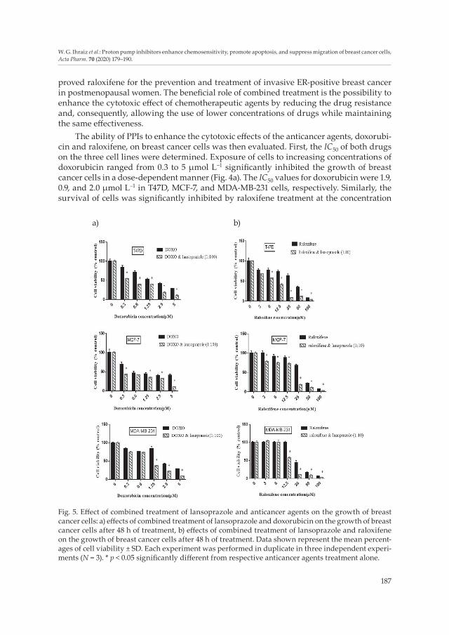
Fig. 5. Effect of combined treatment of lansoprazole and anticancer agents on the growth of breast cancer cells: a) effects of combined treatment of lansoprazole and doxorubicin on the growth of breast cancer cells after 48 h of treatment, b) effects of combined treatment of lansoprazole and raloxifene on the growth of breast cancer cells after 48 h of treatment. Data shown represent the mean percent-ages of cell viability ± SD. Each experiment was performed in duplicate in three independent experi-ments (N = 3). * p < 0.05 significantly different from respective anticancer agents treatment alone.
proved raloxifene for the prevention and treatment of invasive ER-positive breast cancer in postmenopausal women. The beneficial role of combined treatment is the possibility to enhance the cytotoxic effect of chemotherapeutic agents by reducing the drug resistance and, consequently, allowing the use of lower concentrations of drugs while maintaining the same effectiveness.
The ability of PPIs to enhance the cytotoxic effects of the anticancer agents, doxorubi-cin and raloxifene, on breast cancer cells was then evaluated. First, the IC50 of both drugs on the three cell lines were determined. Exposure of cells to increasing concentrations of doxorubicin ranged from 0.3 to 5 μmol L–1 significantly inhibited the growth of breast cancer cells in a dose-dependent manner (Fig. 4a). The IC50 values for doxorubicin were 1.9, 0.9, and 2.0 μmol L–1 in T47D, MCF-7, and MDA-MB-231 cells, respectively. Similarly, the survival of cells was significantly inhibited by raloxifene treatment at the concentration
a) b)

188
W. G. Ihraiz et al.: Proton pump inhibitors enhance chemosensitivity, promote apoptosis, and suppress migration of breast cancer cells, Acta Pharm. 70 (2020) 179–190.
range 3–100 μmol L–1 in a dose-dependent manner (Fig. 4b) with IC50 values 27.3, 32.9, and 25.0 μmol L–1 in T47D, MCF-7, and MDA-MB-231 cells, respectively. Our results demon-strated that the combination of PPIs and doxorubicin significantly increased the cytotoxic effect of doxorubicin and resulted in synergistic inhibition of growth in MDA-MB-231, MCF-7, and T47D cells (CI < 1) (Table II). However, variable results were obtained when PPIs were combined with raloxifene. In T47D cells, combining PPIs to raloxifene signifi-cantly suppressed cell growth compared to respective raloxifene treatment alone with CI values < 1. Interestingly, combined treatment of raloxifene and PPIs in MDA-MB-231 and MCF-7 cells rather demonstrate the additive effect. Thus, the combined effect of PPIs and anticancer drugs is variable and may be affected by the type of cancer cells, anticancer agents, and probably the mechanism that is responsible for drug resistance in the different cell lines.
CONCLUSIONS
PPIs (lansoprazole, omeprazole, and pantoprazole) inhibited the growth of MDA-MB-231, MCF7, and T47D breast cancer cells in a dose-dependent manner. This anti-proli-ferative activity is mediated through the induction of apoptosis. Combined PPIs with doxorubicin resulted in synergistic inhibition of viability in three cell lines, but the combined effect with raloxifene induced a synergistic effect in T47D cells with only additive effect in MDA-MB-231 and MCF-7 cells. In MDA-MB-231 cells, PPIs treatment also significantly inhibited cell migration.
Acknowledgements. – We thank The Deanship of Academic Research at the University of Jordan for supporting this research.
REFERENCES
1. L. A. Torre, F. Bray, R. L. Siegel, J. Ferlay, J. Lortet-Tieulent and A. Jemal, Global cancer statistics, 2012, Ca-Cancer J. Clin. 65 (2015) 87–108; https://doi.org/10.3322/caac.21262
2. C. A. Gabriel and S. M. Domchek, Breast cancer in young women, Breast Cancer Res. 12 (2010) 212–221; https://doi.org/10.1186/bcr2647
Table II. Combination index (CI) values for the combined treatment of PPIs and anticancer agents in breast cancer cell lines. CI < 1, = 1, and > 1 indicate synergism, additive, and antagonistic effects, respectively
Drug combinationCombination index
T47D MCF-7 MDA-MB-231
Doxorubicin + Lansoprazole 0.5 0.3 0.7Doxorubicin + Omeprazole 0.4 0.3 0.7Doxorubicin + Pantoprazole 0.6 0.2 0.5Raloxifene + Lansoprazole 0.7 1.001 1.0Raloxifene + Omeprazole 0.6 1.0 1.08Raloxifene + Pantoprazole 0.7 1.07 1.09
* p < 0.05 significantly different from respective anticancer agents treatment alone.

189
W. G. Ihraiz et al.: Proton pump inhibitors enhance chemosensitivity, promote apoptosis, and suppress migration of breast cancer cells, Acta Pharm. 70 (2020) 179–190.
3. G. Housman, S. Byler, S. Heerboth, K. Lapinska, M. Longacre, N. Snyder and S. Sarkar, Drug resis-tance in cancer: an overview, Cancers 6 (2014) 1769–1792; https://doi.org/10.3390/cancers6031769
4. M. A. Aleskandarany , M. E. Vandenberghe , C. Marchiò , I. O. Ellis, A. Sapino and E. A. Rakha, Tumour heterogeneity of breast cancer: from morphology to personalized medicine, Pathobiology 85 (2018) 23–34; https://doi.org/10.1159/000477851
5. S. Fais, G. Venturi and B. Gatenby, Microenvironmental acidosis in carcinogenesis and metastases: new strategies in prevention and therapy, Cancer Metastasis Rev. 33 (2014) 1095–1108; https://doi.org/10.1007/s10555-014-9531-3
6. E. K. Rofstad, B. Mathiesen, K. Kindem and K. Galappathi, Acidic extracellular pH promotes ex-perimental metastasis of human melanoma cells in athymic nude mice, Cancer Res. 66 (2006) 6699–6707; https://doi.org/10.1158/0008-5472.can-06-0983
7. S. Taylor, E. P. Spugnini, Y. G. Assaraf, T. Azzarito, C. Rauch and S. Fais, Microenvironment acidity as a major determinant of tumor chemoresistance: Proton pump inhibitors (PPIs) as a novel thera-peutic approach, Drug Resist. Updates 23 (2015) 69–78; https://doi.org/10.1016/j.drup.2015.08.004
8. M. Bellone, A. Calcinotto, P. Filipazzi, L. Rivoltini, A. De Milito and S. Fais, The acidity of the tumor microenvironment is a mechanism of immune escape that can be overcome by proton pump in-hibitors, OncoImmunology 2 (2013) e22058; https://doi.org/10.4161/onci.22058
9. D. A. Loeffler, P. L. Juneau and G. H. Heppner, Natural killer- cell activity under conditions reflective of tumor micro-environment, Int. J. Cancer 48 (1991) 895–899; https://doi.org/10.1002/ijc.2910480617
10. S. Fais, A. De Milito, H.You and W.Qin, Targeting vacuolar H+-ATPases as a new strategy against cancer, Cancer Res. 67 (2007) 10627–10630; https://doi.org/10.1158/0008-5472.CAN-07-1805
11. C. A. Stedman and M. L. Barclay, Review article: comparison of the pharmacokinetics, acid suppres-sion and efficacy of proton pump inhibitors, Aliment. Pharmacol. Ther. 14 (2000) 963–978; https://doi.org/10.1046/j.1365-2036.2000.00788.x
12. K. Lindner, C. Borchardt, M. Schopp, A. Burgers, R. Hummel, C. Stock, D. J. Hussey and J. Haier, Proton pump inhibitors (PPIs) impact on tumour cell survival, metastatic potential and chemo-therapy resistance, and affect expression of resistance-relevant miRNAs in esophageal cancer, Exp. Clin. Cancer Res. 33 (2014) 73–85; https://doi.org/10.1186/s13046-014-0073-x
13. J. Patlolla, Y. Zhang, Q. Li, V. Steele and C. Rao, Anti-carcinogenic properties of omeprazole against human colon cancer cells and azoxymethane-induced colonic aberrant crypt foci formation in rats, Int. J. Oncol. 40 (2012) 170–175; https://doi.org/10.3892/ijo.2011.1214
14. A. Udelnow, A. Kreyes, S. Ellinger, K. Landfester, P. Walther, T. Klapperstueck, J. Wohlrab, D. Henne-Bruns, U. Knippschild and P. Würl, Omeprazole inhibits proliferation and modulates autophagy in pancreatic cancer cells, Plos One 6 (2011) e20143; https://doi.org/10.1371/journal.pone.0020143
15. M. Yeo, D. K. Kim, Y. B. Kim, T. Y. Oh, J. E. Lee, S. W. Cho, H. C. Kim and K. B. Hahm, Selective induc-tion of apoptosis with proton pump inhibitor in gastric cancer cells, Clin. Cancer Res. 10 (2004) 8687–8696; https://doi.org/10.1158/1078-0432.CCR-04-1065
16. U. H. Jin, S. O. Lee, C. Pfent and S. Safe, The aryl hydrocarbon receptor ligand omeprazole inhibits breast cancer cell invasion and metastasis, BMC Cancer 14 (2014) 498; https://doi.org/10.1186/1471-2407-14-498
17. Y. Shen, M. Chen, S. Huang and M. Zou, Pantoprazole inhibits human gastric adenocarcinoma SGC-7901 cells by downregulating the expression of pyruvate kinase M2, Oncol. Lett. 11 (2016) 717–722; https://doi.org/10.3892/ol.2015.3912
18. X. Zeng , L. Liu, M. Zheng, H. Sun, J. Xiao, L. Lu, G. Huang, P. Chen, J. Zhang, F. Zhu, H. Li and Q. Duan, Pantoprazole, an FDA-approved proton-pump inhibitor, suppresses colorectal cancer growth by targeting T-cell-originated protein kinase, Oncotarget 7 (2016) 22460–22473; https://doi.org/ 10.18632/oncotarget.7984
19. M. Chen, S. L. Huang, X. Q. Zhang, B. Zhang, H. Zhu, V. W. Yang and X. P. Zou, Reversal effects of pantoprazole on multidrug resistance in human gastric adenocarcinoma cells by down-regulating

190
W. G. Ihraiz et al.: Proton pump inhibitors enhance chemosensitivity, promote apoptosis, and suppress migration of breast cancer cells, Acta Pharm. 70 (2020) 179–190.
the V-ATPases/mTOR/HIF-1a/P-gp and MRP1 signaling pathway in vitro and in vivo, J. Cell. Bio-chem. 113 (2012) 2474–2487; https://doi.org/10.1002/jcb.24122
20. S. K. Bardaweel, H. A. Alsalamat, S. M. Aleidi and R. M. Bashatwah, Glucose deprivation en-hances the antiproliferative effects of oral hypoglycemic biguanides in different molecular sub-types of breast cancer: An in vitro study, Acta Pharm. 68 (2018) 517–524; https://doi.org/10.2478/acph-2018-0031
21. T. C. Chou, Theoretical basis, experimental design, and computerized simulation of synergism and antagonism in drug combination studies, Pharmacol. Rev. 58 (2006) 621–681; https://doi.org/10.1124/pr.58.3.10
22. S. R. Sennoune, K. Bakunts, G. M. Martínez, J. L. Chua-Tuan, Y. Kebir, M. N Attaya and R. Mar-tínez-Zaguilán, Vacuolar H+-ATPase in human breast cancer cells with distinct metastatic poten-tial: distribution and functional activity, Am. J. Physiol. Cell Physiol. 286 (2004) C1443-C1452; https://doi.org/10.1152/ajpcell.00407.2003
23. J. Capecci and M. Forgac, The function of vacuolar ATPase (V-ATPase) a subunit isoforms in inva-siveness of MCF10a and MCF10CA1a human breast cancer cells, J. Biol. Chem. 288 (2013) 32731–32741; https://doi.org/10.1074/jbc.M113.503771
24. E. Iessi, M. Logozzi, D. Mizzoni, R. Di Raimo, C. Supuran and S. Fais, Rethinking the combination of proton exchanger inhibitors in cancer therapy, Metabolites 8 (2018) 2; https://doi.org/10.3390/metabo8010002
25. A. De Milito, E. Iessi, M. Logozzi, F. Lozupone, M. Spada, M. L. Marino, C. Federici, M. Perdicchio, P. Matarrese, L. Lugini, A. Nilsson and S. Fais, Proton pump inhibitors induce apoptosis of human B-cell tumors through a caspase-independent mechanism involving reactive oxygen species, Cancer Res. 67 (2007) 5408–5417; https://doi.org/10.1158/0008-5472.CAN-06-4095
26. L. Scaringi, P. Cornacchione, E. Ayroldi, L. Corazzi, E. Capodicasa, R. Rossi and P. Marconi, Omeprazole Induces apoptosis in jurkat cells, Int. J. Immunopathol. Pharmacol. 17 (2004) 331–342; https://doi.org/10.1177/039463200401700313
27. S. Zhang, Y. Wang and S. J. Li, Lansoprazole induces apoptosis of breast cancer cells through in-hibition of intracellular proton extrusion, Biochem. Biophys. Res. Commun. 448 (2014) 424–429; https://doi.org/10.1016/j.bbrc.2014.04.127
28. C. Cristina, X. S. Renato, C. Susana, C. Sonia, J. O. Paulo, S. S. Maria and I. M. Paula, Doxorubicin: the good, the bad and the ugly effect, Curr. Med. Chem. 16 (2009) 3267–3285; https://doi.org/10.2174/ 092986709788803312
29. K. J. Patel, C. Lee, Q. Tan and I. F. Tannock, Use of the proton pump inhibitor pantoprazole to modify the distribution and activity of doxorubicin: a potential strategy to improve the therapy of solid tumors, Clin. Cancer Res. 19 (2013) 6766–6776; https://doi.org/ 10.1158/1078-0432.CCR-13-0128
30. J. R. Rey, E. V. Cervino, M. L. Rentero, E. C. Crespo, A. O. Alvaro and M. Casillas, Raloxifene: mechanism of action, effects on bone tissue, and applicability in clinical traumatology practice, Open Orthop. J. 3 (2009) 14–21; https://doi.org/10.2174/1874325000903010014