Embed Size (px)
Citation preview
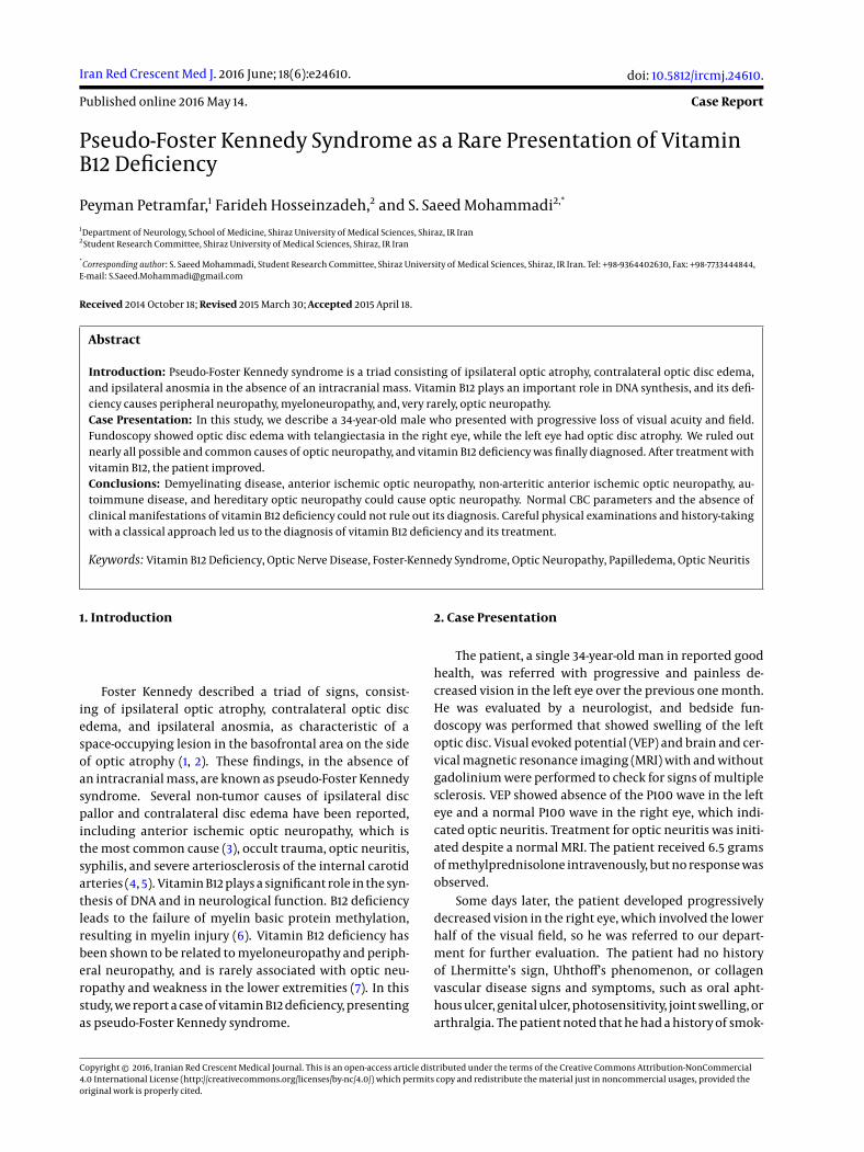
Iran Red Crescent Med J. 2016 June; 18(6):e24610.
Published online 2016 May 14.
doi: 10.5812/ircmj.24610.
Case Report
Pseudo-Foster Kennedy Syndrome as a Rare Presentation of VitaminB12 Deficiency
Peyman Petramfar,1 Farideh Hosseinzadeh,2 and S. Saeed Mohammadi2,*
1Department of Neurology, School of Medicine, Shiraz University of Medical Sciences, Shiraz, IR Iran2Student Research Committee, Shiraz University of Medical Sciences, Shiraz, IR Iran
*Corresponding author: S. Saeed Mohammadi, Student Research Committee, Shiraz University of Medical Sciences, Shiraz, IR Iran. Tel: +98-9364402630, Fax: +98-7733444844,E-mail: [email protected]
Received 2014 October 18; Revised 2015 March 30; Accepted 2015 April 18.
Abstract
Introduction: Pseudo-Foster Kennedy syndrome is a triad consisting of ipsilateral optic atrophy, contralateral optic disc edema,and ipsilateral anosmia in the absence of an intracranial mass. Vitamin B12 plays an important role in DNA synthesis, and its defi-ciency causes peripheral neuropathy, myeloneuropathy, and, very rarely, optic neuropathy.Case Presentation: In this study, we describe a 34-year-old male who presented with progressive loss of visual acuity and field.Fundoscopy showed optic disc edema with telangiectasia in the right eye, while the left eye had optic disc atrophy. We ruled outnearly all possible and common causes of optic neuropathy, and vitamin B12 deficiency was finally diagnosed. After treatment withvitamin B12, the patient improved.Conclusions: Demyelinating disease, anterior ischemic optic neuropathy, non-arteritic anterior ischemic optic neuropathy, au-toimmune disease, and hereditary optic neuropathy could cause optic neuropathy. Normal CBC parameters and the absence ofclinical manifestations of vitamin B12 deficiency could not rule out its diagnosis. Careful physical examinations and history-takingwith a classical approach led us to the diagnosis of vitamin B12 deficiency and its treatment.
Keywords: Vitamin B12 Deficiency, Optic Nerve Disease, Foster-Kennedy Syndrome, Optic Neuropathy, Papilledema, Optic Neuritis
1. Introduction
Foster Kennedy described a triad of signs, consist-ing of ipsilateral optic atrophy, contralateral optic discedema, and ipsilateral anosmia, as characteristic of aspace-occupying lesion in the basofrontal area on the sideof optic atrophy (1, 2). These findings, in the absence ofan intracranial mass, are known as pseudo-Foster Kennedysyndrome. Several non-tumor causes of ipsilateral discpallor and contralateral disc edema have been reported,including anterior ischemic optic neuropathy, which isthe most common cause (3), occult trauma, optic neuritis,syphilis, and severe arteriosclerosis of the internal carotidarteries (4, 5). Vitamin B12 plays a significant role in the syn-thesis of DNA and in neurological function. B12 deficiencyleads to the failure of myelin basic protein methylation,resulting in myelin injury (6). Vitamin B12 deficiency hasbeen shown to be related to myeloneuropathy and periph-eral neuropathy, and is rarely associated with optic neu-ropathy and weakness in the lower extremities (7). In thisstudy, we report a case of vitamin B12 deficiency, presentingas pseudo-Foster Kennedy syndrome.
2. Case Presentation
The patient, a single 34-year-old man in reported goodhealth, was referred with progressive and painless de-creased vision in the left eye over the previous one month.He was evaluated by a neurologist, and bedside fun-doscopy was performed that showed swelling of the leftoptic disc. Visual evoked potential (VEP) and brain and cer-vical magnetic resonance imaging (MRI) with and withoutgadolinium were performed to check for signs of multiplesclerosis. VEP showed absence of the P100 wave in the lefteye and a normal P100 wave in the right eye, which indi-cated optic neuritis. Treatment for optic neuritis was initi-ated despite a normal MRI. The patient received 6.5 gramsof methylprednisolone intravenously, but no response wasobserved.
Some days later, the patient developed progressivelydecreased vision in the right eye, which involved the lowerhalf of the visual field, so he was referred to our depart-ment for further evaluation. The patient had no historyof Lhermitte’s sign, Uhthoff’s phenomenon, or collagenvascular disease signs and symptoms, such as oral apht-hous ulcer, genital ulcer, photosensitivity, joint swelling, orarthralgia. The patient noted that he had a history of smok-
Copyright © 2016, Iranian Red Crescent Medical Journal. This is an open-access article distributed under the terms of the Creative Commons Attribution-NonCommercial4.0 International License (http://creativecommons.org/licenses/by-nc/4.0/) which permits copy and redistribute the material just in noncommercial usages, provided theoriginal work is properly cited.
Petramfar P et al.
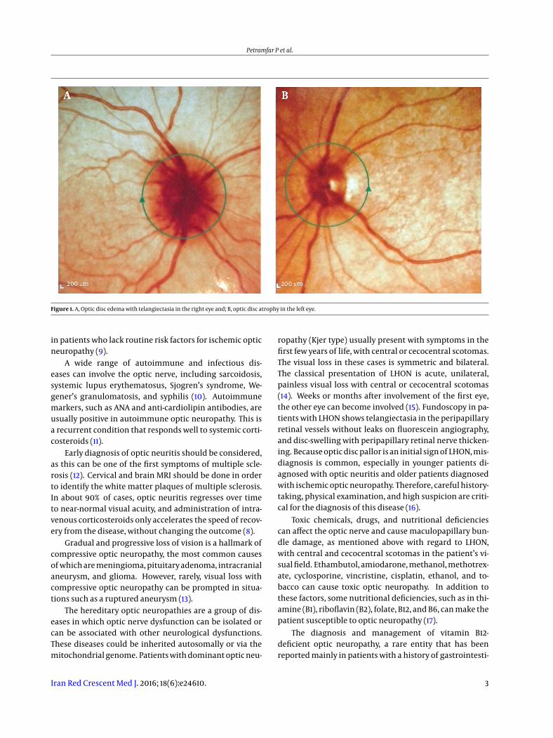
ing crystal methamphetamine until 4 - 5 years prior to hisadmission, but stopped using at that time. There was nohistory of similar or significant diseases in his family. Thepatient’s vital signs and head, ear, nose, throat, chest, heart,abdominal, and neurological exams were all normal, ex-cept for visual acuity. In the left eye, visual acuity was a 3-meter finger-count with a general reduction of sensitivity,and a Marcus Gunn pupil was noted. In the right eye, visualacuity was a 6-meter finger-count, with reduced sensitivityin the inferior visual field. Bedside fundoscopy showed op-tic disc edema with telangiectasia in the right eye, whilethe left eye had optic disc atrophy (Figure 1).
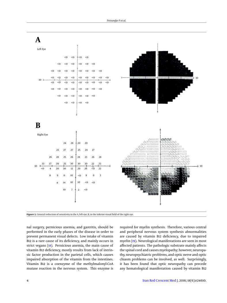
Perimetry was performed, which confirmed the physi-cal examination findings (Figure 2).
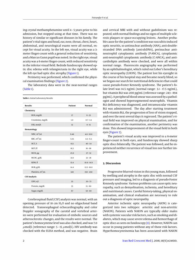
The laboratory data were in the near-normal ranges(Table 1).
Table 1. Initial Laboratory Results
Results Patient Normal
Chemistry
BUN, mg/dL 17 6 - 23
Creatinine, mg/dL 1.0 0.7 - 1.4
ESR, mm/h 12 0 - 15
Hematology
WBC, 103/uL 6.48 4.8 - 10.8
RBC, 106/uL 5.55 4.5 - 6.5
HCT, % 45.1 40 - 54
MCV, fl 81.3 81 - 98
MCH, pg 25.9 27 - 32
MCHC, g/dL 31.9 32 - 36
RDW, fl 15.4 10.8 - 14.9
HGB, g/dL 14.4 13.5 - 18.0
Platelets, 103/uL 328 150 - 450
CSF Analysis
LDH, u/L 25 20 - 72
Protein, mg/dL 35 15 - 60
Sugar, mg/dL 67 50 - 80
Cerebrospinal fluid (CSF) analysis was normal, with anopening pressure of 18 cm H2O and no oligoclonal banddetected. Transesophageal echocardiography and colorDoppler sonography of the carotid and vertebral arter-ies were performed for evaluation of embolic sources andatherosclerotic changes, and the results were normal. Thepatient’s homocysteine level was also checked, and was 7.0µmol/L (reference range: 5 - 15 µmol/L). HIV antibody waschecked with the ELISA method, and was negative. Brain
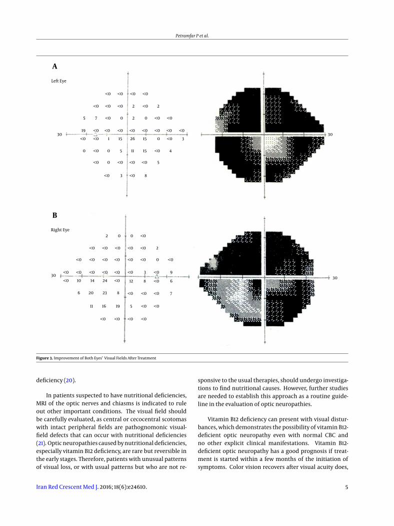
and cervical MRI with and without gadolinium was re-peated, with normal findings and no signs of multiple scle-rosis plaques or space-occupying lesions. Another proba-ble cause for the patient’s condition was vasculitis-inducedoptic neuritis, so antinuclear antibody (ANA), anti-double-stranded DNA antibody (anti-dsDNA), perinuclear anti-neutrophil cytoplasmic antibody (P-ANCA), cytoplasmicanti-neutrophil cytoplasmic antibody (C-ANCA), and anti-cardiolipin antibody were checked, and were all withinnormal range. Fluorescein angiography was performedby an ophthalmologist, which ruled out Leber’s hereditaryoptic neuropathy (LHON). The patient lost his eyesight inthe course of his hospital stay and became nearly blind, sowe began our search for nutritional deficiencies that couldcause pseudo-Foster Kennedy syndrome. The patient’s fo-late level was 14.5 ng/mL (normal range: 3.1 - 17.5 ng/mL),but vitamin B12 was 200 pg/mL (reference range: 243 - 894pg/mL). A peripheral blood smear was assessed by a pathol-ogist and showed hypersegmented neutrophils. VitaminB12 deficiency was diagnosed, and intramuscular vitaminB12 was administered. The day after starting treatmentwith vitamin B12, the progression of loss of vision stopped,and over the next several days it regressed. The patient’s vi-sual field was improved on physical examination, and forconfirmation of the examination, another perimetry wasdone. This showed improvement of the visual field in botheyes (Figure 3).
The patient’s visual acuity was improved to a 6-meterfinger-count in both eyes, and fundoscopy showed sharpoptic discs bilaterally. The patient was followed, and he ex-perienced neither recurrence of visual loss nor further im-provement.
3. Discussion
Progressive blurred vision in this young man, followedby swelling and atrophy in the optic disc with normal CSFpressure and imaging, led to a diagnosis of pseudo-FosterKennedy syndrome. Various problems can cause optic neu-ropathy, such as demyelination, ischemia, and hereditaryand nutritional causes. Careful history-taking, physical ex-amination, and clinical evaluation are necessary to ruleout a diagnosis of optic neuropathy.
Anterior ischemic optic neuropathy (AION) is cate-gorized into two subtypes: arteritic and non-arteritic(NAION). Patients with NAION are typically older maleswith systemic vascular risk factors, such as smoking and di-abetes, which may cause severe edema and hemorrhage ofoptic discs as seen on fundoscopy (8). However, NAION canoccur in young patients without any of those risk factors.Hyperhomocysteinemia has been associated with NAION
2 Iran Red Crescent Med J. 2016; 18(6):e24610.
Petramfar P et al.
Figure 1. A, Optic disc edema with telangiectasia in the right eye and; B, optic disc atrophy in the left eye.
in patients who lack routine risk factors for ischemic opticneuropathy (9).
A wide range of autoimmune and infectious dis-eases can involve the optic nerve, including sarcoidosis,systemic lupus erythematosus, Sjogren’s syndrome, We-gener’s granulomatosis, and syphilis (10). Autoimmunemarkers, such as ANA and anti-cardiolipin antibodies, areusually positive in autoimmune optic neuropathy. This isa recurrent condition that responds well to systemic corti-costeroids (11).
Early diagnosis of optic neuritis should be considered,as this can be one of the first symptoms of multiple scle-rosis (12). Cervical and brain MRI should be done in orderto identify the white matter plaques of multiple sclerosis.In about 90% of cases, optic neuritis regresses over timeto near-normal visual acuity, and administration of intra-venous corticosteroids only accelerates the speed of recov-ery from the disease, without changing the outcome (8).
Gradual and progressive loss of vision is a hallmark ofcompressive optic neuropathy, the most common causesof which are meningioma, pituitary adenoma, intracranialaneurysm, and glioma. However, rarely, visual loss withcompressive optic neuropathy can be prompted in situa-tions such as a ruptured aneurysm (13).
The hereditary optic neuropathies are a group of dis-eases in which optic nerve dysfunction can be isolated orcan be associated with other neurological dysfunctions.These diseases could be inherited autosomally or via themitochondrial genome. Patients with dominant optic neu-
ropathy (Kjer type) usually present with symptoms in thefirst few years of life, with central or cecocentral scotomas.The visual loss in these cases is symmetric and bilateral.The classical presentation of LHON is acute, unilateral,painless visual loss with central or cecocentral scotomas(14). Weeks or months after involvement of the first eye,the other eye can become involved (15). Fundoscopy in pa-tients with LHON shows telangiectasia in the peripapillaryretinal vessels without leaks on fluorescein angiography,and disc-swelling with peripapillary retinal nerve thicken-ing. Because optic disc pallor is an initial sign of LHON, mis-diagnosis is common, especially in younger patients di-agnosed with optic neuritis and older patients diagnosedwith ischemic optic neuropathy. Therefore, careful history-taking, physical examination, and high suspicion are criti-cal for the diagnosis of this disease (16).
Toxic chemicals, drugs, and nutritional deficienciescan affect the optic nerve and cause maculopapillary bun-dle damage, as mentioned above with regard to LHON,with central and cecocentral scotomas in the patient’s vi-sual field. Ethambutol, amiodarone, methanol, methotrex-ate, cyclosporine, vincristine, cisplatin, ethanol, and to-bacco can cause toxic optic neuropathy. In addition tothese factors, some nutritional deficiencies, such as in thi-amine (B1), riboflavin (B2), folate, B12, and B6, can make thepatient susceptible to optic neuropathy (17).
The diagnosis and management of vitamin B12-deficient optic neuropathy, a rare entity that has beenreported mainly in patients with a history of gastrointesti-
Iran Red Crescent Med J. 2016; 18(6):e24610. 3
Petramfar P et al.
A
B
Left Eye
Right Eye
3030
30 30
24 28 20 20
24 27
26232826
30
25
25292723
<0 4 20 30 12
1011
14
10 7
108 10
2
911
29
30 30 22 23
23<0
<0
<0 <0 <0
<0<0<0
<0
<0
<0
<0
<0 <0 <0
<0<0<0
<0 <0<0<0<0 <0<0
<0<0<0<0 <0<0
<0<0<0<0
<0
<0
<0 <0<0<0<0 <0 <0
<0<0<0<0 <0 <0
<0<0<0 <0
<0
<0
<0
<0
<0
26
9 0 3
2826 28
25272723
Figure 2. General reduction of sensitivity in the A, left eye; B, in the inferior visual field of the right eye.
nal surgery, pernicious anemia, and gastritis, should beperformed in the early phases of the disease in order toprevent permanent visual defects. Low intake of vitaminB12 is a rare cause of its deficiency, and mainly occurs instrict vegans (18). Pernicious anemia, the main cause ofvitamin B12 deficiency, mostly results from lack of intrin-sic factor production in the parietal cells, which causesimpaired absorption of the vitamin from the intestines.Vitamin B12 is a coenzyme of the methylmalonyl-CoAmutase reaction in the nervous system. This enzyme is
required for myelin synthesis. Therefore, various centraland peripheral nervous system synthesis abnormalitiesare caused by vitamin B12 deficiency, due to impairedmyelin (19). Neurological manifestations are seen in mostaffected patients. The pathologic substrate mainly affectsthe spinal cord and causes myelopathy; however, neuropa-thy, neuropsychiatric problems, and optic nerve and opticchiasm problems can be involved, as well. Surprisingly,it has been found that optic neuropathy can precedeany hematological manifestation caused by vitamin B12
4 Iran Red Crescent Med J. 2016; 18(6):e24610.
Petramfar P et al.
Left Eye
Right Eye
<0
<0
<05
19
<0
<0
<0<0
<0
<0 <0 <0
<0
<0
<0<0<0
<0
<0
12 8
<0 <0
<0
<0
<0
<0 <0
6
9
7
3
<0
<0
<0
<0
<0
<0
<0
<0 <0 <0<0
2414
6 20
10
23 8
191611 5
<0<0<0<0
<0
<0 <0 <00
2
<0
2 0 0
3
<0
8
5
30 30
30 30
0 0
0
<0 <0 <0 <0
15
15
26
11
15
5
1
<0
<0
<0
<0
<0
3
4
0
<0
<0
<0 <0
<0
<0 <00207
2 2
A
B
Figure 3. Improvement of Both Eyes’ Visual Fields After Treatment
deficiency (20).
In patients suspected to have nutritional deficiencies,MRI of the optic nerves and chiasms is indicated to ruleout other important conditions. The visual field shouldbe carefully evaluated, as central or cecocentral scotomaswith intact peripheral fields are pathognomonic visual-field defects that can occur with nutritional deficiencies(21). Optic neuropathies caused by nutritional deficiencies,especially vitamin B12 deficiency, are rare but reversible inthe early stages. Therefore, patients with unusual patternsof visual loss, or with usual patterns but who are not re-
sponsive to the usual therapies, should undergo investiga-tions to find nutritional causes. However, further studiesare needed to establish this approach as a routine guide-line in the evaluation of optic neuropathies.
Vitamin B12 deficiency can present with visual distur-bances, which demonstrates the possibility of vitamin B12-deficient optic neuropathy even with normal CBC andno other explicit clinical manifestations. Vitamin B12-deficient optic neuropathy has a good prognosis if treat-ment is started within a few months of the initiation ofsymptoms. Color vision recovers after visual acuity does,
Iran Red Crescent Med J. 2016; 18(6):e24610. 5
Petramfar P et al.
and recurrences are rare after the recovery from symptomsis complete. Our patient presented with an unusual mani-festation of vitamin B12 deficiency, but careful physical ex-amination and history-taking with a classical approach ledus to the exact diagnosis and appropriate treatment.
Acknowledgments
We would like to express our special appreciation andthanks to Dr.Nasrin Shokrpour for improving the use of En-glish in the manuscript.
Footnote
Authors’ Contribution: Study concept and design: Pey-man Petramfar, S. Saeed Mohammadi, Farideh Hossein-zadeh; acquisition of data: S. Saeed Mohammadi; analysisand interpretation of data: Peyman Petramfar, and S. SaeedMohammadi; drafting of the manuscript: S. Saeed Mo-hammadi, Farideh Hosseinzadeh; critical revision of themanuscript for important intellectual content: Peyman Pe-tramfar, S. Saeed Mohammadi; study supervision: PeymanPetramfar.
References
1. Kennedy F. Retrobulbar neuritis as an exact diagnostic sign of cer-tain tumors and abscesses in the frontal lobes. Am. J. Med. Sci.1911;142(3):355–68.
2. Kennedy F. A further note on the diagnostic value of retrobulbar neu-ritis in expanding lesions of the frontal lobes: with a report of thissyndrome in a case of aneurysm of the right internal carotid artery. J.Am. Med. Assoc. 1916;67(19):1361–3.
3. Gelwan MJ, Seidman M, Kupersmith MJ. Pseudo-pseudo-FosterKennedy syndrome. J Clin Neuroophthalmol. 1988;8(1):49–52. [PubMed:2972751].
4. Watnick RL, Trobe JD. Bilateral optic nerve compression as amechanism for the Foster Kennedy syndrome. Ophthalmology.1989;96(12):1793–8. [PubMed: 2560156].
5. Schatz NJ, Smith JL. Non-tumor causes of the Foster Kennedy syn-drome. J Neurosurg. 1967;27(1):37–44. doi: 10.3171/jns.1967.27.1.0037.[PubMed: 6028867].
6. Weir DG, Scott JM. Brain function in the elderly: role of vitamin B12and folate. Br Med Bull. 1999;55(3):669–82. [PubMed: 10746355].
7. Saperstein DS, Barohn R. Peripheral neuropathy due to cobal-amin deficiency. Curr Treat Options Neurol. 2002;4(3):197–201. doi:10.1007/s11940-002-0036-y.
8. Beck RW. The optic neuritis treatment trial: three-year follow-up re-sults. Arch Ophthalmol. 1995;113(2):136–7. [PubMed: 7864737].
9. Pianka P, Almog Y, Man O, Goldstein M, Sela BA, Loewenstein A. Hy-perhomocystinemia in patients with nonarteritic anterior ischemicoptic neuropathy, central retinal artery occlusion, and central reti-nal vein occlusion. Ophthalmology. 2000;107(8):1588–92. [PubMed:10919914].
10. Petzold A, Plant GT. Diagnosis and classification of autoim-mune optic neuropathy. Autoimmun Rev. 2014;13(4-5):539–45. doi:10.1016/j.autrev.2014.01.009. [PubMed: 24424177].
11. Frohman L, Turbin R, Bielory L, Wolansky L, Lambert WC, Cook S. Au-toimmune optic neuropathy with anticardiolipin antibody mimick-ing multiple sclerosis in a child.Am JOphthalmol. 2003;136(2):358–60.[PubMed: 12888064].
12. Beck RW, Trobe JD, Moke PS, Gal RL, Xing D, Bhatti MT, et al. High-and low-risk profiles for the development of multiple sclerosiswithin 10 years after optic neuritis: experience of the optic neuritistreatment trial. Arch Ophthalmol. 2003;121(7):944–9. doi: 10.1001/ar-chopht.121.7.944. [PubMed: 12860795].
13. Bulters DO, Shenouda E, Evans BT, Mathad N, Lang DA. Visual re-covery following optic nerve decompression for chronic compres-sive neuropathy. Acta Neurochir (Wien). 2009;151(4):325–34. doi:10.1007/s00701-009-0192-x. [PubMed: 19255716].
14. Kerrison JB. Latent, acute, and chronic Leber’s heredi-tary optic neuropathy. Ophthalmology. 2005;112(1):1–2. doi:10.1016/j.ophtha.2004.10.021. [PubMed: 15629812].
15. Barboni P, Savini G, Valentino ML, Montagna P, Cortelli P, De Ne-gri AM, et al. Retinal nerve fiber layer evaluation by optical coher-ence tomography in Leber’s hereditary optic neuropathy.Ophthalmol-ogy. 2005;112(1):120–6. doi: 10.1016/j.ophtha.2004.06.034. [PubMed:15629831].
16. Newman NJ. Leber hereditary optic neuropathy: somenew observations. J Neuroophthalmol. 2011;31(1):3–5. doi:10.1097/WNO.0b013e31820c511d. [PubMed: 21317730].
17. Hsu CT, Miller NR, Wray ML. Optic neuropathy from folic acid defi-ciency without alcohol abuse.Ophthalmologica. 2002;216(1):65–7. doi:10.1159/000048300. [PubMed: 11901292].
18. Grzybowski A. Problems related to the diagnosis of vitamin B12 de-ficiency optic neuropathy. Acta Ophthalmol. 2014;92(1):e74–5. doi:10.1111/aos.12195. [PubMed: 23826888].
19. Scalabrino G. The multi-faceted basis of vitamin B12 (cobal-amin) neurotrophism in adult central nervous system: Lessonslearned from its deficiency. Prog Neurobiol. 2009;88(3):203–20. doi:10.1016/j.pneurobio.2009.04.004. [PubMed: 19394404].
20. Larner AJ. Visual failure caused by vitamin B12 deficiency optic neu-ropathy. Int J Clin Pract. 2004;58(10):977–8. [PubMed: 15587778].
21. Nicholls P. Formate as an inhibitor of cytochrome c oxidase. BiochemBiophys Res Commun. 1975;67(2):610–6. [PubMed: 1020].
6 Iran Red Crescent Med J. 2016; 18(6):e24610.