Embed Size (px)
Citation preview

11
Psychophysiological Markers of Anxiety Disorders and Anxiety Symptoms
Seung-Hwan Lee and Gewn-Hi Park Psychiatry Department, Inje University, Ilsan Paik Hospital
R. O. Korea
1. Introduction
In anxiety research, relative few psychophysiological studies have been conducted. In this chapter, we presented previous studies that used different psychophsiological markers that can be further utilized in future research. However, there are a few things to be considered when psychophysiological markers are used in anxiety studies, first of which may be genetic factors. Genetic factors influence vulnerability to anxiety disorders. There are several genetic polymorphisms associated with anxiety disorders among which are the serotonin-transporter-linked polymorphic region (5-HTTLPR), the Catechol-O-methyltransferase (COMT), and the brain-derive neurotrophic factor (BDNF) gene variants. We first presented studies that invsestigated the relationship between these genetics variants and anxiety disorders. Also, it has been suggested that anxiety disorders are characterized by abnormal neural activity—amygdala hyperactivity and dysfunctional prefrontal activity—and cognitive bias favoring threat-relevant stimuli (Cisler et al., 2010; McClure et al., 2007; Nitschke et al., 2009; Whalen et al., 2008). We will present different psychophysiological markers that have been used to study dysfunctional neural, serotonergic, cognitive and autonomic activites associated with anxiety disorders. They include: (1) a loudness dependence of the auditory evoked potential (LDAEP) which is proposed to be associated with serotonin activity, (2) various components of the event-related potentials [P1, P2, N300, P3b, early posterior negativity (EPN), late positive potential (LPP), and error-related negativity (ERN)] that reflect altered neural activity in anxiety disorders and (3) the reduced heart rate variability (HRV) which indicates autonomic dysregulation associated with increased sympathetic and decreased vagal control of the heart. Particularly, in this chapter, we introduced the loudness of the auditory evoked potential (LDAEP) as a possible psychophysiological marker that can be utilized in anxiety research. Our previous studies revealed that patients with different subtypes of anxiety disorders produced distinctive LDAEPs and that the LDAEP could play an important role in predicting the efficacy of selective serotonin reuptake inhibitor (SSRI) treatment in anxiety disorders (Park et al., 2010, 2011). We suggest that utilizing the LDAEP along with other various ERP components indicating neural and cognitive dysfunctions associated with anxiety disorders may enhance our understanding of the etiology and maintenance of anxiety disorders. Also, it is important to understand how they interact with each other and with other environmental stressor to reinforce or to exacerbate anxiety symptoms (see Figure 1). Of clinical relevance is whether these psychophysiological markers may play a role in predicting clinical outcome of different treatment.
www.intechopen.com

Anxiety Disorders
204
Fig. 1. Development of anxiety disorders and research methods that can be used in each stage.
2. Genetic predispositions of anxiety symptoms
Anxiety disorders have genetic predispositions and it is critical to consider individuals’ genetic predispositions to develop anxiety prone characteristics and anxiety disorders. Several anxiety-related genetic markers have been identified, one of which includes the serotonin transport promoter polymorphism (5-HTTLPR). The dysfunctional serotonergic system (5-hydroxytryptamine or 5-HT) is known to be implicated in anxiety and fear (Harmer et al., 2004). In human, serotonergic raphe neurons project to different brain structures (e.g., cortex, amygdala, hippocampus) and are associated with integrating various functions including emotion, cognition, motor function, pain, circadian and neuroendocrine functions such as food intake, sleep and sexual activity (Lesch et al., 1997). The 5-HT transporter (5-HTT) plays a vital role in regulating serotonergic neurotransmission by facilitating the reuptake of 5-HT from the synaptic cleft (Lesch et al., 1996; Hariri et al., 2002). Lesch and colleagues (1996, 1997) identified a relatively common polymorphism in the promoter region of the serotonin transporter gene, which results in two different alleles—the short (s) and long (l). Research has showed that the 5-HTTLPR plays a functional role in regulating 5-HTT expression and 5-HTTLPR genotype may modulate 5-HTT expression (Lesch et al., 1996; Lesch et al., 1997). Individuals carrying one or two copies of the s form of 5-HTTLPR were associate with almost 50% reduction in 5-HTT availability compared to individuals homozygous for the l variant (Lesch et al., 1996; Lesch et al., 1997; Hariri et al., 2002). As a result, it has been reported that s-carriers were associate with increased anxiety-related behaviors and greater risk for developing anxiety in stressful life situations compared to individuals homozygous for the l variant (Lesch et al., 1996; Lesch et al., 1997; Hariri et al., 2002). Research also indicated that allelic differences in the 5-HTT may modulate activity of neural circuits (Heinz et al., 2005; Pezawas et al., 2005). Health individuals carrying one or two copies of the s allele showed greater activity in the amygdala in response to fearful stimuli (Heinz et al., 2005). Also, individuals with the s allele showed altered coupling of prefrontal-amygdala feedback circuit—which may lead to dysfunctional amygdala regulation in response to fearful stimuli—compared to individuals homozygous of the l allele (Heinz et al., 2005; Pezawas et al., 2005).
www.intechopen.com

Psychophysiological Markers of Anxiety Disorders and Anxiety Symptoms
205
Another gene associated with anxiety disorders is the Catechol-O-methyltransferase (COMT) genetic variation (Funke et al., 2005). COMT is an enzyme that plays an important role in the metabolism of brain dopamine and norepinephrine (Gadow et al., 2009). The COMT gene can be found in chromosome 22q11 and contains several single nucleotide polymorphisms (SNPs) that are functionally important. For example, Val158Met (rs4680)—associated with encoding either valine (Val) or methionine (Met)—plays an important role in modulating COMT activity in the prefrontal cortex (Harrison et al., 2008). Current evidences suggest that Val158Met may be associated with anxiety disorders, particularly bipolar disorder, via controlling dopamine activity in the prefrontal cortex (Funke et al.,2005). Individuals with Val158 homozygous showed 35-50% higher COMT activity in human dorsolateral prefrontal cortex than those with Mel158 homozygous (Harrison et al., 2008). Although Met-COMT is considered to play an important role in the development of bipolar disorder, there exists evidence that Val-COMT is also associated with bipolar disorder (Funke et al., 2005). Lastly, the brain-derived neurotrophic factor (BDNF) gene variants are suggested to be linked with anxiety disorders (Chen et al., 2006; Gadow et al., 2009). BDNF is a neurotrophin that plays an important role in neuronal growth, differentiation, and synaptic plasticity (Chen et al., 2006; Gadow et al., 2009; Rasmusson et al., 2002). BDNF is also associated with learning and memory and modulates aggression (Rasmusson et al., 2002). It has been reported that BDNF plays a role in mediating effects of stress (Rasmusson et al., 2002). Reduced BDNF expression in the hippocampus was observed in response to stress, which may contribute to hippocampus-dependent memory deficits and the decreases in hippocampal volume associated with patients with post-traumatic stress disorder (PTSD; Rasmusson et al., 2002). Recent studies investigated the relationship between a SNP in the BDNF gene, Val66Met, and psychopathology, which yielded conflicting results (Jiang et al., 2005). In an animal study, when exposed to stress, BDNF Met/Met mice demonstrated anxiety-related behaviors and were not responsive to the antidepressant, fluoxetine (Chen et al., 2006). Studies showed that Val66 allele were associated with greater neuroticisim scores, suggesting that individuals with the Val allele may have increased risk for developing anxiety or depression (Hünnerkopf et al., 2007; Sen et al., 2003). However, no association between BDNF Val66Met genotypes and neuroticisim was observed in Asian female participants (Tsai et al., 2004). BDNF Met66 allele was found to be a risk allele for anxiety and depression (Jiang et al., 2005) whereas other found it to be a protective allele for obsessive-compulsive disorder (OCD; Hall et al., 2003). In sum, BDNF may be related to anxiety disorder though it is yet to be determined which specific variant is responsible for the pathogenesis of anxiety disorders (Gadow et al., 2009). So far, we have presented different candidate genetic marker of anxiety disorders. To have better understanding of anxiety disorders, it would be important to identify genetic polymorphisms associated with anxiety disorders and study together with psychophysiological markers which will be discussed later.
3. Neurophysiological and cognitive characteristics of anxiety disorders
Anxiety disorders are characterized by altered neurophysiological and cognitive functions. Various psychophysiological markers used in anxiety research may reflect these altered neural and cognitive characteristics of anxiety disorders. Here, we briefly described altered neural activity and dysfunctional cognitive processing of threat-relevant information in people with anxiety disorders.
www.intechopen.com

Anxiety Disorders
206
3.1 Amygdala hyperactivity and reduced PFC function
Amygdala hyperactivity has been considered as an important neural characteristic of anxiety disorders (Bar-Haim et al., 2005; Dannlowski et al., 2007; McClure et al., 2007). Previous functional brain imaging studies indicated that anxiety disorders are linked with hyperactivity of the amygdala in response to anxiety provoking tasks (e.g., public speaking), fear-conditioning, and viewing face pictures with emotionally negative expressions (Phan et al., 2006). Also, functional magnetic resonance imaging (fMRI) studies revealed that effective treatment produced significantly reduced amygdala activity in patients with social phobia (Kilts et al., 2006; Furmark et al., 2002). Patients characterized with greater amygdala hyperactivity before treatment responded better to treatments such as SSRI medications and cognitive behavioral therapy (CBT; McClure et al., 2007). More recently, fMRI studies also revealed that high-trait anxiety individuals showed reduced activity in anterior cingulate cortex (ACC)—associated with conflict monitoring—and lateral prefrontal cortex (lateral PFC)—related with attentional control over threat-relevant distractors (Bishop et al., 2004). Patients with generalized anxiety disorder (GAD) patients who showed greater activation in ACC in response to or in anticipation of aversive pictures were associated with better treatment outcome (Nitschke et al., 2009; Whalen et al., 2008). In addition, GAD patients who showed greater activation in the ventrolateral prefrontal cortex—associated with emotional regulation by exerting inhibitory control over subcortical structures—had fewer anxiety symptoms (Monk et al., 2006). Therefore, anxiety disorders may be characterized by amygdala hyperactivity—associated with heighten sensitivity to motivation-relevant stimuli—and reduced PFC activity—resulting in the lack of top-down attentional control and emotional regulation (Bishop et al., 2004; McClure et al., 2007; Monk et al., 2006).
3.2 Cognitive characteristics of anxiety disorders
It has been well established that anxious individuals exhibit attentional biases toward threat-relevant stimuli (Cisler et al., 2010; Mathews et al., 1997). Anxiety-related attentional biases are typically observed in three different ways: (1) faster detection to threat-relevant stimuli relative to nonthreat stimuli, (2) difficulties in disengaging attention away from threat stimuli (sustained attention to threat stimuli), and (3) attentional avoidance of where threat-relevant stimuli are presented (Cisler et al., 2010; Fox et al., 2001; Koster et al., 2004; 2005). Initially, anxiety-related attentional biases were studied in the emotional Stroop task. In the task, threat or neutral words were written in different colors and participants were instructed to name the color of ink while ignoring the meaning of the word (Cisler et al., 2010). Research showed that high-trait anxiety participants were slower to name the color of ink in which threaten words were written, particularly to items relevant to their anxiety conditions. For instance, Vietnam combat veterans with Post-Traumatic Stress Disorder (PTSD) and without PTSD were asked to name the color of PTSD-related words,OCD-related words, positive words, and neutral words (McNally et al., 1993). Veterans with PTSD took longer to name the color in which PTSD-related words were written relative to veterans without PTSD who showed no difference in reaction times across different types of the words. Similarly, slower responses were observed to read threat-relevant words in patients with GAD (Mathews & MacLeod, 1985) and panic disorder (McNally et al., 1994). Highly anxious non-clinical participants showed negativity bias even though they could not consciously aware of the presence of threat-relevant stimuli (MacLeod and Rutherford, 1992). Moreover, performances on the masked emotional Stroop task predicted later emotional reactivity (Den Hout et al., 1995).
www.intechopen.com

Psychophysiological Markers of Anxiety Disorders and Anxiety Symptoms
207
Fox and her colleagues (2001) adapted Posner’s spatial cuing task to systematically
investigate different components of anxiety-related attention bias (Posner & Petersen, 1990).
In the spatial cuing task, a target is preceded by a cue which can be either “central” (e.g., an
arrow presented at the center of the display pointing one of two peripheral boxes in which
the target would subsequently appear), or “peripheral” (e.g., an abrupt luminance of one of
the peripheral boxes; Posner et al., 1990; Posner et al., 2007; Bartolomeo et al., 2001). When
the cue appears on the same display that a target appears, it is considered to be a valid trial
because the cue correctly predicts the location in which the target appears. However, when
the cue fails to predict where the target will appear, it is considered to be invalid. In valid
trials the detection of targets is facilitated (cuing benefits) whereas the detection of targets is
delayed in invalid trials (cuing costs). In the modified emotion spatial cuing task,
emotionally charged words or pictures were used as cues (Fox et al., 2001; Vuilleumier et al.,
2009). If high-trait anxiety participants automatically draw their attention to threat-relevant
stimuli, their response to targets following valid threat-relevant cues should be fast (Fox et
al., 2001). If high-trait anxiety participants have a difficulty in disengaging their attention
from threat-relevant stimuli, then their responses to targets following invalid threat-related
stimuli should be slow (Fox et al., 2001). In the first study, Fox et al. (2001) found that
attentional disengagement from threat-relevant words took longer compared to neutral or
positive words. However, there was no difference between high and low-trait anxiety
participants. In the second study, they used schematic faces with ‘angry,’ ‘neutral,’ and
‘happy’ facial expressions. When a cue duration period increased to 250 ms, only high
anxious individuals showed the delayed attentional disengagement from angry faces. Fox
and colleague (2001) suggested that regardless of anxiety level, people were initially drawn
to threat-relevant information for a brief period of time (about 100 ms). However, low-trait
anxiety participants were capable of quickly disengaging their attention from threat-
relevant, positive and neutral information whereas high-trait anxiety participants were less
successful in disengaging their attention from the location in which threat-related
information was presented.
Recent studies suggested that high-trait anxiety is associated with the “vigilance-avoidance”
attentional patterns in response to threat-relevant information, which may account for the
maintenance of anxiety (Koster et al., 2006). The initial attention to mildly and highly
threatening information may trigger the constant processing of fearful information and
interfere with engaging in goal-directed behaviors (Koster et al., 2006). Faster detection of
mildly and highly threatening information trigger anxious conditions in high-trait anxiety
participants, which reinforces them to avoid threatening information in an attempt to reduce
anxiety (Koster et al., 2006). However, this strategic attentional avoidance may not be a good
coping strategy because it can lead to a failure of habituation to threaten stimuli and
constantly remind of fear (Koster et al., 2006). Koster and colleagues (2005, 2006) used
neutral, mildly and highly threatening pictures as cues and reported that when picture cues
were presented at 100 ms, high-trait anxiety participants exhibited faster attentional
engagement and slower attentional disengagement in response to highly threatening
pictures compared to low-trait anxiety participants. However, when picture cues were
presented longer, 500 ms, high-trait anxiety participants showed slower attentional
engagement to highly and mildly threatening cues, which may suggest attentional
avoidance to highly threatening stimuli (Koster et al., 2006).
www.intechopen.com

Anxiety Disorders
208
Interestingly, there is evidence suggesting that the 5-HTTLPR s allele—genetic predispositions to anxiety—may be related with anxiety-related cognitive bias (Beck, 2008). Twenty-seven psychiatric inpatients who carried s allele of the promoter region of the 5-HTTPR showed anxiety-related attentional biases favoring threat-relevant words compared to patients with homozygous for the l variant (Beevers et al., 2007). Fox and colleagues recently showed that healthy individuals with homozygous for the l allele were characterized by a marked avoidance of negative stimuli and a vigilance for positive stimuli whereas s-allele carriers did not show such protective attentional pattern (Fox et al., 2009). Therefore, allelic variation of the promoter region of the serotonin transporter gene may influence the way in which an individual processes emotional materials.
4. The event-related potentials (ERP) components used to study anxiety disorders
Several ERP components have been used to study serotonergic, neural, and cognitive
dysfunctions associated with anxiety disorders. We provided underlying mechanisms of the
loudness dependence of the auditory evoked potential (LDAEP) and presented studies that
used the LDAEP in anxiety research. Also, researchers have studied other ERP components
that reflect neural mechanisms of cognitive bias toward threat-relevant stimuli commonly
observed in patients with anxiety disorders.
4.1 Loudness of the auditory evoked potential
It has been proposed that the LDAEP—which measures activity in the primary auditory
cortex in response to different tone intensities—indicates the functioning of the central
serotonergic system (Hegerl et al., 1993; Juckel et al., 1999). More specifically, the LDAEP is
defined as the linear regression slope calculated from five amplitudes of N1/P2 components
in response to increasing five auditory tones (Senkowski et al., 2003; see Figure 2). Research
has indicated that the LDAEP is inversely related to central serotonergic activity: a stronger
LDAEP indicates lower serotonergic neurotransmission and vice versa (Juckel et al., 1999,
Park et al. 2010).
Initial evidence that linked the LDAEP and the serotonergic system came from animal
studies (O’Neill et al., 2008). Administrating quipazine maleate—a 5-HT2 receptor agonist—
reduced the amplitude of N1/P2 components whereas administrating spiperone—a 5-HT1A
receptor antagonist—increased the N1/P2 amplitude in rats (Manjarrez et al., 2005).
Administrating the precursor of 5-HT, L-tryptophan, was associated with the reduced
amplitude of N1/P2 components (Manjarrez et al., 2005). Other studies showed that the
LDAEP was inversely correlated with the concentration of 5-hydroxyindoleacetic acid (main
metabolite of serotonin) in cerebrospinal fluid (von Knorring et al, 1981). High scores in the
serotonin syndrome scale were associated with weaker LDAEPs and vice versa in
depressive patients who underwent SSRI treatment (Hegerl et al., 1998). Individuals scored
high on measures of sensation seeking and impulsiveness were associated with stronger
LDAEP—potentially indicating reduced serotonergic function (Brocke et al., 2000; Hegeral
et al., 1995). So far, there have been three studies that investigated the relationship between
allelic variants of the seroton transporter gene and the LDAEP. It has been found that
individuals homozygous for the l variant exhibited lower LDAEP (Gallinat et al., 2003)
whereas others (Strobel et al., 2003; Hensch et al., 2006) reported that the l allele carriers
www.intechopen.com

Psychophysiological Markers of Anxiety Disorders and Anxiety Symptoms
209
showed stronger LDAEP. These studies provide evidence that the LDAEP is linked with the
serotonin transporter polymorphism although there are inconsistencies in predicting
directional changes in serotonin neurotransmission (O’Neill et al., 2008).
Fig. 2. Subject 1 possesses a steep LDAEP (a large increase in N1/P2 amplitude with increasing loudness) whereas subject 2 shows a shallow LDAEP (a small increase in N1/P2 amplitude with increasing loudness). Adapted from O’Neill et al., 2008. (Reprinted by permission of author and publisher).
There is evidence suggesting that the LDAEP is also modulated by dopaminergic
neurotransmission (Juckel et al., 2008). High intensity dependence of auditory and visual
evoked potentials were associated with low levels of dopamine metabolites (i.e., homvanillic
acid) in cerebrospinal fluid and urine (Pogarell et al., 2004; O’Neill et al., 2008). Pogarell and
colleagues (2004) used single photon emission computed tomography (SPECT) and showed
that the LDAEP was positively associated with both serotonin and dopamine transporter
availabilities in patients with OCD. Recently, Juckel and colleagues (2008) found that the
LDAEP is also related with the genetic variants of the cCOMT—implicated in the
inactivation of synaptic dopamine (Stein et al., 2005; Samochowiec et al., 2004). Reduced
COMT activity caused by genetic polymorphisms was associated with a weaker LDAEP
(Juckel et al., 2008).
The LDAEP has been utilized to study dysfunctional serotonergic and dopaminergic activity
in patients with GAD (Senkowski et al., 2003), PTSD (Park et al., 2010), schizophrenia (Juckel
et al., 2003) or depression (Gallinat et al., 2000). Recently, Park and colleagues (2010)
compared the results of the LDAEP in a variety of psychiatric patients including GAD,
PTSD, panic disorder, bipolar depression, major depressive disorder (MDD), and
schizophrenia. Individuals with different anxiety disorders produced different strengths of
LDAEPs (see Fig. 3), which raised a possibility that the differences in the LDAEP may be
associated with distinctive anxiety symptoms and cognitive impairments that characterize
different subtypes of anxiety disorders. However, further studies are needed to explicate the
relationship between different anxiety disorders and the strength of LDAEP.
www.intechopen.com

Anxiety Disorders
210
Fig. 3. Comparison of the LDAEP among panic disorder (PD), generalized anxiety disorder (GAD), and post-traumatic stress disorder (PTSD). Note: Adapted and modified from Park et al. (2011). (Reprinted by permission of publisher).
Furthermore, evidence suggests that the LDAEP can serve as a predictor of responses to
SSRI treatment in GAD patients, which phenomenon was previously observed in patients
with MDD (Gallinat et al., 2000; Linka et al., 2004). Research has indicated that a strong
LDAEP— indicating lower serotonergic activity and turnover rate—is associated with a
favorable response to SSRI treatment in patients with depression (Gallinat et al., 2000; Linka
et al., 2004). Our study (Park et al., 2011) also showed that GAD patients who had stronger
LDAEPs responded favorably to SSRI (escitalopram) treatment. Park et al. (2011) also
confirmed this finding in the brain source activity of the LDAEP, which was measured using
a standardized low resolution brain electro-magnetic tomography (sLORETA; Pascual-
Marqui, 2002). GAD patients who showed greater loudness dependence source activity in
the primary auditory cortex were more responsive to the escitalopram treatment (see Fig. 4).
The study (Park et al., 2011) implies that source activity of the LDAEP, as well as the cortical
LDAEP, may play an important role in predicting the efficacy of SSRI treatment in GAD
patients, which can be used in clinical settings.
4.2 Other ERP components that are associated with neural mechanisms of cognitive bias
Because of high-temporal resolution, the event-related potentials (ERP) method can be
particularly useful to capture the time course of anxiety-related attentional biases (Mercade
et al., 2009) and has been utilized in some studies (Holmes, et al., 2008; Bar-Haim et al.,
2005). Researchers have studied the P1—the first major positive voltage deflection occurring
50-165 ms after the onset of stimulus—and the early posterior negativities (EPNs)—showing
negative deflection over the temporo-occipital sites within a time window between 150 (200)
and 300 ms—in response to emotional stimuli (Schupp et al. 2006; see Figure 5). A number of
studies reliably found that negative faces elicited the significantly higher amplitude of the
www.intechopen.com

Psychophysiological Markers of Anxiety Disorders and Anxiety Symptoms
211
Fig. 4. The source activities of ERP components (N1-P2) on the primary auditory cortex were
obtained by the sLORETA program. And the linear regression slope of the source activities
of five ERP components was considered as the sLORETA - loudness dependence of the
auditory evoked potential (sLORETA-LDAEP). The sLORETA-LDAEP showed the
significant positive correlation [Spearman’s rho = 0.54 (p=0.005)] with symptom response
rates measured by Hamilton Anxiety Rating Scale (HAM-A) in patients with generalized
anxiety disorder treated with escitalopram. Note: Adapted and modified from Park et al.
(2011). (Reprinted by permission of publisher).
exogenous visual P1 component (Pourtois et al., 2005; Holmes et al., 2008). Greater EPNs were
observed in response to negative faces compared to positive and neutral faces over lateral
posterior and occipital areas (Schupp et al., 2004b; Holmes et al., 2008). Similarly, Bublatzky
and colleagues (2010) reported that emotional pictures elicited an enlarged EPN. The P1 and
the EPNs are particularly useful because they are associated with the preferential attentional
processing of negative facial expressions in extrastriate visual cortex, which is extensively
modulated by the amygdala and attentional networks in fronto-parietal cortex (Homles et al.,
2008). Also, the EPN may be associated with a transient stage at which motivationally relevant
stimuli are ‘tagged’ for prioritized processing, which can be useful to study preferential
processing of motivationally relevant stimuli commonly observed in patients with anxiety
disorders (Cuthbert et al. 2000; Michalowski et al. 2009; Schupp et al., 2006). In Holmes et al.
(2008), high-trait anxiety participants who performed a variant of the emotional spatial cuing
task showed an enhanced early P1 component to fearful faces relative to neutral faces at
occipital electrode sites (Holmes et al., 2008). However, high-trait anxiety participants did not
show greater lateral parietal negativities (or EPNs) in response to fearful faces, which may
indicate attentional avoidance following the initial attentional vigilance or the failure to
differentiate threat from non-threat stimuli (Holmes et al., 2008). In contrast, Wieser and
colleagues (2010) reported that that healthy participants who expected to make public
speaking produced enhanced EPN responses for angry facial expressions, suggesting
enhanced early perceptional processing of angry faces.
Furthermore, Bar-Haim and colleagues (2005) used the emotional spatial cuing task similar to Fox et al., (2001) and reported high-trait anxiety participants had greater amplitudes of
www.intechopen.com

Anxiety Disorders
212
the P2 component—the following major positive voltage deflection occurring 50-165 ms after the onset of stimulus—to angry faces compared to low-trait anxiety participants. Greater P2 components indicate greater attentional allocation to threat-related stimuli, which is frequently exhibited in individuals with anxiety disorders (Holmes et al., 2008). Rossignol and colleagues (2005) used an emotional oddball task in which participants were
asked to detect an infrequent emotional target stimulus among a series of frequent neutral
standard stimuli and provided evidence that anxiety modulated the amplitude of N300, a
negative deflexion peaking at central sites around 300ms, and the latency of the P3b
component, occurring at parietal situes around 450 ms. N300 is associated with affective
processing and P3b reflects decision-making and premotor response-related stage
(Rossignol et al., 2005). High-trait anxiety participant showed the reduced amplitude of
N300 suggesting that they were less able to process the emotional content of faces. However,
faster detection of infrequent emotional target stimuli as suggested by faster reaction time
latency and the P3b latency indicated that high-trait anxiety participants made fast decisions
and preparation for actions (Rossignol et al., 2005).
Fig. 5. Illustration of P1, P2, P3, early posterior negativity (EPN), and late positive potential (LPP) in response to fearful target stimuli in the odd ball task.
Another ERP component that has been used to study the abnormal processing of threat-relevant stimuli in anxious individuals is the late positive potential (LPP)—which becomes apparent approximately 300 ms after stimulus onset (Hajcak et al., 2010). Research indicated that greater LPPs are observed in response to emotional compared to neutral stimuli and do not habituate to stimuli that are repeatedly presented (Cuthbert, Schupp, Bradley, Birbaumer, & Lang, 2000; Dillon, Cooper, Grent-’t-Jong, Woldorff, & LaBar, 2006; Foti, & Hajcak, 2008; Hajcak, Dunning, & Foti, 2007; Hajcak & Nieuwenhuis, 2006; Hajcak & Olvet, 2008; Moser, Hajcak, Bukay, & Simons, 2006; Schupp et al., 2000; Schupp, Cuthbert et al., 2004a; Schupp, Ohman et al., 2004b; Schupp, Junghöfer, Weike, & Hamm, 2003). The LPP is associated with sustained attention toward, and elaborative processing of, motivationally relevant stimuli, which phenomenon is commonly observed in patients with anxiety
www.intechopen.com
Psychophysiological Markers of Anxiety Disorders and Anxiety Symptoms
213
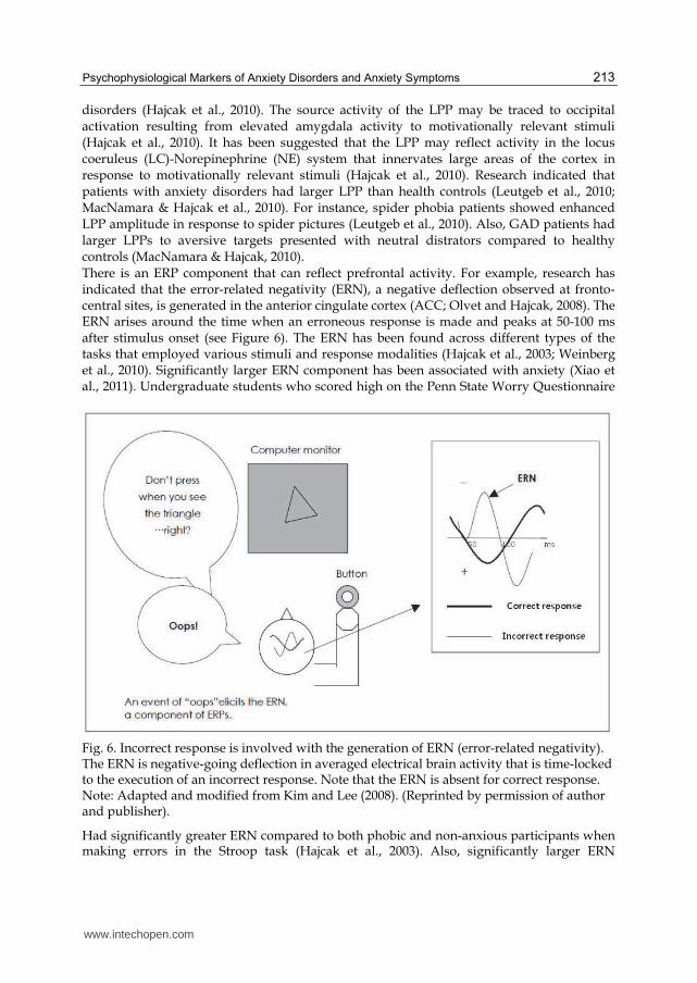
disorders (Hajcak et al., 2010). The source activity of the LPP may be traced to occipital activation resulting from elevated amygdala activity to motivationally relevant stimuli (Hajcak et al., 2010). It has been suggested that the LPP may reflect activity in the locus coeruleus (LC)-Norepinephrine (NE) system that innervates large areas of the cortex in response to motivationally relevant stimuli (Hajcak et al., 2010). Research indicated that patients with anxiety disorders had larger LPP than health controls (Leutgeb et al., 2010; MacNamara & Hajcak et al., 2010). For instance, spider phobia patients showed enhanced LPP amplitude in response to spider pictures (Leutgeb et al., 2010). Also, GAD patients had larger LPPs to aversive targets presented with neutral distrators compared to healthy controls (MacNamara & Hajcak, 2010). There is an ERP component that can reflect prefrontal activity. For example, research has indicated that the error-related negativity (ERN), a negative deflection observed at fronto-central sites, is generated in the anterior cingulate cortex (ACC; Olvet and Hajcak, 2008). The ERN arises around the time when an erroneous response is made and peaks at 50-100 ms after stimulus onset (see Figure 6). The ERN has been found across different types of the tasks that employed various stimuli and response modalities (Hajcak et al., 2003; Weinberg et al., 2010). Significantly larger ERN component has been associated with anxiety (Xiao et al., 2011). Undergraduate students who scored high on the Penn State Worry Questionnaire
Fig. 6. Incorrect response is involved with the generation of ERN (error-related negativity). The ERN is negative-going deflection in averaged electrical brain activity that is time-locked to the execution of an incorrect response. Note that the ERN is absent for correct response. Note: Adapted and modified from Kim and Lee (2008). (Reprinted by permission of author and publisher).
Had significantly greater ERN compared to both phobic and non-anxious participants when making errors in the Stroop task (Hajcak et al., 2003). Also, significantly larger ERN
www.intechopen.com

Anxiety Disorders
214
amplitudes were observed in individuals who had high scores on negative affect scores on negative affect compared to those with low scores on NA (Hajcak et al., 2004). Administrating anxiolytics such as oxazepam and alprazolam has decreased ERN amplitudes (Johannes et al., 2001; Riba et al., 2005). Several studies showed that patients with OCD have been associated with enhanced ERN amplitudes which did not change after successful treatment (Endrass et al., 2010; Gehring et al., 2000; Hajcak et al., 2008). A recent study showed that GAD patients showed larger ERN relative to healthy controls (Weinberg et al., 2010). Olvet and Hajcak (2008) have proposed that error monitoring activity of the ACC indexed by the ERN may play a role as an endophenotype of anxiety disorders.
5. Heart rate variability
Patients with anxiety disorders are characterized by reduced heart rate variability (HRV; Ost et al., 1984; Friedman, 2007). HRV—which refers to the differences in beat-to-beat alterations in heart rate—indicates the dynamic interplay between sympathetic and parasympathetic (vagal) activity in the heart (Berntson et al., 1997; Task Force of the European Society of Cardiology and the North American Society of Pacing and Electrophysiology, 1996; Thayer and Lane, 2000). Under resting conditions, the heart is predominately under the control of the parasympathetic activity (Levy, 1971). Although the intrinsic heart rate is approximately 105 beat per minute, resting heart rate is only 6o-80 beats per minute, indicating that the heart is under the strong vagal control (“vagal dominance”; Brownley et al., 2000; Ellis & Thayer, 2010) There is converging evidence suggesting that reduced HRV—indicating autonomic dysregulation associated with elevated sympathetic activity and reduced vagal activity of the heart—is commonly observed in patients with panic disorder, GAD, and even children of patients with panic disorder (see Friedman, 2007 for a review; Friedman and Thayer, 1998; Srinivasan et al., 2002). Research has indicated that reduced HRV is associated with predispositions to various physical and psychological illnesses and considered to be a predictor of all-cause mortality (Thayer and Lane, 2000). Also, reduced HRV is associated with reduced attentional control, poor emotional regulation, decreased response to various stimuli, and antisocial behavior in adolescents (Mezzacappa et al., 1997; Friedman, 2007; Thayer et al., 2000). Patients who experienced severe panic attacked frequently exhibited reduced HRV in various situations (e.g., quiet rest, shock avoidance, face immersion, isoproterenol infusions; Friedman et al., 1993; Yeragani et al., 1995). High trait anxiety was associated with autonomic dysreguation indexed by reduced HRV (Miu et al., 2009). Thus, reduced HRV may play a critical role in the development of anxiety disorders and can be considered as an important endophenotype of anxiety (Friedman, 2007; Crişan et al., 2009). In a recent review of the literature, Friedman (2007) provided a number of studies that linked reduced HRV with a variety of anxiety disorders over the past 15 years and provided the summary of the Neurovisceral Integration model of anxiety.
5.1 The Neurovisceral Integration Model of anxiety
Several researchers have identified neural networks in the central nervous system associated with autonomic, emotional, and cognitive self-regulatory responses, one of which is the central autonomic network (CAN; Benarroch, 1993; Thayer and Lane, 2000; 2002). The structures of the CAN include the anterior cingulate, insula, ventromedial prefrontal cortices, the central nucleus of the amygdala, the paraventricular and related nuclei of the
www.intechopen.com

Psychophysiological Markers of Anxiety Disorders and Anxiety Symptoms
215
hypothalamus, the periaquaductual gray matter, the parabrachial nucleus, the nucleus of the solitary tract (NTS), the nucleus ambiguous, the ventrolateral medulla, the ventromedial medulla, and the medullary tegmental field (for reviews, Ellis and Thayer, 2010; Thayer and Lane, 2002). Reciprocally interconnected components in the CAN allow information to flow in both top-down and bottom-up fashions (Theyer and Lane, 2000, 2002). Also, these components are loosely connected so that it is possible to recruit additional structures when it is necessary to make specific behavioral adaptations (Thayer and Lane, 2000, 2002). In the CAN, the prefrontal cortical structures—including the orbitofrontal cortex (OFC) and
medial prefrontal cortex (mPFC)—modulates cardiovascular, autonomic, and endocrine
responses by exerting tonic inhibitory control on subcortical structures, such as the central
nucleus of the amygdala (Thayer and Lane, 2000; Thayer et al., 2009). In emotionally
stressful and threatening situations, sympathoexcitatory subcortical circuits are activated to
produce the fight or flight response (Thayer and Lane, 2000; Thayer and Seigle, 2002).
However, the constant activation of sympathoexcitatory subcortical activity is not suitable
for many other situations and will eventually wear and tear the system down. Therefore,
sympathoexcitatory subcortical activity has to be controlled, and research indicated that the
PFC—typically associated with governing higher cognitive functions—is involved in
regulating activity in sympathoexcitatory subcortical circuits (Thayer et al., 2009). In
emotionally stressful situations, the prefrontal cortex disinhibits its inhibitory control over
sympathoexcitatory subcortical circuits and lets subcortical neural structures such as the
amygdala make autonomic and prepotent responses to situations (Thayer et al., 2009).
However, when identifying certain safety signals, the PFC exerts its inhibitory control over
sympathoexcitatory subcortical circuits and makes responses that are appropriate for
contexts in which the signals occur. Therefore, the inhibitory cortical-subcortical circuit is
critical for self-regulation (Thayer and Lane, 2000; 2002). On the other hand, the breakdown
of the inhibitory mechanism can result in the constant activation of sympathoexcitatory
subcortical circuits, which may lead to emotional, attentional, and autonomic dysreguation
and the emergence of perseverative behavior such as worry. Neurochemically, tonic
inhibitory control is achieved by γ-aminobutyric acid (GABA) activity within the NTS and
reduced GABA activity has been also associated with anxiety, perseverative cognition and
poor habituation (Malizia et al., 1998; Friedman, 2007; Thayer and Lane, 2000).
The complex neutral circuits link the inhibitory cortical and subcortical pathways with the
heart via the vagus nerve (for reviews, see Benarroch, 1993; Ellis & Thayer, 2010; Thayer et al.,
2009). High vagally-mediated HRV indicates an exertion of good cognitive, emotional and
physiological self-regulation, which is associated with highly integrated cortical-subcortical
circuits (Thayer et al., 2009). In contrast, low HRV is associated with poor regulatory systems
resulting from the lack of prefrontal regulation over subcortical activity, which is behaviorally
manifested through hypervigilance, the failure to habituate to novel, nonthreatening stimuli,
and perseverative behavior such as worry (Friedman, 2007; Thayer et al., 2009).
There exists the relationship between serotonergic activity and HRV. HRV is positively related to serotonin turnover (DePetrillo et al., 1999). Individuals carrying s allele of serotonin transporter gene showed reduced HRV compared to l-homozygotes (Crişan et al., 2009). Lower levels of serotonin induced by tryptophan depletion were associated with reduced HRV in remitted depressed patients (Booij et al., 2005). Thayer and Ruiz-Padial (2006) suggested that reduced HRV may also indicate the altered coupling or breakdown of the connectivity between the PFC and the amygdala typically exhibited in individuals
www.intechopen.com

Anxiety Disorders
216
carrying s allele (Heinz et al., 2005; Pezawas et al., 2005). Increased vagally-mediated HRV was observed after SSRI treatment in panic patients (Tucker et al., 1997) and PTSD (Cohen et al., 2000).
6. Conclusion
In this chapter, we presented potential psychophysiological markers that have been studied in anxiety research. A weak LDAEP may indicate dysfunctional serotonergic activity associated with patients with anxiety disorders. Patients with different subtypes of anxiety disorders may be associated with distinctive LDAEPs and that the LDAEP may serve as a predictor of SSRI treatment outcome in patients with GAD. Also, other ERP components (e.g., P1, P2, N300, P3b, EPN, LPP and ERN) have been useful in studying attentional biases favoring threat-relevant stimuli in highly anxious individuals. Patients with anxiety disorders typically show reduced HRV—indicating autonomic dysfunction caused by elevated sympathetic and reduced vagal cardiac control (Friedman, 2007). Accumulated evidence suggests that reduced HRV—linked with anxiety disorders—may contribute to poor emotional and cognitive self-regulation, the failure of inhibition at multiple levels and perseverative cognition such as worry (Friedman, 2007). The genetic, cognitive, and psychophysiological characteristics may interact with each other and with other environmental factors such as stress to produce or exacerbate different symptoms in anxiety disorders. Future work may benefit from integrating these markers and exploring the relationships with genetic predispositions to the psychopathology. Of clinical importance is whether these potential psychophysiological markers may play a role in predicting the efficacy of psychological and medical treatments, which is yet to be determined.
7. Acknowledgment
The studies on the LDAEP were supported by a grant of the Korea Healthcare Technology R&D Project, Ministry for Health, Welfare & Family Affairs, Republic of Korea (no. A08-4117-A22023-08N1-00010A).
8. References
Bar-Haim, Y.; Lamy, D. & Glickman, S. (2005). Attentional bias in anxiety: A behavioral and ERP study. Brain and Cognition, Vol.59, No.1, (October 2005), pp. 11-22, ISSN 0278-2626
Bartolomeo, P.; Sieroff, E.; Decaix, C. & Chokron, S. (2001). Modulating the attentional bias in unilateral neglect: the effects of the strategic set. Experimental Brain Research, Vol. 137, No.3-4, (April 2001), pp. 432-444, ISSN 0014-4819
Beck, A. T. (2008). The evolution of the cognitive model of depression and its neurobiological correlates, American Journal of Psychiatry, Vol.165, No.8, (August 2008), pp. 969-977, ISSN 0002-953X
Beevers, C. G.; Gibb, B. E.; McGeary, J. E. & Miller, I. W. (2007). Serotonin transporter genetic variation and biased attention for emotional word stimuli among psychiatric inpatients. Journal of Abnormal Psychology, Vol.116, No. 1, (February 2007), pp. 208-212, ISSN 0021-843X
www.intechopen.com
Psychophysiological Markers of Anxiety Disorders and Anxiety Symptoms
217
Benarroch, E. E. (1993). The central autonomic network: Functional organization, dysfunction, and perspective. Mayo Clinic Proceedings, Vol.68, No.10, (October 1993), pp. 988-1001, ISSN 0025-6196
Berntson, G. G.; Bigger, T.; Eckberg, D. L.; Grossman, P.; Kaufmann, P. G.; Malik, M.; Nagaraja, H. N.; Porges, S. W.; Saul, J. P.; Stone, P. H. & Van Der Molen, M. W. (1997). Heart rate variability: Origins, methods, and interpretive caveats. Psychophysiology, Vol.34, No.6, (November 1997), pp. 623-648, ISSN 0048-5772
Bishop, S.; Duncan, J.; Brett, M. & Lawrence, A. D. (2004). Prefrontal cortical function and anxiety: controlling attention to threat-related stimuli. Nature Neuroscience, Vol.7, No.2, (February 2004), pp. 184-188, ISSN 1097-6256
Booij, L.; Van der Does, A. J. W.; Riedel, W. J.; Fekkes, D. & Blom, M. J. B. (2005). The effects of high-does and low-does tryptophan depletion on mood and cognitive functions of remitted depressed patients. Journal of Psychopharmacology, Vol.19, No.3, (May 2005), pp. 267-275, ISSN 0269-8811
Brocke, B.; Beauducel. A.; John, R.; Debener, S. & Heilemann, H. (2000). Sensation seeking and affective disorders: characteristics in the intensity dependence of acoustic evoked potentials. Neuropsychobiology, Vol.41, No.1, pp. 24-30, ISSN 0302-282X
Brownley, K.A.; Hurwitz, B. E. & Schneiderman, N. (2000). Cardiovascular psychophysiology, In: Handbook of Psychophysiology, 2nd ed., J.T. Cacioppo, L.G. Tassinary, & G.G. Berntson, (Ed.), pp. 224–264, Cambridge University Press, ISBN 0-521-62634-X, New York
Bublatzky, F.; Flaisch, T.; Stockburger, J.; Schmälzle, R. & Schupp, H. T. (2010). The interaction of anticipatory anxiety and emotional picture processing: an event-related brain potential study. Psychophysiology, Vol.47, No.4, (July 2010), pp. 678-696, ISSN 0048-5772
Chen, Z-Y.; Jing, D.; Bath, K. G.; Leraci, A.; Khan, T.; Siao, C-J.; Herrera, D. G.; Toth, M.; Yang, C.; McEwen, B. S.; Hempstead, B. L. & Lee, F. S. (2006), Science, Vol. 314, No.5796, (October 2006), pp. 140-143, ISSN 0036-8075
Cisler, J. M. & Koster, E. H. W. (2010). Mechanisms of attentional biases towards threat in anxiety disorders: An integrative review. Clinical Psychology Review, Vol.30, No.2, (March 2010), pp. 203-216, ISSN 0272-7358.
Cohen, H.; Kotler, M.; Mater, M. & Kaplan, Z. (2000). Normalization of heart rate variability in post-traumatic stress disorder patients following fluoxetine treatment: preliminary results. The Israel Medical Association Journal, Vol.2, (April 2000), pp. 296-301, ISSN 1565-1088
Crişan, L. G.; Pana, S.; Vulturar, R.; Heilman, R. M.; Szekely, R.; Druğa, B.; Dragoş, N. & Miu, A. C. (2009). Genetic contributions of the serotonin transporter to social learning of fear and economic decision making. Social Cognitive and Affective Neuroscience, Vol.4, No.4, (December 2009), pp. 399-408, ISSN 1749-5016
Cuthbert, B. N.; Schupp, H. T.; Bradley, M. M.; Birbaumer, N. & Lang, P. J. (2000). Brain potentials in affective picture processing: Covariation with autonomic arousal and affective report. Biological Psychology, Vol.52, No.2, (March 2000), pp. 95-111, ISSN 0301-0511
Dannlowski, U.; Ohrmann, P.; Bauer, J.; Kugel, H.; Arolt, V.; Heindel, W.; Kersting, A.; Baune, B. T. & Suslow, T. (2007). Amygdala reactivity to masked negative faces is associated with automatic judgmental bias in major depression: a 3 T fMRI study.
www.intechopen.com

Anxiety Disorders
218
Journal of Psychiatry and Neuroscience, Vol.154, No.1, (January 2007), pp. 13-20, ISSN 1180-4882
Den Hout, M. V.; Tenney, N.; Huygens, K.; Merckelbach, H. & Kindt, M. (1995). Responding to subliminal threat cues is related to trait anxiety and emotional vulnerability: a successful replication of MacLeod and Hagan (1992). Behaviour Research and Therapy, 33(4), 451-454. Behavior Research and Therapy, Vol.33, No.4, (May 1995), pp. 451-454, ISSN 0005-7967
DePetrillo, P. B.; White, K. V.; Liu, M.; Hommer, D. & Goldman, D. (1999). Effects of alcohol use and gender on the dynamics of EKG time-series data. Alcoholism: Clinical and Experimental Research, Vol.23, No.4, (April 1999), pp. 745-750, ISSN 1530-0277
Dillon, D. G.; Cooper, J. J.; Grent-’t-Jong, T.; Woldorff, M. G. & LaBar, K. S. (2006). Dissociation of event-related potentials indexing arousal and semantic cohesion during emotional word encoding. Brain and Cognition, Vol.62, No.1, (October 2006), pp. 43-57, ISSN 0278-2626
Endrass, T.; Schuermann, B.; Kaufmann, C.; Spielberg, R.; Kniesche, R. & Kathmann, N. (2010). Performance monitoring and error significance in patients with obsessive-compulsive disorder, Biological Psychology, Vol.84, No.2, (May 2010), pp. 257-263, ISSN 0301-0511
Ellis, R. J. & Thayer, J. F. (2010). Music and autonomic nervous system (dys)function. Music Perception, Vol.27, No.4, (April 2010), pp. 317-326, ISSN 0730-7829
Foti, D. & Hajcak, G. (2008). Deconstructing reappraisal: Descriptions preceding arousing pictures modulate the subsequent neural response. Journal of Cognitive Neuroscience, Vol.20, No.6, (June 2008), pp. 977-988, ISSN 0898-929X
Fox, E.; Ridgewell, A. & Ashwin, C. (2009). Looking on the bright side: biased attention and the human serotonin transporter gene. Proceedings of the Royal Society, Vol.276, No.1663, (May 2009), pp. 1747-1751, ISSN 1364-5021
Fox, E.; Russo, R.; Bowles, R. J. & Dutton, K. (2001). Do threatening stimuli draw or hold visual attention in sub-clinical anxiety? Journal of Experimental Psychology: General, Vol.130, No.4, (December 2001), pp. 681-700, ISSN 0096-3445
Friedman, B. H. (2007). An autonomic flexibility-neurovisceral integration model of anxiety and cardiac vagal tone. Biological Psychology, Vol.74, No.2, (February 2007), pp. 185-199, ISSN 0301-0511
Friedman, B. H. & Thayer, J. F. (1998). Autonomic balance revisited: panic anxiety and heart rate variability. Journal of Psychosomatic Research, Vol.44, No.1, (January 1998), pp. 133-151, ISSN 0022-3999
Friedman, B. H.; Thayer, J. F.; Borkovec, T. D.; Tyrrell, R. A.; Johnsen, B. H. & Colombo, R. (1993). Autonomic characteristics of nonclinical panic and blood phobia. Biological Psychiatry, Vol.34, No.5, (September 1993), pp. 298-310, ISSN 0006-3223
Furmark, T.; Tillfors, M.; Marteinsdottir, I.; Fischer, H.; Pissiotta, A.; Långström, B. & Fredrikson, M. (2002). Common changes in cerebral blood flow in patients with social phobia treated with citalopram or cognitive-behavioral therapy. Archives of General Psychiatry, Vol.59, No.5, (May 2002), pp. 425-433, ISSN 0003-990X
Funke, B.; Malhotra, A. K.; Finn, C. T.; Plocik, A. M.; Lake, S. L.; Lencz, T.; DeRosse, P.; Kane, J. M. & Kucherlapati, R. (2005). Behavioral and Brain Functions, Vol.1, No.19, (October 2005), pp.1-9, ISSN 1744-9081
www.intechopen.com

Psychophysiological Markers of Anxiety Disorders and Anxiety Symptoms
219
Gadow, K. D.; Roohi, J.; DeVincent, C. J.; Kirsch, S. & Hatchwell, E. (2009). Association of COMT (Val158Met) and BDNF (Val166Met) Gene polymorphism with anxiety, ADHD, and Tics in children with autism, spectrum disorder. Journal of Autism and Developmental Disorder, Vol.39, No.11, (November 2009), pp. 1542-1551, ISSN 0162-3257
Gallinat, J.; Bottlender, R.; Juckel, G.; Munke-Puchner, A.; Stotz, G. & Kuss, H. J. (2000). The loudness dependence of the auditory evoked N1/P2-component as a predictor of the acute SSRI response in depression. Psychopharmacology, Vol.148, No.4, (March 2000), pp. 404-411, ISSN 0033-3158
Gallinat, J.; Senkowski, D.; Wernicke, C.; Juckel, G.; Becker, I.; Sander, T.; Smolka, M.; Hegerl, U.; Rommelspacher, H.; Winterer, G. & Herrmann, W. M. (2003). Allelic variants of the functional promoter polymorphism of the human serotonin transporter gene is associated with auditory cortical stimulus processing, Neuropsychopharmacology, Vol.28, No.3, (March 2003), pp. 530-532, ISSN 0893-133X
Gehring, W.; Himle, J. & Nisenson, L. G. (2000). Action-monitoring dysfunction in obsessive-compulsive disorder, Psychological Science, Vol.11, No.1, (January 2000), pp. 1-6, ISSN 0956-7976
Hajcak, G.; Dunning, J. P. & Foti, D. (2007). Neural response to emotional pictures is unaffected by concurrent task difficulty: An event-related potential study. Behavioral Neuroscience, Vol.121, No.6, (December 2007), pp. 1156–1162, ISSN 0735-7044
Hajcak, G.; McDonald, N. & Simons, R. F. (2003). Anxiety and error-related brain activity, Biological Psychology, Vol.64, No.1-2, (October 2003), pp. 77-90, ISSN 0301-0511
Hajcak, G.; McDonald, N. & Simons, R. F. (2004). Error-related psychophysiology and negative affect. Brain and Cognition, Vol.56, No.2, (November 2004), pp. 189-197, ISSN 0278-2626
Hajcak, G.; Moser, J. S. & Simons, R. F. (2006). Attending to affect: Appraisal strategies modulate the electrocortical response to arousing pictures. Emotion, Vol.6, No.3, (August 2006), pp. 517–522, ISSN 1528-3542
Hajcak, G. & Nieuwenhuis, S. (2006). Reappraisal modulates the electrocortical response to negative pictures. Cognitive, Affective, and Behavioral Neuroscience, Vol.6, No.3, (December 2006), pp. 291–297, ISSN 1530-7026 Hajcak, G. & Olvet, D. M. (2008). The persistence of attention to emotion: Brain potentials during and after picture presentation. Emotion, Vol.8, No.2, (August 2008), pp. 250–255, ISSN 1528-3542
Hajcak, G.; MacNamara, A. & Olvet, D. M. (2010). Event-related potentials, emotion, and emotional regulation: An integrative Review. Developmental Neuropsychology, Vol.35, No.2, pp. 129-155, ISSN 8756-5641
Hall, D.; Hilla, A.; charalambous, A.; Gogos, J. A. & Karayiorgou, M. (2003). Sequence variants of the brain-derived neurotropic factor (BDNF) gene are strongly associated with obsessive-compulsive disorder. American Journal of Human Genetics, Vol.73, No.2, (August 2003), pp. 370-376, ISSN 0002-9297
Hariri, A. R.; Mattay, V. S.; Tessitore, A.; Kolachana, B.; Fera, F.; Goldman, D.; Egan, M. F. & Weinberger, D. R. (2002). Serotonin transporter genetic variation and the response of the human amygdala. Science, Vol. 297, No.5580, (July 2002), pp. 400-403, ISSN 0036-8075
www.intechopen.com
Anxiety Disorders
220
Harrison, P. J. & Tunbridge, E. M. (2008). Catechol-O-Methyltransferase (COMT): A gene contributing to sex differences in brain function, and to sexual dimorphism in the predisposition to psychiatric disorders. Neuropsychopharmacology, Vol.33, No.13, (December 2008), pp. 3037-3045, ISSN 0893-133X
Harmer, C. J.; Shelley, N. C.; Cowen, P. & Goodwin, G. M. (2004). Increased positive versus negative affective perception and memory in healthy volunteers following selective serotonin and norepinephrine reuptake inhibition. American Journal of Psychiatry, Vol. 161, pp. 1256-1263, ISSN 0002-953X
Hegerl, U.; Bottlender, R.; Gallinat, J.; Kuss, H. J.; Ackenheil, M. & Holler, H. J. (1998). The serotonin syndrome scale: first results on validity, European Archives of Psychiatry and Clinical Neuroscience, Vol.248, No.2, (May 1998), pp. 96-103, ISSN 1433-8491
Hegerl, U. & Juckel, G. (1993). Intensity dependence of auditory evoked potentials as an indicator of central serotonergic neurotransmission: a new hypothesis. Biological psychiatry, Vol.33, No.3, (February1993), pp. 173-187, ISSN 0006-3223
Hegerl, U.; Gallinat, J. & Mrowinski, D. (1995). Sensory cortical processing and the biological basis of personality, Biological psychiatry, Vol.37, No.7, (April 1995), pp. 467-472, ISSN 0006-3223
Heinz, A.; Braus, D.; Smolka, M. N.; Wrase, J.; Puls, I.; Hermann, D.; Klein, S.; Grusser, S. M.; Flor, H.; Schumann, G.; Mann, K. & Buchel, C. (2005). Amygdala-prefrontal coupling depends on a genetic variation of the serotonin transporter. Nature Neuroscience, Vol.8, No.1, (January 2005), pp. 20-21, ISSN 1097-6256
Hensch, T.; Wargelius, H. L.; Herold, U.; Lesch, K. P.; Oreland, L. & Brocke, B. (2006). Further evidence for an association of 5-HTTLPR with intensity dependence of auditory-evoked potentials, Neuropsychopharmacology, Vol.31, No.9, (September 2006), pp. 530-532, ISSN 0893-133X
Holmes, A.; Nielsen, M. K. & Green, S. (2008). Effects of anxiety on the processing of fearful and happy faces: An event-related potential study. Biological Psychology, Vol.77, No.2, (February 2008), pp. 159-173, ISSN 0301-0511
Hünnerkopf, R.; STrobel, A.; Gutknecht, L.; Brocke, B. & Lesch, K. P. (2007). Interaction between BDNF val66met and dopamine transporter gene variation influences anxiety-related traits. Neuropsychopharmacology, Vol.32, No.12, (December 2007), pp. 2552-2560, ISSN 0893-133X
Jiang, X.; Xu, K.; Hoberman, J.; Tian, F.; Marko, A.; Waheed, J. F.; Harris, C. R.; Marini, A. M.; Enoch, M-A. & Lipsky, R. H. (2005). BDNF variation and mood disorders: A novel functional promoter polymorphism and val66met are associated with anxiety but have opposing effects. Neuropsychopharmacology, Vol.30, No.7, (July 2005), pp. 1353-1361, ISSN 0893-133X
Johannes, S.; Wieringa, B. M.; Nager, W.; Rada, D.; Dengler, R.; Emrich, H. M.; Münte, T. F. & Dietrich, D. E. (2001). Discrepant target detection and action monitoring in obsessive-compulsive disorder, Psychiatry Research, Vol.108, No.2, (November 2001), pp. 101-110, ISSN 0165-1781
Juckel, G.; Gallinat, J.; Riedel, M.; Sokullu, S.; Schulz, C.; Hans-Jurgen, M.; Muller, R. & Hegerl, U. (2003). Serotonergic dysfunction in schizophrenia assessed by the loudness dependence measure of primary auditory cortex evoked activity. Schizophrenia Research, Vol.64, No.2, (November 2003), pp. 115-124, ISSN 0920-9964
www.intechopen.com

Psychophysiological Markers of Anxiety Disorders and Anxiety Symptoms
221
Juckel, G.; Hegerl, U.; Molár, M.; Csépe, V. & Karmos, G. (1999). Auditory evoked potentials reflect serotonergic neuronal activity—a study in behaving cats administered drugs acting on 5-HT1A autoreceptors in the dorsal raphe nuclesus. Neuropsychopharmacology, Vol.21, No.6, (December 1999), pp. 710-716, ISSN 0893-133X
Juckel, G.; Kawohl, W.; Giegling, I.; Mavrogiorgou, P.; Winter, C.; Pogarell, O.; Mulert, C.; Hegerl, U. & Rujescu, C. (2008). Association of catechol-O-methyltransferase variants with loudness dependence of auditory evoked potentials. Human Psychopharmacology Clinical and Experimental, Vol.23, No.2, (September 2004), pp. 115-120, ISSN 0885-6222
Kim, E. Y.; Lee, S. H. (2008). Psychiatric Implications of Error-Related Negativity - Focused on Symptom Severity and Medication Response. Korean J Psychopharmacol, Vol.19, No.1 (January 2008), pp. 19-28, ISSN 1017-5717
Kilts, C. D.; Kelsey, J. E.; Knight, B.; Ely, T. D.; Bowman, F. D.; Gross, R.; Selvig, A.; Gordon, A.; Newport, D. J. & Nemeroff, C. B. (2006). The neural correlates of social anxiety disorder and response to pharmacotherapy. Neuropsychopharmacology, Vol.31, No.10, (October 2006), pp. 2243-2253, ISSN 0893-133X
Koster, E.; Crombez, G.; Van Damme, S.; Van Damme, S.; Verschuere, B. & De Houwer, J. (2004). Does imminent threat capture and hold attention? Emotion, Vol.4, No.3, (September 2004), pp. 312-317, ISSN 1528-3542
Koster, E.; Crombez, G.; Van Damme, S.; Verschuere, B. & De Houwer, J. (2005). Signals for threat modulate attentional capture and holding: Fear-conditioning and extinction during the exogenous cueing task. Cognition and Emotion, Vol.19, No.5, (August 2005), pp771-780, ISSN 0269-9931
Koster, E. H. W.; Crombez, G.; Verschuere, B.; Damme, S. V. & Wiersema, J. R. (2006). Components of attentional bias to threat in high trait anxiety: Facilitated engagement, impaired disengagement, and attentional avoidance. Behaviour Research and Therapy, Vol.44, No.12, (December 2006), pp. 1757–1771, ISSN 0005-7967
Lesch, K. P.; Bengel, D.; Heils, A.; Sabol, S. Z.; Greenberg, B. D.; Petri, S.; Benjamin, J.; Müller, C. R.; Hamer, D. H. & Murphy, D. L. (1996). Association of anxiety-related traits with a polymorphism in the serotonin transporter gene regulatory region. Science, Vol. 274, No.5292, (August 1996), pp. 1527-1531, ISSN 0036-8075
Lesch, K. P.; Meyer, J.; Glatz, K.; Flügge, G.; Hinney, A.; Hebebrand, J.; Klauck, S. M.; Poustka, A.; Poustka, F.; Bengel, D.; Mössner, R.; Riederer, P. & Heils, A. (1997). The 5-HT transporter gene-linked polymorphic region (5-HTTLPR) in evolutionary perspective: alternative biallelic variation in rhesus monkeys. Journal of Neural Transmission, Vol. 104, No.11-12, (November 1997), pp. 1259-1266, ISSN 0300-9564
Levy, M. (1971). Sympathetic-parasympathetic interactions in the heart. Circulation Research, Vol. 29, No.5, pp. 437–445, ISSN 0009-7330
Linka, T.; Müller, B. W.; Bender, S. & Sartory, G. (2004). The intensity dependence of the auditory evoked N1 component as a predictor of response to Citalopram treatment in patients with major depression. Neuroscience Letters, Vol. 367, No.3, (September 2004), pp. 375-278, ISSN 0304-3940
www.intechopen.com

Anxiety Disorders
222
Leutgeb, V.; Schäfer, A.; Köchel, A.; Scharmüller, W. & Schienel, A. (2010). Psychophysiology of spider phobia in 8- to 12-year-old girls. Biological Psychology, Vol.85, No.3, (December 2010), pp. 424-431, ISSN 0301-0511
MacLeod, C. & Rutherford, E. (1992). Anxiety and the selective processing of emotional information: Mediating roles of awareness, trait and state variables, and personal relevance of stimulus materials. Behaviour Research and Therapy, Vol.30, No.5, (September 1992), pp. 479-491, ISSN 0005-7967
MacNamara, A. & Hajcak, G. (2010). Distinct electrocortical and behavioral evidence for increased attention to threat in generalized anxiety disorder. Depression and Anxiety, Vol.27, No.3, (March 2010), pp. 234-243, ISSN 1091-4269
Malizia, A. L.; Cunningham, V. J.; Bell, C. J.; Liddle, P. F.; Jones, T. & Nutt, D. J. (1998). Decreased brain GABAA-Benzodiazepine receptor binding in panic disorder: preliminary results from a quantitative PET study. Archives of General Psychiatry, Vol.55, No.8, (August 2002), pp. 715-720, ISSN 0003-990X
Manjarrez, G.; Hernandez, E.; Robles, A. & Hernandez, J. (2005). N1/P2 component of auditory evoked potential reflect changes of the brain serotonin biosynthesis in rats. Nutritional Neuroscience, Vol.8, No.4, (August 2005), pp. 213-218, ISSN 1028-415X
Mathews, A.; Mackintosh, B. & Fulcher, E. P. (1997). Cognitive biases in anxiety and attention to threat. Trends in Cognitive Sciences, Vol.1, No.9, (December 1997), pp. 340−345, ISSN 1364-6613
Mathews, A. & MacLeod, C. (1985). Selective processing of threat cues in anxiety states. Behavior Research and Therapy, Vol.23, No.5, pp. 563-569, ISSN 0005-7967
McClure, E. B.; Adler, A.; Monk, C. S.; Cameron, J.; Smith, S.; Nelson, E. E.; Leibenluft, E.; Ernst, M. & Pine, D. S. (2007). fMRI predictors of treatment outcome in pediatric anxiety disorders. Psychopharmacology, Vol.191, No.1, (January 2007), pp. 97-105, ISSN 0033-3158
McNally, R. J.; Amir, N.; Louro, C. E.; Lukach, B. M.; Riemann, B. C. & Calamari, J. E. (1994). Cognitive processing of idiographic emotional information in panic disorder. Behavior Research and Therapy, Vol.32, No.1, (January 1994), pp. 119-122, ISSN 0005-7967
McNally, R. J.; English, G. E. & Lipke, H. J. (1993). Assessment of intrusive cognition in PTSD: Use of the modified Stroop paradigm. Journal of Traumatic Stress, Vol.6, No.1, (January 1993), pp. 33-41, ISSN 1573-6598
Mercado, F.; Carretié, L.; Hinojosa, J. A. & Peñacoba, C. (2009). Two successive phases in the threat-related attentional response of anxious subjects: Neural correlates. Depression and Anxiety, Vol. 26, No.12, (December 2009), pp. 1141-1150, ISSN 1091-4269
Mezzacappa, E.; Tremblay, R. E.; Kindlon, D.; Saul, J. P.; Arseneault, L.; Seguin, J.; Pihl, R. O. & Earls, F. (1997). Anxiety, antisocial behavior, and heart rate regulation in adolescent males. Journal of Child Psychology and Psychiatry, Vol. 38, No.4, (May 1997), pp. 457-469, ISSN 0021-9630
Michalowski, J. M.; Melzig, C. A.; Weike, A. I.; Stockburger, J.; Schupp, H. T. & Hamm, A. O. (2009). Brain dynamics in spider-phobic individuals exposed to phobia-relevant and other emotional stimuli. Emotion, Vol.9, No.3, (June 2009), pp. 306-315, ISSN 1528-3542
www.intechopen.com

Psychophysiological Markers of Anxiety Disorders and Anxiety Symptoms
223
Miu, A. C.; Heilman, R. M. & Miclea, M. (2009). Reduced heart rate variability and vagal tone in anxiety: Trait versus state, and the effects of autogenic training. Autonomic Neuroscience: Basic and Clinical, Vol.145, No.1, (January 2009), pp. 99-103, ISSN 1566-0702
Monk, C. S.; Nelson, E. E.; McClure, E. B.; Mogg, K.; Bradley, B. P.; Leibenluft, E.; Blaire, R. J. R.; Chen, G.; Charney, D. S.; Ernst, M. & Pine, D. S. (2006). Ventrolateral prefrontal cortex activation and attentional bias in response to angry faces in adolescents with generalized anxiety disorder, American Journal of Psychiatry, Vol.163, No.6, (June 2006), pp. 1091-1097, ISSN 0002-953X
Moser, J. S.; Hajcak, G.; Bukay, E. & Simons, R. F. (2006). Intentionalmodulation of emotional responding to unpleasant pictures: An ERP study. Psychophysiology, Vol.43, No.3, (May 2006), pp. 292–296, ISSN 0048-5772
Nitschke, J. B.; Sarinopoulos, I.; Oathes, D. J.; Johnstone, T.; Whalen, P. J.; Davidson, R. J. & Kalin, N. H. (2009). Anticipatory activation in the amygdala and anterior cingulate in generalized anxiety disorder and prediction of treatment response. American Journal of Psychiatry, Vol.166, No.3, (March 2009), pp. 302-309, ISSN 0002-953X
Olvet, D. M. & Hajcak, G. (2008). The error-related negativity (ERN) and psychopathology: Toward an endophenotype. Clinical Psychology Review, Vol.28, No.8, (December 2008), pp. 1343-1354
O’Neill, B. V.; Croft, R. J. & Nathan, P. J. (2008). The loudness dependence of the auditory evoked potential (LDAEP) as an in vivo biomarker of central serotonergic function in humans: rationale, evaluation and review of findings. Human Psychopharmacology Clinical and Experimental, Vol.23, No.5, (July 2008), pp. 355-370, ISSN 0885-6222
Ost, L.G.; Sterner, U. & Lindahl, I. (1984). Physiological responses in blood phobics. Behavioral Research Therapy, Vol.22, No.2, pp. 109 – 117, ISSN 0005-7967
Park, Y-M.; Lee, S-H.; Kim, S. & Bae, S-M. (2010). The loudness dependence of the auditory evoked potential (LDAEP) in schizophrenia, bipolar disorder, major depressive disorder, anxiety disorder, and healthy controls. Progress in Neuro-Pscyhopharmacology & Biological Psychiatry, 34,. Vol.34, No.2, (March 2010), pp. 313-316, ISSN 0278-5846
Park, Y-M.; Kim, D-W.; Kim, S.; Im, C-H. & Lee, S-H. (2011). The loudness dependence of the auditory evoked potential (LDAEP) as a predictor of the response to escitalopram in patients with generalized anxiety disorder. Psychopharmacology, Vol.213, No.3, (April 2011), pp. 625-632, ISSN 0033-3158
Pascual-Marqui, R. D. (2002) Standardized low resolution brain electro-magnetic tomography (sLORETA): technical details. Methods and Findings in Experimental Clinical Pharmacology, Vol.24 (Suppl.D), pp. 5-12 ISSN 0379-0355
Pezawas, L.; Meyer-Lindenberg, A.; Drabant, E. M.; Verchinski, B. A.; Munoz, K.; Kolachana, B. S.; Egan, M. F.; Mattay, V. S.; Hariri, A, R. & Weinberger, D. R. (2005). 5-HTTLPR polymorphism impacts human cingulate-amygdala interactions: A genetic susceptibility mechanism for depression. Nature Neuroscience, Vol.8, No.7, (July 2005), pp. 828-834, ISSN 1097-6256
Phan, K. L.; Fitzgerald, D. A.; Nathan, P. J. & Tancer, M. E. (2006). Association between amygdala hyperactivity to harsh faces and severity of social anxiety in generalized social phobia. Biological Psychiatry, Vol.59, No.5, (March 2006), pp. 424-429, ISSN 0006-3223
www.intechopen.com

Anxiety Disorders
224
Pogarell, L.; Tatsch, L.; Juckel, G.; Hamann, C.; Mulert, C.; Pöpperl, G.; Folkerts, M.; Choukèr, M.; Riedel, M.; Zaudig, M.; Möller, H-J. & Hegerl, U. (2004). Serotonin and dopamine transporter availabilities correlates with the loudness dependence of auditory evoked potentials in patiens with obsessive- compulsive disorder, Neuropsychopharmacology, Vol.29, No.10, (October 2004), pp. 1910-1917, ISSN 0893-133X
Posner, M. I. & Petersen, S. E. (1990). The attention system of the human brain, Annual Review of Neuroscience, Vol.13, pp. 25-42, ISSN 0147-0006X
Posner, M. I. & Rothbart, M. K. (2007). Research on attention Networks as a model for the integration of psychological science, Annual Review of Psychology, Vol. 58, (January 2007), pp. 1-23, ISSN 0066-4308.
Pourtois, G.; Dan, E. S.; Grandjean, D.; Sander, D. & Vuilleumier, P. (2005). Enhanced extrastriate visual response to bandpass spatial frequency filtered faearful faces: Time course and topographic evoked-potential mapping. Human Brain Mapping, Vol.26, No.1, (September 2005), pp. 65-79, ISSN 1065-9471
Rasmusson, A. M.; Shi, L. & Duman, R. (2002). Downregulation of BDNF mRNA in the hippocampal dentate gyrus after re-exposure to cures previously associated with footshock. Neuropsychopharmacology, Vol.27, No.2, (August 2002), pp. 133-142, ISSN 0893-133X
Riba, J.; Rodríguez-Fornells, A.; Münte, T. F. & Barbanoj, M. J. (2005). A neurophysiological study of the detrimental effects of alprazolam on human action monitoring, Cognitive Brain Research, Vol.25, No.2, (October 2005), pp. 554-565, ISSN 0926-6410
Rossignol, M.; Philippot, P.; Douilliez, C.; Crommelinck, M. & Campanella, S. (2005). The perception of fearful and happy facial expression is modulated by anxiety: an event-related potential study. Neuroscience Letters, Vol. 377, No.2, (March 2005), pp. 114-120, ISSN 0304-3940
Samochowiec, J.; Hajduk, A.; Samochowiec, A.; Horodnicki, J.; Stepien, G. & Grzywacz, A. et al (2004). Association studies of MAO-A, COMT, and 5-HTT genes polymorphisms in patients with anxiety disorders of the phobic spectrum. Psychiatry Research, Vol.128, No.1, (January 2004), pp. 21–26, ISSN 0022-3956
Schupp, H. T.; Cuthbert, B. N.; Bradley, M. M.; Cacioppo, J. T.; Ito, T. & Lang, P. J. (2000). Affective picture processing: The late positive potential is modulated by motivational relevance. Psychophysiology, Vol.39, No.2, (March 2000), pp. 257–261, ISSN 0048-5772
Schupp, H. T.; Cuthbert, B. N.; Bradley, M. M.; Hillman, C. H.; Hamm, A. O. & Lang, P. J. (2004a). Brain processes in emotional perception: Motivated attention. Cognition and Emotion, Vol.13, No.5, (August 2004), pp. 593–611, ISSN 0269-9931
Schupp, H. T.; Flaisch, T.; Stockburger, J. & Junghöfer, M. (2006). Emotion and attention: Event-related brain potential studies. In: Progress in brain research: Vol. 156. Understanding emotions, S. Anders, G. Ende, M. Junghöfer, J. Kissler, & D. Wildgruber (Vol. Eds.), pp. 31–51, Elsevier, ISBN 0-444-80104-9, Amsterdam
Schupp, H. T.; Junghöfer, M.; Weike, A. I. & Hamm, A. O. (2003). Attention and emotion: An ERP analysis of facilitated emotional stimulus processing. NeuroReport, Vol.14, No.8, (June 2003), pp. 1107–1110, ISSN 0959-4965
www.intechopen.com

Psychophysiological Markers of Anxiety Disorders and Anxiety Symptoms
225
Schupp, H. T.; Ohman, A.; Junghöfer, M.; Weike, A. I.; Stockburger, J. & Hamm, A. O. (2004b). The facilitated processing of threatening faces: An ERP analysis. Emotion, 4, 189–200. Vol.4, No.3, (September 2004), pp. 312-317, ISSN 1528-3542
Sen, S.; Nesse, R. M.; Stoltenberg, S. F.; Li, S.; Gleiberman, L.; Chakravarti, A. et al. (2003). A BDNF coding variant is associated with the NEO personality inventory domain neuroticism, a risk factor for depression. Neuropsychopharmacology, Vol.28, No.2, (February 2003), pp. 397-401, ISSN 0893-133X
Senkowski, D.; Linden, M.; Zubrägel, D.; Bär, T. & Gallinat, J. (2003). Evidence for disturbed cortical signal processing and altered serotonergic neurotransmission in generalized anxiety disorder. Biological Psychiatry, 53, 304-314. Vol.34, No.5, (September 1993), pp. 298-310, ISSN 0006-3223
Srinivasan, K.; Ashok, M. V.; Vaz, M. & Yeragani, V. K. (2002). Decreased chaos of heart rate time series in children of patients with panic disorder. Depression and Anxiety, Vol.15, No.4, (June 2002), pp. 159-167, ISSN 1091-4269
Stein, M. B.; Fallin, M. D.; Schork, N. J. & Gelernter, J. (2005). COMT polymorphisms and anxiety related personality traits. Neuropsychopharmacology, Vol.30, No.11, (November 2005), pp. 2092–2102, ISSN 0893-133X
Strobel, A.; Debener, S.; Schmidt, D.; Hunnerkopf, R.; Lesch, K. P. & Brocke, B. (2003). Allelic variation in serotonin transporter function associated with the intensity dependence of the auditory evoked potential. American Journal of Medical Genetics Part B: Neuropsychiatry Genetics, Vol.118B, No.1, (April 2003), ISSN 1552-485X
Task Force of the European Society of Cardiology and the North American Society of Pacing and electrophysiology. (1996). Heart rate variability: Standards of measurement, physiology interpretation, and clinical use. Circulation, Vol.93, No.5, (March 1996), pp. 1043-1065, ISSN 0009-7322
Thayer, J. F.; Hansen, A. L.; Saus-Rose, E. & Johnsen, B. H. (2009). Heart rate variability, prefrontal neural function and cognitive performance: The neurovisceral integration perspective on self-regulation, adaptation, and health. Annals of Behavioral Medicine, Vol.37, No.2, (April 2009), pp. 141-153, ISSN 1532-4796
Thayer, J. F. & Lane, R. D. (2000). A model of neurovisceral integration in emotion Regulation and dysregulation. Journal of Affective Disorder, Vol.61, No.3, (December 2000), pp. 201-216, ISSN 0165-0327
Thayer, J. F. & Lane, R. D. (2002). Perseverative thinking and health: Neurovisceral concomitants. Psychology and Health, Vol.17, No.5, (January 2002), pp. 685-695, ISSN 0887-0446
Thayer, J. F. & Ruiz-Paidal, E. (2006). Neruovisceral integration, emotions and health: An update. International Congress Series, Vol. 1287, (October 2006), pp. 122-127, ISSN 0531-5131
Thayer, J. F. & Siegle, G. J. (2002). Neurovisceral integration in cardiac and emotional regulation. IEEE Engineering in Medicine and Biology, Vol.21, No.4, (July-Aug. 2002), pp. 24-28, ISSN 0739-5175
Thayer, J. F. & Sternberg, E. (2006). Beyond heart rate variability: vagal regulation of allostatic system. Annuals of the New York Academy of Sciences, Vol.1088, (November 2006), pp. 361-372, ISSN 0077-8923
Tsai, S-J.; Hong, C-J.; Yu, Y. W. & Chen, T-J. (2004). Association study of a Brain-Derived Neurotrophic Factor (BDNF) val66met polymorphism and personality trait and
www.intechopen.com

Anxiety Disorders
226
intelligence in healthy young females, Neuropsychobiology, 49, No. 1, pp. 13-16, ISSN 0302-282X
Tucker, P.; Adamson, P.; Miranda, R. J.; Scarborough, A.; Williams, D., Groff, J. & McClean, H. (1997). Paroxetine increase heart rate variability in panic disorder. Journal of Clinical Psychopharmacology, Vol.17, No.5, (October 1997), pp. 370–376, ISSN 0271-0749
Von Knorring, L. & Perris, C. (1981). Biochemistry of the augmenting/reducing response in visual evoked potential. Neuropsychobiology, Vol.7, No.1, (January 1981), pp. 1-8, ISSN 0302-282X
Vuilleumier, P. & Huang, Y-M. (2009). Emotional attention: Uncovering the mechanisms of affective biases in perception. Current Directions in Psychological Science, Vol. 18, No.3, (June 2009), pp. 148-152, ISSN 0963-7214
Whalen P., J.; Johnstone, T.; Somerville, L. H.; Nitschke, J. B.; Polis, S.; Alexander, A. L.; Davidson, R. J. & Kalin, N. H. (2008). A functional magnetic resonance imaging predictor of treatment response to venlafaxine in generalized anxiety disorder, Biological psychiatry 63 (9),. Biological Psychiatry, Vol.63, No.9, (May 2008), pp. 858-663, ISSN 0006-3223
Weinberg, A.; Olvet, D. M. & Hajcak, G. (2010). Increased error-related brain activity in generalized anxiety disorder, Biological Psychology, Vol.85, No.3, (December 2010), pp. 472-480, ISSN 0301-0511
Wieser, M. J.; Pauli, P.; Reicherts, P. & Mühlberger, A. (2010). Don’t look at me in anger! Enhanced processing of angry faces in anticipation of public speaking. Psychophysiology, Vol.47, No.2, (March 2010), pp. 271-280, ISSN 0048-5772
Xiao, Z.; Wang, J.; Zhang, M.; Li, H.; Tang, Y.; Wang, Y.; Fan, Q. & Fromson, J. A. (2011). Error-related negativity abnormalities in generalized anxiety disorder and obsessive-compulsive disorder. Progress in Neuro-Pscyhopharmacology & Biological Psychiatry, Vol.35, No.1, (January 2011), pp. 265-272, ISSN 0278-5846
Yeragani, V. K. (1995). Heart rate and blood pressure variability: implications for psychiatry research. Neuropsychobiology, Vol.32, No.4, pp. 182 – 191, ISSN 0302-282X
www.intechopen.com

Anxiety DisordersEdited by Prof. Vladimir Kalinin
ISBN 978-953-307-592-1Hard cover, 324 pagesPublisher InTechPublished online 01, August, 2011Published in print edition August, 2011
InTech EuropeUniversity Campus STeP Ri Slavka Krautzeka 83/A 51000 Rijeka, Croatia Phone: +385 (51) 770 447 Fax: +385 (51) 686 166www.intechopen.com
InTech ChinaUnit 405, Office Block, Hotel Equatorial Shanghai No.65, Yan An Road (West), Shanghai, 200040, China
Phone: +86-21-62489820 Fax: +86-21-62489821
During the last 2-3 decades drastic research progress in anxiety issues has been achieved. It concerns mostlythe study of different subtypes of anxiety and their treatment. Nevertheless, the data on anxiety pathogenesisis less elaborated, although here a multidimensional approach exists. It includes neurochemistry,pathophysiology, endocrinology and psychopharmacology. Again, we are able to recognize the multifarioussense of anxiety, and the present collective monograph composed of 16 separate chapters depicting thedifferent aspects of anxiety. Moreover, a great part of book includes chapters on neurochemistry, physiologyand pharmacology of anxiety. The novel data on psychopathology and clinical signs of anxiety and itsrelationship with other psychopathological phenomena is also presented. The current monograph mayrepresent an interest and be of practical use not only for clinicians but for a broad range of specialists,including biochemists, physiologists, pharmacologists and specialists in veterinary.
How to referenceIn order to correctly reference this scholarly work, feel free to copy and paste the following:
Seung-Hwan Lee and Gewn-Hi Park (2011). Psychophysiological Markers of Anxiety Disorders and AnxietySymptoms, Anxiety Disorders, Prof. Vladimir Kalinin (Ed.), ISBN: 978-953-307-592-1, InTech, Available from:http://www.intechopen.com/books/anxiety-disorders/psychophysiological-markers-of-anxiety-disorders-and-anxiety-symptoms

© 2011 The Author(s). Licensee IntechOpen. This chapter is distributedunder the terms of the Creative Commons Attribution-NonCommercial-ShareAlike-3.0 License, which permits use, distribution and reproduction fornon-commercial purposes, provided the original is properly cited andderivative works building on this content are distributed under the samelicense.


![Research Article Psychophysiological Associations between ... · etiological factors in comorbid tinnitus and sleep disorders [ ]. Primary or psychophysiological insomnia insomnia](https://img.pdfslide.net/doc/110x75/5e988286df96d863364cf528/research-article-psychophysiological-associations-between-etiological-factors.jpg)