Embed Size (px)
Citation preview
Bohn et al. Diagnostic Pathology 2012, 7:154http://www.diagnosticpathology.org/content/7/1/154
CASE REPORT Open Access
Pulmonary artery sarcoma with angiosarcomaphenotype mimicking pleomorphic malignantfibrous histiocytoma: a case reportOlga L Bohn1,5*, Eric Acosta-Ponce de León2, Oscar Lezama3, Nina P Rios-Luna1, Sergio Sánchez-Sosa1
and Antonio Llombart-Bosch4
Abstract: Primary sarcomas of the major blood vessels can be classified based on location in relationship to thewall or by histologic type. Angiosarcomas are malignant neoplasms that arise from the endothelial lining of theblood vessels; those arising in the intimal compartment of pulmonary artery are rare. We report a case ofpulmonary artery angiosarcoma in a 36-year old female with pulmonary masses. The patient had no other primarymalignant neoplasm, thus excluding a metastatic lesion. Gross examination revealed a thickened right pulmonaryartery and a necrotic and hemorrhagic tumor, filling and occluding the vascular lumen. The mass extended distally,within the pulmonary vasculature of the right lung. Microscopically, an intravascular undifferentiated tumor wasidentified. The tumor cells showed expression for vascular markers VEGFR, VEGFR3, PDGFRa, FGF, Ulex europaeus,FVIII, FLI-1, CD31 and CD34; p53 was overexpressed and Ki67 proliferative rate was increased. Intravascularangiosarcomas are aggressive neoplasms, often associated with poor outcome.
Virtual slide: The virtual slide(s) for this article can be found here: http://www.diagnosticpathology.diagnomx.eu/vs/2315906377648045.
Keywords: Pulmonary artery, Sarcoma, Angiosarcoma, Immunohistochemistry
BackgroundPrimary sarcomas of the major blood vessels can be clas-sified based on location in relationship to the wall(mural or intraluminal (also known as “intimal”) or byhistologic type. Angiosarcomas are malignant tumorsthat arise from the endothelial lining of the blood vessels[1]. Pulmonary artery sarcomas (PAS) include two types:intimal sarcomas, presenting as intraluminal growingexcrescences, and mural sarcomas, involving the pul-monary artery wall. PAS are rare tumors, with unknownand probably, underestimated incidence, which mayshow myofibroblastic, leiomyosarcomatous, osteosarco-matous, rhabdomyosarcomatous or angiosarcomatousdifferentiation [2]. Angiosarcoma of the pulmonary arteryis a distinctive tumor and few cases have been reportedto the date [2,3].
* Correspondence: [email protected] of Pathology, Christus Mugerza UPAEP University Hospital,Puebla 72000, Mexico5Current affiliation: Memorial Sloan-Kettering Cancer Center, New York, NY,USAFull list of author information is available at the end of the article
© 2012 Bohn et al.; licensee BioMed Central LCommons Attribution License (http://creativecreproduction in any medium, provided the or
Case presentationA 36-year-old female presented to the clinic with a3-month history of non-productive cough and weight loss.Her past medical history was unremarkable. Chest X-raysand a computed tomography (CT) scan showed two rightperihilar lung masses, each measuring 3.5 and 2.5 cm ingreater diameter, adjacent to the main right pulmonary ar-tery (Figure 1). No extrapulmonary masses were identifiedby PET scan and CT; therefore, metastatic disease wasexcluded. A bronchoscopy was performed and transbron-chial biopsies were taken but were non diagnostic. Shehad a thoracoscopic exploration and an open-lung biopsy.A pathology report showed a malignant fibrous histiocy-toma (MFH) of the lung. A right pneumonectomy wasperformed and she required 2 days in the ICU; pulmonaryartery hypertension and tachycardia developed, whichresolved with digoxin and diltiazem.
Pathological findingsThe specimen consisted of a total right lung, weighing994 grams and measuring 35 × 11 × 7 cm. On the
td. This is an Open Access article distributed under the terms of the Creativeommons.org/licenses/by/2.0), which permits unrestricted use, distribution, andiginal work is properly cited.
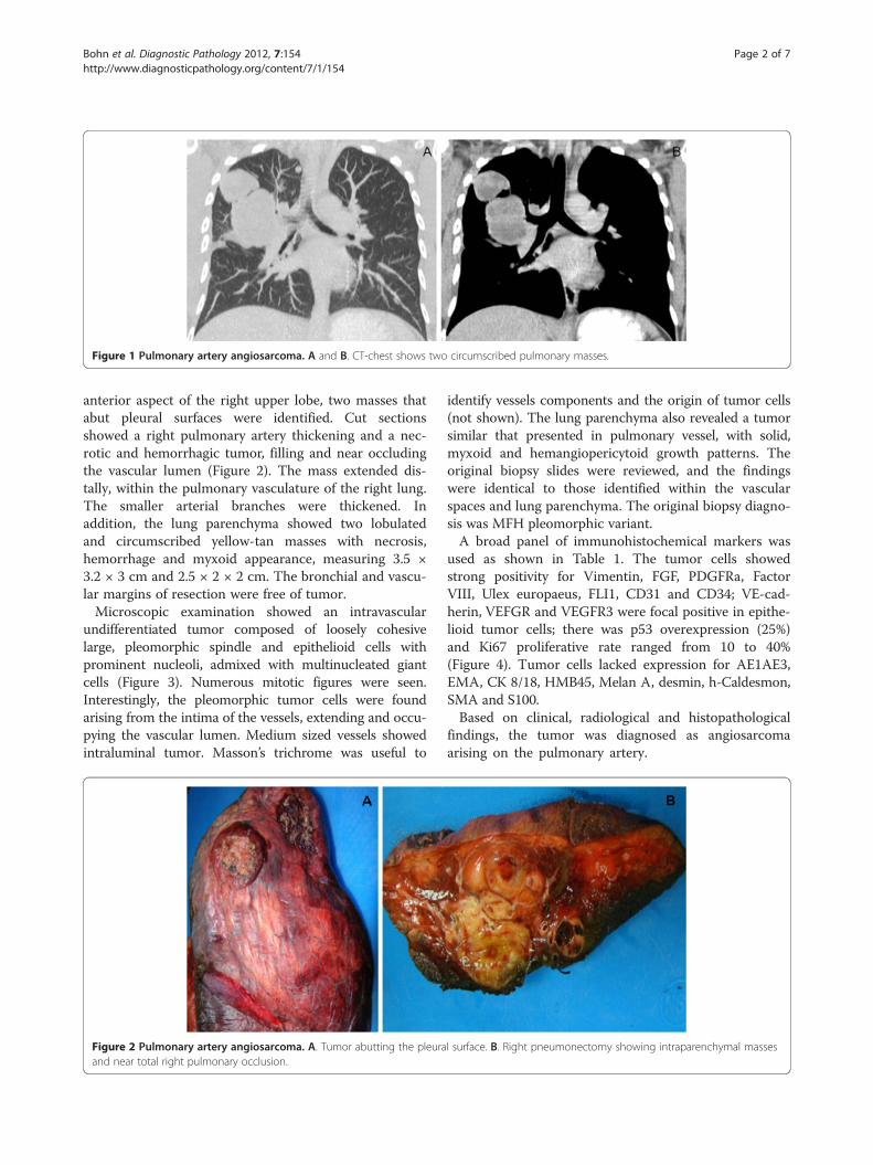
Figure 1 Pulmonary artery angiosarcoma. A and B. CT-chest shows two circumscribed pulmonary masses.
Bohn et al. Diagnostic Pathology 2012, 7:154 Page 2 of 7http://www.diagnosticpathology.org/content/7/1/154
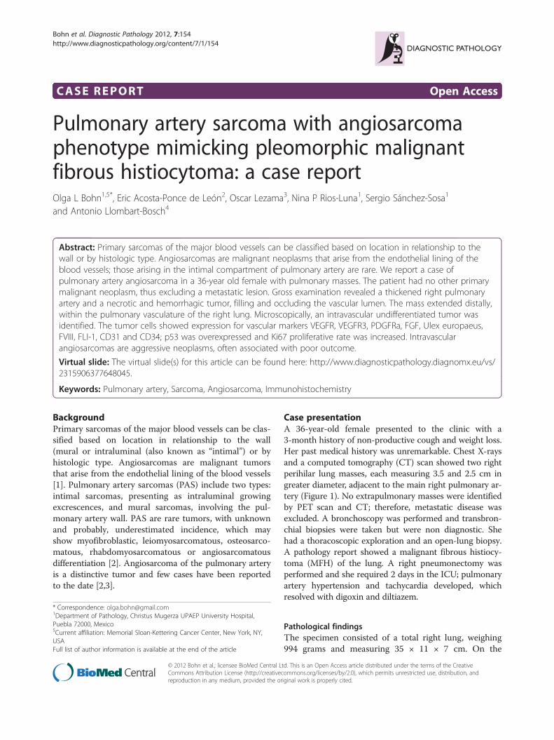
anterior aspect of the right upper lobe, two masses thatabut pleural surfaces were identified. Cut sectionsshowed a right pulmonary artery thickening and a nec-rotic and hemorrhagic tumor, filling and near occludingthe vascular lumen (Figure 2). The mass extended dis-tally, within the pulmonary vasculature of the right lung.The smaller arterial branches were thickened. Inaddition, the lung parenchyma showed two lobulatedand circumscribed yellow-tan masses with necrosis,hemorrhage and myxoid appearance, measuring 3.5 ×3.2 × 3 cm and 2.5 × 2 × 2 cm. The bronchial and vascu-lar margins of resection were free of tumor.Microscopic examination showed an intravascular
undifferentiated tumor composed of loosely cohesivelarge, pleomorphic spindle and epithelioid cells withprominent nucleoli, admixed with multinucleated giantcells (Figure 3). Numerous mitotic figures were seen.Interestingly, the pleomorphic tumor cells were foundarising from the intima of the vessels, extending and occu-pying the vascular lumen. Medium sized vessels showedintraluminal tumor. Masson’s trichrome was useful to
Figure 2 Pulmonary artery angiosarcoma. A. Tumor abutting the pleuraand near total right pulmonary occlusion.
identify vessels components and the origin of tumor cells(not shown). The lung parenchyma also revealed a tumorsimilar that presented in pulmonary vessel, with solid,myxoid and hemangiopericytoid growth patterns. Theoriginal biopsy slides were reviewed, and the findingswere identical to those identified within the vascularspaces and lung parenchyma. The original biopsy diagno-sis was MFH pleomorphic variant.A broad panel of immunohistochemical markers was
used as shown in Table 1. The tumor cells showedstrong positivity for Vimentin, FGF, PDGFRa, FactorVIII, Ulex europaeus, FLI1, CD31 and CD34; VE-cad-herin, VEFGR and VEGFR3 were focal positive in epithe-lioid tumor cells; there was p53 overexpression (25%)and Ki67 proliferative rate ranged from 10 to 40%(Figure 4). Tumor cells lacked expression for AE1AE3,EMA, CK 8/18, HMB45, Melan A, desmin, h-Caldesmon,SMA and S100.Based on clinical, radiological and histopathological
findings, the tumor was diagnosed as angiosarcomaarising on the pulmonary artery.
l surface. B. Right pneumonectomy showing intraparenchymal masses
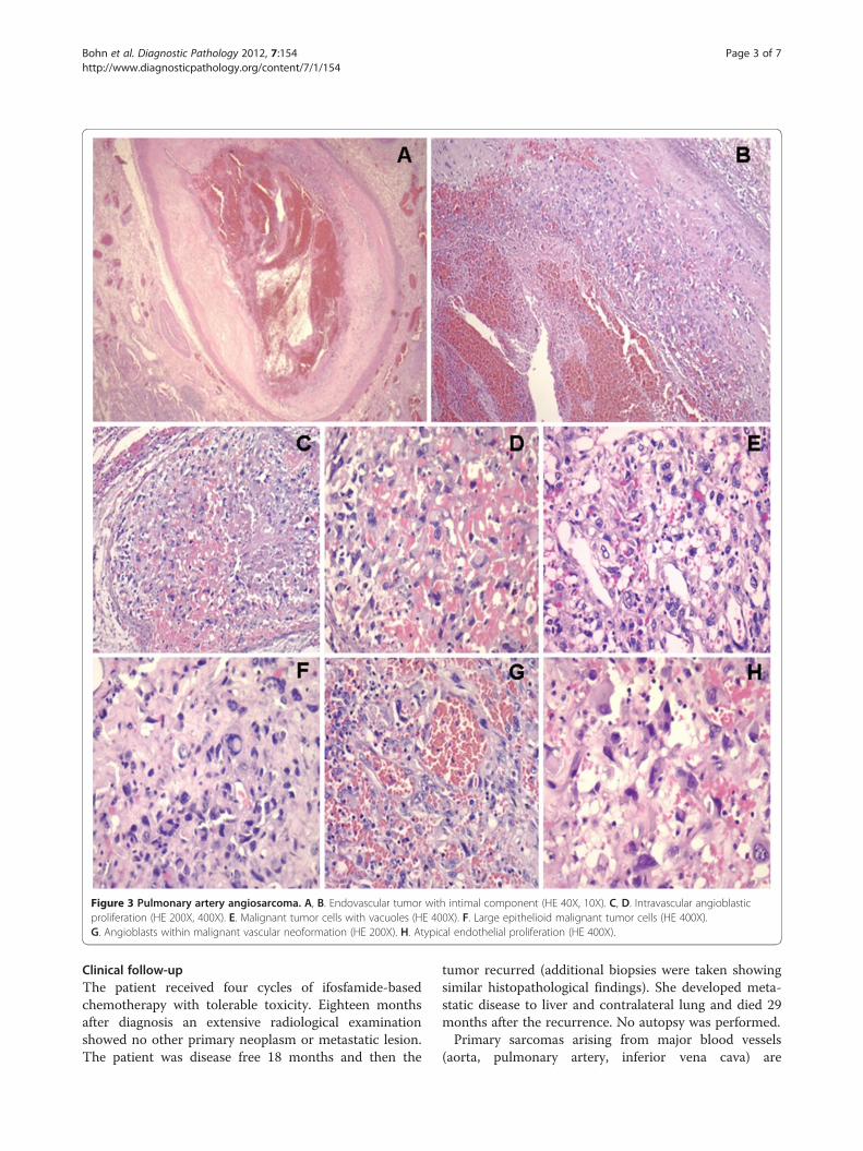
Figure 3 Pulmonary artery angiosarcoma. A, B. Endovascular tumor with intimal component (HE 40X, 10X). C, D. Intravascular angioblasticproliferation (HE 200X, 400X). E. Malignant tumor cells with vacuoles (HE 400X). F. Large epithelioid malignant tumor cells (HE 400X).G. Angioblasts within malignant vascular neoformation (HE 200X). H. Atypical endothelial proliferation (HE 400X).
Bohn et al. Diagnostic Pathology 2012, 7:154 Page 3 of 7http://www.diagnosticpathology.org/content/7/1/154
Clinical follow-upThe patient received four cycles of ifosfamide-basedchemotherapy with tolerable toxicity. Eighteen monthsafter diagnosis an extensive radiological examinationshowed no other primary neoplasm or metastatic lesion.The patient was disease free 18 months and then the
tumor recurred (additional biopsies were taken showingsimilar histopathological findings). She developed meta-static disease to liver and contralateral lung and died 29months after the recurrence. No autopsy was performed.Primary sarcomas arising from major blood vessels
(aorta, pulmonary artery, inferior vena cava) are
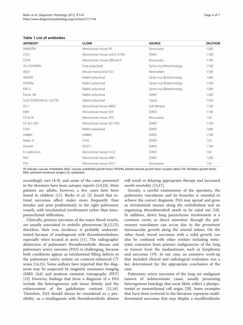
Table 1 List of antibodies
ANTIBODY CLONE SOURCE DILUTION
VIMENTIN Monoclonal mouse V9 Novocastra 1:300
CD31 Monoclonal mouse anti-h JC70A DAKO 1:100
CD34 Monoclonal mouse QBEnd/10 Novocastra 1:100
VE-CADHERIN Goat polyclonal Santa cruz Biotechnology 1:100
VEGF Mouse monoclonal VG1 Neomarkers 1:100
VEGFR3 Rabbit polyclonal Santa cruz Biotechnology 1:400
PDGFRa Rabbit polyclonal Santa cruz Biotechnology 1:100
FGF-2 Rabbit polyclonal Santa cruz Biotechnology 1:200
Factor VIII Rabbit polyclonal DAKO 1:200
ULEX EUROPAEUS-I LECTIN Rabbit polyclonal Sigma 1:500
FLI-1 Monoclonal mouse MRQ1 Cell Marque 1:100
EMA Monoclonal mouse E29 DAKO 1:100
CK 8/18 Monoclonal mouse 5D3 Novocastra 1:50
CK AE1-AE3 Monoclonal mouse AE1-AE3 DAKO 1:100
S100 Rabbit polyclonal DAKO 1:600
HMB45 HMB45 DAKO 1:100
Melan A A103 DAKO 1:50
Desmin DE-R11 DAKO 1:100
h-Caldesmon Monoclonal mouse h-CD DAKO 1:50
Ki67 Monoclonal mouse MIB-1 DAKO 1:200
P53 Monoclonal mouse DO-7 Novocastra 1:50
VE indicates vascular endothelial; VEGF, vascular endothelial growth factor; PDGFRa, platelet-derived growth factor receptor alpha; FGF, fibroblast growth factor;EMA, epithelial membrane antigen; CK, cytokeratin.
Bohn et al. Diagnostic Pathology 2012, 7:154 Page 4 of 7http://www.diagnosticpathology.org/content/7/1/154
exceedingly rare [4-8], and some of the cases presentedin the literature have been autopsy reports [5,9,10]. Mostpatients are adults, however, a few cases have beenfound in children [11]. Burke et al. [5] found that in-timal sarcomas affect males more frequently thanfemales and arise predominatly in the right pulmonaryvessels, with intraluminal involvement rather than intra-parenchymal infiltration.Clinically, primary sarcomas of the major blood vessels,
are usually associated to embolic phenomena [6,12,13];therefore, their true incidence is probably underesti-mated because of misdiagnosis with thromboembolism,especially when located at aorta [11]. The radiographicdistinction of pulmonary thromboembolic disease andpulmonary artery sarcoma (PAS) is challenging, becauseboth conditions appear as intraluminal filling defects inthe pulmonary artery system on contrast-enhanced CTscans [14,15]. Some authors have reported that the diag-nosis may be suspected by magnetic resonance imaging(MRI) [4,6] and positron emission tomography (PET)[15]. However, findings that favor a diagnosis of a PASinclude the heterogeneous soft tissue density and theenhancement of the gadolinium contrast [11,16].Therefore, PAS should always be considered as a pos-sibility, as a misdiagnosis with thromboembolic disease
will result in delaying appropriate therapy and increasedmorbi-mortality [15,17].Grossly, a careful examination of the specimen, the
pulmonary vasculature and its branches is essential toachieve the correct diagnosis. PAS may spread and growas intraluminal masses along the endothelium and anorganizing thromboemboli needs to be ruled out [18].In addition, direct lung parenchyma involvement is acommon event, as direct extension through the pul-monary vasculature can occur due to the prominentintravascular growth along the arterial intima. On theother hand, mural sarcomas with a solid growth canalso be confused with other entities including meta-static extension from primary malignancies of the lungor tumors from the mediastinum, such as lymphomaand sarcomas [19]. In our case, an extensive work-upthat included clinical and radiological evaluation was akey determinant for the appropriate conclusion of thecase.Pulmonary artery sarcomas of the lung are malignant
tumors of indeterminate cause, usually presentingheterogenous histology that most likely reflect a pluripo-tential or mesenchymal cell origin [20]. Some examplesthat have been reviewed in the literature represent undif-ferentiated sarcomas that may display a myofibroblastic
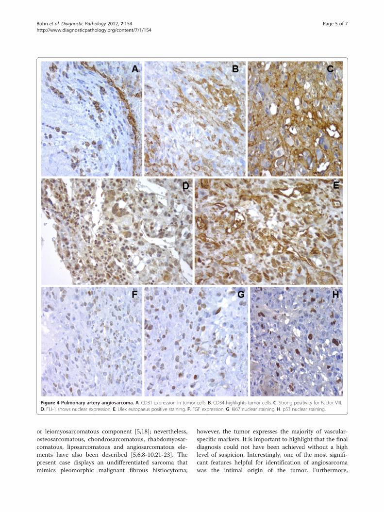
Figure 4 Pulmonary artery angiosarcoma. A. CD31 expression in tumor cells. B. CD34 highlights tumor cells. C. Strong positivity for Factor VIII.D. FLI-1 shows nuclear expression. E. Ulex europaeus positive staining. F. FGF expression. G. Ki67 nuclear staining. H. p53 nuclear staining.
Bohn et al. Diagnostic Pathology 2012, 7:154 Page 5 of 7http://www.diagnosticpathology.org/content/7/1/154
or leiomyosarcomatous component [5,18]; nevertheless,osteosarcomatous, chondrosarcomatous, rhabdomyosar-comatous, liposarcomatous and angiosarcomatous ele-ments have also been described [5,6,8-10,21-23]. Thepresent case displays an undifferentiated sarcoma thatmimics pleomorphic malignant fibrous histiocytoma;
however, the tumor expresses the majority of vascular-specific markers. It is important to highlight that the finaldiagnosis could not have been achieved without a highlevel of suspicion. Interestingly, one of the most signifi-cant features helpful for identification of angiosarcomawas the intimal origin of the tumor. Furthermore,
Bohn et al. Diagnostic Pathology 2012, 7:154 Page 6 of 7http://www.diagnosticpathology.org/content/7/1/154
undifferentiated tumors as primary pulmonary angiosar-comas have been confused with malignant neoplasms(primary or metastatic malignancies) with dedifferen-tiated component, such as carcinomas, sarcomas andlymphomas [24-27]. Differential diagnoses in this specificcase include high grade sarcomas with leiomyosarcoma-tous, fibro or myofibroblastic differentiation. For thatreason, an appropriate and consecutive utilization of abroad panel of antibodies, and occasionally, electron mi-croscopy (not used in this case) are often necessary todistinguish the origin [8,13,28]. We found in this case,that co-expression of endothelial markers such as VEGF,PDGFR, Ulex europaeus, FLI1, CD31 and CD34 is usefulin classifying the neoplasm, representing an unusualpresentation of a true angiosarcoma similar to thoseobserved in soft tissues.Although in this case it was not possible to identify
specific exposure, angiosarcomas have been reportedassociated to previous irradiation fields [29] or arterio-venous fistulas [30]. Comparative genomic hybridization(CGH) analysis of intimal pulmonary sarcomas haveshown gains and amplifications in the 12q13–14 region,with other less consistent findings including losses on3p, 3q, 4q, 9p, 11q, 13q, Xp, and Xq, gains on 7p, 17p,and 17q, and amplifications on 4q, 5p, 6p, and 11q [18].Currently, no prognostic markers have been extensivelystudied; however, in one study, Gaumann et al. [31]found that osteopontin expression may contribute tometastasis due to its role in cell attachment and there-fore to poor prognosis. In addition, Bode-Lesniewskaet al. [18] reported that dysregulation of the cell cycleproteins in p53 pathway and overexpression of mdm2might be implicated in the pathogenesis. At this point,further gene profiling studies, including a larger groupof this specific type of vascular malignancy can be usedto identify predictive and prognostic markers for intra-vascular angiosarcoma of the pulmonary vessels.Overall, PAS are considered aggressive neoplasms,
often associated to poor outcome irrespective of treat-ment [9,10,32,33], and with no exception for pulmonaryartery angiosarcomas. Only few patients have survivedmore than 12 months [5,34]; surgery and complete re-section of the tumor provide local control. Adjuvantchemotherapy and radiotherapy have a controversial rolein the management of this disease [5,33]. The role ofchemotherapy is unclear, but a 50% response rate hasbeen found to palliative chemotherapy with anthracy-clines and ifosfamide in patients with advanced PAS[11,35].
ConclusionIn summary, we report an unusual case of pulmonaryartery sarcoma with angiosarcoma phenotype thatmimics pleomorphic malignant fibrous histiocytoma. A
high level of suspicion and identification of the intimallocation of the tumor, in addition to the use of a broadpanel of immunohistochemical markers were helpfulfor the identification of the vascular origin. A multidis-ciplinary clinical and radiological approach and an exten-sive work-up were key determinant for the appropriateconclusion of the case.
ConsentWritten informed consent was obtained from thepatient’s family members for publication of this CaseReport and any accompanying images. A copy of thewritten consent is available for review by the Editor-inChief of this journal.
Competing interestsThe authors declare that they have no competing interests.
Authors’ contributionsOB, EA, OL, NR, SS and AL have been directly involved in diagnosis andinterpretation of patient’s diagnosis. OB, SS and AL were responsible for theconception and design of the Case Report. All authors read and approvedthe final manuscript.
Author details1Department of Pathology, Christus Mugerza UPAEP University Hospital,Puebla 72000, Mexico. 2Department of Surgical Oncology, Hospital Angeles,5 Poniente 715, Puebla, Mexico. 3Department of Radiology, Hospital Angeles,Puebla, México. 4Department of Pathology, Valencia University, Valencia,Spain. 5Current affiliation: Memorial Sloan-Kettering Cancer Center, New York,NY, USA.
Received: 14 July 2012 Accepted: 31 October 2012Published: 7 November 2012
References1. Rosai J (Ed): Rosai and Ackerman's Surgical Pathology, 10th edition.
Philadelphia: Mosby Elservier; 2011.2. Huo L, Moran CA, Fuller GN, Gladish G, Suster S: Pulmonary artery sarcoma:
a clinicopathologic and immunohistochemical study of 12 cases. Am JClin Pathol 2006, 125:419–424.
3. Huo L, Lai S, Gladish G, Czerniak BA, Moran CA: Pulmonary arteryangiosarcoma: a clinicopathologic and radiological correlation. AnnDiagn Pathol 2005, 9:209–214.
4. Seelig MH, Klingler PJ, Oldenburg WA, Blackshear JL: Angiosarcoma of theaorta: report of a case and review of the literature. J Vasc Surg 1998,28:732–737.
5. Burke AP, Virmani R: Sarcomas of the great vessels. A clinicopathologicstudy. Cancer 1993, 71:1761–1773.
6. Santonja C, Costa-Subias J, Martin-Hita AM, Dotor A: Intimal angiosarcomaof the aorta with tumour embolisation causing mesenteric ischaemia.Report of a case diagnosed using CD31 immunohistochemistry in anintestinal resection specimen. Virchows Arch 2001, 438:404–407.
7. Hottenrott G, Mentzel T, Peters A, Schroder A, Katenkamp D: Intravascular("intimal") epithelioid angiosarcoma: clinicopathological andimmunohistochemical analysis of three cases. Virchows Arch 1999,435:473–478.
8. Miracco C, Laurini L, Santopietro R, De Santi MM, Sassi C, Neri E, Pepi F, LuziP: Intimal-type primary sarcoma of the aorta. Report of a case withevidence of rhabdomyosarcomatous differentiation. Virchows Arch 1999,435:62–66.
9. Nonomura A, Kurumaya H, Kono N, Nakanuma Y, Ohta G, Terahata S,Matsubara F, Matsuda T, Asaka T, Nishino T: Primary pulmonary arterysarcoma. Report of two autopsy cases studied by immunohistochemistryand electron microscopy, and review of 110 cases reported in theliterature. Acta Pathol Jpn 1988, 38:883–896.
Bohn et al. Diagnostic Pathology 2012, 7:154 Page 7 of 7http://www.diagnosticpathology.org/content/7/1/154
10. Gaumann A, Tews DS, Mayer E, Dahm M, Petrow PK, Otto M, Kirkpatrick CJ,Kriegsmann J: Expression of apoptosis-related proteins, p53, and DNAfragmentation in sarcomas of the pulmonary artery. Cancer 2001,92:1237–1244.
11. Chappell T, Creech CB, Parra D, Strauss A, Scholl F, Whitney G: Presentationof pulmonary artery intimal sarcoma in an infant with a history ofneonatal valvular pulmonic stenosis. Ann Thorac Surg 2008, 85:1092–1094.
12. Nishida N, Yutani C, Ishibashi-Ueda H, Tsukamoto Y, Ikeda Y, Nakamura Y:Histopathological characterization of aortic intimal sarcoma withmultiple tumor emboli. Pathol Int 2000, 50:923–927.
13. Goldblum JR, Rice TW: Epithelioid angiosarcoma of the pulmonary artery.Hum Pathol 1995, 26:1275–1277.
14. Scheffel H, Stolzmann P, Plass A, Weber A, Pretre R, Marincek B, Alkadhi H:Primary intimal pulmonary artery sarcoma: a diagnostic challenge.J Thorac Cardiovasc Surg 2008, 135:949–950.
15. Thurer RL, Thorsen A, Parker JA, Karp DD: FDG imaging of a pulmonaryartery sarcoma. Ann Thorac Surg 2000, 70:1414–1415.
16. Yi ES: Tumors of the pulmonary vasculature. Cardiol Clin 2004, 22:431–440.vi-vii.
17. Scheidl S, Taghavi S, Reiter U, Troster N, Kovacs G, Rienmuller R, Lang S,Klepetko W, Olschewski H: Intimal sarcoma of the pulmonary valve. AnnThorac Surg 2010, 89:e25–e27.
18. Bode-Lesniewska B, Zhao J, Speel EJ, Biraima AM, Turina M, Komminoth P,Heitz PU: Gains of 12q13-14 and overexpression of mdm2 are frequentfindings in intimal sarcomas of the pulmonary artery. Virchows Arch 2001,438:57–65.
19. Sebenik M, Ricci A Jr, DiPasquale B, Mody K, Pytel P, Jee KJ, Knuutila S,Scholes J: Undifferentiated intimal sarcoma of large systemic bloodvessels: report of 14 cases with immunohistochemical profile and reviewof the literature. Am J Surg Pathol 2005, 29:1184–1193.
20. McGlennen RC, Manivel JC, Stanley SJ, Slater DL, Wick MR, Dehner LP:Pulmonary artery trunk sarcoma: a clinicopathologic, ultrastructural, andimmunohistochemical study of four cases. Mod Pathol 1989, 2:486–494.
21. Johansson L, Carlen B: Sarcoma of the pulmonary artery: report of fourcases with electron microscopic and immunohistochemicalexaminations, and review of the literature. Virchows Arch 1994,424:217–224.
22. Baker PB, Goodwin RA: Pulmonary artery sarcomas. A review and reportof a case. Arch Pathol Lab Med 1985, 109:35–39.
23. Tavora F, Miettinen M, Fanburg-Smith J, Franks TJ, Burke A: Pulmonaryartery sarcoma: a histologic and follow-up study with emphasis on asubset of low-grade myofibroblastic sarcomas with a good long-termfollow-up. Am J Surg Pathol 2008, 32:1751–1761.
24. Hisaoka M, Tsuji S, Hashimoto H, Aoki T, Uriu K: Dedifferentiatedliposarcoma with an inflammatory malignant fibrous histiocytoma-likecomponent presenting a leukemoid reaction. Pathol Int 1997, 47:642–646.
25. Hollowood K, Holley MP, Fletcher CD: Plexiform fibrohistiocytic tumour:clinicopathological, immunohistochemical and ultrastructural analysis infavour of a myofibroblastic lesion. Histopathology 1991, 19:503–513.
26. Kao YC, Chow JM, Wang KM, Fang CL, Chu JS, Chen CL: Primary pleuralangiosarcoma as a mimicker of mesothelioma: a case report **VS**.Diagn Pathol 2011, 6:130.
27. Jinghong X, Lirong C: Pulmonary epithelioid hemangioendotheliomaaccompanied by bilateral multiple calcified nodules in lung. Diagn Pathol2011, 6:21.
28. Emmert-Buck MR, Stay EJ, Tsokos M, Travis WD: Pleomorphicrhabdomyosarcoma arising in association with the right pulmonaryartery. Arch Pathol Lab Med 1994, 118:1220–1222.
29. Davies JD, Rees GJ, Mera SL: Angiosarcoma in irradiated post-mastectomychest wall. Histopathology 1983, 7:947–956.
30. Byers RJ, McMahon RF, Freemont AJ, Parrott NR, Newstead CG: Epithelioidangiosarcoma arising in an arteriovenous fistula. Histopathology 1992,21:87–89.
31. Gaumann A, Petrow P, Mentzel T, Mayer E, Dahm M, Otto M, Kirkpatrick CJ,Kriegsmann J: Osteopontin expression in primary sarcomas of thepulmonary artery. Virchows Arch 2001, 439:668–674.
32. Anderson MB, Kriett JM, Kapelanski DP, Tarazi R, Jamieson SW: Primarypulmonary artery sarcoma: a report of six cases. Ann Thorac Surg 1995,59:1487–1490.
33. Gaumann A, Bode-Lesniewska B, Zimmermann DR, Fanburg-Smith JC,Kirkpatrick CJ, Hofstadter F, Woenckhaus M, Stoehr R, Obermann EC,
Dietmaier W, Hartmann A: Exploration of the APC/beta-catenin (WNT)pathway and a histologic classification system for pulmonary arteryintimal sarcoma. A study of 18 cases. Virchows Arch 2008, 453:473–484.
34. Mayer E, Kriegsmann J, Gaumann A, Kauczor HU, Dahm M, Hake U, SchmidFX, Oelert H: Surgical treatment of pulmonary artery sarcoma. J ThoracCardiovasc Surg 2001, 121:77–82.
35. Manso L, Alvarez E, Quintela M, Cortes-Funes H, Hitt R: Primary pulmonaryartery sarcoma: report of three cases and review of the literature.Clin Lung Cancer 2007, 8:277–281.
doi:10.1186/1746-1596-7-154Cite this article as: Bohn et al.: Pulmonary artery sarcoma withangiosarcoma phenotype mimicking pleomorphic malignant fibroushistiocytoma: a case report. Diagnostic Pathology 2012 7:154.
Submit your next manuscript to BioMed Centraland take full advantage of:
• Convenient online submission
• Thorough peer review
• No space constraints or color figure charges
• Immediate publication on acceptance
• Inclusion in PubMed, CAS, Scopus and Google Scholar
• Research which is freely available for redistribution
Submit your manuscript at www.biomedcentral.com/submit