Embed Size (px)
Citation preview
Remedy Publications LLC.
Journal of Dermatology and Plastic Surgery
2016 | Volume 1 | Issue 1 | Article 10031
AbbreviationsAKE: Acrokeratoelastoidosis; FAH: Focal acral hyperkeratosis; MPA: Marginal
papularacrokeratodermas; PK: Punctate Keratoderma; PPK: Palmoplantar Keratoderma; Punctate PPK: Punctate; Palmoplantar Keratoderma
IntroductionPalmoplantar keratodermas (PPK) may be categorized as acquired or hereditary. Acquired
PPK have been associated with paraneoplastic syndromes and HIV/AIDS [1]. Inherited PPK are further classified by their distribution of epidermal involvement. The three classes of inherited PPKs are diffuse keratodermas, focal keratodermas and punctate keratodermas. These three classes of inherited PPKs can manifest in three ways; simple manifestation remains limited to the skin; complex manifestation includes lesions of nonvolar skin, hair, teeth, nails and sweat glands and syndromic manifestation can involve other organ systems, deafness, and/or cancer [2].
There are several types of punctate palmoplantar keratodermas (punctate PPK) described in the literature; however, classification is complicated by the misuse of names and redundant terminology. Type 1 punctate PPK, also known as Buschke-Fischer-Brauer disease, has been referred to in the literature as punctate PPK, lending to further confusion. Type 2 punctate PPK was referred to as porokeratosispunctata palmaris etplantaris; however, spiny keratoderma of the palms and soles is now favored (discussion to follow). Type 3 punctate PPK, or the marginal keratodermas, consist of focal acral hyperkeratosis and acrokeratoelastoidosis (Table 1).
We present two cases of punctate PPK that were clinically and histologically consistent with the diagnosis of focal acral hyperkeratosis. In addition, the second patient presented with the rare association of FAH with keratosis punctata of the palmar creases. Recent literature demonstrates a
Punctate Palmoplantar Keratodermas: Case Reports and A Review of the Literature and Terminology
OPEN ACCESS
*Correspondence:Eve J Lowenstein, SUNY Downstate
Dermatology, 450 Clarkson Ave, Box46, Brooklyn, USA, Tel: 718-270-2791;
E-mail: [email protected] Date: 17 Aug 2016Accepted Date: 26 Aug 2016
Published Date: 23 Sep 2016
Citation: Lieberman MR, Kober M, Lowenstein
EJ, Heilman E. Punctate Palmoplantar Keratodermas: Case Reports and
A Review of the Literature and Terminology. J Dermatol Plast Surg.
2016; 1(1): 1003.
Copyright © 2016 Lowenstein EJ. This is an open access article distributed
under the Creative Commons Attribution License, which permits unrestricted
use, distribution, and reproduction in any medium, provided the original work
is properly cited.
Case ReportPublished: 23 Sep, 2016
AbstractKeratodermas encompass a wide spectrum of disorders of keratinization that may be acquired or hereditary. We present two cases of focal acral hyperkeratosis (FAH), a subtype of punctate palmoplantar keratoderma. We review the literature and attempt to clarify the confusing classification of the heritable punctate palmoplantar keratodermas.
Capsule Summary:
1. (First bullet) What is already known on this topic
a. Punctate PPK is an esoteric topic within the field of dermatology that has been discussed both clinically and histologically.
2. (Second bullet) What this article adds to our knowledge
a. Despite this, the nosography and classification of punctate PPKs have been confused in published literature.
3. (Third bullet) How this information impacts clinical practice and/or changes patient care
a. This article hopes to clarify the terminology and classification of punctate PPKs, thereby increasing awareness of this disease, with the hope of advancing treatment options.
Keywords: Punctate palmoplantar keratoderma; Palmoplantar keratoderma; Punctate keratoderma; Buschke-fischer-brauer syndrome; Acrokeratoelastoidosis; Focal acral hyperkeratosis
Lieberman MR1, Kober M1, Lowenstein EJ1* and Heilman E1
Department of Dermatology, State University of New York Downstate Medical Center, USA
Lowenstein EJ, et al. Journal of Dermatology and Plastic Surgery
Remedy Publications LLC. 2016 | Volume 1 | Issue 1 | Article 10032
renewed interest in these entities; however, ambiguous terminologies persist [3,4]. We therefore present an overview of hereditary punctate PPK, focusing on clarifying the terminology.
Case Presentation OneA healthy 29-year old African American woman presented to the
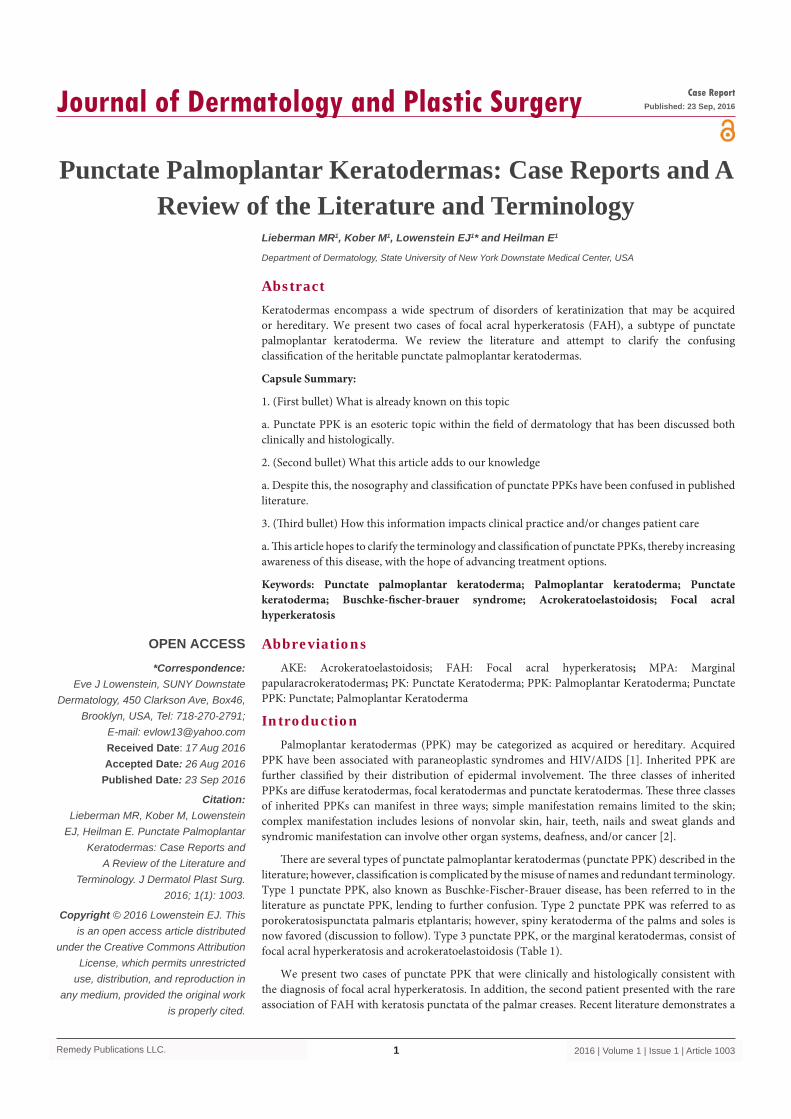
clinic for evaluation of an asymptomatic lesions. The lesions were present for a number of years with a progressive worsening over this time. She received no previous treatment of the condition. The lesions remained localized to the areas of friction located over the posterior and posteriolateral ankles. The patient denied other skin concerns and had no past dermatologic history, including no history of atopic dermatitis or nail complaints. Of note, the patient reported that a sibling had similar lesions, but less severe invovement.
On physical examination, the patient was well-nourished. There were multiple papules with a central keratotic core and 2-5 mm
circular depressions overlying hyperpigmented plaques, with mild lichenification located bilaterally on her posterior ankles, overlying the Achilles tendon (Figure 1a and b). Her palms and soles were spared and her fingers and toenails were normal in appearance.
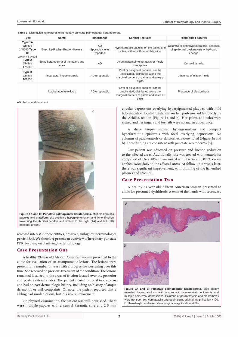
A shave biopsy showed hypergranulosis and compact hyperkeratotic epidermis with focal overlying depressions. No columns of parakeratosis or elastorrhexis were noted (Figure 2a and b). These finding are consistent with punctate keratoderma [5].
Our patient was educated on pressure and friction reduction to the affected areas. Additionally, she was treated with keratolytics comprised of Urea 40% cream mixed with Tretinoin 0.025% cream applied twice daily to the affected areas. At follow-up 6 weeks later, there was significant improvement, with thinning of the lichenified plaques and spicules.
Case Presentation TwoA healthy 51 year old African American woman presented to
clinic for presumed dyshidrotic eczema of the hands with secondary
Type Name Inheritance Clinical Features Histologic FeaturesType 1AOMIM#
148600 Type 1B
OMIM# 614936
Buschke-Fischer-Brauer diseaseAD
Sporadic cases reported
Hyperkeratotic papules on the palms and soles, with or without umbilication
Columns of orthohyperkeratosis, absence of epidermal dyskeratosis or hydropic
change
Type 2OMIM#175860
Spiny keratoderma of the palms and soles AD Acuminata (spiny) keratosis or music
box spines Cornoid lamella
Type 3OMIM#101850
Focal acral hyperkeratosis AD or sporadic
Oval or polygonal papules, can be umbilicated, distributed along the
marginal borders of palms and soles or digits
Absence of elastorrhexis
Acrokeratoelastoidosis AD or sporadicOval or polygonal papules, can be umbilicated, distributed along the
marginal borders of palms and soles or digits
Presence of elastorrhexis
Table 1: Distinguishing features of hereditary punctate palmoplantar keratodermas.
AD: Autosomal dominant
A
B
Figure 1A and B: Punctate palmoplantar keratoderma. Multiple keratotic papules and crateiform pits overlying hyperpigmentation and lichenification traversing the Achilles tendon and limited to the right (1A) and left (1B) posterior ankles.
A
B
Figure 2A and B: Punctate palmoplantar keratoderma. Skin biopsy revealed hypergranulosis with a compact hyperkeratotic epidermis and multiple epidermal depressions. Columns of parakeratosis and elastorhexis were not seen (A: Hematoxylin and eosin stain, original magnification x100, B: Hematoxylin and eosin stain, original magnification x200).
Lowenstein EJ, et al. Journal of Dermatology and Plastic Surgery
Remedy Publications LLC. 2016 | Volume 1 | Issue 1 | Article 10033
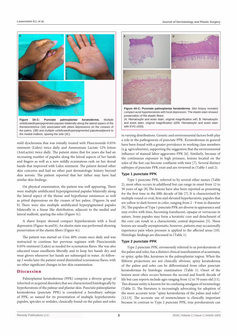
mild dyschromia that was initially treated with Fluocinonide 0.05% ointment (Lidex) twice daily and Ammonium Lactate 12% lotion (AmLactin) twice daily. The patient states that for years she had an increasing number of papules along the lateral aspects of her hands and fingers as well as a new mildly eczematous rash on her dorsal hands that improved with Lidex ointment. The patient denied other skin concerns and had no other past dermatologic history beyond skin xerosis. The patient reported that her father may have had similar skin findings.
On physical examination, the patient was well appearing. There were multiple umbilicated hyperpigmented papules bilaterally along the lateral aspect of the thenar and hypothenar eminences as well as pitted depressions on the creases of her palms. (Figures 3a and b) There were also multiple umbilicated hyperpigmented papules bilaterally in a linear like distribution, adjacent to the medial and lateral malleoli, sparing the soles (Figure 3c).
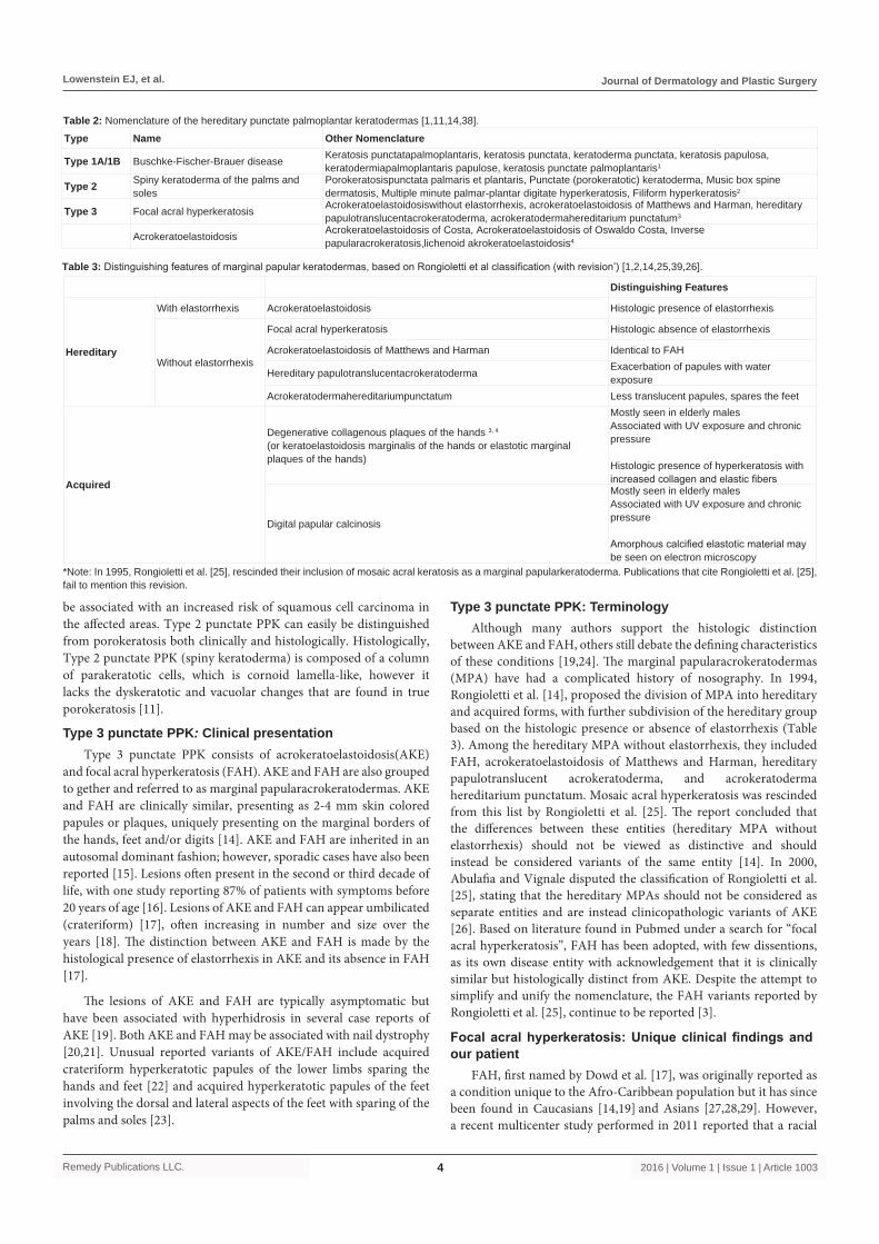
A shave biopsy showed compact hyperkeratosis with a focal depression (Figure 4a and b). An elastin stain was performed showing preservation of the elastin fibers (Figure 4c).
The patient was started on Urea 40% cream once daily and was instructed to continue her previous regimen with Fluocinonide 0.05% ointment (Lidex) as needed for eczematous flares. She was also educated touse emollients liberally and to keep her hands dry and wear gloves whenever her hands are submerged in water. At follow-up 2 weeks later the patient noted diminished eczematous flares, with no other significant changes in her skin examination.
DiscussionPalmoplantar keratodermas (PPK) comprise a diverse group of
inherited or acquired disorders that are characterized histologically by hyperkeratosis of the palmar and plantar skin. Punctate palmoplantar keratoderma (punctate PPK) is considered a hereditary subtype of PPK, so named for its presentation of multiple hyperkeratotic papules, spicules or nodules, classically found on the palms and soles
in varying distributions. Genetic and environmental factors both play a role in the pathogenesis of punctate PPK. Keratodermas in general have been found with a greater prevalence in working class members (e.g. agriculturists), supporting the suggestion that the environmental influence of manual labor aggravates PPK [6]. Similarly, because of the continuous exposure to high pressure, lesions located on the soles of the feet can become confluent with time [7]. Several distinct subtypes of punctate PPK exist and are reviewed in (Table 1 and 2).
Type 1 punctate PPKType 1 punctate PPK, referred to by several other names (Table
2), most often occurs in adulthood but can range in onset from 12 to 30 years of age [8].The lesions have also been reported as presenting for the first time in the fifth decade of life [7]. It is characterized by multiple round or oval, firm and elevated hyperkeratotic papules that are yellow to dark brown in color, ranging from 2 - 9 mm in diameter [9]. The papules of Type 1 punctate PPK are diverse in appearance and may evolve with time, becoming translucent, opaque or verrucous in nature. Some papules may form a keratotic core and detachment of the core can result in a characteristic central depression [5]. These lesions are usually asymptomatic; however, patients may occasionally experience pain when pressure is applied to the affected areas [10]. Histologic findings are discussed in (Table 1).
Type 2 punctate PPKType 2 punctate PPK, erroneously referred to as porokeratosis of
the palms and soles, has a distinct clinical manifestation of acuminata, or spiny, spike-like, keratoses in the palmoplantar region. When the filiform projections are not clinically obvious, spiny keratoderma of the palms and soles can be differentiated from other punctate keratodermas by histologic examination (Table 1). Onset of the lesions most often occurs between the second and fourth decade of life but case reports include ages ranging from 12 to 70 years old [11]. This disease entity is known for its confusing amalgam of terminology (Table 2). The literature is increasingly advocating for adoption of the more accurate term “spiny keratoderma of the palms and soles” [12,13]. The accurate use of nomenclature is clinically important because in contrast to Type 2 punctate PPK, true porokeratosis can
A B
C
Figure 3A-C: Punctate palmoplantar keratoderma. Multiple umbilicatedhyperpigmented papules bilaterally along the lateral aspect of the thenareminence (3A) associated with pitted depressions on the creases of the palms. (3B) and multiple umbilicatedhyperpigmented papulesadjacent to the medial malleoli, sparing the sole (3C).
A B
C
Figure 4A-C: Punctate palmoplantar keratoderma. Skin biopsy revealed compact acral hyperkeratosis with focal depression. The elastin stain showed preservation of the elastin fibers.(A: Hematoxylin and eosin stain, original magnification x40, B: Hematoxylin and eosin stain, original magnification x200, Hematoxylin and eosin stain with EVG x200).
Lowenstein EJ, et al. Journal of Dermatology and Plastic Surgery
Remedy Publications LLC. 2016 | Volume 1 | Issue 1 | Article 10034
be associated with an increased risk of squamous cell carcinoma in the affected areas. Type 2 punctate PPK can easily be distinguished from porokeratosis both clinically and histologically. Histologically, Type 2 punctate PPK (spiny keratoderma) is composed of a column of parakeratotic cells, which is cornoid lamella-like, however it lacks the dyskeratotic and vacuolar changes that are found in true porokeratosis [11].
Type 3 punctate PPK: Clinical presentationType 3 punctate PPK consists of acrokeratoelastoidosis(AKE)
and focal acral hyperkeratosis (FAH). AKE and FAH are also grouped to gether and referred to as marginal papularacrokeratodermas. AKE and FAH are clinically similar, presenting as 2-4 mm skin colored papules or plaques, uniquely presenting on the marginal borders of the hands, feet and/or digits [14]. AKE and FAH are inherited in an autosomal dominant fashion; however, sporadic cases have also been reported [15]. Lesions often present in the second or third decade of life, with one study reporting 87% of patients with symptoms before 20 years of age [16]. Lesions of AKE and FAH can appear umbilicated (crateriform) [17], often increasing in number and size over the years [18]. The distinction between AKE and FAH is made by the histological presence of elastorrhexis in AKE and its absence in FAH [17].
The lesions of AKE and FAH are typically asymptomatic but have been associated with hyperhidrosis in several case reports of AKE [19]. Both AKE and FAH may be associated with nail dystrophy [20,21]. Unusual reported variants of AKE/FAH include acquired crateriform hyperkeratotic papules of the lower limbs sparing the hands and feet [22] and acquired hyperkeratotic papules of the feet involving the dorsal and lateral aspects of the feet with sparing of the palms and soles [23].
Type 3 punctate PPK: TerminologyAlthough many authors support the histologic distinction
between AKE and FAH, others still debate the defining characteristics of these conditions [19,24]. The marginal papularacrokeratodermas (MPA) have had a complicated history of nosography. In 1994, Rongioletti et al. [14], proposed the division of MPA into hereditary and acquired forms, with further subdivision of the hereditary group based on the histologic presence or absence of elastorrhexis (Table 3). Among the hereditary MPA without elastorrhexis, they included FAH, acrokeratoelastoidosis of Matthews and Harman, hereditary papulotranslucent acrokeratoderma, and acrokeratoderma hereditarium punctatum. Mosaic acral hyperkeratosis was rescinded from this list by Rongioletti et al. [25]. The report concluded that the differences between these entities (hereditary MPA without elastorrhexis) should not be viewed as distinctive and should instead be considered variants of the same entity [14]. In 2000, Abulafia and Vignale disputed the classification of Rongioletti et al. [25], stating that the hereditary MPAs should not be considered as separate entities and are instead clinicopathologic variants of AKE [26]. Based on literature found in Pubmed under a search for “focal acral hyperkeratosis”, FAH has been adopted, with few dissentions, as its own disease entity with acknowledgement that it is clinically similar but histologically distinct from AKE. Despite the attempt to simplify and unify the nomenclature, the FAH variants reported by Rongioletti et al. [25], continue to be reported [3].
Focal acral hyperkeratosis: Unique clinical findings and our patient
FAH, first named by Dowd et al. [17], was originally reported as a condition unique to the Afro-Caribbean population but it has since been found in Caucasians [14,19] and Asians [27,28,29]. However, a recent multicenter study performed in 2011 reported that a racial
Type Name Other Nomenclature
Type 1A/1B Buschke-Fischer-Brauer disease Keratosis punctatapalmoplantaris, keratosis punctata, keratoderma punctata, keratosis papulosa, keratodermiapalmoplantaris papulose, keratosis punctate palmoplantaris1
Type 2 Spiny keratoderma of the palms and soles
Porokeratosispunctata palmaris et plantaris, Punctate (porokeratotic) keratoderma, Music box spine dermatosis, Multiple minute palmar-plantar digitate hyperkeratosis, Filiform hyperkeratosis2
Type 3 Focal acral hyperkeratosis Acrokeratoelastoidosiswithout elastorrhexis, acrokeratoelastoidosis of Matthews and Harman, hereditary papulotranslucentacrokeratoderma, acrokeratodermahereditarium punctatum3
Acrokeratoelastoidosis Acrokeratoelastoidosis of Costa, Acrokeratoelastoidosis of Oswaldo Costa, Inverse papularacrokeratosis,lichenoid akrokeratoelastoidosis4
Table 2: Nomenclature of the hereditary punctate palmoplantar keratodermas [1,11,14,38].
Distinguishing Features
Hereditary
With elastorrhexis Acrokeratoelastoidosis Histologic presence of elastorrhexis
Without elastorrhexis
Focal acral hyperkeratosis Histologic absence of elastorrhexis
Acrokeratoelastoidosis of Matthews and Harman Identical to FAH
Hereditary papulotranslucentacrokeratoderma Exacerbation of papules with water exposure
Acrokeratodermahereditariumpunctatum Less translucent papules, spares the feet
Acquired
Degenerative collagenous plaques of the hands 3, 4
(or keratoelastoidosis marginalis of the hands or elastotic marginal plaques of the hands)
Mostly seen in elderly malesAssociated with UV exposure and chronic pressure
Histologic presence of hyperkeratosis with increased collagen and elastic fibers
Digital papular calcinosis
Mostly seen in elderly malesAssociated with UV exposure and chronic pressure
Amorphous calcified elastotic material may be seen on electron microscopy
Table 3: Distinguishing features of marginal papular keratodermas, based on Rongioletti et al classification (with revision*) [1,2,14,25,39,26].
*Note: In 1995, Rongioletti et al. [25], rescinded their inclusion of mosaic acral keratosis as a marginal papularkeratoderma. Publications that cite Rongioletti et al. [25], fail to mention this revision.
Lowenstein EJ, et al. Journal of Dermatology and Plastic Surgery
Remedy Publications LLC. 2016 | Volume 1 | Issue 1 | Article 10035
predominance of FAH in patients of African descent does exist. The study also noted that patients of African descent can have punctate keratosis limited to the creases of the palms, fingers and soles. This unique finding is referred to as keratosis punctata of the palmar creases, and is seen more often than FAH alone [30]. Furthermore, the coexistence of FAH with keratosis punctata of the palmar creases has rarely been reported in the literature [31,32]. Our second patient presents a case of FAH associated with keratosis punctate of the palmar creases.
Differential diagnosis of hereditary punctate PPKThe differential diagnosis of palmoplantar hyperkeratos is
includes consideration of digitate keratoses of the palmoplantar surface, which includes spiny keratoderma, verrucae vulgaris and arsenical keratosis [13]. In contrast to Type 1 and Type 3 punctate PPK, digitate keratoses have a characteristic finding of minute fingerlike projections, descriptively referred to as “spiked,” “spiny,” “filiform” and so forth, or by the more imaginative name, “music box spines.” As discussed for spiny keratoderma of the palms and soles, the clinical distinction of spiny projections versus punctate papules is not always obvious. Furthermore, verrucae vulgaris and arsenical keratosis can manifest as both filiform and papular, making histologic examination essential for diagnosis. Verrucae vulgaris papules develop pinpoint bleeding when the lesions are pared [33]. Furthermore, dermoscopy reveals thrombosed capillaries in verrucae vulgaris, but not in punctate PPK. Arsenical keratoses may also appear clinically similar to punctate PPK, but a history of long-term arsenic exposure is required [34]. Arsenical keratoses lesions differ histologically with thick, compact hyperkeratosis and parakeratosis. Some epidermal keratinocytes may show atypia histologically. While the presence of numerous vacuolated keratinocytes and the absence of solar elastosis suggest arsenical keratoses, these findings are not definitive [35]. Important considerations in the differential diagnosis of palmoplantar pits also include nevoid basal cell carcinoma syndrome and plantar pitted keratolysis, hereditary and acquired disorders, respectively. Both disorders are easily distinguished from punctate PPK by histologic focal areas of absent or reduced stratum corneum [9].
Treatment of focal acral hyperkeratosisBecause it is an asymptomatic and benign condition, treatment of
FAH is largely dependent on the patient’s desires and is often indicated for cosmetic reasons alone. A therapeutic goal in the treatment of FAH is to decrease hyperkeratosis with topical keratolytics such as urea, salicyclic acid and topical retinoids. Liquid nitrogen cryotherapy and prednisone have also been attempted, with poor results [36]. There is one report in the literature of acitretin successfully being used for treatment [29]. An earlier report published successful treatment of AKE with etretinate [37-39]. Our first patient responded to twice daily application of Urea 40% cream mixed with Tretinoin 0.025% cream, with significant improvement after 6 weeks. The second patient reported no significant improvement at 2 weeks follow-up with Urea 40% cream applied once daily. Longer follow-up of treatment for this patient is necessary to determine treatment efficacy.
ConclusionThis reportpresents two cases of a unique type of hereditary
punctate PPK known as FAH, or Type 3 punctate PPK. Owing to its asymptomatic nature, FAH has been underreported in the literature, therefore leading to a decreased awareness of its existence. With this
case we hope to further elucidate an understanding of the classification of hereditary punctate PPK, thereby bringing greater awareness of FAH as a diagnostic potential. Furthermore, we propose inclusion of the Type 1A/B, 2 and 3 designations, as outlined in (Table 1), when referring to the various forms of hereditary punctate PPK. Although more cumbersome, it avoids ambiguity. It is our hope that a uniformly adopted terminology will facilitate further study and research of the punctate PPK group, thereby advancing our understanding of the disease and improving potential treatment options.
AcknowledgmentAuthor Contributions: Lieberman, Kober and Lowenstein had
full access to all of the data in the study and take responsibility for the integrity of the data and the accuracy of the data analysis.
Study concept and design: Lieberman, Kober and Lowenstein.
Analysis and interpretation of data: Lieberman, Kober and Lowenstein.
Drafting of the manuscript: Lieberman, Kober and Lowenstein.
Critical revision of the manuscript for important intellectual content: Lieberman, Kober and Lowenstein.
References1. James WD, Berger TG, Elston DM. Andrews’ diseases of the skin: clinical
dermatology. 2011; 207-212.
2. Freedberg IM, Eisen AZ, Wolff K, et al, eds. Fitzpatrick’s Dermatology in General Medicine. McGraw-Hill. 2003; 505-514.
3. Mervak JE, Lowe L, Cha KB. Chronic translucent papules of the palms and soles. JAMA Dermatol. 2014; 150: 1001-1002.
4. Sano DT, Melo LV, Tebcherani AJ, et al. Case for diagnosis. An Bras Dermatol. 2014; 89: 835-836.
5. Oztas P, Alli N, Polat M, Dagdelen, Ustun H, Artuz F, et al. Punctate palmoplantar keratoderma (Brauer-Buschke-Fischer syndrome). Am J ClinDermatol. 2007; 8: 113-116.
6. Murthy SC, Raghu TY, Suresh T. A study of palmoplantar keratodermas in South India. Int J Dermatol. 2008; 47: 762-764.
7. Emmert S, Küster W, Hennies HC, et al. 47 individuals in 14 families with the rare genodermatosis keratosis puncatatapalmopantarisBuschke-Fischer-Brauer. Eur J Dermatol. 2003; 13: 16-20.
8. Stevens HP, Kelsell DP, Bryant SP, Bishop DT, Spurr NK, Weissenbach J, et al. Linkage of an American pedigree with palmoplantar keratoderma and malignancy (palmoplantar ectodermal dysplasia type III) to 17q24. Literature survey and proposed updated classification of the keratodermas. Arch Dermatol. 1996; 132: 640-651.
9. Bolognia JL, Jorizzo J, Schaffer J. Dermatology. 3rd ed. 2012; 777-789.
10. Scott MJ, Costello MJ, Simuangco S. Keratosis punctata palmaris etplantaris. AMA Arch DermSyphilol. 1951; 64: 301-308.
11. Osman Y, Daly TJ, Don PC. Spiny keratoderma of the palms and soles. J Am Acad Dermatol. 1992; 26: 879-881.
12. Torres G, Behshad R, Han A, Castrovinci AJ, Gilliam AC. “I forgot to shave my hands”: a case of spiny keratoderma. J Am Acad Dermatol. 2008; 58: 344–348.
13. Caccetta TP, Dessauvagie B, McCallum D, Kumarasinghe SP. Multiple minute digitate hyperkeratosis: A proposed algorithm for the digitate keratoses. J Am AcadDermatol. 2012; 67: 49-55.
14. Rongioletti F, Betti R, Crosti C, Rebora A. Marginal popular acrokeratodermas: a unified nosography for focal acral hyperkeratosis,
Lowenstein EJ, et al. Journal of Dermatology and Plastic Surgery
Remedy Publications LLC. 2016 | Volume 1 | Issue 1 | Article 10036
acrokeratoelastoidosis and related disorders. Dermatology. 1994; 188: 28-31.
15. AlKahtani HS, AlHumidi AA, Al-Hargan AH, AI-Sayed AA. A sporadic case of unilateral acrokeratoelastoidosis in Saudi Arabia: a case report. J Med Case Rep. 2014; 8: 143.
16. Waxtein-Morgenstern L, Teixeira F, Cortes-Franco R, Vega-Memije ME, Ortiz-Plata A, Zamora-Hernández C, et al. Lenticular acral keratosis in washerwomen. Int J Dermatol. 1998; 37: 532-537.
17. Dowd PM, Harman RRM, Black MM. Focal acral hyperkeratosis. Br J Dermatol. 198; 109: 97-103.
18. Shbaklo Z, Jamaleddine NF, Kibbi AG, Salman SM, Zaynoun ST. Acrokeratoelastoidosis. Int J Dermatol. 1990; 29: 333-336.
19. Erkek E, Kocak M, Bozdogan O, Atasoy P, Birol A. Focal acral hyperkeratosis: a rare cutaneous disorder within the spectrum of Costa acrokeratoelastoidosis. PediatrDermatol. 2004; 21: 128-130.
20. vanSteensel MA, Verstraeten VL, Frank J. Acrokeratoelastoidosis with nail dystrophy: a coincidence or a new entity? Arc Dermatol. 2006; 142: 939–941.
21. Khamaysi Z, Bergman R, Sprecher E. Nail dystrophy in focal acral hyperkeratosis: a distinctive feature? J Eur Acad Dermatol Venereol. 2008; 22: 891-893.
22. Helbling I, Tucker SC, Chalmers RJ. Acquired crateriform hyperkeratotic papules of the lower limbs: an unusual variant of acrokeratoelastoidosis of Costa. Clin Exp Dermatol. 2001; 26: 263-265.
23. Ballambat SP, Pai K. Acquired crateriform hyperkeratotic papules of the feet: an unusual variant of focal acral hyperkeratosis. Indian J Dermatol VenereoI Leprol. 2007; 73: 359-361.
24. Rubegni P, De Aloe G, Romano C, Flori ML, Fimiani M. Acrokeratoelastoidosis: a report of two sporadic cases. Clin Exp Dermatol. 1997; 22: 54-64.
25. Rongioletti F, Betti R, Crosti C, et al. Reply. Dermatology. 1995; 190: 179.
26. Abulafia J, Vignale RA. Degenerative collagenous plaques of the hands and acrokeratoelastoidosis: pathogenesis and relationship with knuckle pads. Int J Dermatol. 2000; 39: 424-432.
27. Lee SE, Kim SC. Focal acral hyperkeratosis. Clin Exp Dermatol. 2007; 32: 608-610.
28. Lee FA, Kim HS, Kim HO, Park YM. A case of focal acral hyperkeratosis. Ann Dermatol. 2009; 21: 426–428.
29. Lambiris AG, Newman PL. Marginal papularacrokeratodermas: no racial limitations for a clinical spectrum that responds to acitretin. Dermatology. 2001; 203: 63-65.
30. Bourrat E, Cabotin PP, Baccard M, Fitoussi C, Eyraud D, Eudes AM, et al. Palmoplantar keratodermas in black patients (Fitzpatrick skin phototype V-VI) of African descent: a multicentre comparative and descriptive series. Br J Dermatol. 2011; 165: 219-221.
31. Khera P, Shiferman G, English III JC. Concurrent punctate keratosis of the palmar creases and focal acral hyperkeratosis. Cutis. 2008; 81: 348-350.
32. Morand JJ, Lightburn E, Civatte M. [Atypical acral hyperkeratosis in a black woman]. [Article in French] Med Trop (Mars). 2002; 62: 85-88.
33. Laube S. Skin infections and ageing. Ageing Res Rev. 2004; 3: 69-89.
34. Paul PC, Chattopadhyay A, Dutta SK, Mazumder DN, Santra A. Histopathology of skin lesions in chronic arsenic toxicity--grading of changes and study of proliferative markers. Indian J Pathol Microbiol. 2000; 43: 257-64.
35. WashingtonDeceit. Dermatopathology of arsenical keratosis. Available at. 2015.
36. Natow, Steven. Focal Acral Hyperkeratosis. Dermatology Online Journal.2001; 7: 10.
37. Handfield-Jones S, Kennedy CT. Acrokeratoelastoidosis treated with etretinate. J Am Acad Dermatol. 1987; 17: 881-882.
38. Marques LP, Trope BM, Pina JC, Cuzzi T, Ramos E Silva M. Inverse papularacrokeratosis of Oswaldo Costa. a case report. J Clin Aesthet Dermatol. 2010; 3: 51–53.
39. Rahbari H. Acrokeratoelastoidosis and keratoelastoidosismarginalis-any relation?. J Am Acad Dermatol. 1981; 5: 348-350.