Embed Size (px)
Citation preview

1
UNIVERSIDADE FEDERAL DE PERNAMBUCO
CENTRO DE CIÊNCIAS BIOLÓGICAS
DEPARTAMENTO DE BIOQÚIMICA
PROGRAMA DE PÓS-GRADUAÇÃO EM BIOQUÍMICA
PURIFICAÇÃO DA LECTINA DE
SEMENTES DE Moringa oleifera (WSMoL) E
AVALIAÇÃO DA GENOTOXICIDADE
LUCÍOLA A. D. M. M. ROLIM
RECIFE 2007
2
UNIVERSIDADE FEDERAL DE PERNAMBUCO CENTRO DE CIÊNCIAS BIOLÓGICAS DEPARTAMENTO DE BIOQÚIMICA
PROGRAMA DE PÓS-GRADUAÇÃO EM BIOQUÍMICA
PURIFICAÇÃO DA LECTINA DE SEMENTES DE Moringa oleifera (WSMoL) E AVALIAÇÃO DA
GENOTOXICIDADE
Dissertação apresentada ao Programa de Pós-
Graduação em Bioquímica da Universidade
Federal de Pernambuco, como parte dos requisitos
para obtenção do grau de Mestre em Bioquímica.
ORIENTADOR: Profª Drª Patrícia Maria Guedes Paiva
CO-ORIENTADOR: Profª Drª Silvia Regina B. de Medeiros
RECIFE
2007

3
Rolim, Lucíola A. D. M. M.
Purificação da lectina de sementes de Moringa oleifera (WSMoL) e avaliação da genotoxidade / Lucíola A. D. M. M. Rolim. – Recife: O Autor, 2007. vii, 67 folhas : il., fig.
Dissertação (mestrado) – Universidade Federal de Pernambuco. CCB. Bioquímica, 2007.
Inclui bibliografia e anexo.
1. Proteínas 2. Lectinas 3. Moringa oleifera 4. Genotoxidade I. Título. 577.112 CDU (2.ed.) UFPE
572.6 CDD (22.ed.) CCB – 2007-075

4
(Shaskespeare-Romeu e Julieta, ato III, cena 3)
“Que mágico poder é aquele encontrado
nas plantas, nas ervas...! Em tudo aquilo que
nasce da terra nada é isento de algo que tenha
em si qualquer coisa de bom; e nada daquilo que
é bom está isento de poder se transformar em
veneno!”

5 5

6

7

8
AGRADECIMENTOS
A Deus que está sempre presente em minha vida.
À minha orientadora Professora Dra. Patrícia Maria Guedes Paiva pelo inestimável
apoio científico, incentivo, amizade e carinho.
À Professora Dra. Silvia Regina Batistuzzo de Medeiros pela orientação nos testes
de genotoxicidade e pelo gentil acolhimento no Laboratório de Biologia Molecular
e Genômica (LBMG) e no Laboratório de mutagênese ambiental (LAMA).
À Professora Dra. Luanna C. B. B. Coelho pelas sugestões e revisão do trabalho.
À Maria Barbosa Reis pelo suporte técnico e prestimosidade indispensáveis para a
realização deste trabalho.
Aos amigos da turma de mestrado, em especial a Juliene, Raquel, Jayra, Cynthia e
Fernanda, pelo companheirismo e convivência muito agradável.
À grande família do Laboratório de Glicopoteínas pela união e solidariedade,
especialmente a Roberto, Gyseli, Lidiane, Romero, Adriana, Andréa, Regina e
Mariana, que sempre colaboraram de diferentes formas na realização desse trabalho.
À Tiago, Cynthia e Fernando pela disponibilidade e colaboração na confecção dos
gráficos e figuras.
Ao apoio e colaboração do pessoal do LBMG, em especial a Herbert e Fernanda,
fundamentais na realização dos experimentos de genotoxicidade/mutagenicidade,
todo o meu carinho e reconhecimento.

9
Aos amigos do LAMA, que contribuíram bastante para a realização do teste de
Kado e Allium cepa, em especial a Jefferson e Jana Dara.
Aos funcionários do departamento de bioquímica da Universidade Federal de
Pernambuco pela constante disponibilidade e simpatia, especialmente a Miron.
Ao meu esposo, Jânio, o maior incentivador desse projeto, pela força transmitida
nos momentos difíceis, o amor, o carinho e o entusiasmo. Todo o meu amor.
Aos meus pais, Bosco e Lúcia, que sempre estiveram presentes em minha vida,
como alicerce e exemplos de vida, acompanhando cada passo, dando equilíbrio e
incentivo.
Às minhas irmãs, Luanna e Layse Julia, minhas melhores amigas e companheiras de
todas as horas que vibram comigo a cada conquista.
À todos que de alguma forma contribuíram para a realização deste trabalho.

10
LISTA DE FIGURAS Figura 1: Moringa oleifera, aspectos gerais. Folhas (A), vagem (B), vagem com sementes
(C) e sementes (D).............................................................................................6

11
LISTA DE ABREVIATURA
AH AHE AS-PTA CFUs DNA IPA
Atividade hemaglutinante Atividade hemaglutinante específica Accesoria e Serviços a Projetos em Agricultura Alternativa Unidade formadora de colônia do inglês Colony-Forming Units. Ácido desoxirribonucléico Instituto de Pesquisa Agropecuária de Pernambuco
PAGE SDS
WSMoAC
WSMoL
Eletroforese em gel de poliacrilamida do inglês polyacrylamide gel electrophoresis Dodecil sulfato de sódio Composto antioxidante de Moringa oleifera solúvel em água, do inglês Water Soluble Moringa oleifera antioxidant compound Lectina de Moringa oleifera solúvel em água, do inglês Water Soluble Moringa oleifera lectin.

12
SUMÁRIO
AGRADECIMENTOS...................................................................................................... I
LISTA DE FIGURAS.....................................................................................................III
LISTA DE ABREVIATURAS.......................................................................................IV
SUMÁRIO........................................................................................................................V
RESUMO........................................................................................................................VI
ABSTRACT...................................................................................................................VII
1. Introdução................................................................................................................... 1
1.1 Moringa. Oleifera .................................................................................1
1.2 Lectinas... .............................................................................................................7
1.3 Genotoxicidade ........................................................................................................11
2.0 Objetivos...................................................................................................................17
2.1 Geral……………………….....................................................................................17
2.2 Específicos……………………………………………............................................17
3.0 Referências...............................................................................................................18
4.0 ARTIGO “Moringa oleifera seed lectin had coagulant property and was not a
genotoxic agent, however extracts of seeds promoted micronucleus
formation”……………………………………………………………………………...30
5.0 Conclusões ...............................................................................................................60
6.0 Anexos ......................................................................................................................61
6.1 Abstract apresentado em congresso.......................................................................61
6.2 Certificado de Menção honrrosa............................................................................62
6.3 Normas para publicação na Water Research.......................................................63

13
RESUMO
O trabalho reporta o isolamento e caracterização da lectina de sementes de Moringa
oleifera (WSMoL). Seu efeito sobre a turbidez da água bem como o potencial
genotoxico/mutagênico da água tratada com diferentes concentrações de sementes de M.
oleifera (MoW1, MoW2, MoW3 and MoW4) ou WSMoL foram também discutidos.
WSMoL, aglutinou diferentes eritrócitos, foi principalmente ativa em pH 6,5 a 7,5 e foi
inibida por azocaseína. Cromatografia de gel filtração e perfis eletroforéticos revelaram
WSMoL como uma proteína de 102 kDa constituída por subunidades de 5 kDa.
Glicosilação não foi detectada. WSMoL apresentou propriedade coagulante similar ao
sulfato de alumínio, o coagulante mais comumente usado. As preparações MoW e WSMoL
foram negativas no teste de DNA plasmidial, no ensaio de transformação bacteriana e no
ensaio Salmonella typhimurium usando TA97, TA98 e TA100 na presença ou ausência de
metabolização hepática. MoW1 e MoW2 foram também negativas no ensaio Allium cepa,
contudo formação de micronúcleos foi induzida por MoW3 e MoW4. Os resultados
sugerem WSMoL como um componente ativo no processo de purificação da água pelas
sementes. WSMoL não foi um agente genotóxico porém genotoxicidade was detected na
água tratada com maior concentração de sementes. Este estudo é uma etapa positive no
sentido da determinação do uso seguro de sementes de M. oleifera para o tratamento da
água.
Key Words: Moringa oleifera, genotoxicidade, mutagenicidade.
14
ABSTRACT
The work reports the isolation and characterization of Moringa oleifera seed lectin
(WSMoL). Its effects on water turbidity as well as genotoxic/mutagenic potential of water
treated with different concentrations of M. oleifera seeds (MoW1, MoW2, MoW3 and
MoW4) or WSMoL were also discussed. WSMoL, agglutinated different erythrocytes, was
mainly active at pH 6.5 to 7.5 and inhibited by azocasein. Gel filtration chromatography
and electrophoretic profiles revealed WSMoL as 102 kDa protein constituted by 5 kDa
subunits. Glycosylation was not detected. WSMoL had coagulant property similar to the
most commonly used coagulant alluminum sulfate. MoW preparations and WSMoL were
negative in a cell-free plasmid DNA test, bacterial transformation assay and in Salmonella
typhimurium assay using TA97, TA98 and TA100 at presence or absence of hepatic
metabolization. MoW1 and MoW2 were also negative by Allium cepa assay however
micronucleus formation was induced by MoW3 and MoW4. The results to suggest
WSMoL is an active component in the water purification process by seeds. WSMoL was
not a genotoxic agent but genotoxicity was detected in water treated with more
concentration of seeds. This study is a positive step forward in determining the safe use of
M. oleifera seeds for water treatment.
Key Words: Moringa oleifera, genotoxicity, mutagenicity.

15
1. INTRODUÇÃO
1.1 Moringa oleifera
Moringa oleifera Lam pertence à família Moringaceae. Um gênero único com
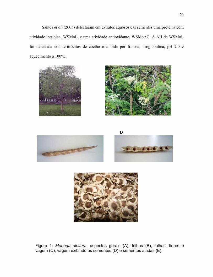
quatorze espécies conhecidas, sendo a M. oleifera (figura 1) a mais conhecida e utilizada de
todas (JANH, 1988a). É uma planta tropical nativa do norte da Índia, largamente cultivada
nos trópicos e encontrada em muitos países da África, Ásia e América do Sul
(SUTHERLAND et al., 1994). Ela cresce rapidamente, através de sementes e mudas, até
em solos marginais, demanda pouca ou nenhuma atenção horticultural e possui uma
resistência que permite a sua sobrevivência em períodos de seca prolongados
(SUTHERLAND et al., 1994). Bezerra et al. (2004) relataram que o peso da semente
favoreceu a germinação e as sementes mais pesadas proporcionaram plântulas mais
vigorosas.
Quase todas as partes da arvore possuem utilidade. As folhas da M. oleifera (figura
1B) são usadas como alimentos em alguns países (NDABIGENGESERE & NARASIAH,
1998), possuindo cerca de 23.000 UI de vitamina A sobressaindo-se entre olerículas
consagradas como brócolis, cenoura, couve, espinafre e alface, que possuem,
respectivamente, 5.000; 3.700; 2.200; 1.900; 1.000 UI de vitamina A (SILVA &
KERR,1999) e para o tratamento de doenças infecciosas da pele, mucosa e trato
respiratório (CÁCERES et al., 1991). Algumas escolas de regiões carentes estão usando as
folhas da moringa na merenda escolar como no Instituto de Permacultura da Bahia em
Salvador (DELDUQ, 2000). Os frutos que correspondem a vagens com três faces (figura 1,
C e D) possuem cerca de 50 cm de comprimento, contendo sementes em forma triangular,

16
com o miolo circundado por uma delicada casca alada (figura 1E) (ABDULKARIM et al.,
2005). Estudo realizado em coelhos sugere que os frutos têm ação hipolipidêmica, anti-
aterosclerótica e promove redução do peso corpóreo (MEHTA et al., 2003).
A semente de M. oleifera possui 40% do peso constituído por óleo que pode ser
usado para cozinhar, na indústria de sabão e como base para cosméticos (SUTHERLAND
et al., 1994). O óleo da semente possui propriedades físicas e químicas equivalentes ao
azeite de oliva e contém uma grande quantidade de tocoferóis (TSAKNIS et al., 1999). As
cascas de M. oleifera podem ser usadas para produzir carvão ativado a baixo custo e o
processo possui viabilidade tecnológica (POLLARD et al., 1995). A diferença na dosagem
ideal entre sementes secas descascadas e com cascas sugere que a proteína ativa esta
contida apenas no miolo, portanto sementes descascadas são mais apropriadas para o
processo do que as não descascadas (NDABIGENGESERE & NARASIRAH,1998;
NDABIGENGESERE et al., 1995 ).
Coagulação ou floculação seguido de sedimentação, filtração e desinfecção,
geralmente com cloro, é usado mundialmente na indústria de tratamento da água antes da
distribuição da água tratada para os consumidores. Durante o processo de desinfecção com
cloro, o material orgânico pode agir como precursor de trialometanos, que podem ser
carcinogênicos (AWWA, 1990). Estudos sugerem que sementes de M. oleifera podem ser
usadas no tratamento industrial da água apenas após a purificação adequada das proteínas
ativas, tornando a preparação mais segura e estável para o tratamento da água.
Tem sido demonstrado que extratos de M. oleifera têm um largo efeito na remoção
da turbidez (redução de 92-99%) e de microorganismos da água (JAHN, 1986; MUYIBI &
EVINSON, 1995a; GHEBREMICHAEL et al., 2005). Coagulantes naturais de origem
vegetal ou mineral eram utilizados, no processo de tratamento da água, antes do advento

17
dos sais químicos, mas eles não estavam aptos a competir efetivamente pelo fato de sua
efetividade e seu mecanismo de ação não serem bem entendidos cientificamente
(NDABIGENGESERE & NARASIAH, 1998). O custo e os efeitos ambientais provocados
pelos sais iônicos têm aumentado o interesse no uso de coagulantes orgânicos derivado de
plantas, como as sementes de M. oleifera, em países em desenvolvimento (JAHN, 1986;
MUYIBI & EVINSON, 1995). A técnica foi trazida da África para o Brasil em 1994 e está
sendo difundida por organizações não governamentais nas regiões brasileiras. O uso como
purificador da água vem sendo divulgado principalmente por entidades ligadas a AS-PTA -
ASSESSORIA E SERVIÇOS A PROJETOS EM AGRICULTURA ALTERNATIVA, que
publicou uma cartilha sobre o uso de sementes de M. oleifera para o tratamento da água
baseado nos trabalhos da pesquisadora alemã Dra. Samia Ao Azharia Jahn.
Essa alternativa oferece vantagens em relação aos coagulantes inorgânicos ou
polímeros orgânicos sintéticos que estão associados a processos patológicos em humanos
(OKUDA et al., 2001a) como a doença de Alzheimer e outras doenças similares
relacionadas com problemas associados com o alumínio residual na água tratada
(AWWA,1990). Além disso, produzem menos volume de lama comparado com água
tratada com alumínio (NDABIGENGESERE & NARASIAH, 1998). A maior desvantagem
no uso da M. oleifera para o tratamento da água é um significativo aumento do material
orgânico (NDABIGENGESERE & NARASIAH, 1998; OKUDA et al., 2001b), não
podendo, por isso ser armazenada por mais de 24 horas (JAHN, 1988). Quando extrato
bruto da água é estocado por períodos maiores que dois dias, pode ser desenvolvido odor
desagradável devido à decomposição microbiana do material orgânico
(NDABIGENGESERE & NARASIRAH,1998).

18
Para remover esse material orgânico pode ser usado adsorção com carbono ou,
alternativamente, o coagulante ativo pode ser extraído das sementes e usado na forma pura
ou semi-pura, reduzindo, assim, a quantidade total de material orgânico adicionado ao
processo de tratamento. Foi observado que após retirar do óleo das sementes, o material
residual ainda contém o coagulante ativo para o tratamento da água (SUTHERLAND et al.,
1994). Os resíduos sólidos podem ser usados como alimentos para animais e fertilizantes,
enquanto a casca pode ser ativada e usada como adsorvente, sendo o coagulante produzido
a custo extremamente baixo ou mesmo zero (GHEBREMICHAEL et al., 2005).
As sementes de M. oleifera não afetam significativamente o pH, a condutividade e a
alcalinidade da água tratada não havendo assim a necessidade de ajustes, já com o sulfato
de alumínio é necessário acrescentar bicarbonato ou cal, o que aumenta o volume de
material residual, o custo de tratamento, assim como ações corrosivas. No tratamento com
M. oleifera não é necessário adicionar nenhuma outra substância química. Uma vantagem
adicional no problema do material residual é o fato de todos os subprodutos da moringa
serem organicamente biodegradáveis, logo a lama pode ser usada como fertilizante
contanto que a água tratada não apresente metais pesados (NDABIGENGESERE &
NARASIAH, 1998).
O extrato aquoso de sementes de moringa esta longe de ser puro. É uma
solução consistindo principalmente de proteínas, lipídios e carboidratos. Uma série de
experimentos foi realizada para caracterizar os agentes coagulantes e floculantes da
mistura. A coagulação usando moringa é causada pela desestabilização dos colóides
negativamente carregados por polieletrólitos catiônicos. O mecanismo predominante de
coagulação parece ser adsorção e neutralização de cargas (NDABIGENGESERE et
al,1995). Proteínas positivamente carregadas se ligam a partes da superfície de partículas

19
negativamente carregadas. Isso leva a formação de partes carregadas negativamente e
positivamente nas superfícies das partículas. Devido à colisão das partículas, saturação
interpartículas entre diferentes setores carregados, a formação de flocos acontecem
(GASSENSCHNIDT et al., 1995).
Estudos têm sido realizados com as proteínas das sementes de M. oleifera.
GHEBREMICHAEL et al. (2005) detectou que o coagulante ativo não é uma proteína
única, mas sim uma mistura de proteínas com características físicas similares. Foi
demonstrado que uma proteína ativa da semente de M. oleifera é dimérica com massa
molecular de 13 kDa e subunidades de aproximadamente 6.5 kDa. A proteína possui ponto
isoelétrico entre 10 e 11, e tanto os monômeros quanto os dímeros apresentam atividade
anticoagulante (NDABIGENGESERE et al., 1995). Uma proteína floculante com massa
molecular de aproximadamente 6.5 kDa e ponto isoelétrico acima de 10 foi também isolada
das sementes (GASSENSCHMIDT et al.,1995). OKUDA et al. (2001a,b) isolaram um
coagulante ativo das sementes de M. oleifera em extrato salino e caracterizou esse
composto como um polieletrólito com peso molecular em torno de 3 kDa, sendo mais
eficaz na remoção da turbidez do que o coagulante extraído em água.
Compostos não protéicos foram isolados de sementes de moringa tostadas. Dentre
eles 4(α-L-raminosiloxi)-fenilacetonitrila, 4-hidroxifenilacetonitrila e 4-hidroxifenil-
acetamida exibiram atividade mutagênica no teste de micronúcleo e em ensaio in vivo
usando camundongo albino (VILLASENOR et al., 1989). Os compostos são facilmente
solúveis na água e ainda não foi determinado se os mesmos estão presentes em sementes
não tostadas (NDABIGENGESERE & NARASIAH, 1998).
20
Santos et al. (2005) detectaram em extratos aquosos das sementes uma proteína com
atividade lectínica, WSMoL, e uma atividade antioxidante, WSMoAC. A AH de WSMoL
foi detectada com eritrócitos de coelho e inibida por frutose, tiroglobulina, pH 7.0 e
aquecimento a 100ºC.
D
Figura 1: Moringa oleifera, aspectos gerais (A), folhas (B), folhas, flores e vagem (C), vagem exibindo as sementes (D) e sementes aladas (E).

21
1.2 Lectinas
A primeira descrição a respeito das lectinas foi feita por Peter Hermann Stillmark,
em 1888 em sua tese de doutorado, onde isolou das folhas de Ricinus communis, a ricina,
uma proteína altamente tóxica e que possuía a capacidade de aglutinar eritrócitos.
Subsequentemente H. Helin demonstrou a presença de uma hemaglutinina tóxica, a abrina,
em extrato de Abrus precatorius. Dessa forma, essa classe de proteínas, inicialmente, foi
chamada de hemaglutininas ou fitoaglutininas, porque foram originalmente encontradas em
extratos de plantas. Paul Erlich utilizou essas hemaglutininas, como modelos de antígenos
para estudos imunológicos e conseguiu demonstrar em 1890 inúmeros princípios
fundamentais da imunologia, como a especificidade da resposta de anticorpos, o fenômeno
da memória imunológica e a transferência da imunidade humoral da mãe para os seus
descendentes. Em 1919 James B. Sumner isolou a Concanavalina A da Canavalia
ensiformis, sendo a primeira lectina pura a ser obtida. Em 1936 foi descoberta a
especificidade das lectinas por carboidratos e em 1940 por grupos sangüíneos (SHARON &
LIS, 2004). Devido à capacidade das aglutininas das plantas em distinguir eritrócitos dos
grupos sangüíneos diferentes, em 1954, Boyde e Shapleigh propuseram o termo lectina, do
latim legere, que significa reconhecer, escolher (SHARON, 2005).
Atualmente o termo lectina engloba todas as proteínas que possuem pelo menos um
sítio não-catalítico capaz de ligar-se reversivelmente a mono e oligossacarídeos específicos
(PEUMANS & VAN DAMME, 1996). Representam uma classe heterogênea de proteínas
que variam amplamente em tamanho, estrutura, organização molecular, e constituição dos
seus sítios de combinação (DRICKAMER, 1988). Suas características em comum são
habilidade de se ligar reversivelmente a carboidratos específicos e aglutinar células

22
(SHARON, 1993). Precipitam polissacarídeos, glicoproteínas e glicolipídeos ligando-se aos
açúcares específicos, atuando assim como um reconhecedor celular (SINGH et al., 1999).
As lectinas estão amplamente distribuídas na natureza, sendo encontradas em
microorganismos, animais, plantas (NGAI & NG, 2004; SANTOS et al., 2004; MOURA et
al., 2006; FENG et al., 2006; CHUMKHUNTHOD et al., 2006).
As plantas têm constituído o material de excelência para a obtenção de lectinas
isoladas e caracterizadas, sendo a maioria proveniente de sementes de leguminosas
(VATTUONE et al., 2001).
Considerando a estrutura global das lectinas de plantas, elas podem ser subdivididas
em quatro grupos pricipais: merolectinas possuem um único domínio de ligação a
carboidratos; hololectinas compreendem todas as lectinas que possuem sítios de ligação a
carboidratos di- ou multivalente; quimerolectinas proteínas que possuem além de um ou
mais sítios de ligação a carboidratos, mais um domínio catalítico adicional ou com outra
atividade independente e as superlectinas que também possuem ao menos dois domínios de
ligação a carboidratos, mas diferem das hololectinas porque seus sítios são capazes de
reconhecer carboidratos estruturalmente não relacionados (PEUMANS & VAN DAMME
et al., 1998).
Lectinas de plantas são também subdivididas em cinco grupos de acordo com o
monossacarídeo pelo qual exibem maior especificidade: manose/glucose, galactose/N-
acetilgalactosamina, N-acetilglicosamina, L-fucose, ácido siálico (SHARON & LIS, 1990).
A ligação da lectina com segmentos do carboidrato específico ocorre através de
pontes de hidrogênio e interações de Van Der Waals (LIS & SHARON, 1998), e
principalmente, interações hidrofóbicas (QUIOCHO, 1986). Os sítios de ligação a
carboidrato de lectinas reconhecem e ligam carboidratos através de um mecanismo do tipo

23
chave e fechadura (KENNEDY et al., 1995). Algumas lectinas também podem conter um
segundo tipo de sítio de ligação que pode interagir com um ligante não carboidrato (SINGH
et al., 1999).
A presença de lectinas é detectada através de ensaios de hemaglutinação (SANTOS
et al., 2005) e a sua especificidade a carboidratos através de ensaios de inibição da
atividade hemaglutinante (AH) com diferentes monossacarídeos, oligossacarídeos ou
glicoproteínas (NGAI & NG, 2004) ou por ensaios de precipitação de moléculas glicídicas
(SHARON & LIS, 1990). O ensaio de inibição da AH é um método indispensável para
creditar à ação aglutinante a uma lectina.
Para purificar lectinas o passo inicial é o preparo de extratos salinos ou em solução
tampão (KENNEDY et al., 1995). Posteriormente, utiliza-se o fracionamento por salting
out, onde comumente usa-se o sulfato de amônio ((NH4)2SO4), a fim de se precipitar à
proteína de interesse, seguido de diálise exaustiva (BASZKIN et al., 2000). São utilizadas
também técnicas cromatográficas convencionais como a cromatografia de troca iônica e gel
filtração que se baseiam, respectivamente, na carga e no tamanho e forma das moléculas. A
aplicação da cromatografia de afinidade é uma das técnicas mais poderosas para a
purificação de lectinas. Esta técnica baseia-se na habilidade da proteína de se ligar a
carboidratos de forma específica e reversível e a escolha da matriz de afinidade para a
lectina é realizada de acordo com sua especificidade a carboidratos (COELHO & SILVA,
2000). A eluição da proteína da matriz de afinidade é feita bioespecificamente, usando o
carboidrato ou não-bioespecificamente pela mudança do pH ou da força iônica
(KENNEDY et al., 1995).
Para a caracterização de lectinas, utiliza-se eletroforese em condições nativas para
proteínas ácidas ou básicas a fim de se avaliar o grau de pureza (SANTOS et al., 2005), e

24
eletroforese em presença de dodecil sulfato de sódio (SDS), que promove desnaturação
protéica, para se estimar a massa molecular (CORREIA & COELHO, 1995).
As lectinas possuem uso significativo na elucidação da estrutura de carboidratos
(KENNEDY et al., 1995), são uma ferramenta inestimável para a detecção, isolamento e
caracterização de glicoconjugados (LIMA et al., 1997; PAIVA et al., 2003), para
histoquímica de células e tecidos (BELTRÃO et al., 2003) e para avaliação das mudanças
que ocorrem na superfície celular durante processos fisiológicos e patológicos desde a
diferenciação celular até o câncer (MAZUMDAR et al., 1993).
A ligação de lectinas à superfície celular dota as mesmas de diversas propriedades
biológicas. A lectina isolada das sementes de Cratylia mollis, Cramoll 1,4, estimulou a
proliferação de linfócitos humanos (MACIEL et al., 2004) e encapsulada em lipossoma foi
avaliada contra o sarcoma 180 (ANDRADE et al., 2004).A BmoLL, lectina das folhas de
Bauhinia monandra, tem potencial para ser usada como inseticida contra pragas que
reduzem a produção agrícola (MACEDO et al., 2006). As lectinas dos cogumelos
Armillaria lúteo-virens, Ganoderma capense e Schizophyllum commune apresentaram
atividade antiproliferativa contra células de tumor (NGAI & NG, 2004;
CHUMKHUNTHOD et al., 2006; FENG et al., 2006). A lectina da esponja marinha cliona
varians apresentou atividade contra bactérias Gram positiva como Bacilus subtilis e
Sttaphylococcus aureus (MOURA et al., 2006).
25
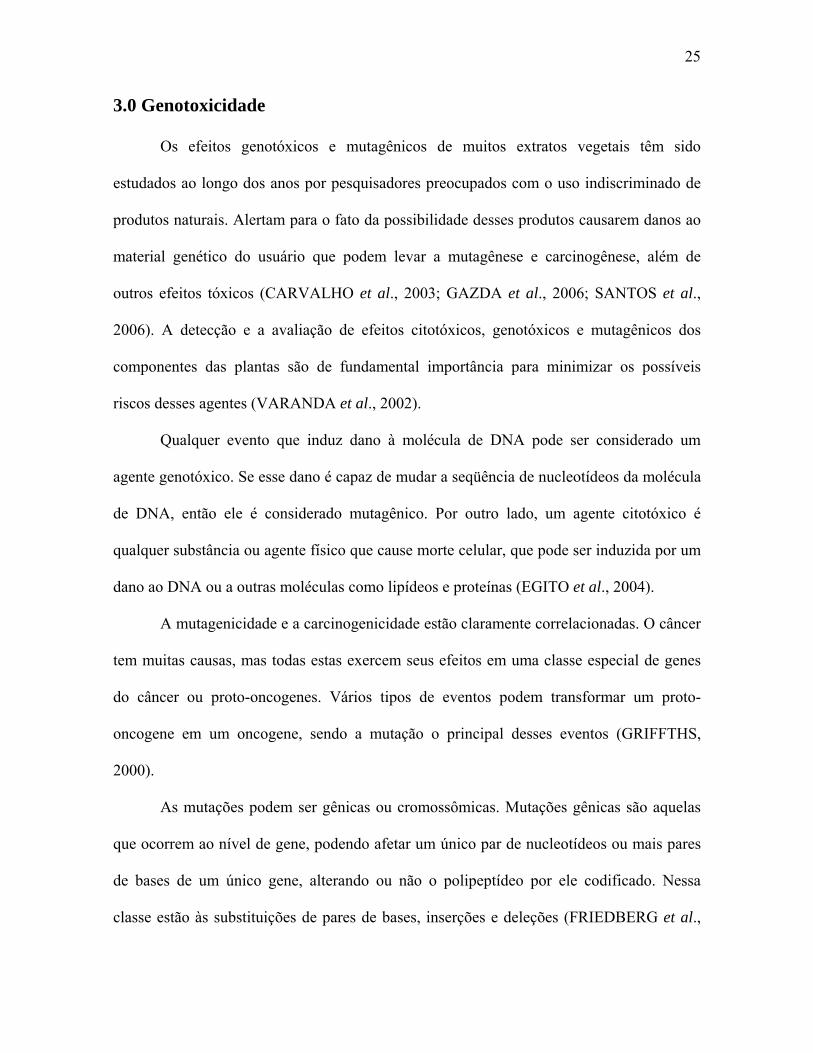
3.0 Genotoxicidade
Os efeitos genotóxicos e mutagênicos de muitos extratos vegetais têm sido
estudados ao longo dos anos por pesquisadores preocupados com o uso indiscriminado de
produtos naturais. Alertam para o fato da possibilidade desses produtos causarem danos ao
material genético do usuário que podem levar a mutagênese e carcinogênese, além de
outros efeitos tóxicos (CARVALHO et al., 2003; GAZDA et al., 2006; SANTOS et al.,
2006). A detecção e a avaliação de efeitos citotóxicos, genotóxicos e mutagênicos dos
componentes das plantas são de fundamental importância para minimizar os possíveis
riscos desses agentes (VARANDA et al., 2002).
Qualquer evento que induz dano à molécula de DNA pode ser considerado um
agente genotóxico. Se esse dano é capaz de mudar a seqüência de nucleotídeos da molécula
de DNA, então ele é considerado mutagênico. Por outro lado, um agente citotóxico é
qualquer substância ou agente físico que cause morte celular, que pode ser induzida por um
dano ao DNA ou a outras moléculas como lipídeos e proteínas (EGITO et al., 2004).
A mutagenicidade e a carcinogenicidade estão claramente correlacionadas. O câncer
tem muitas causas, mas todas estas exercem seus efeitos em uma classe especial de genes
do câncer ou proto-oncogenes. Vários tipos de eventos podem transformar um proto-
oncogene em um oncogene, sendo a mutação o principal desses eventos (GRIFFTHS,
2000).
As mutações podem ser gênicas ou cromossômicas. Mutações gênicas são aquelas
que ocorrem ao nível de gene, podendo afetar um único par de nucleotídeos ou mais pares
de bases de um único gene, alterando ou não o polipeptídeo por ele codificado. Nessa
classe estão às substituições de pares de bases, inserções e deleções (FRIEDBERG et al.,

26
1995). As lesões no DNA originam respostas celulares, designadas de reparo, que podem
ser classificadas em: reversão direta do dano, excisão do dano e tolerância do dano
(FRIEDBERG et al., 1995). As mutações cromossômicas podem afetar vários genes. Essa
classe se constitui por alterações tanto no número quanto na estrutura dos cromossomos
(THERMAN & SUSMAN, 1996).
Vários testes têm sido utilizados para a avaliação do potencial genotóxico e/ou
mutagênico de extratos vegetais, entre eles destacam-se o teste de kado (KADO et
al.,1983), o teste in vitro com DNA plasmidia, transformação bacterianal e o teste com
Allium cepa (RANK, 2003).
Dentre os vários ensaios microbianos para avaliação da genotoxicidade, o mais
comumente utilizado tem sido o teste desenvolvido por Ames et al. (1973) e modificado
por Maron e Ames (1983). Esse teste avalia o potencial do composto químico ou misturas
complexas de induzirem mutações no genoma de linhagens bacterianas mutantes
(auxotróficas) revertendo-as para o estado selvagem. Para aumentar a sensibilidade do teste
de Ames, Kado et al. (1983) desenvolveram uma metodologia baseada no aumento da
quantidade de bactérias utilizadas no ensaio e na redução da quantidade de amostra
utilizada.
O teste de Kado et al. (1983) utiliza as linhagens de Salmonella typhimurium,
especialmente construídas para esse ensaio, que apresentam mutações pontuais específicas
de vários tipos como transições, transversões e modificação do quadro de leitura, de modo
que o evento mutacional de reversão permite estabelecer a freqüência e o tipo de mutação
ocorrido. A mutação leva à inativação de uma enzima da via biossintetizante da histidina
(his) impedindo, portanto, seu crescimento em meio desprovido de tal aminoácido. Dessa
forma, em meio mínimo, somente conseguirão crescer aquelas colônias que foram capazes
27
de reverter à mutação, passando, portanto, à condição selvagem, ou seja, sendo capaz de
sintetizar a histidina (his+).
Essas cepas também apresentam outras modificações genéticas que aumentam a
sensibilidade do teste tais como a mutação no gene rfa que gera modificações na camada
lipopolissacarídica da parede celular, aumentando a permeabilidade bacteriana a entrada de
compostos e a mutação uvrB que torna as bactérias incapazes de reparar lesões no DNA por
excisão de nucleotídeos. A deleção no gene uvrB estende-se até o gene da biotina, de modo
que essas linhagens também são auxotróficas para esta vitamina (MARON & AMES,
1983). As linhagens mais utilizadas para avaliação de compostos mutagênicos são TA 97a e
TA 98, que detectam deslocamento do quadro de leitura do DNA, e TA 100 que detecta
substituição de pares de base no DNA (CETESB, 1991).
O teste de Kado também pode ser realizado com uma preparação de fígado de ratos
pré-tratados com Aroclor 1254, a fração microssomal S9 (MARON & AMES, 1983). S9
induz a síntese de enzimas microssomais, simulando, assim, a metabolização hepática em
um sistema in vitro, visando à detecção de substâncias pró-mutagênicas ou de metabólitos
com essa potencialidade.
O teste in vitro com DNA plasmidial detecta agentes capazes de quebrar ligações
fosfodiéster do DNA (CARVALHO et al., 2003), através da análise da conformação
plasmidial. Em virtude de se poder efetuar transformação bacteriana com o DNA
plasmidial tratado, é também possível analisar se outras lesões que não quebras estão sendo
geradas bem como obter informações a respeito da letalidade dessas lesões geradas no
plasmídeo após tratamento com o agente mutagênico.
A eletrofose em gel de agarose é uma forma simples e rápida para se visualizar e
separar fragmentos de DNA, valendo-se do fato dessa molécula apresentar carga negativa.

28
Para se avaliar as possíveis quebras no DNA, usa-se como parâmetro a forma e o tamanho
do DNA plasmidial. O plasmídeo íntegro se apresenta sob a forma superenovelada (forma
I), a qual migra rapidamente, colocando-se em uma posição mais distal em relação ao ponto
de aplicação do DNA plasmidial no gel. Se o plasmídeo sofrer quebras em uma das fitas do
seu DNA, ele assume a forma circular-relaxada (forma II), a qual, por expor uma maior
superfície de contato, migra mais lentamente e se coloca em uma posição mais proximal em
relação ao ponto de aplicação do DNA. Dessa forma, um aumento da forma II e
correspondentes reduções da forma I indicam quebra na cadeia fosfodiéster. Em casos
extremos, onde são geradas duas quebras na ligação fosfodiéster uma em cada fita de DNA,
o plasmídeo assume a forma linear (forma III) que apesar de expor uma maior superfície de
contato, é favorecida por sua conformação e migra mais rápido que a forma II, colocando-
se em uma posição intermediária entre as formas I e II no gel de agarose. Com base nesses
dados o perfil eletroforético no gel identifica os diferentes tipos de quebras que ocorreram
na cadeia do DNA plasmidial.
A conversão de um genótipo em outro pela introdução de DNA exógeno é
chamada de transformação. O DNA exógeno é incorporado ao cromossomo bacteriano por
um processo de quebra e inserção (GRIFFTHS et al., 2000). A maioria das espécies de
bactérias, inclusive a Escherichia coli, incorpora apenas quantidades limitadas de DNA sob
circunstâncias normais. Para transformar tais espécies eficientemente, as bactérias devem
passar pro alguma forma de tratamento físico e/ou químico que aumente as suas
capacidades de captação de DNA. Células submetidas a esse tratamento são ditas
competentes (BROWN, 2003).
Para a transformação bacteriana foi utilizada a
linhagem bacteriana DH10B. Essa linhagem só

29
pode ser transformada por eletroporação e não por
choque térmico (CALVIN & HANWALT, 1988;
DOWER et al., 1988). O vetor pBC foi derivado
do pBluescript® II phagemid, sendo o gene que
confere resistência a ampicilina trocado por um
gene de resistência a cloranfenicol. Elas contêm
também o gene lacZΔM15 que codifica parte da
enzima β-galactosidase. Genes que não possuem
inserto, não vão crescer como colônias azuis na
linhagem de bactéria apropriada que contenha o
gene lacZΔM15 como a E. coli DH10B. Caso não
tenha inserto, a mesma linhagem irá crescer como
colônias brancas, porque o inserto rompe a região
que codifica o fragmento do gene LacZ
(INVITROGEN,2003). Essa enzima é uma das
várias enzimas envolvidas na quebra de lactose
em glicose mais galactose. A clonagem com pBC
envolve a seleção de transformantes em agar com
cloranfenicol, seguida pela identificação de
recombinantes com base na atividade de β-
galactosidase.
A identificação da presença ou da ausência da β-galactosidase é realizada em um
ensaio um pouco diferente, também catalisada pela enzima. Essa reação envolve um
análogo da lactose, chamado de X-gal (5-bromo-4-cloro-3-indolil- β-D-
galactopiranosídeo), o qual é degradado pela β-galactosidase em um produto que tem
coloração azul-escuro. Se X-gal (mais um indutor da enzima, como o
isopropiltiogalactosídeo, IPTG) for adicionado ao ágar juntamente com cloranfenicol, as
colônias não-recombinantes, formada por células que sintetizam β-galactosidase, terão cor
azul, enquanto as recombinantes, com o gene lacZ interrompido e incapazes de produzir β-

30
galactosidase, serão brancas. Esse sistema, chamado de seleção lac. Os genes bacteriano e
plasmidial complementam-se um ao outro para produzirem uma molécula de β-
galactosidase funcional. Os recombinantes são selecionados a partir do plaqueamento em
agar contendo X-gal e IPTG (BROWN, 2003).
Outro ensaio biológico utiliza a cebola Allium cepa. As células mitóticas
meristemáticas de raízes de plantas são células indicadoras apropriadas para a detecção de
clastogenicidade dos poluentes ambientais, especialmente para o monitoramento dos
contaminantes da água e do solo (MA et al., 1995; RANK & NIELSON, 1997). Muitos
pontos podem ser monitorados nessas células em rápida divisão, como aberrações
cromossômicas, troca de cromátides irmãs e micronúcleo. A formação de micronúcleos é o
mais freqüentemente usado, o mais efetivo e o mais simples indicador de dano ao DNA
(MIGID et al., 2007). Em geral, a introdução de micronúcleo em meristemas de raízes é a
manifestação de quebra cromossômica ou distúrbio no processo mitótico devido a
anormalidades no fuso (GROVER & KAUR, 1999). Células que portam micronúcleo são
observadas em diferentes estágios do ciclo celular, embora a maioria dos micronúcleos seja
encontrada em células em interfase ou prófase. Em geral o micronúcleo observado é
sincrônico com a divisão do núcleo principal, mas em alguns casos esse sincronismo não
está presente (FERNANDES et al., 2007).

31
2. OBJETIVOS
2.1 Objetivo geral
Purificar a lectina solúvel em água da semente de Moringa oleifera (WSMoL) e
avaliar a genotoxicidade na água tratada com as sementes e da lectina isolada.
2.2 Objetivos específicos
Purificar WSMoL através de métodos cromatográficos;
Caracterizar bioquimicamente a lectina isolada;
Determinar a propriedade coagulante de WSMoL
Avaliar a genotoxicidade da WSMoL e da água tratada com M. oleifera, utilizando o
teste de Kado, in vitro com DNA plasmidial, transformação bacteriana e Allium cepa.

32
3. REFERÊNCIAS
ABDULKARIM, S. M.; LONG, K.; LAI, O. M.; MUHAMMAD, S. K. S.; GHAZALI, H.
M. Some physico-chemical properties of Moringa Oleifera seed oil extracted using solvent
and aqueous enzymatic methods. Food Chemistry, v.93, p. 253-263, 2005.
AMES, B. N.; McCANN, J.; YAMASAKI, E. Methods for detecting carcinogens and
mutagens with Salmonella/mammalian microsome mutagenesis test. Mut. Res, n. 31, 347-
364, 1973.
ANDRADE, C. A. S.; CORREIA, M. T. S.; COELHO, L. C. B. B.; NASCIMENTO, S. C.;
MAGALHÃES, N. S. S. Antitumor activity of Cratylia mollis lectin ancapsulated into
liposomes. International Journal of Pharmaceutics, v. 278, p. 435-445, 2004.
AMERICAN WATER WORKS ASSOCIATION (AWWA) Water quality and treatment; a
handbook of community water supplies, McGraw Hill Publishing Company, 4th edition,
New York, 1990
BASZKIN, A. BOISSONNADE, M., SANTOS-MAGALHAES, N. S., CARVALHO JR.,
L. B., CORREIA, M. T. S., COELHO, L. C. B. B. Cratylia mollis lectin at the air-aqueous
solution interface: adsorption and lectin-lipid interactions. Colloids and Surfaces B:
Biointerfaces, 17 191-201, 2000.

33
BELTRÃO, E. I. C., MEDEIROS, P. L., RODRIGUES, O. G., FIGUEIREDO-SILVA, J.,
VALENÇA, M. M., COELHO, L. C. B. B., CARVALHO JR, L. B. Parkia pendula lectin
as histochemistry marker for meningothelial tumor. European Journal of Histochemistry,
v.47, p. 139-142, 2003.
BEZERRA, A. M. E.; MOMENTE, V. G.; FILHO, S. M. Germinação de sementes e
desenvolvimento de plântulas de moringa (Moringa oleifera Lam.) em função do peso da
semente e do tipo de substrato. Horticultura Brasileira, Brasília, v.22, n.2, p.295-299,
abril-junho 2004.
BROWN, T. A. Clonagem gênica e análise de DNA: uma introdução. 4a ed. Artmed. 240p.
CACERES, A., CABRERA, O.; MORALES, O.; MOLLINEDO, P.; MENDIA, P.
Pharmacological properties of Moringa oleifera. 1: Preliminary screening for antimicrobial
activity. Journal of Ethnopharmacology, v. 33, n.3, p.213-216, 1991.
CALVIN, N. M. and HANWALT, P. C. J. Bacteriol. 170, 2796, 1988.
CARVALHO, M. C. R. D.; BARCA, F. N. T. V.; AGNEZ-LIMA, L. F.; AND
MEDEIROS, S. R.B. Evaluation of mutagenic activity in na extract of pepper tree stem
bark (Schinus terebinthifolius Raddi). Environmental and Molecular Mutagenegis, v.42,
185-191, 2003.
CETESB - Companhia de Tecnologia de Saneamento Ambiental. Teste de Kado- método
do ensaio.Norma CETESB L5 241, São Paulo, 1991.

34
COELHO, L. C. B. B.; SILVA, M. B. R. Simple method to purify milligram quantities of
the galactose-specific lectin from the leaves of Bauhinia monandra. Phytochemical
analysis, 11, p. 295-300, 2000.
CORREIA, M. T. S.; COELHO, L. C. B. B. Purification of a Glucose/ Mannose Specific
Lectin, Isoform 1, from Seeds of Cratylia mollis Mart. (Caramatu Bean). Applied
Biochemistry and Biotechnology, v. 55, p. 261-273, 1995.
CHUMKHUNTHOD, P et al. Purification and characterization of an N-acetil-D-
galactosamine-specific lectin from the edible musroom Schizophyllum commune.
Biochimica et Biophysica Acta. v. 1760, p. 326-332, 2006.
DELDUQUE, M (2000) Moringa. GloboRural, fascículo maio 2000, p. 89-91, 2000.
DOWER, W. J. et al. Nucl. Acids Research, 16, 6127, 1988. DRICKAMER, K. Two distinct classes of carbohydrate-recognition domains in animal
lectins. J. Biol. Chem. 263, 9557-9560,1988.
EGITO, L. C. M.; MEDEIROS, S. R. B.; MEDEIROS, M. G.; PRICE, J. C.; EGITO, S. T.
Evaluation of the relationship of the molecular aggregation state of amphotericin B in
medium to its genotoxic potencial. Journal of Pharmaceutical Sciences, v. 93, n. 6, p.
1557-1565, 2004.

35
FENG, K et al. Isolation and characterization of a novel lectin from the musroom
Armillaria luteo-virens. Biochemical and Biophysical Research Comunications, v. 345, p.
1573-1578, 2006.
FERNANDES, T. C. C.; MAZZEO, D. E. C. AND MORALES, M. A. M. Mechanism of
micronuclei formation in polyploidizated cells of Allium cepa exposed to trifluralin
herbicide. Pesticid Biochemistry and Phisiology, doi:10.1016/j.pestbp.2006.12.003, article
in press, 2007.
FRIEDBBERG, E. C.; WALLKER,G. C.; SIEDE, W. DNA repair and mutagenesis.
Washington: ASM Press, v.1, p. 407-437, 1995.
GASSENSCHMIDT, U.; JANY, K.D.; TAUSCHER, B.; NIEBERGALL, H. Isolation and
characterization of a flocculating protein from Moringa oleifera Lam. Biochemistry
Biophysical Acta, v.1243, p.477-481, 1995.
GAZDA, V. E.; CARNEIRO, M. R. G.; BARBI, N. S.; PAUMGARTTEN, F. J. R.
Toxicological evaluation of ethanolic extract from Chiocacca alba roots. Journal of
Ethonopharmacology, n. 105, p.187-195, 2006.
GHEBREMICHAEL, K. A., GUNARATNA, K. R., HENRIKSSON H., BRUMER, H.,
DALHAMMAR G. A simple purification and activity assay of the coagulant protein from
Moringa oleifera seed. Water Research 39, 2338-2344, 2005.

36
GRIFFTHS, A. J. F.; MILLER, J. H.; SUZUKI, D. T.; LEWONTIN, R. C.; GELBART, W.
M. Introdução à Genética, Guanabara koogan, 7a ed., Rio de Janeiro, 2000
GROVER, I. S. & KAUR, S. Genotoxicity of wastewater samples from sewage and
industrial effluent detected by the Allium root anaphase aberration and micronucleus
assays. Mutat. Res. n. 426, p. 183-188, 1999.
INVITROGEN. Discover a start-studded cast of competent cells: for all your
transformation – Catalogue of techniques. USA, 2003.
JANH, S. A. Proper use of African coagulants for rural water supply: Research in the
Sudan and a guide for new projects. Deutsche Gesellschaft für technische
Zusammenarbeit (German society for technical co-operation) (GTZ), Manual 191,
Eschborn,1986.
JANH, S. A. Chemotaxonomy of flocculating plant materials and their application for rural
water purification in developing countries. Acta Univ. Ups. Symb. Bot. Ups XXVIII 3,171-
185, 1988a.
JANH, S. A. Using Moringa oleifera seeds as coagulants in developing countries. J. Am.
Wat. Wks. Ass. 90, 43-50, 1988b.
KADO, N. Y.; LANGLEY, D.; EISENSTADT, E. A simple modification of the
Salmonella liquid incubation assay. Mut. Res. n. 121, p. 25-32, 1983.

37
KENNEDY, J. F., PAIVA, P. M. G., CORREA, M.T.S., CAVALCANTI, M. S. M.,
COELHO, L.C.B.B. Lectins, versatile proteins of recognition: a review. Carbohydrate
Polymers, Great Yarmouth, v.26, n.3, p.219-230, 1995.
LIMA, V. L. M.; CORREIA, M. T. S.; CHACHINEL, Y. M. N.; SAMPAIO, C. A. M.;
OWEN, J. S.; COELHO, L. C. B. B. Immobilized Cratylia mollis lectin as a potential
matrix to isolate plasma glycoproteins, including lecithin-cholesterol acyltransferase.
Carbohydrate Polymers, v. 33, p. 27-32, 1997.
LIS, H. & SHARON, N. Lectins: carbohydrate specific proteins that mediate cellular
recognition. Chem. Revs. 98, 637-674, 1998.
MA, T. H.; XU, Z.; XU, C.; MCCONNELL, H.; RABAGO, E. V.; ARREOLA, G. A.
ZHANG, H. The improvrd Allium/Vicia root tip micronucleus assay for clastogenicity of
environmental pollutants. Mutat. Res. n. 334, p. 185-195, 1995.
MACEDO, M. L. R.; FREIRE, M. G. M.; SILVA, M. B. R.; COELHO, L. C. B. B. (2006)
Insecticidal actino of Bauhinia monandra leaf lectin (BmoLL) against Anagasta kuehniella
(Lepidoptera: Pyralidae), Zabrotes subfasciatus and Callosobruchus maculates
(Coleoptera: Bruchidae). Comparative Biochemistry and Physiology, Part A, p. 13-25,
2006.
MACIEL, E. V. M.; FILHO, V.S.A.; NAKAZAWA, M.; GOMES, Y. M.; COELHO, L. C.
B. B.; CORREIA, M. T. S.; (2003) Mitogenic activity af Cratylia mollis lectin on human
lymphocytes. Biologicals, v. 32, p. 57-60, 2004.

38
MARON, D. M.; AMES, B. N. Revised methods for the Salmonella mutagenicity test.
Mut. Res, n. 113, p. 173-215, 1983.
MAZUMDAR, S; SENGUPTA, S. K.; PARAM, R.; SINHA, S. N. Binding pattern of eight
different lectins in healthy subjects and patients with dysplastic and malignant lesions of
the oral cavity. Int J Oral Maxillofac Surg. v. 22, n. 5, 301-305, 1993.
METHA, L. K.; BALARAMAN, R.; AMIN, A. H.; BAFNA, P. A.; GULATI, O. D. (2003)
Efects Of fruits of Moringa Oleifera on the lipid profile of normal and
hypercholesterolaemic rabbits. Journal or Etno-Pharmacology, v.86, p.191-195, 2003.
MIGID, H. M. A.; AZAB, Y, A.; IBRAHIM, W. M. Use of plant genotoxicity bioassay for
the evaluation of efficiency of algal biofilters in bioremediation of toxic industrial effluent.
Ecotoxicology and Enviromental Safety, n. 66, p. 5764, 2007.
MOURA, R. M.; QUEIROZ, A. F. S.; FOOK, J. M. S. L. L.; DIAS, A. S. F., MONTEIRO,
N. K. V.; RIBEIRO, J. K. C.; MOURA, G. E. D. D.; MACEDO, L. L. P.; SANTOS, E. A.
AND SALES, M. P. CvL, a lectin from the marine sponge Clione varians: Isolation,
characterization and its effects on pathogenic bacteria and Leishmania promastigotes.
Comparative Biochemistry and Physiology, Part A, doi: 10.1016/j.cbpa.2006.08.028. in
press, 2006.
MUYIBI, S. A. & EVISION, L. M.; Optimizing physical parameters affecting coagulation
of turbid water with Moringa oleifera seeds, Water Ressearch, vol. 29, n. 12, p. 2689-
2695, 1995.

39
NDABIGENGESERE, A., NARASIAH, K. S. (1998) Quality of water treated by
coagulation using Moringa oleifera seeds. Water Research 32, 781-791.
NDABIGENGESERE, A., NARASIAH, K. S., TALBOT, B. G. Active agents and
mechanism of coagulation of turbid waters using Moringa oleifera. Water Research 29,
nº2, 703-710, 1995.
NGAI, P. H. K. & NG, T. B. A mushroom (Ganoderma capense) lectin with spetacular
thermostability, potent mitogenic activity on splenocytes, and antiproliferative activity
toward tumor cells. Biochemical and Biophysical Research Communications, n. 314, p.
988-993, 2004.
OKUDA, T.; BAES, A. U.; NISHIJIMA, W.; OKADA, M. Isolation and characterization
of coagulant extracted from Moringa oleifera seed by salt solution. Water Research, v. 35,
p. 405-410, 2001a.
OKUDA, T.; BAES, A. U.; NISHIJIMA, W. AND OKADA, M. Coagulation mechanism
of salt solution-extracted active component in Moringa oleifera seeds. Water Research, 35
(3), 830-834, 2001b.
PEUMANS, W. J., VAN DAMME, E. J. M. Plant lectins: proteins with important
perspectives in biotechnology. Biotechnology and Genetic Engineering Reviews, v. 15, p.
199-228, 1998.

40
PEUMANS, W.J., VAN DAMME, E.J.M. Prevalence, biological activity and genetic
manipulation of lectins in foods. Trends Food Science Technology, Cambridge, v.7, n.4,
p.132-138, 1996.
POLLARD, S. J. T.; THOMPSON, F. E. AND McCONNACHE, G. L. Microporous
carbons from Moringa Oleifera husks for water purification in less developed countries.
Water Research, v.29, n.1, p. 337-347, 1995.
QUIOCHO, F. A. (1986) A. Rev. Biochem. 55, 287-315.
RANK, J; NIELSEN, M. H. Allium cepa anaphase-telophase root tip chromosome
aberration assay on N-methyl-N-nitrosourea, maleic hydrazide, sodium azide, and ethyl
methanesulfonate. Mutation Research, n. 390, p. 121-127, 1997.
RANK, J. The method of Allium anapase-telophase chromosome aberration assay.
Ekologija, n. 1, 2003.
SANTOS, F.V; COLUS, I. M. S.; SILVA, M. A.; VILEGAS, W.; VARANDA, E. A.
Assessment of DNA damage by extracts and fractions of Strychnos pseudoquina, a
Brazilian medicinal plant with antiulcerogenic activity. Food and Chemical Toxicology, n.
44, p. 1585-1589, 2006.

41
SANTOS, A. F. S.; ARGOLO, A. C. C.; COELHO, L. C. B. B.; PAIVA, P. M. G.
Detection of water soluble lectin and antioxidant component from Moringa oleifera seeds.
Water Research, v.39, p. 975-980, 2005.
SANTOS, A.C.O., PEIXOTO, C.A., COELHO, L.C.B.B. Ultrastructural analysis and
imunocytochemical localization of isolectins in Cratylia mollis seeds. Micron. v.35, p. 613-
618, 2004.
SHARON, N.; LIS, H. History of lkectins: from hemaggutininins to biological recognition
molecules. Glicobiology, v.14, n. 11, p. 53R-62R, 2004.
SHARON, N. Memories of a Sênior Scientist – A life with lectins. Cellular and Molecular
Life Sciences, v. 62, p. 1057-1062, 2005.
SHARON, N. Lectin-carbohydrate complexes of plants and animals: an atomic view.
Trends in Biochemical Science, v.18, n. 6, p. 221-226,1993.
SHARON, N. and LIS, H. ´Legum Lectins – A large family of homologous proteins´ in
FASEB J. 4,3198-3208, 1990.
SILVA, A. R.; KERR, W. E. (1999) Moringa: uma nova hortaliça para o Brasil.
Uberlândia: UFU/DIRIU, p 95.

42
SINGH, R. S., TIWARY, A. K., KENNEDY, J. F. (1999) Lectins: Sources, Activities and
Applications. Critical Reviews in Biotecnolog, v. 19, n. 2, p.145-178, 1999.
SUTHERLAND, J. P.; FOLKARD, G.K.; MTAWALI, M. A.; GRANT, W. D. (1994)
Moringa oleifera as a natural coagulant. Affordable water supply and sanitation, 20th
WEDC Conference, Colombo, Sri Lanka, p. 297-299, 1994.
THERMAN, E. E. & SUSMAN, M. Cromosomas humanos: estructura, comportamiento y
efectos. 3a ed. Ribeirão Preto: SBG, p. 335-337, Cromossomos, 1996.
TSAKNIS, J., LALAS, S., GERGIS, V., DOURTOGLOU, V., SPILIOTIS, V.
Characterization of Moringa oleifera variety Mbololo seed oil of Kenya. Journal of
Agricultural and Food Chemistry 47, 4495– 4499, 1999.
VARANDA, E. A.; POZETTI, G. L.; LOURENÇO, M. V.; VILEGAS, W.;
RADDI, M. S. G. Genotoxicity of Brosimum gaudichaudii measured by the
Salmonella/microsome assay and chromosomal aberrations in CHO cells. Journal of
Ethnopharmacology, v. 81, p. 257-264, 2002.
VATTUONE, M. A., PRADO, F. E., SAYAGO, J. E. & SAMPIETERO, A. R.
Phytochemistry, 30, 419-422, 2001.

43
VILLASENOR, I. M.; LIM-SYLIANCO, C. Y.; DAYRIT, F. Mutagens from roasted
seeds of Moringa oleifera seeds. Mutation Research/ Genetic toxicology, v. 224(2), p.
209-212, 1989.
THERMAN, E. E. & SUSMAN, M. Cromosomas humanos: estructura, comportamiento y
efectos. 3a ed. Ribeirão Preto: SBG, p. 335-337, Cromossomos, 1996.
44
4. Artigo a ser submetido ao periódico Water Research
Moringa oleifera seed lectin had coagulant property and was not a genotoxic agent, however extracts of seeds promoted micronucleus formation.
L.A.D.M.M. Rolim1, M.F.S. Macedo2, H.A.A.A.C.N. Sisenando2, A.F.S. Santos1,
L.C.B.B. Coelho1, S.R.B. de Medeiros2, P.M. G. Paiva1∗
1Departamento de Bioquímica, CCB/UFPE, Av. Prof. Moraes Rego, S/N, Cidade Universitária, Recife-PE, 50670-420, Brasil. 2Laboratório de Biologia Molecular e Genômica, CCB/UFRN, Natal-RN, Brasil.
∗ Corresponding author: Tel./fax: +5508121268540. e-mail address: [email protected] (P.M.G. Paiva).

45
Abstract
This paper reports the isolation and characterization of Moringa oleifera seed lectin
(WSMoL). Its effects on water turbidity as well as genotoxic/mutagenic potential of
water treated with different concentrations of M. oleifera seeds (MoW1, MoW2,
MoW3 and MoW4) or WSMoL were also discussed. WSMoL, agglutinated different
erythrocytes, was mainly active at pH 4.5 to 7.5 and inhibited by azocasein. Gel
filtration chromatography and electrophoretic profiles revealed WSMoL as 102 kDa
protein constituted by 5 kDa subunits. Glycosylation was not detected. WSMoL had
coagulant property similar to the most commonly used coagulant alluminum
sulfate. MoW preparations and WSMoL were negative in a cell-free plasmid DNA
test, bacterial transformation assay and in Salmonella typhimurium assay using
TA97, TA98 and TA100 at presence or absence of hepatic metabolization. MoW1
and MoW2 were also negative by Allium cepa assay however micronucleus
formation was induced by MoW3 and MoW4. The results to suggest WSMoL is an
active component in the water purification process by seeds. WSMoL was not a
genotoxic agent but genotoxicity was detected in water treated with more
concentration of seeds. This study is a positive step forward in determining the
safe use of M. oleifera seeds for water treatment.
Key Words: Moringa oleifera, lectin, coagulant property, genotoxicity, mutagenicity.

46
1. Introduction
Moringa oleifera Lam belongs to the family Moringaceae. Seeds are widely
used in developing countries as a natural coagulant for water treatment. It can
reduce in 92-99% the turbidity (Muyubi and Evinson, 1995) and the microbial
population (Ghebremichael et al., 2005). The major disadvantage in the use of M.
oleifera for water treatment is a significant increase of organic material
(Ndabigengesere and Narasiah, 1998; Okuda et al., 2001b). The use of pure or
semi-pure form of active coagulant component has been suggested for reducing
the total amount of organic material added to the treatment process
(Ghebremichael et al., 2005).
Studies have demonstrated coagulant activity of cationic proteins isolated
from M. oleifera seeds and Ghebremichael et al. (2005) suggested the evolvement
of protein mixture in the process. Monomeric or dimeric protein structures with
molecular mass of 6.5 to 13 kDa and isoelectric point in the range of 9.6 to 11 have
been described (Gassenschmidt et al., 1995; Ndabigengesere et al., 1995;
Ghebremichael et al., 2005).
Santos et al. (2005) detected the presence of a water soluble M. oleifera
lectin (WSMoL) in water obtained after soaking intact seeds. Lectins are
hemagglutinating proteins possessing carbohydrate binding sites (Peumans and
Van Damme, 1996). The availability of a great number of lectins with distinct
carbohydrate specificity makes these proteins tools in medical and biological
research (Kennedy et al., 1995).
Genotoxic and mutagenic effects have been reported in vegetable extracts.
The indiscriminate consumes of such products by people has stimulated studies

47
that to evaluate cytotoxic, genotoxic or mutagenic effects of plant compounds
aiming to minimize the possible risks of these agents (Varanda et al., 2002).
Damage to the users’ genetic material could lead to mutagenesis and
carcinogenesis as well as other toxic effects (Carvalho et al., 2003; Gazda et al.,
2006; Santos et al., 2006).
This paper reports upon the results of WSMoL isolation and its coagulant
property. Another objective of the present study was to evaluate the DNA damage
potential of WSMoL and of water treated with M. oleifera seeds. This water is used
as a source of drinking water around the world. Thus its quality for assessment of
genotoxic and mutagenic agents is important since plant compounds can have
adverse health effects on human including damage to DNA.
2. Materials and Methods
2.1. Protein evaluation
The protein concentration was estimated in all samples according to Lowry
et al. (1951) using bovine serum albumin (31-500 µg ml-1) as standard.
Absorbance at 280 nm was also measured.
2.2. Hemagglutinating activity (HA)
Hemagglutinating activity (HA) was evaluated according to Correia and
Coelho (1995) using glutaraldehyde-treated rabbit or human erythrocytes (types A,
AB and O). The HA was obtained by mixing a twofold serial dilution of samples (50
µl) in 0.15 M NaCl followed by the addition of a 2.5% (v/v) suspension of

48
erythrocytes (50 µl), in microtiter plates (Kartell S. P. A., Italy) and incubation (45
min). Titer was defined as the lowest sample concentration, which showed
hemagglutination. Specific HA (SHA) was calculated from the ratio of titer to
protein concentration (mg ml-1).
A HA inhibitory assay was performed by incubation (45 min) of lectin sample
with 200 mM carbohydrates (N-acetylglucosamine, D(+)-fructose, D(+)-galactose,
D(+)-glucose and raffinose) or 0.5 mg ml-1 glycoprotein solutions (azocasein, fetuin
and ovalbumin), before rabbit erythrocyte suspension addition. HA was also
evaluated at different temperatures (30-100ºC, 15 min) and pH values (10 mM
citrate phosphate buffer containing 0.15 M NaCl, pH 4.5, 5.0, 6.0, 6.5 or 10 mM
sodium phosphate buffer containing 0.15 M NaCl, pH 7.0, 7.5 or 10mM Tris-HCl
containing 0.15 M NaCl, pH 8.0, 8.5, 9.0, 9.5).
2.3. M. oleifera water (MoW)
Mature seeds from M. oleifera were collected in Recife city, State of
Pernambuco, Brazil Northeast. Taxonomic identification was performed and
voucher specimens were deposited under number 73,345 (IPA – Instituto de
Pesquisas Agropecuárias de Pernambuco).
M. oleifera water was obtained according to protocol for treatment of turbid
water used by people (Gerdes, 1996) except the filtration steps. Macerated shelled
seeds (2.0 g) were added to distilled water (100 ml) and manual agitation (5 min)
was made. Following, this M. oleifera suspension was through on filter paper and
sterile millipore filter (0.20 μm). The filtered suspension was then added to milli-Q

49
water to obtain M. oleifera waters (MoW) of concentration (g l-1) 0.0125 (MoW1),
0.05 (MoW2), 0.2 (MoW3) and 0.8 (MoW4). MoW3 corresponds to concentration
consumed by people.
2.4. Purification of WSMoL
Mature seeds were milled to a fine powder. The meal (10 g) was then
homogenised in a magnetic stirrer (16 h at 4°C) with distilled water (100 ml),
filtered through gauze and centrifuged at 3,000 x g (15 min). The supernatant,
crude extract (CE), was taken as starting material. The initial SHA of CE was
considered as the initial activity of the isolation procedure. The proteins in the CE
were precipitated using ammonium sulphate (saturation of 60%) at 4°C under
magnetic agitation (Green and Hughes, 1955). The sediment separated by
centrifugation (0-60 fraction) was dissolved in 0.15 M NaCl. The 0-60 (80 mg of
proteins) was applied onto a chitin column (23 x 0.75 cm) equilibrated with 0.15 M
NaCl (0.3 ml min-1 flow rate). After extensive washing with the equilibrium solution,
elution was performed with 1.0 M acetic acid. Spectrophotometry at 280 nm was
used to follow protein elution. Fractions with lectin activity (WSMoL) were pooled,
dialysed against 0.15 M NaCl (6 h at 4°C). Purification was measured as the ratio
between the SHA in the stage and SHA of CE. Yield was measured by the ratio of
HA values.

50
2.5. Polyacrylamide gel electrophoresis (PAGE)
SDS-PAGE was performed on 10% (w/v) gel according to the method
described by Laemmli (1970) with or without β-mercaptoethanol. Peptide of
WSMoL (100 μg of protein) and solution of molecular mass standard constituted by
four peptides with 6.5, 12.5, 21 and 29 kDa fragments (Q-BIOgene, USA) were
stained with Coomassie Brilliant Blue. Glycoprotein staining was also performed
with Schiff´s reagent (Pharmacia Fine Chemicals, 1980). PAGE for native basic
[7.5% (w/v) gel] and acidic [7.5% (w/v) gel] proteins was carried out according to
the protocols of Reisfeld et al. (1962) and Davis (1964), respectively.
2.6. Gel filtration chromatography
WSMoL was chromatographed by gel filtration on a Hiprep 16/60 Sephacryl
S-300 column (16 mm x 60 cm)/Äkta FPLC system (Amersham Pharmacia
Biotech, Sweden) pre-equilibrated at 24 ºC with 500 mM NaCl. Samples (2.0 ml
containing 4 mg of protein) were injected and eluted (3.0 ml fraction) with 500 mM
NaCl at a flow rate of 0.5 ml/min. Bovine serum albumin (66 kDa), fetuin (64 kDa),
ovalbumin (45 kDa) and the trypsin inhibitor type III-O chicken (28 kDa) standards
were similarly chromatographed.
2.7. Coagulation assay
Coagulation assay was performed according Ghebremichael et al. (2005).
Initially, synthetic turbid water was prepared. Distilled water (1 L) was treated with
kaolin clay (10 g), stirred for 30 min and allowed to settle for 24 h to achieve

51
complete hydration. Desired turbidity was obtained by dilution. Aliquot (0.3 ml) of
WSMoL (1 mg ml-1) or positive control (5% aluminium sulphate) was added to clay
suspension (2.7 ml, 250-300 NTU, Nephelometric Turbidity Units). Samples were
allowed to settle for 1 h at 27 °C. In order to reduce background effect, a sample
volume of 900 μl from the top was transferred to the cuvette and absorbance
measured at 500 nm using a UV-Visible spectrophotometer FEMTO 700 S
corresponded to initial absorbance (time 0). Then, absorbance was determined
every 5 min up to 60 min and to each 10 min up to 140 min. Reduction in
absorbance relative to control defines coagulation activity. The assays were
performed three times.
2.8. Kado test
Mutagenicity was measured according Kado et al. (1983) without and with
addition of an extrinsic metabolic activation system, S9 mixture (Aroclor-induced
rat liver S9 from Moltox), using Salmonella typhimurium TA97, TA98 and TA100.
Aliquots (5 μl) of MoW1, MoW2, MoW3, MoW4 or WSMoL (0.0125, 0.05, 0.2 and
0.8 mg ml-1) were analyzed. Before Kado test, cytotoxicity screening was
performed with the TA98 strain using the same sample concentrations. The
positive controls used in the assays performed without metabolic activation were:
4-nitroquinoline-N-oxide (0.5 μg per plate) for TA97 and TA98 and sodium azide (5
μg per plate) for TA 100. In the assays with metabolic activation, the positive
control was 2-aminoanthracene (5 μg per plate) for all strains.

52
The bacteria/sample/top agar mixture was poured onto a Petri dish
containing minimal agar (1.5% agar, Vogel-Bonner E medium, with 2% glucose).
His+ revertants were counted after 66 h of incubation at 37° C. The test substance
was considered mutagenic if the number of revertants was at least double
spontaneous frequency. The assay was repeated three times.
2.9. Plasmid DNA test
Genotoxicity was evaluated by plasmid DNA assay. The pBC plasmid with
cloranphenicol-resistance gen was used in the test. Plasmid (1 μg) was incubated
(1 h at 37°C) with aliquots (20 μl) of MoW1, MoW2, MoW 3, MoW4 or WSMoL
(0.0125, 0.05, 0.2 and 0.8 mg/ml). The assays included a negative control
(supercoiled or form I plasmid) and a positive control (plasmid incubated with
hydrogen peroxide by 30 min at 37°C following 30 min at 60°C). The treated
plasmids were submitted to electrophoresis on 0.7% (w/v) agarose in 1x TBE
buffer (90 mM Tris, 90 mM boric acid, 2 mM EDTA buffer, pH 7.0) and analyzed at
UV light.
2.10. Bacterial transformation test
Competent bacteria Escherichia coli DH10B (F- mcrA Δ(mrr-hsdRMS-
mcrBC) φ80lacZΔ.M15 ΔlacX74 deoR recA1 endA1 araΔ139 Δ(ara, leu)7697 galU
galK λ- rpL(StrR) nupG tonA) from Invritogen (USA) were obtained according
Sambrook et al. (1989). Plasmid pBC (2 μl) was incubated (1 h) with 48 μl of
MoW1, MoW2, MoW3, MoW4 or WSMoL (0.0125, 0.05, 0.2 or 0.8 mg ml-1) then 1
μl of each mixture (10 ng μl-1) was mixed with DH10B (50 μl). Following,

53
electroporation according to Gene Pulser electroprotocols (BIO-RAD) was
performed to permit the insertion of plasmid DNA pretreated with M. oleifera
samples in the bacteria. All of these procedures were carried out on ice. After E.
coli electroporated was added to Lennox Broth (LB) medium and putted in shacker
(200 rpm) for 1 h at 37°C. Then, aliquots were plated onto LB-agar containing 0.1%
of cloranphenicol and 0.08% of X-Gal. Plates were incubated overnight at 37°C. A
mean value of number of colony-forming units (CFUs) from each M. oleifera
sample employed was obtained, and the transformation efficiency was expressed
as the percentage of the maximal value (100%), attributed to the negative control.
Value lowest that 50% to indicate cytotoxicity. The colonies were also observed in
relation to its coloration for mutagenicity detection. Blue and white colors identify
non-mutagenic and mutagenic effect, respectively. Negative (distilled and
autoclaved water) and positive (1 μl of Bam HI endonuclease restriction enzyme, 5
μl of buffer for Bam HI and 40 μl distilled and autoclaved water) controls were
used. This experiment was carried out in triplicate and the results presented are
the mean average.
2.11. Allium cepa test
Allium cepa test was carried out according Rank (2003). Allium cepa onions
bulbs were purchased from the local market in Natal city, Brazil. The yellow
shallows and the dry parts inside the root primordial were removed before the test.
Roots were initially exposed to distilled water (24 h at 27°C) following to MoW
preparations (48 h at 27°C). The roots were then submitted to acid hydrolysis (1N

54
HCl at 60ºC for 10 min) and incubated with Schiff´s reactive (30 min at dark). Root
tips were cut onto a slide for extraction of meristematic region. The negative control
was performed only in distilled water. 5,000 cells were counted per treatment to
observe the presence or not of micronucleus. Results were expressed as number
of micronuclei per 1,0000 cells.
2.12. Statistical analysis
The computer package GraphPad InStat, version 3.0 was used for statistical
analysis. For Kado test, analysis of variance (ANOVA) and Dunnett test were
utilized. For bacterial transformation and Allium cepa tests values were evaluated
by analysis of variance (ANOVA) and Tukey test.
3. Results and discussion
The coagulant property of M. oleifera seeds has been indicated as an alternative process for water purification in Brazilian rural regions. The excellent seed availability, facility of methodology recommended and quality of water obtained have contributed for its acceptance by people. Additional advantage for Moringa use is the antimicrobial activity detected in seeds (Cáceres et al., 1991).
Although M. oleifera seeds are efficient in water treatment, due to its usage has a major impact on the health of a large number of people it is important to characterize the seeds in relation to source of others bioactive compounds as well as their potential effect on DNA molecule. This paper to contribute for diagnosis of water treated with M. oleifera.
Santos et al. (2005) detected HA in extracts obtained by soaking
of M. oleifera intact seeds and named WSMoL. This paper describes the
protocol for lectin isolation. Water extract obtained using seeds powder
presented SHA of 22 with rabbit erythrocytes. The extract proteins were
than concentrated in the 0-60 ammonium sulphate fraction that was
chromatographed on chitin column. WSMoL, adsorbed on matrix and eluted
with acetic acid (Figure 1A), showed SHA of 2,985 corresponding to
purification of 137 folds. Each chromatographic experiment yielded 3.4
mg of lectin.

55
The HA of WSMoL was inhibited by carbohydrates and glycoproteins
and stable to pH and temperature (Table 1). Gel filtration
chromatography profile showed unique peak of 102 kDa revealing the high
native molecular mass of WSMoL. The high molecular mass leaded to low
protein migration on 7.5% polyacrylamide gel for native acid protein.
Protein band was not detected on PAGE for native basic protein. SDS-
PAGE (Figure 1B) resolved WSMoL as a diffuse peptide band with
molecular mass between 20 and 5 kDa. After treatment with reducing
agent β-mercaptoethanol was detected a 5 kDa band. The peptides were
not marked on gel by the glycoprotein staining procedure. Glycoproteins
are identified by a magenta coloration (Moura et al., 2006).
Comparison of results from gel filtration chromatography (native
protein) with SDS-PAGE with and without disulfide bonds reducing agent
indicates the molecular structure of WSMoL as multiple subunit aggregate
maintained by non-covalent and covalent bonds. Thus, a possible structure
of WSMoL may be a polypeptide constituted by arrangement of five 20
kDa peptides that were decomposed to four subunits of 5 kDa subunits.
Oligomeric structure was similarly proposed for the lectin isolated from
marine sponge Cliona varians, a 114 kDa protein constituted by 28 kDa
subunits (Moura et al., 2006). Generally, protein constituted by multiple
subunits presents stability to pH and temperature variations
(Chumkhunthod et al., 2006) probably due to high number of forces that
maintains the native structure.
Comparison of isolated WSMoL with HA before described (Santos et
al., 2005) reveals WSMoL similarly active with different erythrocytes and
inhibited by carbohydrates and glycoproteins. However differences were
56
detected in relation to activity at pH 7 and after heating at 100 °C.
Also, the developed purification approach resulted in most active lectin.
The highest SHA detected by Santos et al. (2005) was 0.9. The
purification process can have caused the differences searched since
modification in protein activity has been described after chromatography
procedures (Quiocho, 1986). Indeed, slight alterations in lectin structure
may modify the protein activity (Ng et al., 1996).
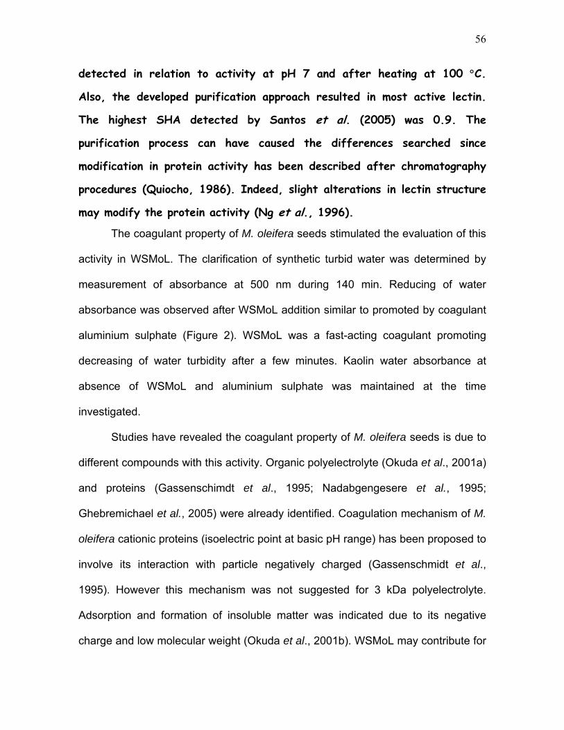
The coagulant property of M. oleifera seeds stimulated the evaluation of this
activity in WSMoL. The clarification of synthetic turbid water was determined by
measurement of absorbance at 500 nm during 140 min. Reducing of water
absorbance was observed after WSMoL addition similar to promoted by coagulant
aluminium sulphate (Figure 2). WSMoL was a fast-acting coagulant promoting
decreasing of water turbidity after a few minutes. Kaolin water absorbance at
absence of WSMoL and aluminium sulphate was maintained at the time
investigated.
Studies have revealed the coagulant property of M. oleifera seeds is due to
different compounds with this activity. Organic polyelectrolyte (Okuda et al., 2001a)
and proteins (Gassenschimdt et al., 1995; Nadabgengesere et al., 1995;
Ghebremichael et al., 2005) were already identified. Coagulation mechanism of M.
oleifera cationic proteins (isoelectric point at basic pH range) has been proposed to
involve its interaction with particle negatively charged (Gassenschmidt et al.,
1995). However this mechanism was not suggested for 3 kDa polyelectrolyte.
Adsorption and formation of insoluble matter was indicated due to its negative
charge and low molecular weight (Okuda et al., 2001b). WSMoL may contribute for
57
coagulant activity of seeds by charge interaction inducing the formation of
interparticle bridging due to both its negative charge and high molecular mass.
To determine whether M. oleifera water (MoW) or WSMoL cause mutation
the reverse mutagenesis test (Kado test) was made using S. typhimurium, strains
TA97, TA98 and TA100 in the presence and absence of the S9 microssome
fraction (hepatic metabolization). Frameshift-type (TA97 and TA98) or for base pair
substitution (TA100) mutagens were not detected. As shown in Table 2, the
samples at different concentrations did not cause any increase in the number of
his+ revertant colonies over double of the negative control values obtained for the
3 tester strains corresponding to mutagenic index < 2. The highest MoW or
WSMoL concentration used in this work (0.8 g l-1) is closely 4-fold higher than the
dose used by people for water treatment (0.2 g l-1). Even in this concentration
negative results were obtained in the Kado test. The evaluation in a range of
concentration was made because concentrated fractions of non-mutagenic plant
presented mutagenic activity (Cardoso et al., 2006). The Kado test has been used
to evaluate water under the impact of genotoxic agents and enabled the detection
of S9 activated frameshift mutagens (Cardozo et al., 2006). Also, provided
experimental evidence for the non-mutagenic (Marques et al., 2003; Gazda et al.,
2006) or mutagenic (Santos et al., 2006) effects of Brazilian medicinal plants.
The genotoxic potential of MoW or WSMoL was evaluated in a cell-free
plasmid DNA test. The conformational changes in plasmid DNA treated with M.
oleifera samples were analyzed through agarose gel electrophoresis. MoW1, 2 and
3 or WSMoL did not generate breaks in the phosphodiester bonds of DNA since
changes in plasmid supercoiled form was not detected on gel (Figure 3A and 3B).

58
Plasmid treated with MoW4 did not migrate on gel (Figure 3A) probably due to its
high molecular weight resulting of its complexion with MoW4 constituents.
Interaction of plant compounds with DNA has been already described (Sengupta et
al., 2005; Giri et al., 2006). Carvalho et al. (2003) using plasmid DNA test
additionally to S. typhimurium DNA damage assay, detected interaction of
compounds with DNA and suggested that the popular medicinal use of Schinus
terebinthifolius extract as anti-inflammatory agent may present a health risk to
humans.
Cytotoxicity of MoW samples and WSMoL was evaluated by bacterial
transformation test (Figure 4). Bacterial survival percentages determined for
MoW1, MoW2, MoW3 as well as WSMoL in all tested concentrations (0.0125,
0.05, 0.2 and 0.8 g l-1) were higher than 50% indicating absence of cytotoxicity.
MoW4 promoted the lowest bacterial survival percentage, 47% was detected. In
this assay X-gal was used aiming to detect DNA alteration by colony coloration
modification of blue for white. Only blue CFUs were observed indicating
maintaining of bacteria ability to promote X-gal degradation. Thus mutagen agent
was not detected in M. oleifera samples. The later results suggesting that the
bacterial survival decrease in presence of MoW4 probably was not due to DNA
damage. A hypothesis to explain the phenomenon may be increase in the
membrane cell permeability induced by MoW4 constituents favoring the entry of
molecules that leads to bacterial death similar to describe to Aloe vera extract
(Paes-leme et al., 2005). Indeed alteration of plasmid molecular weight was
detected only after MoW4 treatment (Figure 3A).

59
Genotoxicity was evaluated by A. cepa test that detect alterations in the
cells from meristematic regions of roots (Rank and Nielsen, 1997). MoW
preparations and distilled water (negative control) were evaluated. It was observed
that micronuclei were not detected with MoW1 and MoW2 induced micronucleus
formation (0.6±0.5) similar to negative control (0.6±0.9). The number of
micronucleus after MoW3 and MoW4 treatments was 2.4±1.1 (p<0.05) and 4.8±1.3
(p<0.001), respectively. Although, detection of micronuclei in negative control can
be explained by spontaneous micronucleus formation (Fernandes et al., 2007) in
MoW2 this can be occurred or compounds present in MoW2 induced micronuclei
formation. In fact, in most high concentrations of M. oleifera extracts significant
increase of micronuclei was detected and additionally to this, a dose response
effect was observed with Mow4 promoting more effect than MoW3.
It has been recommended to Brazilian rural people that according to grade
of water turbidity the number of M. oleifera seeds used to promote effective
coagulant effect can be increased. Mow3 and MoW4 promoted micronucleus
formation in A. cepa test and since this plant assay has been considered as the
most effective and the simplest indicator of DNA damage (Migid et al., 2007) the
number of seeds used for water treatment should be monitored.
4. Conclusions
The purpose of this study was the isolation of water soluble M.
oleifera seed lectin (WSMoL), determination of its coagulant property and
evaluation of potential genotoxic and mutagenic of water treated with M.
oleifera seeds or WSMoL.

60
1. The results showed that seeds of M. oleifera were source of lectin. WSMoL is
stable to pH and temperature, biochemical properties proper for its
biotechnological utilization. The detected coagulant property of WSMoL to
suggests its involvement in the reduction of water turbidity. WSMoL was not
genotoxic/mutagenic agent in the concentrations studied. This result is of interest
due to its coagulant property. Antimicrobial activity of WSMoL is in development
aiming to define its potential role in water treatment.
2. The mutagenicity/genotoxicity evaluations revealed that water obtained
according to recommended for human consumption (MoW3) induced genotoxicity
in the in vivo assay. The effect was highest in the more concentrated MoW4 and
not detected in most diluted MoW1 and MoW2. In our opinion aiming to minimize
risk for human health, treatment of turbid water with lesser seed concentration
should be evaluated by its coagulant property. This study is a positive step forward
in determining the safe use of M. oleifera seeds for water treatment.
Acknowledgements
The authors express their gratitude to the Conselho Nacional de
Desenvolvimento Científico e Tecnológico (CNPq) for research grants and
fellowship (LCBBC). Also, the Fundação de Amparo à Ciência e Tecnologia do
Estado de Pernambuco (FACEPE) and the Coordenação de Aperfeiçoamento de
Pessoal de Nível Superior (CAPES) for financial support. The authors are deeply
grateful for the technical assistance of Maria Barbosa Reis da Silva.
61
References Caceres, A., Cabrera, O.; Morales, O.; Mollinedo, P. and Mendia, P. (1991)
Pharmacological properties of Moringa oleifera. 1: Preliminary screening for
antimicrobial activity. Journal of Ethnopharmacology 33(3), 213-216.
Cardoso, C. R. P.; Cólus, I. M. S.; Bernardi, C. C.; Sannomiya, M.; Vilegas, W. and
Varanda, E. A. Mutagenic activity promoted by amentoflavone and methanolic
extract of Byrsonima crassa Niedenzu. Toxicology 225, 55-63.
Cardozo, T. R.; Rosa, D. P.; Feiden, I. R.; Rocha, J. A. V.; Oliveira, N. C. D. A.;
Pereira, T. S.; Pastoriza, T. F.; Marques, D. M.; Lemos, C. T.; Terra, N. R. and
Vargas, V. M. F. (2006) Genotoxicity and toxicity assessment in urban
hydrographic basins. Mutation Research 603, 83-96.
Carvalho, M. C. R. D.; Barca, F. N. T. V.; Agnez-Lima, L. F.; and Medeiros, S. R.B.
(2003) Evaluation of mutagenic activity in na extract of pepper tree stem bark
(Schinus terebinthifolius Raddi). Environmental and Molecular Mutagenegis 42,
185-191.
Correia, M. T. S. and Coelho, L. C. B. B. (1995) Purification of a Glucose/ Mannose
Specific Lectin, Isoform 1, from Seeds of Cratylia mollis Mart. (Caramatu Bean).
Applied Biochemistry and Biotechnology 55, 261-273.
62
Davis, B.J. (1964) Disc electrophoresis II: methods and application to human
serum proteins. Annals of the New York Academy of sciences 121, 404-427.
Fernandes, T. C. C.; Mazzeo, D. E. C. and Morales, M. A. M. (2007) Mechanism of
micronuclei formation in polyploidizated cells of Allium cepa exposed to trifluralin
herbicide. Pesticid Biochemistry and Phisiology, doi:10.1016/j.pestbp.2006.12.003,
article in press.
Gassenschmidt, U.; Jany, K.D.; Tauscher, B. and Niebergall, H. (1995) Isolation
and characterization of a flocculating protein from Moringa oleifera Lam.
Biochemistry Biophysical Acta 1243, 477-481.
Gazda, V. E.; Carneiro, M. R. G.; Barbi, N. S. and Paumgartten, F. J. R. (2006)
Toxicological evaluation of ethanolic extract from Chiocacca alba roots. Journal of
Ethonopharmacology 105, 187-195.
Gerdes, G. (1997) Como limpar e tratar água suja com sementes da moringa.
ESPLAR - Centro de Pesquisa e Assessoria, Fortaleza, 18 (Boletim Técnico).
Ghebremichael, K. A.; Gunaratna, K. R.; Henriksson H.; Brumer, H. and
Dalhammar, G. (2005) A simple purification and activity assay of the coagulant
protein from Moringa oleifera seed. Water Research 39, 2338-2344.

63
Giri, P.; Hossain, M. and Kumar, G. S. (2006) Molecular Aspects on the specific
interaction of cytotoxic plant alkaloid palmatine to poly(A). International Journal of
Biological Macromolecules 39, 210-231.
Green, A. A. and Hughes, W. L. (1955) Protein fraction on the basis of solubility in
aqueous of salts and organic solvents. In Colowick, S. and Kaplan, N.- Methods in
Enzymology. New York, Academic Press, 67-90.
Kado, N. Y.; Langley, D. and Eisenstadt, E. (1983) A simple modification of the
Salmonella liquid incubation assay. Mut. Res 121, 25-32.
Kennedy, J. F.; Paiva, P. M. G.; Correia, M.T.S.; Cavalcanti, M. S. M. and coelho,
L.C.B.B. (1995) Lectins, versatile proteins of recognition: a review. Carbohydrate
Polymers, Great Yarmouth, 26(3), 219-230.
Laemmli, U. K. (1970) Cleavage of structural proteins during the assembly of the
head of bacteriophage T4. Nature 227, 680-685.
Lowry, O. H.; Rousebrought, N. J. Farr, A. L. and Randal, R. J. (1951) Protein
measurement with the folin phenol reagent. Journal of Biological Chemistry 193,
265-275.
Moura, R. M.; Queiroz, A. F. S.; Fook, J. M. S. L. L.; Dias, A. S. F., Monteiro, N. K.
V.; Ribeiro, J. K. C.; Moura, G. E. D. D.; Macedo, L. L. P.; Santos, E. A. and Sales,
M. P. (2006) CvL, a lectin from the marine sponge Clione varians: Isolation,

64
characterization and its effects on pathogenic bacteria and Leishmania
promastigotes. Comparative Biochemistry and Physiology, Part A, doi:
10.1016/j.cbpa.2006.08.028.
Muyibi, S. A. and Evinson, L. M. (1995) Optimizing physical parameters affecting
coagulation of turbid water with Moringa oleifera seeds. Water Ressearch 29(12),
2689-2695.
Ndabigengesere, A. and Narasiah, K. S. (1998) Quality of water treated by
coagulation using Moringa oleifera seeds. Water Research 32, 781-791.
Ndabigengesere, A.; Narasiah, K. S. and Talbot, B. G. (1995) Active agents and
mechanism of coagulation of turbid waters using Moringa oleifera. Water Research
29(2), 703-710.
Ng, K. K. S.; Drickamer, K. and Weis, W. I. (1996) Structural analysis of
monosaccharide recognition by rat liver mannose-binding protein. J. Biol. Chem.
271, 663-674.
Okuda, T.; Baes, A. U.; Nishijima, W. and Okada, M. (2001b) Coagulation
mechanism of salt solution-extracted active component in Moringa oleifera seeds.
Water Research, 35 (3), 830-834.

65
Okuda, T.; Baes, A. U.; Nishijima, W.; Okada, M. (2001a) Isolation and
characterization of coagulant extracted from Moringa oleifera seed by salt solution.
Water Research 35,. 405-410.
Paes-Leme, A. A.; Mota, E. S.; Mattos, J. C. P.; Dantas, F. J. S.; Bezerra, R. J. A.
C. and Caldeira-de-Araujo, A. (2005) Assessment of Aloe vera (L.) genotoxic
potencial of Escherichia coli and plasmid DNA. Journal of Ethnopharmacology 102,
197-201.
Peumans, W.J., Van Damme, E.J.M.(1996) Prevalence, biological activity and
genetic manipulation of lectins in foods. Trends Food Science Technology.
Cambridge 7 (4), 132-138.
Pharmacia Fine Chemicals (1980) Polyacrylamide Electrophoresis; Laboratory
Techniques. Pharmacia, Uppsala.
Quiocho, F. A. (1986) A. Rev. Biochem. 55, 287-315.
Rank, J. (2003) The method of Allium anaphase-telophase chromosome
aberration assay. Ekologija, 1.
Rank, J. and Nielsen, M. H. (1997) Allium cepa anaphase-telophase root tip
chromosome aberration assay on N-methyl-N-nitrosourea, maleic hydrazide,
sodium azide, and ethyl methanesulfonate. Mutation Research 390, 121-127.

66
Reisfeld, R. A.; Lewis, U. J. and Williams, D. E. (1962) Disk electrophoresis of
basic proteins and peptides on polyacrilamide gels. Nature 195, 281-283.
Sambrook, J.; Fritsch, E. F. and Maniatis, T. (1989) Molecular cloning – a
laboratory manual. Cold Spring Harbor Laboratory Press 1, 2nd edition.
Santos, A. F. S.; Argolo, A. C. C.; Coelho, L. C. B. B. and Paiva, P. M. G. (2005)
Detection of water soluble lectin and antioxidant component from Moringa oleifera
seeds. Water Research 39, 975-980.
Santos, F.V; Colus, I. M. S.; Silva, M. A.; Vilegas, W. and Varanda, E. A. (2006)
Assessment of DNA damage by extracts and fractions of Strychnos pseudoquina,
a Brazilian medicinal plant with antiulcerogenic activity. Food and Chemical
Toxicology 44, 1585-1589.
Sengupta, B.; Banerjee, A. and Sengupta, P. K. (2005) Interactions of plant
flavonoid fisetin with macromolecular targets: Insights from fluorescence
spectroscopic studies. Journal of Photochemistry and Photobiology B: Biology 80
(2), 79-86.
Varanda, E. A.; Pozetti, G. L.; Lourenço, M. V.; Vilegas, W.; Raddi, M. S. G.
(2002) Genotoxicity of Brosimum gaudichaudii measured by the
Salmonella/microsome assay and chromosomal aberrations in CHO cells. Journal
of Ethnopharmacology 81, 257-264.

67

68
Table 1.Characterization of WSMoL
Characteristic Determination
SHA Rabbit (2,985)
Human types: A (746), AB and O (1,492)
SHA at inhibitors presence* Carbohydrates (200 mM): galactose (1,492),
glucose (0), fructose and N-acetylglucosamine
(746), raffinose (0)
Glycoproteins (0.5 mg ml-1): azocasein (12),
ovalbumin (82), fetuin (2,985)
SHA at pH values* 4.5 (5,971), 5.0 and 6.0 (373), 6.5 to 7.5 (1,492),
8.0 to 9.5 (186)
SHA at temperature values* 30 to 80 °C (1,492), 90 and 100 °C (93)
Native molecular mass 102 kDa
Subunit molecular mass 20 kDa (SDS), 5 kDa (SDS + β mercaptoethanol)
Glycosylation No detected
Specific hemagglutinating activity (SHA) was calculated from the ratio of titer to
protein concentration (mg ml-1). * HA assay using rabbit erythrocytes.

69
MoW TA 97 TA 98 TA 100 (µg/plate) -S9 +S9 -S9 +S9 -S9 +S9
NC 407 ±34 (1.0) 204±06 (1,0) 65±07 (1.0) 37±04 (1.0) 33±09 (1.0) 23±04 (1.0)
MoW1 294±29 (0.72) 232±11 (1.13) 53±02 (0.82) 51±05 (1.40) 37±03 (1.13) 20±03 (0.90)
MoW2 247±22 (0.61) * 261±24 (1.28) 43±12 (0.70) 47±05 (1.28) 31±03 (0.95) 23±02 (1.04)
MoW3 476±141 (1.17) 247±09 (1.21) 53±02 (0.81) 33±01 (0.89) 45±07 (1.38) 21±01 (0.93)
MoW4 374±38 (0.92) 172±06 (0.84) 50±11 (0.77) 39±08 (1.05) 36±05 (1.10) 19±04 (0.84) PC 1955±67 (4.81) ** 888±155 (4.35) ** 325±129 (5.03) ** 268±22 (7.32) ** 1451±300 (44.52) ** 172±19 (7.62) **
WSMoL (µg/plate) -S9 +S9 -S9 +S9 -S9 +S9
Table 2. Mutagenic activity expressed as the mean and standard deviation of the number of revertants/plate in bacterial strains TA97, TA98 and TA100 exposed to MoW and WSMoL at various concentrations, with (+S9) or without (-S9) metabolic activation.
NC 251±14 (1.0) 214 ±32 (1.0) 29±10 (1.0) 32±08 (1.0) 33±06 (1.0) 23±05 (1.0)
0.0125 186±08 (0.74) 263±55 (1.23) 41±02 (1.41) 30±07 (0.93) 22±02 (0.66) 22±05 (0.93)
0.05 207±08 (0.82) 257±04 (1.20) 34±06 (1.17) 32±01 (0.99) 39±08 (1.18) 34±09 (1.47)
0.2 159±25 (0.63) 304±29 (1.42) 39±08 (1.34) 32±02 (1.02) 41±06 (1.24) 18±03 (0.75)
0.8 182±06 (0.72) 275±67 (1.28) 32±10 (1.11) 30±08 (0.94) 49±06 (1.47) 18±02 (0.77) PC 818±99 (3.26) ** 888±155 (4.15) ** 332±109 (11.55) ** 168±42 (5.27) ** 3277±515 (99.31) ** 172±19 (7.33) **
NC: Negative control. PC: Positive control. The values in parenthesis are equal to mutagenic index. * = p< 0.05 ** = p< 0.01 (ANOVA + Dunnett test).

70
A B
KDa
29 21
12.5
6.5
0
0.5
1
1.5
2
2.5
1 6 11 16 21 263136 41 46 5156 61
Fractions
)
00.511.522.533.5
Log
AH
AB
S (2
80 n
m
1
2
Acetic Acid 1.0 M Log AHC
Fig. 1. (A) Purification of WSMoL on a chitin column (23.0 x 0.75 cm).
(B) SDS-PAGE (A) Sample of 0-60 % fraction (80 mg of protein) was
applied to the column. Fractions of 2.0 ml were collected. Absorbance at
280 nm (□) and HA ( ) are represented. (B) Polyacrylamide gel
electrophoresis (10%) of WSMoL (50 μg) β-mercapthoethanol treated (1).
(C) Probably structure of WSMoL. ( ) 102 kDa, ( ) 20 kDa, ( ) 5 kDa.
0
0,5
1
1,5
2
52,
OD
500
nm
0 5 10 15 20 25 30 35 40 45 50 55 60 70 80 90 100 110 120 130 140

71
Fig. 3. Agarose gel electrophoresis of plasmid DNA (pBC, 1 μg). MoW samples (A): Mow1 (01),
Mow2 (02), Mow3 (03) and Mow4 (04). WSMoL (B): 0.0125 (01), 0.05 (02), 0.2 (03) and 0.8
(04). Negative control, NC, Positive control, PC.

72
Fig. 4. Effect of WSMoL (A) and MoW samples (B) on the survival of E. coli DH10B.
Each bar represents the mean + S.D. of three experiments.

73
5. Conclusões
A finalidade do estudo foi o isolamento da lectina solúvel em água de
sementes de M. oleifera (WSMoL), determinação de sua propriedade
coagulante e avaliação do potencial genotóxico e mutagênico da água
tratada com as sementes de M. oleifera ou WSMoL.
1. Os resultados mostraram que as sementes de M. oleifera foram fontes de
lectina. WSMoL é estável a pH e temperatura, propriedades bioquímicas próprias
para a sua utilização biotecnológica. A propriedade coagulante de WSMoL sugere
o seu envolvimento na redução da turbidez da água. WSMoL não foi um agente
genotóxico/mutagênico nas concentrações estudadas. Este resultado é de
interesse devido a sua propriedade coagulante. Atividade antimicrobiana de
WSMoL está sendo avaliada visando definir o seu papel potencial no tratamento
da água.
2. As avaliações de mutagenicidade e genotoxicidade revelaram que a água
obtida de acordo ao recomendado para consumo humano (MoW3) indiziu
genotoxicidade no ensaio in vivo. O efeito foi maior no mais concentrado MoW4 e
não foi detectado nos mais diluídos MoW1 e MoW2. Visando minimizar riscos para

74
a saúde humana, tratamento de água com menor concentração de sementes
devia ser avaliado em relação a sua atividade coagulante.
6. Anexos
6.1 Abstract apresentado em congresso
Abstract Apresentado a VIII Reunião Regional da SBBq e 3rd International
Symposium in Biochemistry of Macromolecules and Biotechnology
EVALUATION OF GENOTOXIC AND MUTAGENIC POTENTIALS FROM WATER TREATED WITH Moringa oleifera SEED FLOUR
Lucíola A. D. M. M. Rolim1, Márcia F. S. Macedo2, Herbert A. A. A. C. N.
Sisenando2, Luana C. B. B. Coelho1, Silvia R. Batistuzzo de Medeiros2, Patrícia M. G. Paiva1
1Laboratório de Glicoproteínas, UFPE, Recife; 2Laboratório de Biologia Molecular e Genômica, UFRN, Natal.
Seed flour from Moringa oleifera is widely used in developing countries as a natural coagulant for water treatment. Lectin and antimicrobial activities were already detected in water treated with M. oleifera seeds. The aims of this work were to evaluate the genotoxic effect and mutagenic potential of water treated with M. oleifera seed flour according to established protocol. Aliquots of water containing 0.0125, 0.05, 0.2 or 0.8 µg of seed flour/µl were analyzed. Genotoxicity was evaluated by DNA plasmid assay. Mutagenicity was measured according to Kado test using Salmonella typhimurium TA97, TA98, TA100 and TA102. Plasmid DNA assay revealed by electrophoresis that there was no break of phosphodiester bonds in DNA molecule. Kado test did not show any mutational effect, by frameshift (TA97 and TA98) or base substitution (TA100 and TA102). These preliminary results indicated that water treated with M. oleifera seed flour was not genotoxic nor mutagenic in studied concentrations. However, other mutagenic assays must be performed with soluble active principles to guarantee the security to population of this water treatment.

75
Supported by: CNPq and CAPES. Key Words: Moringa oleifera, genotoxicity, mutagenicity, Kado test, plasmid DNA.
6.2 Certificado de Menção honrosa

76

77
6.3 Normas para publicação na Water Research
A Journal of the International Water Association (IWA) http://www.iwapublishing.com//template.cfm?name=iwapwatres
Guide for Authors 1. Submission All manuscripts should be submitted electronically through Elsevier Editorial System (EES) which can be accessed at http://ees.elsevier.com/wr. With the submitted manuscript authors should provide the names, addresses and e-mail addresses of four potential reviewers. Submission of a paper implies that it has not been published previously, that it is not under consideration for publication elsewhere, and that if accepted it will not be published elsewhere in the same form, in English or in any other language, without the written consent of the publisher. 2. Types of Contribution Papers are published either as a Full Paper or a Review Paper. Comments on these papers are also welcome. (a) A FULL PAPER is a contribution describing original research, including theoretical exposition, extensive data and in-depth critical evaluation, and is peer reviewed. The total length of a manuscript including figures, tables and references must not exceed 8000 words (40 pages). (b) REVIEW PAPERS are encouraged, but the Editor-in-Chief must be consulted beforehand, in order to decide if the topic is relevant. Only critical review papers will be considered. The format and length of review papers are more flexible than for a full paper. Review papers are peer reviewed. (c) COMMENTS on papers already published are welcome, subject to the criteria of interest, originality and the approval of the appropriate Editor. Comments can include extensions to, or criticisms of, those papers. They must provide arguments that are reasoned, and not presented in a confrontational fashion. They will be sent to the author of the original paper for reply, the outcome of which may be

78
publication in a future issue. Comments and Authors' Replies should not exceed 1200 words each and will be received until 4 months after publication. They will be accepted or rejected without corrections. 3. Paper Submission (a) All types of accepted submissions will have been peer reviewed. (b) Papers must be in English. Use professional help if English is not your mother tongue. (c) Manuscripts must be in double-spaced form with wide margins and line numbering. A font size of 12 pt is required. The corresponding author should be identified (include a Fax number and E-mail address). Full postal addresses (including e-mail addresses) must be given for all co-authors. Authors should consult a recent issue of the journal or the journal's website (http://www.elsevier.com/locate/watres) for style if possible. The Editors reserve the right to adjust style to certain standards of uniformity. (d) Multi-part papers are not to be considered. (e) Papers that are requested by the editors to be revised must be returned within 4 weeks or they will be regarded as withdrawn. (f) No page charges apply for Water Research. (g) The corresponding author, at no cost, will be provided with a PDF file of the article via e-mail or, alternatively, 25 free paper offprints (additional copies can be ordered at current printing prices). The PDF file is a watermarked version of the published article and includes a cover sheet with the journal cover image and a disclaimer outlining the terms and conditions of use. Additional copies can be ordered at current printing prices. (h) Submitted papers should be accompanied by a list of 4 potential referees with names and addresses. 4. Content All pages must be numbered consecutively. Words normally italicised must be typed in italics or underlined. A manuscript would normally include a title, abstract, key words, introduction, materials and methods, results, discussion, conclusions and references. (a) Title page. The title page must state the names and full addresses of all authors. Telephone, fax and E-mail numbers must also be included for the corresponding author to whom proofs will be sent.

79
(b) Abstract. Authors are requested to ensure that abstracts for all types of contribution give concise factual information about the objectives of the work, the methods used, the results obtained and the conclusions reached. A suitable length is about 150 words. (c) Key words. Authors must list immediately below the abstract up to 6 key words (not phrases) that identify the main points in their paper. (d) Abbreviations and Notations. Nomenclature must be listed at the beginning of the paper and must conform to the system of standard SI units. Acronyms and abbreviations must be spelled out in full at their first occurrence in the text. Authors should consult - Notation for Use in the Description of Wastewater Treatment Processes', Water Res. 1987;(21)2:135-9. (e) Conclusions. Papers must end with a listing of major conclusions, preferably in a list form. (f) References. References to published literature must be cited in the text as follows: Li and Gregory (2006) -The date of publication in parentheses after the authors' names.References must be listed together at the end of each paper and must not be given as footnotes. For other than review papers authors should aim to give no more than 20-30 recent, relevant references. They must be listed alphabetically starting with the surname of the first author, ( year ) followed by the title of the referenced paper and the full name of the periodical, as Li, G. and Gregory, J. (2006) Flocculation and sedimentation of high-turbidity waters. Water Research 25(9), 1137-1143. It is particularly requested that (i) authors' initials, (ii) the title of the paper, and (iii) the volume, part number and first and last page numbers are given for each reference. References to books, reports and theses must be cited in the narrative. They must include the author(s), date of publication, title of book, editor(s) name(s) if applicable, page numbers, name of publisher, and place of publication. The abbreviation et al. may be used in the text. However, the names of all authors must be given in the list of references.Personal communications and other unpublished works must be included in the reference list, giving full contact details (name and address of communicator). Personal communications must be cited in the text as, for example, Champney (2006). References in languages other than English must be referred to by an English translation (with the original language indicated in parentheses).
follows:

80
Citing and listing of web references. As a minimum, the full URL should be given. Any further information, if known (author names, dates, reference to a source publication etc.), should also be given. Web references can be listed separately (e.g., after the reference list) under a different heading if desired, or can be included in the reference list. (g) Illustrations and Tables. The total number of all illustrations and tables should not exceed 10. If illustrations need to take up more space than 2 printed pages in Water Research (1 page for shorter contributions) the number of words must be reduced accordingly.All illustrations must be clear and of good quality. Scale bars should be used instead of magnifications, as these change if the photograph is reduced. Tables and their headings must be typed on a separate sheet. Type must be clear and even across columns. Particular care must be taken with nomenclature and sub- and superscripts to ensure correct alignment. Horizontal and vertical lines must be inserted to define rows and columns, and column headings must be correctly a (h) Colour Illustrations: If, together with your accepted article, you submit usable colour figures then Elsevier will ensure, at no additional charge, that these figures will appear in colour on the web (e.g., ScienceDirect and other sites) regardless of whether or not these illustrations are reproduced in colour in the printed version. For colour reproduction in print, you will receive information regarding the costs from Elsevier after receipt of your accepted article. For further information on the preparation of electronic artwork, please see
ligned.
http://authors.elsevier.com/artwork. (i)Supplementary data. Elsevier accepts electronic supplementary material to support and enhance your scientific research. Supplementary files offer the author additional possibilities to publish supporting applications, movies, animation sequences, high-resolution images, background datasets, sound clips and more. Supplementary files supplied will be published online alongside the electronic version of your article in Elsevier web products, including ScienceDirect: http://www.sciencedirect.com. In order to ensure that your submitted material is directly usable, please ensure that data is provided in one of our recommended file formats. Authors should submit the material in electronic format together with the article and supply a concise and descriptive caption for each file. For more detailed instructions please visit our Author Gateway at http://authors.elsevier.com. 5. Proofs Corrections to proofs must be restricted to printer's errors. Please check proofs carefully before return, because late corrections cannot be guaranteed for inclusion in the printed journal. Authors are particularly requested to return their corrected proofs to Elsevier as quickly as possible to maintain their place in the printing schedule.

81
6. Transfer of Copyright Upon acceptance of a paper, authors will be asked to sign a Transfer of Copyright Agreement releasing copyright of the paper to Elsevier Ltd. Provision is made on the form for work performed for the United States Government (which is not subject to copyright restriction) and some United Kingdom Government work (which may be Crown Copyright). Reprints The corresponding author, at no cost, will be provided with a PDF file of the article via e-mail or, alternatively, 25 free paper offprints (additional copies can be ordered at current printing prices). The PDF file is a watermarked version of the published article and includes a cover sheet with the journal cover image and a disclaimer outlining the terms and conditions of use. Additional copies can be ordered at current printing prices. Revised August 2004