Embed Size (px)
Citation preview
www.elsevier.com/locate/foodchem
Food Chemistry 102 (2007) 343–350
FoodChemistry
Purification and characterization of trypsin from the viscera ofsardine (Sardina pilchardus)
Ali Bougatef, Nabil Souissi, Nahed Fakhfakh, Yosra Ellouz-Triki, Moncef Nasri *
Laboratoire de Genie Enzymatique et de Microbiologie – Ecole Nationale d’Ingenieurs de Sfax, B.P. ‘‘W’’ 3038 Sfax, Tunisia
Received 24 February 2006; received in revised form 5 May 2006; accepted 17 May 2006
Abstract
Trypsin from the viscera of Sardina pilchardus was purified by fractionation with ammonium sulphate, heat treatment and SephadexG-100 gel filtration with a ninefold increase in specific activity and 9% recovery. The molecular weight of the enzyme was estimated to be25,000 Da on SDS–PAGE. This enzyme showed esterase-specific activity on Na-benzoyl-L-arginine ethyl ester (BAEE). The purifiedenzyme was inhibited by benzamidine, a synthetic trypsin inhibitor, and phenylmethylsulphonyl fluoride (PMSF) a serine-protease inhib-itor, but was not inhibited by the b-mercaptoethanol. The optimum pH and temperature for the enzyme activity were pH 8.0 and 60 �C,respectively. The relative activity at pH 9.0 was 95.5% and the enzyme showed pH stability between 6.0 and 9.0. The N-terminal aminoacid sequence of the first 12 amino acids of the purified trypsin was IVGGYECQKYSQ. S. pilchardus trypsin, which showed high homol-ogy to other fish trypsins, had a charged Lys residue at position 9, where Pro or Ala are common in fish trypsins. The enzyme wasstrongly inhibited by Zn2+ and Cu2+.� 2006 Elsevier Ltd. All rights reserved.
Keywords: Trypsin; Purification; Biochemical characterization; Viscera; Sardina pilchardus
1. Introduction
Proteases constitute the most important group of indus-trial enzymes used in the world today, accounting for about50% of the total industrial enzyme market (Rao, Tanksala,Ghatge, & Deshpande, 1998). They have diverse applica-tions in a wide variety of industries such as detergent, food,agrochemical and pharmaceutical (Gupta, Beg, & Lorenz,2002; Zukowski, 1992). Proteases are mainly derived fromanimal, plant and microbial sources.
Today, there is an increasing demand for fish proteolyticenzymes in food processing. Viscera, one of the mostimportant by-products of fishing industry, are recognisedas a potential source of digestive enzymes, especially prote-ases with high activity over a wide range of pH and temper-
0308-8146/$ - see front matter � 2006 Elsevier Ltd. All rights reserved.
doi:10.1016/j.foodchem.2006.05.050
* Corresponding author. Tel.: +216 74 274 088; fax: +216 74 275 595.E-mail addresses: [email protected], [email protected] (M.
Nasri).
ature conditions (Cancre et al., 1999; Gildberg, 1992;Shahidi & Janak Kamil, 2001). A variety of digestive pro-teolytic enzymes have been isolated from the internalorgans of fish. The most important proteolytic enzymesfrom fish viscera are the aspartic protease pepsin, and ser-ine proteases trypsin, chymotrypsin and elastase. Acidicproteases from fish stomach display high activity betweenpH 2.0 and 4.0, while alkaline digestive proteases, such astrypsin, are most active between pH 8.0 and 10.0. Trypsinhas many industrial applications, and it is a very importantenzyme in the food industry. Trypsin has been extracted,purified and characterized in several fish species includingtrue sardine (Sardinops melanostictus) and arabesquegreenling (Pleuroprammus azonus) (Kishimura, Hayashi,Miyashita, & Nonami, 2006a), jacopever (Sebastes schle-gelii) and elkhorn sculpin (Alcichthys alcicornis) (Kishim-ura et al., 2006b), yellowfin tuna (Thunnus albacores)(Klomklao et al., 2006a), skipjack tuna (Katsuwonus pela-
mis) (Klomklao, Benjakul, Visessanguan, Kishimura, &Simpson, 2006b) and Monterey sardine (Sardinops sagax
344 A. Bougatef et al. / Food Chemistry 102 (2007) 343–350
caerulea) (Castillo-Yanez, Pacheco-Aguilar, Garcıa-Car-reno, & Navarrete-Del Toro, 2005).
In Tunisia Sardina pilchardus catches were about 12,465tonnes in 2002 (FAO, 2004). It has been exploited as a rawmaterial for canning industries. During processing, largequantities of waste, including heads and viscera, are gener-ated and discarded. They can represent about 30% of theoriginal raw material. These wastes, which represent anenvironmental problem to the fishing industry, constitutean important source of proteolytic enzymes and protein.Traditionally, these materials have been converted to pow-dered fish flour used as animal feed (Strom & Eggum,1981), but the abundance of proteolytic enzymes and pro-teins in sardine viscera open the possibility of furtherutilisation.
One alternative to convert the sardine processing wastesinto more marketable and acceptable products is to pro-duce fish powders or fish protein hydrolysates that maybe used as carbon and/or nitrogen source for biomassand metabolite production. In a previous paper, we haveshown that protease synthesis was strongly induced whenBacillus subtilis strain was grown in media containing onlysardinella (Sardinella aurita) heads and viscera powder(Ellouz, Bayoudh, Kammoun, Gharsallah, & Nasri,2001). Moreover, several studies revealed that fish proteinhydrolysates performed effectively as a nitrogenous sourcein microbial growth and enzyme production (Clausen,Gildberg, & Raa, 1985; Dufosse, De la Broise, & Guerard,2001; Ghorbel et al., 2005; Gildberg, Batista, & Strom,1989; Triki-Ellouz, Ghorbel, Souissi, Kammoun, & Nasri,2003).
An interesting alternative is to isolate and purify prote-olytic enzymes which can be used in the food industry or infish protein hydrolysate preparation. The recovery of pro-teases from sardine viscera might be a solution to the pol-lution problem generated by the sardine processingindustries. This paper describes the purification procedureand some biochemical characterization of trypsin from S.pilchardus viscera.
2. Materials and methods
2.1. Sardine viscera
Sardine (S. pilchardus) was purchased from the localmarket at Sfax City, Tunisia. It was washed twice withwater and viscera were separated, and then stored in sealedplastic bags at �20 �C until used for enzyme extraction.
2.2. Preparation of enzyme extract
Viscera from S. pilchardus (250 g) were homogenizedfor 30 s with 500 ml extraction buffer A (10 mM Tris–HCl pH 8.0, 10 mM CaCl2). The mixture was centrifugedat 10,000g for 15 min at 4 �C. The pellet was discardedand the supernatant was collected and used as crude pro-tease extract.
2.3. Enzyme purification
2.3.1. Ammonium sulphate precipitation
The crude enzyme extract was subjected to ammoniumsulphate fractionation and the precipitate in the 30–60%saturation range was collected by centrifugation 15 minat 10,000g. The precipitate was suspended in buffer Aand dialyzed 24 h at 4 �C against repeated changes in thesame buffer.
2.3.2. Heat treatment
The dialyzed precipitate between 30% and 60% satura-tion was heat-treated in a water bath at 50 �C for 30 minwith continuous stirring, followed by immediate coolingin ice-water. The resulting precipitate was discarded aftercentrifugation at 10,000g for 10 min at 4 �C.
2.3.3. Gel filtration
The heat-treated enzyme was then subjected to gel filtra-tion on a Sephadex G-100 column (2.6 · 100 cm) pre-equili-brated with buffer B (25 mM Tris–HCl buffer pH 8.0containing 0.5& Triton X-100). Enzyme fractions of 5 mlwere eluted at a flow rate of 28 ml/h with the same buffer. Pro-tein content (Abs 280 nm) and protease activity were mea-sured. Fractions showing protease activities were collectedand stored at�20 �C for further analysis. All the purificationsteps were conducted at temperatures not exceeding 4 �C.
2.3.4. Polyacrylamide gel electrophoresis
Sodium dodecyl sulphate-polyacrylamide gel electro-phoresis (SDS–PAGE) was carried out for the determina-tion of purity and molecular weight of the enzyme asdescribed by Laemmli (1970), using a 5% (w/v) stackinggel and a 15% (w/v) separating gel. Samples were preparedby mixing the purified enzyme at 1:5 (v/v) ratio with dis-tilled water containing 10 mM Tris–HCl pH 8.0, 2.5%SDS, 10% glycerol, 5% b-mercaptoethanol and 0.002%bromophenol blue. Samples were heated at 100 �C for5 min before electrophoresis. Gels were stained with0.25% Coomassie Brilliant Blue R250 in 45% ethanol–10% acetic acid and destained with 5% ethanol–7.5% aceticacid. The molecular weight of the enzyme was estimatedusing a low molecular weight calibration kit as markers(Sigma). The molecular mass markers used are: Bovineserum albumin (66,000 Da); ovalbumin (45,000 Da);carbonic anhydrase (29,000 Da); trypsin inhibitor(20,100 Da) and a-lactoalbumin (14,200 Da).
2.3.5. Protein concentration
Protein concentration was determined by the method ofBradford (1976) using bovine serum albumin (BSA) as astandard.
2.4. Determination of protease activity
Protease activity was measured by the method ofKembhavi, Kulkarni, and Pant (1993) using casein as a
A. Bougatef et al. / Food Chemistry 102 (2007) 343–350 345
substrate. A 0.5 ml aliquot of the enzyme, suitably diluted,was mixed with 0.5 ml 100 mM Tris–HCl (pH 8.0) contain-ing 1% casein, and incubated for 15 min at 60 �C. The reac-tion was stopped by addition of 0.5 ml 20% trichloroaceticacid. The mixture was allowed to stand at room tempera-ture for 15 min and then centrifuged at 10,000g for15 min to remove the precipitate. The absorbance was mea-sured at 280 nm. A standard curve was generated usingsolutions of 0–50 mg/l tyrosine. One unit of protease activ-ity was defined as the amount of enzyme required to liber-ate 1 lg tyrosine/millilitre in 1 min under the experimentalconditions used.
2.5. Biochemical properties
2.5.1. Specific activity
Esterase activity was evaluated according to Blanco andGuisan (1988) using BAEE as substrate. Two hundred andfifty microlitres of enzyme solution were added to 2.5 ml0.5 mM BAEE in 50 mM phosphate buffer, pH 7.6 andthe increase in absorbance at 253 nm was measured every30 s for 10 min. One BAEE unit was calculated as theamount of enzyme that hydrolyses 1 lmol of BAEE/minunder the conditions described.
2.5.2. N-terminal amino acid sequence of the enzyme
The purified enzyme was electrophoretically transferredfrom SDS–PAGE to a polyvinylidene difluoride membrane(PVDF). The region containing the protease band on thePVDF was excised and the protein N-terminal amino acidsequence was determined by the Edman degradationmethod on an ABI Procise 494 protein sequencer (AppliedBiosystems).
2.5.3. Effect of pH on activity and stability of trypsin
The effect of pH was determined with casein as a sub-strate. Protease activity was studied in the pH range of6.0–11.0 at the optimal temperature (60 �C). For the mea-surement of pH stability, the enzyme was incubated at40 �C for 1 h in different buffers and then the residual pro-teolytic activity was determined under standard assay con-ditions. The following buffer systems were used: 100 mM
Table 1A summary of the purification of trypsin from sardine (S. pilchardus)
Purification steps Total activity (U) Total protein (mg
Crude extract 335,636 331.7
Step 1: Ammonium sulphate precipitation
F1 (NH4)2SO4 (0–30%) 134,254.4 117F2 (NH4)2SO4 (30–60%) 139,020.43 38.33
Step 2: Heat treatment
Heated 30–60% precipitate 68,872.50 13.28
Step 3: gel filtration
Chromatography Sephadex G-100 31,147 3.31
All operations were carried out at 4 �C. Only precipitate formed between 30% aon Sephadex G-100.
sodium acetate, pH 6.0; phosphate buffer, pH 7.0; Tris–HCl buffer, pH 8.0; glycine-NaOH buffer, pH 9.0–11.0.
2.5.4. Optimum temperature and thermal stability
The effect of temperature on trypsin activity was studiedfrom 40 to 70 �C for 15 min at pH 8.0. Thermal stability ofthe purified trypsin was determined by incubating theenzyme 240 min at 40, 50, 60 and 70 �C at pH 8.0. Aliquotswere withdrawn at desired time intervals to test the remain-ing activity at standard conditions. The non-heated enzymewas considered as control (100%).
2.5.5. Effects of enzyme inhibitors
The effects of inhibitors on protease activity were stud-ied using phenylmethylsulfonyl fluoride (PMSF), benzami-dine, b-mercaptoethanol and ethylenediaminetetraaceticacid (EDTA). The purified enzyme was preincubated withinhibitors for 30 min at 20 �C and then the remainingenzyme activity was estimated using casein as a substrate.
2.5.6. Effect of metal ions
The effects of various metal ions (2 mM) on enzymeactivity were investigated using Ca2+, Mn2+, Zn2+, Na+,K+, Cu2+, Ba2+ and Mg2+.
3. Results and discussion
3.1. Purification of trypsin
Trypsin from viscera of S. pilchardus was purified by thethree-step procedure described in Section 2. In the firststep, the crude enzyme extract was fractionated withammonium sulphate. The fraction F2 (30–60% saturation)showed higher specific activity (3627 U/mg of protein) thanF1 (0–30% saturation; 1147 U/mg of protein). No activitywas detected in final supernatant. The 30–60% ammoniumsulphate precipitate, which gave the highest specific activity(Table 1), was then heat treated at 50 �C for 30 min. Thisstep resulted in a considerable increase in the specific activ-ity. Heat treatment has proven to be an important strategyfor purification of thermostable enzymes. Heat treatmentdenatures thermolabile proteins in the crude extract (Bez-
) Specific activity (U/mg protein) Recovery (%) Purification fold
1011.86 100 1
1147.47 40 1.123626.93 41.42 3.58
5186.18 20.52 5.12
9409.97 9.28 9.29
nd 60% saturation with ammonium sulphate was subjected to gel-filtration
Sardine: S. pilchardus
Cod
Chum salmon
Japanese anchovy (TR- I)
Japanese anchovy (TR- II)
True sardine (TR-S)
Arabesque greenling (TR-P)
Jacopever (TR-J)
Elkhorn sculpin (TR-E)
Skipjack tuna (A, B & C)
Yellowfin tuna (TR-A)
I V G G Y E C Q K Y S Q
I V G G Y E C T K H S Q
I V G G Y E C K A Y S Q
I V G G Y E C Q A H S Q
I V G G Y E C Q P Y S Q
I V G G Y E C K A Y S Q
I V G G Y E C T P H T Q
I V G G Y E C K P Y S Q
I V G G Y E C T P H S Q
I V G G Y E C Q A H S Q
I V G G Y E C Q A H S Q
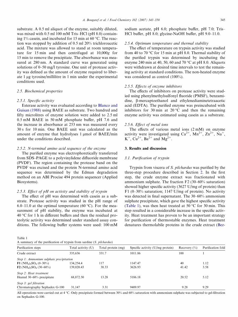
Fig. 2. Comparison of N-terminal amino acid sequence of the purifiedtrypsin from sardine (S. pilchardus) with other enzymes: cod (Gudmunds-dottir et al., 1993), chum salmon (Toyota et al., 2002), Japanese anchovy(TR-I and TR-II) (Kishimura et al., 2005), True sardine (TR-S) andarabesque greenling (TR-P) (Kishimura et al., 2006a), jacopever (TR-J)and elkhorn scuplin (TR-E) (Kishimura et al., 2006b), skipjack tuna (A, Band C) (Klomklao et al., 2006a) and yellowfin tuna (TR-A) (Klomklaoet al., 2006b).
346 A. Bougatef et al. / Food Chemistry 102 (2007) 343–350
erra et al., 2001). Moreover, it is responsible for a signifi-cant breakdown of other undesired thermostable proteins.The heated enzyme was then subjected to gel filtration on aSephadex G-100 column. The elution profiles of trypsinactivity and proteins from Sephadex G-100 are shown inFig. 1. This procedure yielded a single peak of proteaseactivity.
The results of the purification procedure are summarisedin Table 1. After the final purification step, the enzyme waspurified ninefold with a recovery of 9% and a specific activ-ity of 9410 U/mg protein.
3.2. N-terminal amino acid sequence of S. pilchardus trypsin
The N-terminal amino acid sequence of the purifiedtrypsin was determined by the automated Edman methodafter SDS–PAGE and electroblotting. The 12 N-terminalamino acid sequence was IVGGYECQKYSQ. The N-ter-minal amino acid sequence of S. pilchardus trypsin wasaligned with the sequences of other fish trypsins (Gudm-undsdottir et al., 1993; Kishimura, Hayashi, Miyashita, &Nonami, 2005; Kishimura et al., 2006a, 2006b; Klomklaoet al., 2006a, 2006b; Toyota et al., 2002) (Fig. 2). The N-terminal seven amino acid sequence of S. pilchardus trypsin(IVGGYEC) was identical with those of fish trypsins. S.
pilchardus trypsin had a charged Lys residue at position9, where Pro or Ala are common in fish trypsin. The high-est homology was observed with trypsin TR-II from Japa-nese anchovy Engraulis japonica (Kishimura et al., 2005).S. pilchardus trypsin differs from that of Japanese anchovywith only one amino acid in the first 12 amino acids. TheLys-9 in S. pilchardus trypsin was replaced by Pro-9 in Jap-anese anchovy trypsin.
0
0.2
0.4
0.6
0.8
1
1.2
1 25 49 73 97 121 145
Fraction number
Abs
orba
nce
(280
nm
)
0
100
200
300
400
500
600
Prot
ease
act
ivity
(U
/ml)
Absorbance (280 nm)
Protease activity (U/ml)
Fig. 1. Purification profile of trypsin from sardine (S. pilchardus) by gelfiltration on Sephadex G-100 column. The enzyme preparation (30–60%saturation with ammonium sulphate) was applied to a 2.6 · 100 cmcolumn, equilibrated and eluted with buffer B. Fractions (5 ml) collectedfrom the column were assayed for proteins content at 280 nm and proteaseactivity as described in Section 2. Flow rate = 28 ml h�1.
3.3. Biochemical characterization
3.3.1. Molecular weight
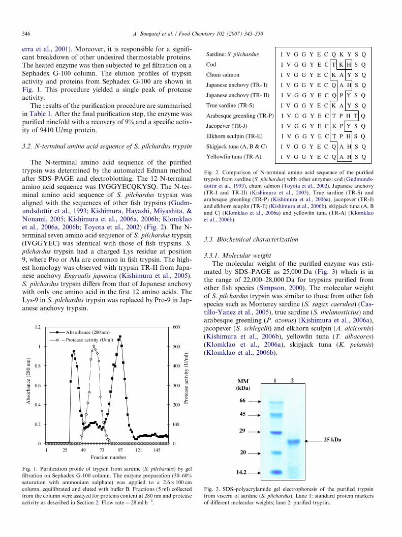
The molecular weight of the purified enzyme was esti-mated by SDS–PAGE as 25,000 Da (Fig. 3) which is inthe range of 22,000–28,000 Da for trypsins purified fromother fish species (Simpson, 2000). The molecular weightof S. pilchardus trypsin was similar to those from other fishspecies such as Monterey sardine (S. sagax caerulea) (Cas-tillo-Yanez et al., 2005), true sardine (S. melanostictus) andarabesque greenling (P. azonus) (Kishimura et al., 2006a),jacopever (S. schlegelii) and elkhorn sculpin (A. alcicornis)(Kishimura et al., 2006b), yellowfin tuna (T. albacores)(Klomklao et al., 2006a), skipjack tuna (K. pelamis)(Klomklao et al., 2006b).
1 2MM(kDa)
66
45
29
20
14.2
25 kDa
Fig. 3. SDS–polyacrylamide gel electrophoresis of the purified trypsinfrom viscera of sardine (S. pilchardus). Lane 1: standard protein markersof different molecular weights; lane 2: purified trypsin.
A. Bougatef et al. / Food Chemistry 102 (2007) 343–350 347
3.3.2. Specific activity
Esterase (BAEE)-specific activity in the pure enzymewas evaluated. S. pilchardus trypsin showed a specific activ-ity of 5.88 U/mg enzyme on the ester substrate benzoyl-L-arginine ethyl ester.
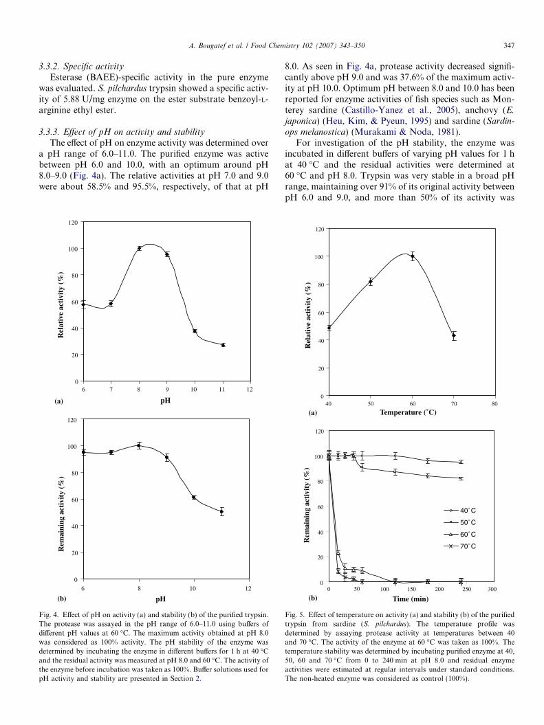
3.3.3. Effect of pH on activity and stability
The effect of pH on enzyme activity was determined overa pH range of 6.0–11.0. The purified enzyme was activebetween pH 6.0 and 10.0, with an optimum around pH8.0–9.0 (Fig. 4a). The relative activities at pH 7.0 and 9.0were about 58.5% and 95.5%, respectively, of that at pH
0
20
40
60
80
100
120
8 10 11 12
pH
Rel
ativ
e ac
tivi
ty (
%)
0
20
40
60
80
100
120
6 10 12
pH
Rem
aini
ng a
ctiv
ity
(%)
6 7
8
9
(a)
(b)
Fig. 4. Effect of pH on activity (a) and stability (b) of the purified trypsin.The protease was assayed in the pH range of 6.0–11.0 using buffers ofdifferent pH values at 60 �C. The maximum activity obtained at pH 8.0was considered as 100% activity. The pH stability of the enzyme wasdetermined by incubating the enzyme in different buffers for 1 h at 40 �Cand the residual activity was measured at pH 8.0 and 60 �C. The activity ofthe enzyme before incubation was taken as 100%. Buffer solutions used forpH activity and stability are presented in Section 2.
8.0. As seen in Fig. 4a, protease activity decreased signifi-cantly above pH 9.0 and was 37.6% of the maximum activ-ity at pH 10.0. Optimum pH between 8.0 and 10.0 has beenreported for enzyme activities of fish species such as Mon-terey sardine (Castillo-Yanez et al., 2005), anchovy (E.
japonica) (Heu, Kim, & Pyeun, 1995) and sardine (Sardin-ops melanostica) (Murakami & Noda, 1981).
For investigation of the pH stability, the enzyme wasincubated in different buffers of varying pH values for 1 hat 40 �C and the residual activities were determined at60 �C and pH 8.0. Trypsin was very stable in a broad pHrange, maintaining over 91% of its original activity betweenpH 6.0 and 9.0, and more than 50% of its activity was
0
20
40
60
80
100
120
40 50 60 70 80
Temperature (˚C)
Rel
ativ
e ac
tivi
ty (
%)
0
20
40
60
80
100
120
150 200 250 300
Time (min)
Rem
aini
ng a
ctiv
ity
(%)
40˚C
50˚C
60˚C
70˚C
0 50 100
(a)
(b)
Fig. 5. Effect of temperature on activity (a) and stability (b) of the purifiedtrypsin from sardine (S. pilchardus). The temperature profile wasdetermined by assaying protease activity at temperatures between 40and 70 �C. The activity of the enzyme at 60 �C was taken as 100%. Thetemperature stability was determined by incubating purified enzyme at 40,50, 60 and 70 �C from 0 to 240 min at pH 8.0 and residual enzymeactivities were estimated at regular intervals under standard conditions.The non-heated enzyme was considered as control (100%).
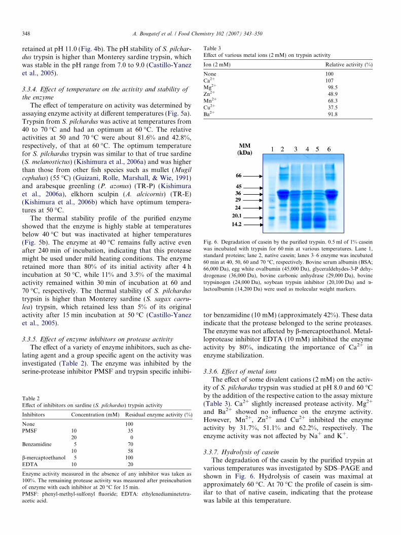
Table 3Effect of various metal ions (2 mM) on trypsin activity
Ion (2 mM) Relative activity (%)
None 100Ca2+ 107Mg2+ 98.5Zn2+ 48.9Mn2+ 68.3Cu2+ 37.5Ba2+ 91.8
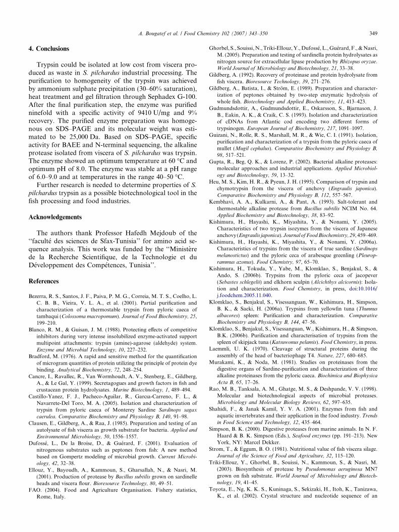
Fig. 6. Degradation of casein by the purified trypsin. 0.5 ml of 1% caseinwas incubated with trypsin for 60 min at various temperatures. Lane 1,standard proteins; lane 2, native casein; lanes 3–6 enzyme was incubated60 min at 40, 50, 60 and 70 �C, respectively. Bovine serum albumin (BSA;66,000 Da), egg white ovalbumin (45,000 Da), glyceraldehydes-3-P dehy-drogenase (36,000 Da), bovine carbonic anhydrase (29,000 Da), bovinetrypsinogen (24,000 Da), soybean trypsin inhibitor (20,100 Da) and a-lactoalbumin (14,200 Da) were used as molecular weight markers.
348 A. Bougatef et al. / Food Chemistry 102 (2007) 343–350
retained at pH 11.0 (Fig. 4b). The pH stability of S. pilchar-
dus trypsin is higher than Monterey sardine trypsin, whichwas stable in the pH range from 7.0 to 9.0 (Castillo-Yanezet al., 2005).
3.3.4. Effect of temperature on the activity and stability ofthe enzyme
The effect of temperature on activity was determined byassaying enzyme activity at different temperatures (Fig. 5a).Trypsin from S. pilchardus was active at temperatures from40 to 70 �C and had an optimum at 60 �C. The relativeactivities at 50 and 70 �C were about 81.6% and 42.8%,respectively, of that at 60 �C. The optimum temperaturefor S. pilchardus trypsin was similar to that of true sardine(S. melanostictus) (Kishimura et al., 2006a) and was higherthan those from other fish species such as mullet (Mugil
cephalus) (55 �C) (Guizani, Rolle, Marshall, & Wie, 1991)and arabesque greenling (P. azonus) (TR-P) (Kishimuraet al., 2006a), elkhorn sculpin (A. alcicornis) (TR-E)(Kishimura et al., 2006b) which have optimum tempera-tures at 50 �C.
The thermal stability profile of the purified enzymeshowed that the enzyme is highly stable at temperaturesbelow 40 �C but was inactivated at higher temperatures(Fig. 5b). The enzyme at 40 �C remains fully active evenafter 240 min of incubation, indicating that this proteasemight be used under mild heating conditions. The enzymeretained more than 80% of its initial activity after 4 hincubation at 50 �C, while 11% and 3.5% of the maximalactivity remained within 30 min of incubation at 60 and70 �C, respectively. The thermal stability of S. pilchardus
trypsin is higher than Monterey sardine (S. sagax caeru-
lea) trypsin, which retained less than 5% of its originalactivity after 15 min incubation at 50 �C (Castillo-Yanezet al., 2005).
3.3.5. Effect of enzyme inhibitors on protease activity
The effect of a variety of enzyme inhibitors, such as che-lating agent and a group specific agent on the activity wasinvestigated (Table 2). The enzyme was inhibited by theserine-protease inhibitor PMSF and trypsin specific inhibi-
Table 2Effect of inhibitors on sardine (S. pilchardus) trypsin activity
Inhibitors Concentration (mM) Residual enzyme activity (%)
None 100PMSF 10 35
20 0Benzamidine 5 70
10 58b-mercaptoethanol 5 100EDTA 10 20
Enzyme activity measured in the absence of any inhibitor was taken as100%. The remaining protease activity was measured after preincubationof enzyme with each inhibitor at 20 �C for 15 min.PMSF: phenyl-methyl-sulfonyl fluoride; EDTA: ethylenediaminetetra-acetic acid.
tor benzamidine (10 mM) (approximately 42%). These dataindicate that the protease belonged to the serine proteases.The enzyme was not affected by b-mercaptoethanol. Metal-loprotease inhibitor EDTA (10 mM) inhibited the enzymeactivity by 80%, indicating the importance of Ca2+ inenzyme stabilization.
3.3.6. Effect of metal ions
The effect of some divalent cations (2 mM) on the activ-ity of S. pilchardus trypsin was studied at pH 8.0 and 60 �Cby the addition of the respective cation to the assay mixture(Table 3). Ca2+ slightly increased protease activity. Mg2+
and Ba2+ showed no influence on the enzyme activity.However, Mn2+, Zn2+ and Cu2+ inhibited the enzymeactivity by 31.7%, 51.1% and 62.2%, respectively. Theenzyme activity was not affected by Na+ and K+.
3.3.7. Hydrolysis of casein
The degradation of the casein by the purified trypsin atvarious temperatures was investigated by SDS–PAGE andshown in Fig. 6. Hydrolysis of casein was maximal atapproximately 60 �C. At 70 �C the profile of casein is sim-ilar to that of native casein, indicating that the proteasewas labile at this temperature.
A. Bougatef et al. / Food Chemistry 102 (2007) 343–350 349
4. Conclusions
Trypsin could be isolated at low cost from viscera pro-duced as waste in S. pilchardus industrial processing. Thepurification to homogeneity of the trypsin was achievedby ammonium sulphate precipitation (30–60% saturation),heat treatment and gel filtration through Sephadex G-100.After the final purification step, the enzyme was purifiedninefold with a specific activity of 9410 U/mg and 9%recovery. The purified enzyme preparation was homoge-nous on SDS–PAGE and its molecular weight was esti-mated to be 25,000 Da. Based on SDS–PAGE, specificactivity for BAEE and N-terminal sequencing, the alkalineprotease isolated from viscera of S. pilchardus was trypsin.The enzyme showed an optimum temperature at 60 �C andoptimum pH of 8.0. The enzyme was stable at a pH rangeof 6.0–9.0 and at temperatures in the range 40–50 �C.
Further research is needed to determine properties of S.
pilchardus trypsin as a possible biotechnological tool in thefish processing and food industries.
Acknowledgements
The authors thank Professor Hafedh Mejdoub of the‘‘faculte des sciences de Sfax-Tunisia’’ for amino acid se-quence analysis. This work was funded by the ‘‘Ministerede la Recherche Scientifique, de la Technologie et duDeveloppement des Competences, Tunisia’’.
References
Bezerra, R. S., Santos, J. F., Paiva, P. M. G., Correia, M. T. S., Coelho, L.C. B. B., Vieira, V. L. A., et al. (2001). Partial purification andcharacterization of a thermostable trypsin from pyloric caeca oftambaqui (Colossoma macropomum). Journal of Food Biochemistry, 25,199–210.
Blanco, R. M., & Guisan, J. M. (1988). Protecting effects of competitiveinhibitors during very intense insolubilized enzyme-activated supportmultipoint attachments: trypsin (amine)-agarose (aldehyde) system.Enzyme and Microbial Technology, 10, 227–232.
Bradford, M. (1976). A rapid and sensitive method for the quantificationof microgram quantities of protein utilizing the principle of protein dyebinding. Analytical Biochemistry, 72, 248–254.
Cancre, I., Ravallec, R., Van Wormhoudt, A. V., Stenberg, E., Gildberg,A., & Le Gal, Y. (1999). Secretagogues and growth factors in fish andcrustacean protein hydrolysates. Marine Biotechnology, 1, 489–494.
Castillo-Yanez, F. J., Pacheco-Aguilar, R., Garcıa-Carreno, F. L., &Navarrete-Del Toro, M. A. (2005). Isolation and characterization oftrypsin from pyloric caeca of Monterey Sardine Sardinops sagax
caerulea. Comparative Biochemistry and Physiology B, 140, 91–98.Clausen, E., Gildberg, A., & Raa, J. (1985). Preparation and testing of an
autolysate of fish viscera as growth substrate for bacteria. Applied and
Environmental Microbiology, 50, 1556–1557.Dufosse, L., De la Broise, D., & Guerard, F. (2001). Evaluation of
nitrogenous substrates such as peptones from fish: A new methodbased on Gompertz modeling of microbial growth. Current Microbi-
ology, 42, 32–38.Ellouz, Y., Bayoudh, A., Kammoun, S., Gharsallah, N., & Nasri, M.
(2001). Production of protease by Bacillus subtilis grown on sardinelleheads and viscera flour. Bioresource Technology, 80, 49–51.
FAO. (2004). Food and Agriculture Organisation. Fishery statistics,Rome, Italy.
Ghorbel, S., Souissi, N., Triki-Ellouz, Y., Dufosse, L., Guerard, F., & Nasri,M. (2005). Preparation and testing of sardinella protein hydrolysates asnitrogen source for extracellular lipase production by Rhizopus oryzae.World Journal of Microbiology and Biotechnology, 21, 33–38.
Gildberg, A. (1992). Recovery of proteinase and protein hydrolysate fromfish viscera. Bioresource Technology, 39, 271–276.
Gildberg, A., Batista, I., & Strom, E. (1989). Preparation and character-ization of peptones obtained by two-step enzymatic hydrolysis ofwhole fish. Biotechnology and Applied Biochemistry, 11, 413–423.
Gudmundsdottir, A., Gudmundsdottir, E., Oskarsson, S., Bjarnason, J.B., Eakin, A. K., & Craik, C. S. (1993). Isolation and characterizationof cDNAs from Atlantic cod encoding two different forms oftrypsinogen. European Journal of Biochemistry, 217, 1091–1097.
Guizani, N., Rolle, R. S., Marshall, M. R., & Wie, C. I. (1991). Isolation,purification and characterization of a trypsin from the pyloric caeca ofmullet (Mugil cephalus). Comparative Biochemistry and Physiology B,
98, 517–521.Gupta, R., Beg, Q. K., & Lorenz, P. (2002). Bacterial alkaline proteases:
molecular approaches and industrial applications. Applied Microbiol-
ogy and Biotechnology, 59, 13–32.Heu, M. S., Kim, H. R., & Pyeun, J. H. (1995). Comparison of trypsin and
chymotrypsin from the viscera of anchovy (Engraulis japonica).Comparative Biochemistry and Physiology B, 112, 557–567.
Kembhavi, A. A., Kulkarni, A., & Pant, A. (1993). Salt-tolerant andthermostable alkaline protease from Bacillus subtilis NCIM No. 64.Applied Biochemistry and Biotechnology, 38, 83–92.
Kishimura, H., Hayashi, K., Miyashita, Y., & Nonami, Y. (2005).Characteristics of two trypsin isozymes from the viscera of Japaneseanchovy (Engraulis japonica). Journal of Food Biochemistry, 29, 459–469.
Kishimura, H., Hayashi, K., Miyashita, Y., & Nonami, Y. (2006a).Characteristics of trypsins from the viscera of true sardine (Sardinops
melanostictus) and the pyloric ceca of arabesque greenling (Pleurop-
rammus azonus). Food Chemistry, 97, 65–70.Kishimura, H., Tokuda, Y., Yabe, M., Klomklao, S., Benjakul, S., &
Ando, S. (2006b). Trypsins from the pyloric ceca of jacopever(Sebastes schlegelii) and elkhorn sculpin (Alcichthys alcicornis): Isola-tion and characterization. Food Chemistry, in press, doi:10.1016/j.foodchem.2005.11.040.
Klomklao, S., Benjakul, S., Visessanguan, W., Kishimura, H., Simpson,B. K., & Saeki, H. (2006a). Trypsins from yellowfin tuna (Thunnus
albacores) spleen: Purification and characterization. Comparative
Biochemistry and Physiology B, 144, 47–56.Klomklao, S., Benjakul, S., Visessanguan, W., Kishimura, H., & Simpson,
B.K. (2006b). Purification and characterisation of trypsins from thespleen of skipjack tuna (Katsuwonus pelamis). Food Chemistry, in press.
Laemmli, U. K. (1970). Cleavage of structural proteins during theassembly of the head of bacteriophage T4. Nature, 227, 680–685.
Murakami, K., & Noda, M. (1981). Studies on proteinases from thedigestive organs of Sardine-purification and characterization of threealkaline proteinases from the pyloric caeca. Biochimica and Biophysica
Acta B, 65, 17–26.Rao, M. B., Tanksala, A. M., Ghatge, M. S., & Deshpande, V. V. (1998).
Molecular and biotechnological aspects of microbial proteases.Microbiology and Molecular Biology Reviews, 62, 597–635.
Shahidi, F., & Janak Kamil, Y. V. A. (2001). Enzymes from fish andaquatic invertebrates and their application in the food industry. Trends
in Food Science and Technology, 12, 435–464.Simpson, B. K. (2000). Digestive proteases from marine animals. In N. F.
Haard & B. K. Simpson (Eds.), Seafood enzymes (pp. 191–213). NewYork, NY: Marcel Dekker.
Strom, T., & Eggum, B. O. (1981). Nutritional value of fish viscera silage.Journal of the Science of Food and Agriculture, 32, 115–120.
Triki-Ellouz, Y., Ghorbel, B., Souissi, N., Kammoun, S., & Nasri, M.(2003). Biosynthesis of protease by Pseudomonas aeruginosa MN7grown on fish substrate. World Journal of Microbiology and Biotech-
nology, 19, 41–45.Toyota, E., Ng, K. K. S., Kuninaga, S., Sekizaki, H., Itoh, K., Tanizawa,
K., et al. (2002). Crystal structure and nucleotide sequence of an

350 A. Bougatef et al. / Food Chemistry 102 (2007) 343–350
anionic trypsin from chum salmon (Oncorhynchus keta) in comparisonwith Atlantic salmon (Salmo salar) and bovine trypsin. Journal of
Molecular Biology, 324, 391–397.
Zukowski, M. M. (1992). Production of commercially valuable products.In R. H. Doi & M. Mc. Gloughlin (Eds.), Biology of bacilli: application
to industry (pp. 311–337). London: Butterworth-Heinemann.