Embed Size (px)
Citation preview

Protocol
Purification of Oligodendrocyte Precursor Cells fromRat Cortices by Immunopanning
Jason C. Dugas1,2,4 and Ben Emery3
1Myelin Repair Foundation, Saratoga, California 95070; 2Department of Neurobiology, School of Medicine,Stanford University, Stanford, California 94305-5125; 3Department of Anatomy and Neuroscience and the FloreyInstitute of Neuroscience and Mental Health, University of Melbourne, Melbourne, Victoria 3010, Australia
This protocol describes how to purify oligodendrocyte precursor cells (OPCs) from postnatal rodentbrains. The method utilizes an immunopanning technique to first remove unwanted cells by negativeselection and then purify OPCs by positive selection and subsequent enzymatic release from the finalpanning plate. Included are modifications that allow for purification and culturing of OPCs frommouse instead of rat tissue and for use of optic nerves instead of whole brains. The method for isolatingOPCs from whole brain can be used for isolating OPCs from any specific region of the brain, providedthat the area can be dissected away from the rest of the tissue. Suggested culture media for maintainingproliferating OPCs or inducing oligodendrocyte (OL) differentiation are also described.
MATERIALS
It is essential that you consult the appropriate Material Safety Data Sheets and your institution’s EnvironmentalHealth and Safety Office for proper handling of equipment and hazardous material used in this protocol.
RECIPES: Please see the end of this article for recipes indicated by <R>. Additional recipes can be found online athttp://cshprotocols.cshlp.org/site/recipes.
Reagents
AntibodiesAnti-mouse Thy 1.2 monoclonal (Serotec MCA02R) (for mouse OPCs only)Anti-O1 antibody (Millipore MAB344)Anti-O4 antibody (Millipore MAB345)Goat anti-mouse IgG + IgM (Jackson ImmunoResearch 115-005-044)Goat anti-mouse IgM, µ chain specific (Jackson ImmunoResearch 115-005-020)
BSA (4%)To prepare a stock of 4% BSA in Dulbecco’s phosphate-buffered saline (D-PBS), dissolve 8 g of BSA(Sigma-Aldrich A4161) in 150 mL of D-PBS (HyClone SH30264.01) at 37˚C. Adjust the pH to 7.4 with �1mLof 1NNaOH.Bring thevolume to200 mL. Filter througha0.22-μmfilter. Store in1-mLaliquots at−20˚C.
DMEM-Sato base growth medium <R>DNase I
4Correspondence: [email protected]
© 2013 Cold Spring Harbor Laboratory PressCite this article as Cold Spring Harb Protoc; 2013; doi:10.1101/pdb.prot070862
745
Cold Spring Harbor Laboratory Press on May 22, 2018 - Published by http://cshprotocols.cshlp.org/Downloaded from

On ice, dissolve 12,500 U of DNase I (Worthington Biochemical LS002007) per 1 mL of chilled Earle’sBalanced Salt Solution (EBSS; Invitrogen 14155-063). Filter-sterilize on ice. Aliquot (e.g., 200 µL/tube) andfreeze overnight at −20˚C. Store aliquots at −20 to −30˚C.
Dulbecco’s phosphate-buffered saline (D-PBS) with phenol redAdd 500 µL of 0.5% phenol red (Sigma-Aldrich P0290) per 500 mL bottle of Dulbecco’s phosphate-buffered saline (D-PBS; Invitrogen 14287-080).
Dulbecco’s phosphate-buffered saline (D-PBS) without Ca2+/Mg2+ (Invitrogen 14190-144)IMPORTANT: D-PBSwithout Ca2+/Mg2+ isONLY used for tissue dissection (see Steps 9 and 15). All othersteps in the protocol refer to the standard D-PBS with phenol red. Cells will not stick to the panningplates in the Ca2+/Mg2+-free D-PBS.
Earle’s balanced salt solution (EBSS) (Invitrogen 14155-063)Ethanol (70%)Ethanol-washed glass coverslips (optional) <R>Fetal calf serum (FCS) (heat-inactivated)
Prepare 50-mL aliquots of fetal calf serum (Invitrogen 16000-036) in sterile conical tubes. Heat-inactivatealiquots for 30 min in a 55˚C water bath, and then store at −20˚C.
High-ovomucoid (high-ovo) stock solution (6×) <R>Insulin stock (0.5 mg/mL) <R>L-cysteine (Sigma-Aldrich C7477)Low-ovomucoid (low-ovo) stock solution (10×) <R>NaOH (1 M, sterile)OPC culture medium <R>
Addition of PDGF and NT-3 to medium in the absence of T3 will promote OPC proliferation, whereasaddition of T3 to medium in the absence of PDGF and NT-3 will promote rapid OL differentiation.Robust differentiation will also commence in medium lacking both mitogens (PDGF and NT-3) and T3,but OLs might be slightly less healthy. For the study of OPCs in the complete absence of T3-mediatedsignaling, prepare DMEM-Sato base growth medium without B-27. We have found that FGF is notrequired in the medium to inhibit differentiation and maintain OPCs as immature, proliferating cells.
Papain (Worthington Biochemical LS003126)Papain buffer <R>Phenol red solution (0.5%) (Sigma-Aldrich P0290)Phosphate-buffered saline (PBS) (filter- or autoclave-sterilized)Poly-D-lysine (PDL) stock (1 mg/mL) <R>
The 1 mg/mL solution is a 100× stock. Dilute it to 1× in sterile H2O before use.
Ran-2 hybridoma cells (supernatant for rat OPC prep) (ATCC TIB-119)Rat pups (Sprague–Dawley) (postnatal day 6–8)
One rat brain should yield�2–3 × 10⁶ OPCs, whereas one mouse brain should yield�0.8–1 × 10⁶ OPCs.Yields and initial viability of purified OPCs will progressively decline in correlation with the age ofanimals used: Older animals yield lower numbers of less healthy OPCs. Mouse pups at postnatal day6–8 can also be used.
Tris-HCl (50 mM, pH 9.5) (filter- or autoclave-sterilized)Trypsin stock
Dissolve trypsin (Sigma-Aldrich T9935) at 50,000 U/mL in Earle’s Balanced Salt Solution (EBSS) (Invitro-gen 14155-063) on ice. If the powder does not go into solution easily, stir with a magnetic stir bar or tryaltering the pH with NaOH or HCl as appropriate. When fully dissolved, aliquot (e.g., 200 µL/tube) andstore at −80˚C. For each new batch of trypsin stock, the optimal trypsinization time required to releaseOPCs from the final panning plate (Step 46) should be redetermined.
Trypan blue (Invitrogen 15250-061)
Equipment
Centrifuge (tabletop, with 15-mL/50-mL conical tube adaptors)Conical tubes (15- and 50-mL, sterile)
746 Cite this article as Cold Spring Harb Protoc; 2013; doi:10.1101/pdb.prot070862
J.C. Dugas and B. Emery
Cold Spring Harbor Laboratory Press on May 22, 2018 - Published by http://cshprotocols.cshlp.org/Downloaded from

Dissection microscopeForceps (#5 or #55) (for optic nerve preparation)Glass spreader (sterile)Heat block base (with a flat insert set to 34˚C in a sterile hood)Hemocytometer slide (Hausser Scientific 3110)Laminar flow tissue culture hoodMonoject syringes (sterile) (Fisher Scientific)Needles (21- and 23-gauge) (for optic nerve preparation)Nitex mesh filters
Nitex mesh 20-μmopening filters can be ordered from Amazon. Cut filters into approximately 3 inch × 3inch squares. Wrap sets of 8–12 in aluminum foil and autoclave to sterilize.
Petri dish lid (6-cm, with a hole in the center to accommodate a 0.22-μm filter)Create the hole using flamed forceps to melt an opening into the center of the lid, or use a drill.
Petri dishes (6-, 10-, and 15-cm) (Falcon or Nunc)Scalpel blade handleScalpel blades (#10)Scissors (large, for decapitation) (e.g., Roboz RS-6820)Scissors (small, curved) (e.g., Roboz RS-5675)Scissors (very fine) (for optic nerve preparation) (e.g., Roboz RS-5602)Source of carbon dioxide (5% CO2/95% O2) with a line leading to a heat block in a sterile hoodSyringe filters (0.22-μm)Tissue-culture incubator (set to 37˚C, 10% CO2)Tissue-culture plates (plastic) (6- and 24-well) (Falcon or Nunc)Water bath (set to 37˚C)
METHOD
An overview of the method is provided in Figure 1. For any steps that require filter sterilization of reagents before use,prerinse the filter with 5–10 mL of base liquid (e.g., D-PBS) to remove any residual surfactants that may be detrimentalto cell health. Discard the prefilter liquid before filtering the solution to be sterilized.
Preparation of Plates and Reagents
Perform Step 1 on the day before cell purification. Steps 2–5 can be performed the day before or the same day as cellpurification. (PDL-coated coverslips and tissue culture plates are usable up to 6 d following preparation.) For usersfamiliar with the protocol, Steps 2–5 also may be performed during the 90-min papain digestion (Step 17) or panningsteps (Steps 35–39) to save time.
1. Coat the panning dishes with secondary antibodies as follows.
i. Coat two 15-cm Petri dishes with 60 µL of goat anti-mouse IgG + IgM plus 20 mL of sterile50 mM Tris-HCl (pH 9.5) per dish.
ii. Coat one 10-cm Petri dish with 30 µL of goat anti-mouse IgM plus 10 mL of sterile 50 mM
Tris-HCl (pH 9.5).
iii. Swirl the plates until the surfaces are evenly coated with the antibody-Tris solution.
iv. Incubate the panning plates overnight at 4˚C.One set of panning plates and reagents is sufficient for one postnatal rat brain, one to three postnatalmouse brains, or one to three litters of rodent optic nerves.
2. Prepare the culture dishes as follows.
i. Add 5 mL/1 mL/250 µL of 1× PDL per well to 10-cm/6-well/24-well tissue culture plates,respectively.
ii. Swirl to evenly coat plastic.
Cite this article as Cold Spring Harb Protoc; 2013; doi:10.1101/pdb.prot070862 747
Purification of OPCs from Rat Cortices
Cold Spring Harbor Laboratory Press on May 22, 2018 - Published by http://cshprotocols.cshlp.org/Downloaded from

iii. Incubate for 20–60 min at room temperature.
iv. Rinse the plates three times with sterile H2O.
v. Allow the plates to completely air-dry before use.
3. Prepare the coverslips as follows.
i. Rinse the ethanol-washed glass coverslips three times with sterile H2O in a Petri dish.
ii. After the last rinse, suction away the remaining H2O and separate the coverslips in the platesuch that they are not touching each other or the sides of the plate.
iii. Allow the coverslips to completely air-dry.This should take 5–10 min after suctioning.
Obtain rodents andprepare all solutions, etc.
Steps 1–101–2 h, day 1; 30–60 min, day 2
O2-CO2 line
Dissect tissue(brain or optic nerve)
Steps 11–1515 min brain; 45 min ON
Dice tissueStep 15
5 min brain; 15 min ON
Preparation& DissectionSteps 1–15
1–2 h
Papain digest tissueSteps 16–17
90 min
TriturationSteps 18–2515–30 min
Rinsing cellsSteps 26–32
2x 15-min spins; 45–60 min total
DissociationSteps 16–32
3 h
Astrocytes Oligodendrocytes OPCs
PanningSteps 33–42
2–2.5 h
CellSuspension
Ran-2 panningSteps 33–35
30 min
01 panningSteps 36–37
30 min
04 panningSteps 38–42
45 min plate + 15 min rinsing
Trypsin digestionSteps 43–46
6–10 min
Squirt cells off plateSteps 47–5315–30 min
Collect and plate cellsSteps 54–59
15 min spin + 10–45 min plating
Trypsinization& Plating
Steps 43–591–2 h
FIGURE 1. Immunopanning of OPCs. ON, optical nerves.
748 Cite this article as Cold Spring Harb Protoc; 2013; doi:10.1101/pdb.prot070862
J.C. Dugas and B. Emery
Cold Spring Harbor Laboratory Press on May 22, 2018 - Published by http://cshprotocols.cshlp.org/Downloaded from

iv. Carefully add 100 µL of 1× PDL to the center of each coverslip.The solution should remain as a bubble on the coverslip.
v. Incubate for 30–60 min at room temperature.
vi. Rinse the coverslips three times with sterile H2O.
vii. Transfer the coverslips to 24-well tissue culture plates, placing one coverslip per well.
viii. Suction any remaining H2O from the wells, being careful to keep the coverslips centered andnot touching the sides of the wells.
If the coverslips contact the sides of the wells, the preplating of OPCs will not work.
ix. Allow the coverslips to completely air-dry before use.
4. Prepare 40 mL of 0.2% BSA solution by adding 2 mL of 4% BSA stock to 38 mL of D-PBS. Storeat 4˚C.
5. Prepare the DMEM-Sato base medium (or the desired medium) for the cells.
Preparation for Cell Purification
Perform Steps 6–10 on the day of cell purification. For users familiar with the protocol, Step 10 also may be performedduring the 90-min papain digestion (Step 17) to save time.
Do not allow the plates to dry at any stage.
6. Coat the panning dishes with primary antibodies as follows.
i. Rinse each antibody-coated plate (prepared in Step 1) three times with PBS.
ii. To prepare for use of rat cells, coat one 15-cm plate from Step 1 with 4 mL of Ran-2hybridoma supernatant plus 8 mL of 0.2% BSA (prepared in Step 4).
iii. To prepare for use of mouse cells, coat one 15-cm plate from Step 1 with 20 µL of Thy 1.2antibody plus 12 mL of 0.2% BSA.
iv. Coat one 15-cm plate from Step 1 with 12 µL (12 µg) of O1 antibody plus 12 mL of 0.2%BSA.
v. Coat one 10-cm plate from Step 1 with 5 µL (5 µg) of O4 antibody plus 5 mL of 0.2% BSA.
vi. Swirl to evenly coat plastic.
vii. Incubate plates >2 h at room temperature.
7. Equilibrate 10 mL of papain buffer in a 6-mL Petri dish as follows.
i. Presterilize a 6-cm dish lid with a central hole by spraying with 70% EtOH. Let air-dry.
ii. Place the 6-cm dish containing papain buffer on a 34˚C heat block in a sterile hood.
iii. Place a 0.22-μm filter at the end of a 5% CO2 / 95% O2 line and place the sterile end of thefilter through the hole in the sterilized lid.
iv. Place the lid supplying gas over the papain buffer dish in the heat block and supply a gentleflow of gas.
8. Place 10 mL of EBSS containing 0.0005% phenol red into a 10% CO2 incubator to equilibrate.
9. Add �300 µL of D-PBS without Ca2+/Mg2+ to a 6-cm Petri dish. Alternatively, if OPCswill be isolated from optic nerves rather than whole brain, use �5 mL of D-PBS withoutCa2+/Mg2+.
10. Prepare the solutions for dissociation, panning and trypsinization as follows.
i. Prepare low-ovo working solution by adding 1 mL of 10× low-ovo stock to 9 mL of D-PBS.
ii. Prepare high-ovo working solution by adding 1 mL of 6× high-ovo stock to 5 mL of D-PBS.
iii. Prepare panning buffer by combining 1.5 mL of 0.2% BSA solution, 13.5 mL of D-PBS, and150 µL of insulin stock (500 µg/mL).
Cite this article as Cold Spring Harb Protoc; 2013; doi:10.1101/pdb.prot070862 749
Purification of OPCs from Rat Cortices
Cold Spring Harbor Laboratory Press on May 22, 2018 - Published by http://cshprotocols.cshlp.org/Downloaded from

iv. Add 3 mL of heat-inactivated FCS to 7 mL of D-PBS and filter through a 0.22-μm filter tosterilize.The phenol red color of the low- and high-ovo solutions should be orange (neutral pH) rather than red (toobasic) or yellow (too acidic). If necessary, sterile 1MNaOH can be used to equilibrate both solutions back toneutral pH.
Dissection
See online Movie 1 at cshprotocols.cshlp.org for an illustration of Steps 12–15.
11. Decapitate one Sprague–Dawley rat pup (or mouse pup) (P6-8) with sharp scissors.
12. Cut the skin along the top midline of the head, and peel back to reveal the skull.
13. Use curved scissors to open the skull: Insert the scissors at the opening at the back of the skull andcut along the left and right edge of the skull, above the ear to just above the eye socket.
14. Lift the skull bone to reveal the brain.
15. Dissect and harvest the brain or optic nerves as follows.
To Harvest the Brain
i. Cut away the olfactory bulbs at the front of the brain, then remove the rest of the brain.The specific brain region of interest can also be dissected at this stage.
ii. Place the tissue in 300 µL of D-PBS without Ca2+/Mg2+ in the dish prepared in Step 9.
iii. Dice the brain with a #10 scalpel into �1-mm3 chunks.
To Harvest the Optic Nerve
iv. After cutting the olfactory bulbs, lift the front of the brain up to reveal the optic nerves/optictracts stretching from the base of the brain to the eye sockets. Cut the optic tracts imme-diately adjacent to the optic chiasm to leave the optic chiasm attached to the two opticnerves.
v. Cut at the base of each eyeball by inserting the curved scissors into the eye orbit behind theeyeball to detach the optic nerve from the eyeball.
vi. Grab the optic chiasm with tweezers and lift the optic chiasm plus the two optic nerves fromthe base of the skull.
vii. Place the tissue in the dish of 5 mL D-PBS without Ca2+/Mg2+ prepared in Step 9.
viii. Dice the nerves with very small dissection scissors. Discard the optic chiasms.Optic nerves should be harvested from approximately 10 pups to obtain �500,000 OPCs.
Tissue Dissociation
16. Prepare the papain solution.
i. Move the equilibrated papain buffer prepared in Step 7 to a 15-mL conical tube and add200 units of papain.
ii. Place this papain solution in a 37˚C water bath for 5–15 min to allow the papain to dissolve.
iii. Weigh out and add 2 mg of L-cysteine to the solution.
iv. When fully dissolved, filter the papain solution through a 0.22-μm filter to sterilize.
v. Add 200 µL of DNase I stock solution to the papain solution.
17. Digest the tissue as follows.
Brain
i. Add the papain solution from Step 16 directly to the diced brain tissue in the 6-cmPetri dish.
750 Cite this article as Cold Spring Harb Protoc; 2013; doi:10.1101/pdb.prot070862
J.C. Dugas and B. Emery
Cold Spring Harbor Laboratory Press on May 22, 2018 - Published by http://cshprotocols.cshlp.org/Downloaded from

ii. Place the dish of tissue in papain solution in an empty heat block at 34˚C, and then coverwith the same lid used to equilibrate the papain solution.
Brain dissociation digestions performed at 34˚C slightly increase the health of the cells obtained com-pared to digestions at 37˚C.
iii. Keep the tissue under 5% CO2/95% O2 gas flow at 34˚C for 90 min, gently agitating thetissue every 15 min to ensure complete tissue digestion.
Optic Nerve
iv. Remove all of the D-PBS solution from the diced optic nerve tissue and add the papainsolution.
v. Transfer the diced tissue to a sterile 50-mL conical tube and digest for 90 min in a 34–37˚Cwater bath, gently agitating the tissue every 15 min.
18. Just before the digestion step is complete, add 100 µL of DNase I stock solution to the low-ovoworking solution prepared in Step 10.
19. When the digestion is complete, transfer the tissue in papain solution to a sterile 15-mL conicaltube and allow the tissue pieces to settle.
20. Carefully remove as much papain solution as possible from the tissue.
21. Gently add 2 mL of low-ovo solution with DNase I from Step 18 to the tissue to stop papaindigestion. Allow the tissue to settle once again.
22. Remove and discard the low-ovo solution from the tissue.
23. Add 2 mL of fresh low-ovo solution with DNase I to the tissue.Dissociation is performed in low-ovo solution because the cells survive trituration better in a lower proteinsolution. High-ovo is subsequently used to fully quench any residual papain enzymatic activity.
24. Dissociate the tissue as follows.
BrainSee online Movie 2 at cshprotocols.cshlp.org for an illustration of Steps 24.i–24.v.
i. Gently pipette the tissue six to eight times through a 5-mL pipette.
ii. Let the tissue chunks settle for 1–2 min.
iii. Remove 1–1.5 mL of the resulting cell suspension, trying to avoid large tissue chunks. Placethe collected cell suspension in a new, sterile 15-mL conical tube.
iv. Add 1 mL of low-ovo solution with DNase I and repeat Steps 24.i–24.iii.
v. Add 1 mL of low-ovo solution with DNase I, then follow Steps 24.i–24.iv, now trituratingtissue through a 1-mL pipette tip; repeat until all of the tissue is dissociated and the low-ovosolution prepared in Step 18 is used up.
Optic Nerve
vi. Pipette the tissue six to eight times through a 1-mL pipette tip.
vii. Let the tissue chunks settle for 1–2 min.
viii. Remove 1–1.5 mL of the resulting cell suspension, trying to avoid large tissue chunks. Placethe collected cell suspension in a new, sterile 15-mL conical tube.
ix. Add 1 mL of low-ovo solution with DNase I and repeat Steps 24.vi–24.viii.
x. Add 1 mL of low-ovo solution with DNase I, then follow Steps 24.vi–24.ix, now trituratingthe tissue three times through a 21-gauge needle followed by a 23-gauge needle, until thelow-ovo solution prepared in Step 18 is used up.
25. (Optional)Todetermine cell viability and the effectiveness of the dissociation at this stage, remove asmall aliquot (50–100 µL) of cell suspension for counting. Count as described in Step 55.
See Troubleshooting.
Cite this article as Cold Spring Harb Protoc; 2013; doi:10.1101/pdb.prot070862 751
Purification of OPCs from Rat Cortices
Cold Spring Harbor Laboratory Press on May 22, 2018 - Published by http://cshprotocols.cshlp.org/Downloaded from

26. Pellet the cells at �220g for 15 min in a tabletop centrifuge at room temperature.
27. Aspirate the supernatant, being careful not to disturb the cell pellet.
28. Resuspend the cell pellet in 6 mL of high-ovo solution prepared in Step 10.
29. Immediately pellet the cells at �220g for 15 min in a tabletop centrifuge at room temperature.Leaving cells for an extended period of time in high-ovo solution will reduce cell viability.
30. Aspirate the supernatant, being careful not to disturb the cell pellet.
31. Resuspend the cell pellet in 6 mL of panning buffer prepared in Step 10.
32. Prewet a sterileNitexmeshfilter byfiltering 2 mLof panning buffer into a sterile 50-mL tube. Filterthe cells into the tube 1 mL at a time, and then rinse the filter with the remaining panning buffer.
Panning
Do not allow plates to dry out at any stage.
33. Immediately before panning dish use, rinse the panning dish three times with D-PBS.
34. Add the cell suspension from Step 32 to a rinsed Ran-2 (rat) or Thy1.2 (mouse) panning plateprepared in Step 6.
35. Incubate the plate for 30 min at room temperature, agitating at 15 min to ensure that all cells havean opportunity to adhere to the plate surface.
36. Gently shake the plate to loosen the nonadherent cells, and transfer the cell suspension to a rinsedO1 panning plate prepared in Step 6.
37. Incubate the plate for 30 min at room temperature, agitating for 15 min to ensure that all cellshave an opportunity to adhere to the plate surface.
38. Gently shake the plate to loosen the nonadherent cells, and transfer the cell suspension to a rinsedO4 panning plate prepared in Step 6.
39. Incubate the plate for 45 min at room temperature, agitating every 15 min to ensure that all cellshave an opportunity to adhere to the plate surface.
40. Shake the plate to loosen the nonadherent cells and pour off the cell suspension.
41. Rinse the plate six to eight times with D-PBS, shaking the plate at each rinse to loosen thenonadherent cells.
OPCs adhere very strongly to the O4 panning plate, so shaking the final plate vigorously should not result ina significant loss of OPCs.
42. Before trypsinization, confirm visually under the microscope that nearly all nonadherent cellshave been removed by rinsing. If needed, perform additional rinsing steps.
There will always be a small number (<0.5%) of floating cells representing dislodged OPCs at the end of therinsing steps.
See Troubleshooting.
Trypsinization
See online Movie 3 at cshprotocols.cshlp.org for an illustration of Steps 48 and 49.
43. Once the OPCs are in their final D-PBS rinse, transfer 4 mL of the equilibrated EBSS prepared inStep 8 to a sterile tube and add 400 µL of trypsin stock solution.
44. Remove the D-PBS from the O4 plate and rinse the plate with the remaining 6 mL of equilibratedEBSS.
45. Pour off the EBSS and add the 4 mL of trypsin-EBSS solution prepared in Step 43.
46. Incubate the plate for 6–8 min in a 37˚C incubator.Optimal trypsinization time should be determined with each batch of stock trypsin solution generated andshould correspond to the time at which a large percentage of cells on the plate can be easily released bygentle pipetting. Trypsinization times that are either too short or too long can result in reduced cell viability.
752 Cite this article as Cold Spring Harb Protoc; 2013; doi:10.1101/pdb.prot070862
J.C. Dugas and B. Emery
Cold Spring Harbor Laboratory Press on May 22, 2018 - Published by http://cshprotocols.cshlp.org/Downloaded from

47. Add 2 mL of the filtered 30% FCS solution prepared in Step 10 to the plate to stop trypsindigestion.
48. Dislodge the OPCs from the plate surface by squirting the FCS solution around the plate using a1-mL pipette tip: Squirt the solution once around the entire circumference toward the center ofthe dish, once along the edge of the entire dish, and then once concentrating on center. Avoidscraping the tip of the pipette along the surface of the plate; instead squirt just above the surface.Avoid generating excess bubbles in the solution during squirting.
49. Remove the cell suspension from plate and place in a sterile 15-mL conical tube.
50. Place 5 mL of fresh 30% FCS in the plate and visualize the plate under the microscope todetermine if there are particular regions of the plate that still contain adherent cells.
Adherent cells are often found along the edges or in the exact center of the plate.
51. Repeat Steps 48 and 49 to dislodge and collect the remaining OPCs.
52. Rinse the plate with the remaining FCS and add the solution to the collection tube to collect thelast remaining OPCs.
53. Remove a 50- to 100-µL aliquot of the cell suspension for counting.
54. Pellet the OPCs at �220g for 15 min in a tabletop centrifuge at room temperature.
55. During Step 54, determine yield by adding an equal volume of trypan blue to the cell suspensionaliquot from Step 53 and count the cells on a hemocytometer slide.
Expected yields are as follows:
• One P6-8 rat brain, 2–2.5 × 10⁶ OPCs
• One P6-8 mouse brain, 0.8–1 × 10⁶ OPCs
One litter (10 pups) P6-8 rat optic nerves, 250 × 103 OPCs
See Troubleshooting.
56. After centrifugation, aspirate the supernatant and resuspend the cell pellet in a small volume ofDMEM-Sato base growth medium.
Plating
OPCs are typically plated at a density of 10,000–20,000/coverslip in 24-well plates, 750,000–1,500,000/10-cm platefor immediate differentiation or short-term proliferation, or 1,000–5,000/50,000–200,000 for long term (5–7 d in vitro)proliferation.
57. Preplate the OPCs as follows.
Coverslips
i. Adjust the volume of the cell suspension in DMEM-Sato base growth medium to thenumber of desired cells per well/50 µL medium.
ii. Place a 50-µL spot of cell suspension at the center of the individual coverslips prepared inStep 3.
iii. Incubate for 20–45 min at 37˚C to let the OPCs adhere to the coverslips.
iv. Add the desired medium at 500 µL/well (see Step 58).
Tissue culture plates
v. Adjust the volume of the cell suspension in DMEM-Sato base growth medium to thenumber of desired cells per plate/300 µL medium.
vi. Pipette 300 µL of cell suspension into a 10-cm PDL-coated tissue culture plate prepared inStep 2 and carefully spread the liquid with a sterile glass spreader, trying to avoid scrapingthe glass spreader on the plastic bottom of the plate.
vii. Incubate for 7 min at 37˚C to let the OPCs adhere to plate.
viii. Add the desired medium at 10 mL/plate (see Step 58).
Cite this article as Cold Spring Harb Protoc; 2013; doi:10.1101/pdb.prot070862 753
Purification of OPCs from Rat Cortices
Cold Spring Harbor Laboratory Press on May 22, 2018 - Published by http://cshprotocols.cshlp.org/Downloaded from

ix. Alternatively, add the number of OPCs to be plated to 10 mL of the desiredmedium and addthe solution directly to the PDL-coated 10-cm tissue culture plate.
This method results in slightly lower initial OPC viability.
58. Prepare OPC culture medium according to the cells used and the desired result.PDGF, NT-3, and/or T3 may be added to the culture medium to promote OPC proliferation and/or differ-entiation (Fig. 2).We have found that, in general, withdrawal of mitogens to promoteOL differentiation fromOPCs leads to the rapid generation (3–4 d) of cultures that are predominantly (>90%) mature OLs, and5%–10% type-2 astrocytes (by GFAP staining and morphology) if rat OPCs are used, and cultures that are60%–70% mature OLs and 30%–40% type-2 astrocytes if mouse OPCs are used.
59. Incubate the OPC cultures at 37˚C, 10% CO2; replace 50% of the medium with fresh mediumevery 2–3 d.
To generate highly dense cultures of OPCs, we recommend using 20 ng/mL of PDGF (2× normal concen-tration) as the OPC cultures begin to get dense (> 25% confluency) to avoid unwanted differentiation.In addition, supplemental PDGF can be added directly to the cultures between feedings.
TROUBLESHOOTING
Problem (Steps 25 and 55): The final yield of OPCs is low.Solution: Check the cell count of the total brain supernatant collected in Step 25. For whole brain,
the yield at this step should be 20–30 × 106 total cells. If the yield is much lower, the dissociationsteps (Steps 17–24) may have been the problem: Either the tissue was not sufficiently dissociated(if there are very few cells), or the trituration was too rough (if there is a very high percentageof trypan blue-positive dead cells). In addition, if the papain enzyme is bad, dissociation will beunsuccessful.
Problem (Steps 42 and 55): The final yield of OPCs is low and the cells were not very dense on the finalplanning plate at Step 42.
Solution: If the dissociation count in Step 25 is satisfactory, the next most likely source of the problemis the panning plates. If the cells were not very dense on the final panning plate at Step 42, there areseveral potential reasons.
Proliferation media
A
C D
B
MBP DAPI
Differentiation media
FIGURE 2. Purified OPCs cultured in proliferation- or differentiation-inducing media. OPCs purified from postnatalday-7 rats were cultured on glass coverslips for 4 d in defined, serum-free media that either contained saturatingamount of mitogens (proliferation media [A,C]) or T3 with no mitogens (differentiation media [B,D]). Cells wereimaged live on an inverted phase contrast microscope (A,B), or fixed, immunostained for expression of myelinbasic protein (MBP, green), and mounted with DAPI to reveal nuclei (blue) (C,D).
754 Cite this article as Cold Spring Harb Protoc; 2013; doi:10.1101/pdb.prot070862
J.C. Dugas and B. Emery
Cold Spring Harbor Laboratory Press on May 22, 2018 - Published by http://cshprotocols.cshlp.org/Downloaded from

• The cells were unhealthy and did not stick to the plate, possibly because of subtle problems inthe trituration steps.
• The cells were rinsed too robustly. (Usually, however, the cells stick strongly to the O4 plate.)
• The panning buffer was made with D-PBS lacking Ca2+/Mg2+, or insulin was not added to thepanning buffer. Either of these errors will lead to the cells not adhering strongly to the panningplates.
• The protocol was initiated using too much tissue and, thus, there was too much tissue in thepanning plates. Excessive cells will block the surface of the plate and prevent the target cells(OPCs) from contacting and adhering to the plate surface.
Problem (Step 55): The final yield of OPCs is low, but the pretrypsinization density of OPCs on thefinal panning plate was high.
Solution: If the pretrypsinization density of OPCs was high at Step 42, then the trypsinization and finalrinsing step might be a problem.
• If the trypsin solution is too weak, or if the trypsinization time is too short, it is very difficult todislodge the OPCs from the final panning plate. In addition, cells that are dislodged will beunhealthy, leading to a low final yield. If after 10 min the OPCs are still stuck very strongly tothe plate, try adding more trypsin to the plate or making a fresh batch of trypsin.
• Alternatively, greatly overtrypsinizing the OPCs will also be detrimental to cell health, so if theOPCs come off very easily with one squirt, try reducing the trypsinization time slightly.
RECIPES
CNTF Stock (10 μg/mL)
To prepare, dilute ciliary neurotrophic factor (CNTF; Peprotech 450-02) to 10 μg/mL withsterile 0.2% BSA in Dulbecco’s phosphate-buffered saline (D-PBS; HyClone SH30264.01).Make 20-μL aliquots, flash freeze in liquid nitrogen, and store –80˚C.
DMEM-Sato Base Growth Medium
1. Combine the following:
ReagentAmount
(for 20 mL) Final
Dulbecco’s Modified Eagle’s Medium(Invitrogen 11960-044)
19.5 mL 1×
Sato supplement (100×) <R> 200 µL 1×Glutamine (200 mM; Invitrogen 25030-081) 200 µL 2 mM
Penicillin-streptomycin (Gibco/LifeTechnologies 15140-122)
200 µL 100 U/mL (penicillin)100µg/mL (streptomycin)
Sodium pyruvate (100 mM; Invitrogen11360-070)
200 µL 1 mM
Insulin stock (0.5 mg/mL) <R> 200 µL 5 µg/mLN-Acetyl-L-cysteine (Sigma-Aldrich A8199)stock (5 mg/mL, prepared in DMEM)
20 µL 5 µg/mL
Trace Elements B (1000×; Cellgro 99-175-CI) 20 µL 1×d-Biotin (Sigma-Aldrich B4639) stock(50 µg/mL)
4 µL 10 ng/mL
Cite this article as Cold Spring Harb Protoc; 2013; doi:10.1101/pdb.prot070862 755
Purification of OPCs from Rat Cortices
Cold Spring Harbor Laboratory Press on May 22, 2018 - Published by http://cshprotocols.cshlp.org/Downloaded from
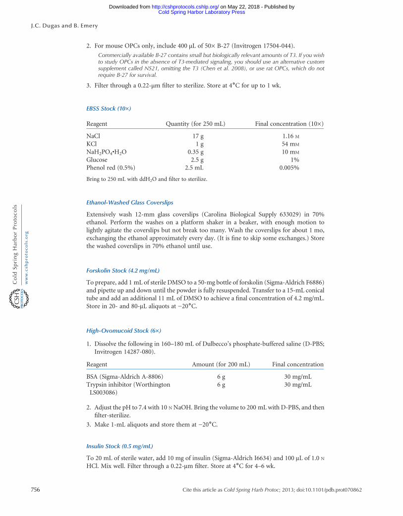
2. For mouse OPCs only, include 400 µL of 50× B-27 (Invitrogen 17504-044).
Commercially available B-27 contains small but biologically relevant amounts of T3. If you wishto study OPCs in the absence of T3-mediated signaling, you should use an alternative customsupplement called NS21, omitting the T3 (Chen et al. 2008), or use rat OPCs, which do notrequire B-27 for survival.
3. Filter through a 0.22-µm filter to sterilize. Store at 4˚C for up to 1 wk.
EBSS Stock (10×)
Reagent Quantity (for 250 mL) Final concentration (10×)
NaCl 17 g 1.16 M
KCl 1 g 54 mM
NaH2PO4•H2O 0.35 g 10 mM
Glucose 2.5 g 1%Phenol red (0.5%) 2.5 mL 0.005%
Bring to 250 mL with ddH2O and filter to sterilize.
Ethanol-Washed Glass Coverslips
Extensively wash 12-mm glass coverslips (Carolina Biological Supply 633029) in 70%ethanol. Perform the washes on a platform shaker in a beaker, with enough motion tolightly agitate the coverslips but not break too many. Wash the coverslips for about 1 mo,exchanging the ethanol approximately every day. (It is fine to skip some exchanges.) Storethe washed coverslips in 70% ethanol until use.
Forskolin Stock (4.2 mg/mL)
To prepare, add 1 mL of sterile DMSO to a 50-mg bottle of forskolin (Sigma-Aldrich F6886)and pipette up and down until the powder is fully resuspended. Transfer to a 15-mL conicaltube and add an additional 11 mL of DMSO to achieve a final concentration of 4.2 mg/mL.Store in 20- and 80-μL aliquots at −20˚C.
High-Ovomucoid Stock (6×)
1. Dissolve the following in 160–180 mL of Dulbecco’s phosphate-buffered saline (D-PBS;Invitrogen 14287-080).
Reagent Amount (for 200 mL) Final concentration
BSA (Sigma-Aldrich A-8806) 6 g 30 mg/mLTrypsin inhibitor (WorthingtonLS003086)
6 g 30 mg/mL
2. Adjust the pH to 7.4 with 10 N NaOH. Bring the volume to 200 mL with D-PBS, and thenfilter-sterilize.
3. Make 1-mL aliquots and store them at −20˚C.
Insulin Stock (0.5 mg/mL)
To 20 mL of sterile water, add 10 mg of insulin (Sigma-Aldrich I6634) and 100 μL of 1.0 N
HCl. Mix well. Filter through a 0.22-μm filter. Store at 4˚C for 4–6 wk.
756 Cite this article as Cold Spring Harb Protoc; 2013; doi:10.1101/pdb.prot070862
J.C. Dugas and B. Emery
Cold Spring Harbor Laboratory Press on May 22, 2018 - Published by http://cshprotocols.cshlp.org/Downloaded from

Low-Ovomucoid Stock Solution (10×)
To prepare, add 3 g of BSA (Sigma-Aldrich A8806) to 150 mL D-PBS. Mix well. Add 3 g oftrypsin inhibitor (Worthington LS003086) and mix to dissolve. Add �1 mL of 1 N NaOHto adjust the pH to 7.4. Bring the volume to 200 mL with D-PBS. Filter-sterilize through a0.22-μm filter. Make 1.0-mL aliquots and store at −20˚C.
NT-3 Stock (1 µg/mL)
1. Prepare a master stock of neurotrophin-3 (NT-3; Peprotech 450-03) at 1–0.1 mg/mLaccording to manufacturer’s instructions. (Instructions may vary from lot to lot; gener-ally, NT-3 is dissolved in buffer [e.g., Dulbecco’s phosphate-buffered saline] plus 0.2%BSA.) Store at −80˚C.
2. Prepare 0.2% BSA (Sigma-Aldrich A4161) in Dulbecco’s phosphate-buffered saline(D-PBS; Invitrogen 14287-080). Filter-sterilize and chill.
3. Dilute an aliquot of NT-3 master stock to 1 µg/mL in sterile, chilled 0.2% BSA preparedin Step 2.
4. Aliquot the NT-3 working stock from Step 3 (e.g., 20 µL/tube) and snap-freeze in liquidnitrogen.
5. Store aliquots at −80˚C.
OPC Culture Medium
1. To prepare full OPC culture medium, combine the following:20 mL DMEM-Sato base growth medium <R>20 µL Forskolin stock (4.2 mg/mL) <R>20 µL CNTF stock (10 µg/mL) <R>
2. Add growth factors as appropriate.• To promote OPC proliferation, add:20 µL PDGF stock (10 µg/mL) <R>20 µL NT-3 stock (1 µg/mL) <R>
• To promote OL differentiation, omit PDGF and add:200 µL T3 stock (4 µg/mL) <R>
3. Store at 4˚C for up to 3 d.
Papain Buffer
Reagent Amount (for 250 mL) Final concentration
EBSS stock (10×) <R> 25 mL 1×MgSO4 (100 mM) 2.5 mL 1 mM
Glucose (30%) 3 mL 0.46 %EGTA (0.5 M) 1 mL 2 mM
NaHCO3 (1 M) 6.5 mL 26 mM
Bring volume up to 250 mL with ddH2O and filter to sterilize.
PDGF Stock (10 µg/mL)
1. Prepare a platelet-derived growth factor (PDGF) master stock by diluting PDGF (Pepro-tech 100-13A) at 1–0.1 mg/mL according to manufacturer’s instructions. (Instructionsmay vary from lot to lot; generally, PDGF is dissolved in buffer [e.g., Dulbecco’s phos-phate-buffered saline] plus 0.2% BSA.) Store at −80˚C.
Cite this article as Cold Spring Harb Protoc; 2013; doi:10.1101/pdb.prot070862 757
Purification of OPCs from Rat Cortices
Cold Spring Harbor Laboratory Press on May 22, 2018 - Published by http://cshprotocols.cshlp.org/Downloaded from

2. Prepare 0.2% BSA (Sigma-Aldrich A4161) in Dulbecco’s phosphate-buffered saline(D-PBS; Invitrogen 14287-080). Filter-sterilize and chill.
3. Dilute an aliquot of PDGFmaster stock to 10 µg/mL in sterile, chilled 0.2% BSA preparedin Step 2.
4. Aliquot the PDGF working stock from Step 3 (e.g., 20 µL/tube) and snap-freeze in liquidnitrogen.
5. Store aliquots at −80˚C.
Poly-D-Lysine (PDL) Stock (1 mg/mL)
1. Resuspend poly-D-lysine (PDL; Sigma-Aldrich P6407; molecular weight 70–150 kDa) at1 mg/mL in borate buffer by combining 50 mg of PDL and 50 mL of 0.15 M boric acid(pH 8.4).
2. Filter to sterilize, and then aliquot (e.g., 100 µL/tube). Store aliquots at −20˚C.
SATO Supplement (100×)
1. Prepare the following stock solutions (these should be made fresh; do not reuse):• Combine 5 mg of progesterone (Sigma-Aldrich P8783) and 200 μL of ethanol to makea progesterone stock solution.
• Combine 4 mg of sodium selenite (Sigma-Aldrich S5261), 10 μL of 1 N NaOH, and 10mL of Dulbecco’s modified Eagle’s medium (DMEM; Gibco/Life Technologies 11960-044) to make a sodium selenite stock solution.
2. Combine the following:
ReagentQuantity
(for 200 mL)Final concentration
(100×)
BSA (Sigma-Aldrich A4161) 2 g 10 mg/mLTransferrin (Sigma-Aldrich T1147) 2 g 10 mg/mLPutrescine (Sigma-Aldrich P5780) 320 mg 1.6 mg/mLProgesterone stock solution 50 μL 6 μg/mLSodium selenite stock solution 2 mL 4 μg/mL
3. Bring to a total volume of 200 mL in DMEM, and then filter-sterilize. Aliquot and storeat –20˚C.
T3 Stock (4 µg/mL)
1. Dissolve 4 mg of thyroid hormone (T3; Sigma-Aldrich T6397) in 500 µL of 1 N NaOH toprepare a solution of 0.8 mg/100 µL.
2. Add 75 µL of the T3 solution from Step 1 to 150 mL of Dulbecco’s phosphate-bufferedsaline (D-PBS; Invitrogen 14287-080).
3. Filter solution through a filter-sterilization unit, discarding the first 10 mL.4. Aliquot (e.g., 200 µL/tube) and then store at −20˚C.
REFERENCE
Chen Y, Stevens B, Chang J, Milbrandt J, Barres BA, Hell JW. 2008. NS21:Re-defined and modified supplement B27 for neuronal cultures. J Neu-rosci Methods 171: 239–247.
758 Cite this article as Cold Spring Harb Protoc; 2013; doi:10.1101/pdb.prot070862
J.C. Dugas and B. Emery
Cold Spring Harbor Laboratory Press on May 22, 2018 - Published by http://cshprotocols.cshlp.org/Downloaded from

doi: 10.1101/pdb.prot070862Cold Spring Harb Protoc; Jason C. Dugas and Ben Emery ImmunopanningPurification of Oligodendrocyte Precursor Cells from Rat Cortices by
ServiceEmail Alerting click here.Receive free email alerts when new articles cite this article -
CategoriesSubject Cold Spring Harbor Protocols.Browse articles on similar topics from
(63 articles)Other Laboratory Organisms (298 articles)Neuroscience, general
(56 articles)Neural Cell Culture (407 articles)Mouse
(31 articles)Immunoseparation (43 articles)Immunoaffinity Purification
http://cshprotocols.cshlp.org/subscriptions go to: Cold Spring Harbor Protocols To subscribe to
© 2013 Cold Spring Harbor Laboratory Press
Cold Spring Harbor Laboratory Press on May 22, 2018 - Published by http://cshprotocols.cshlp.org/Downloaded from