Embed Size (px)
Citation preview
Puri¢cation of Saccharomyces cerevisiae RNase H(70) and identi¢cationof the corresponding gene
Peter Franka, Christa Braunshofer-Reitera, Anneliese Karwana, Rudolf Grimm1;b,Ulrike Wintersbergera;*
aDepartment of Molecular Genetics, Institute of Tumor Biology and Cancer Research, University of Vienna, Borschkegasse 8a, A-1090 Vienna, AustriabHewlett-Packard, Waldbronn Analytical Division, Hewlett-Packard-Str. 8., D-76337 Waldbronn, Germany
Received 24 March 1999
Abstract We purified Saccharomyces cerevisiae RNase H(70)to homogeneity, using an optimized chromatographic purificationprocedure. Renaturation gel assay assigned RNase H activity toa 70 kDa polypeptide. Sequencing of tryptic peptides identifiedthe open reading frame YGR276c on chromosome VII of the S.cerevisiae genome as the corresponding gene, which encodes aputative polypeptide of molecular mass of 62 849. We thereforerenamed this gene RNH70. Immunofluorescence microscopyusing a RNH70-EGFP fusion construct indicates nuclearlocalization of RNase H(70). Deletion of RNH70 from the yeastgenome did not result in any serious phenotype under theconditions tested. Homology searches revealed striking similaritywith a number of eukaryotic proteins and open reading frames,among them the chimpanzee GOR protein, a homolog of ahuman autoimmune antigen, found to elicit autoimmune responsein patients infected with hepatitis C virus.z 1999 Federation of European Biochemical Societies.
Key words: Ribonuclease H; RNA-DNA hybrid;Saccharomyces cerevisiae ; DNA replication; Nuclease;Evolution
1. Introduction
According to current knowledge RNA-DNA hybrids occurtransiently in living cells, necessarily during DNA replicationand transcription, and during the probably very rare events ofretrotransposition. Whereas no special mechanism for dealingwith the short hybrids arising during regular transcription isknown, RNA primers of Okazaki fragments ^ which are in-termediates of lagging strand DNA synthesis and are hy-bridized to the DNA template strand ^ have to be removedenzymatically [1]. Enzymes hydrolyzing speci¢cally the RNAstrand of RNA-DNA hybrids, called ribonucleases H
(RNases H), were found in all organisms yet examined [2].Nevertheless, the most intensively studied and best under-stood proteins exhibiting RNase H activity are retroviral re-verse transcriptases [3]. Their RNase H activity catalyzes anessential step in the life cycle of retroviruses, i.e. the destruc-tion of the RNA genomes after their retrotranscription intoDNA. Another well studied RNase H is RNase HI of Esche-richia coli, which is evolutionarily related to the RNase Hdomain of reverse transcriptases [4]. Interestingly, this enzymewas found to play a regulatory role during DNA replication:in wild type cells it seems to be on the watch for super£uousRNA-DNA hybrids which might have arisen from discontin-ued transcripts (for review see [5]). In E. coli, RNA primers ofOkazaki fragments are removed by the RNase H activity ofDNA polymerase I [1]. Nevertheless, still another enzyme withRNase H activity exists in E. coli, RNase HII [6], the primarystructure of which is not related to the RNase H domain ofreverse transcriptases, and the physiological role of which is asyet unknown. Remarkably, open reading frames, character-ized by their sequences them as orthologs of E. coli RNaseHII, were found in all bacterial genomes examined. Genesencoding orthologs of the two bacterial proteins were recentlycloned from eukaryotic organisms, e.g. from the buddingyeast Saccharomyces cerevisiae [7,8], from the fruit £y Droso-phila melanogaster [9] and from human cells [10^13].
S. cerevisiae RNase H1 is structurally related to E. coliRNase HI [7] ; the biological role of that enzyme, however,is as yet unknown. The yeast protein representing the evolu-tionary relative of bacterial RNase HII was, on the basis of itsmolecular weight, called RNase H(35) by us [8]. It is probablythe main RNase H of that organism because deletion of therespective gene causes a drastic reduction of RNase H activityin yeast cell extracts [8]. Surprisingly, when RNase H activityoriginally was enriched by then classical puri¢cation stepsfrom yeast cell extracts, an enzyme of molecular weightaround 70 kDa was found and called RNase H(70) [14]. Fol-lowing studies intended to isolate the gene encoding RNaseH(70) revealed that the original preparation contained copuri-¢ed proteins, the most prominent of which was poly(A) bind-ing protein (PABP [15]) possessing a very similar molecularweight.
In this communication we report an improved puri¢cationprocedure for RNase H(70), which removed PABP as well asa having reverse transcriptase activity which earlier was con-sidered a property of RNase H(70) [16]. The new procedureresulted in a small amount of homogeneous protein whichcould be used for sequencing of several tryptic peptides which¢nally paved the way for the identi¢cation of the gene,RNH70.
0014-5793 / 99 / $20.00 ß 1999 Federation of European Biochemical Societies. All rights reserved.PII: S 0 0 1 4 - 5 7 9 3 ( 9 9 ) 0 0 5 1 2 - 8
*Corresponding author. Fax: (43) (1) 4277 9651.E-mail: [email protected]
1 Present address: Toplab GmbH, Am Klopferspitz 19, D-82152Martinsried, Germany.
Abbreviations: PCR, polymerase chain reaction; RNase H, ribonu-clease H; SDS-PAGE, sodium dodecyl sulfate polyacrylamide gelelectrophoresis; HPLC, high performance liquid chromatography;EST, expressed sequence tag; ORF, open reading frame; PMSF,phenylmethylsulfonyl £uoride; PEG, polyethylene glycol; EGFP, en-hanced green £uorescent protein; DAPI, 4P,6-diamino-2-phenylin-dole; TPCK, N-tosyl-L-phenylalanine chloromethylketone; TLCK,NK-p-tosyl-L-lysine chloromethylketone; pI, isoelectric point
FEBS 21973 4-5-99
FEBS 21973 FEBS Letters 450 (1999) 251^256
2. Materials and methods
2.1. Yeast strains and growth mediaStrains MZ3 (MatK ; pep4-3 trp1 leu2-v1 ura3-v1) and MZ3vPABP
(derived from MZ3 by deleting a 405 bp SpeI/SphI fragment from theC-terminus of the PABP gene and introducing the URA3 marker [17])were used for enzyme puri¢cation. Strain K699 (Mata ura3 ade 2-1trp1-1 can1-100 leu2-3,112 his3-11,15 ssdl [8]) was used for construct-ing strain BC70 (Mata ura3 ade 2-1 trp1-1 can1-100 leu2-3,112 his3-11,15 ssdl rnh70v : :URA3); strain AK606 (Mata ura3 leu2 trp1 his3),an adenine-prototrophic meiotic segregant from a cross betweenstrains K699 and MZ3, was employed for transformation with plas-mid pUG36-RNH70. YPD growth medium (1% yeast extract, 2%peptone, 2% dextrose) and synthetic complete medium (SC) were asdescribed [18].
2.2. Enzyme activity assays, standard techniques for protein analysis,materials and enzymes
The standard RNase H assay in solution was performed as de-scribed [14], and the reverse transcriptase assay was performed asreported earlier [16]. The renaturation gel assay for the in situ detec-tion of RNase H activity was performed according to Frank et al. [19]using Mn2� as a divalent cation. Protein concentration was deter-mined by the method of Bradford [20]. Discontinuous SDS-PAGEwas performed according to Laemmli [21]; protein bands were visual-ized with Coomassie brilliant blue G250 or by silver staining [22]. [K-32P]ATP, 3000 Ci/mmol, 10 mCi/ml and [5-3H]uridine 5P-triphosphate,15.6 Ci/mmol, 1 mCi/ml, were from American Radiolabeled Chem-icals (ARC). DNA cellulose was prepared according to [23]. Oli-go(dT), RNA polymerase (E. coli), phenyl-Sepharose, and Mono Pfor chromatofocusing were purchased from Pharmacia-LKB. Hydrox-yapatite and A¤-Gel Blue were from Bio-Rad. Restriction enzymes,antipain, pepstatin A and PCR-related products were from Boeh-ringer Mannheim. PMSF, TPCK and TLCK were purchased fromFluka.
2.3. Puri¢cation of RNase H(70)To minimize proteolysis, all puri¢cation steps were carried out at
4³C, and protease inhibitors (0.2 mM PMSF and 1 mM sodium sul-¢te, pH 8.0; 0.1 mM sodium tetrathionate; 1 WM each of TLCK,TPCK, pepstatin A, and antipain) and 0.1% L-mercaptoethanolwere included in all bu¡ers. After having used strain MZ3vPABPfor working out a puri¢cation procedure by which RNase H activitycould unambiguously be separated from PABP the following large-scale preparation was carried out (see Fig. 1A). Cells (strain MZ3)were grown in a 4 l fermenter as a continuous culture at pH 6.2 and30³C using YPD as a medium, and starting with a cell density of1.6U106/ml. The cells were harvested by centrifugation, washedonce with bu¡er A (20 mM Tris-HCl pH 7.9, 1 mM EDTA, 10%glycerol) containing 2 M NaCl, and frozen in liquid nitrogen. Thecell pellet (120 g wet weight per frozen aliquot) was resuspended in180 ml of bu¡er A containing 2 M NaCl, and after addition of anequal volume of acid washed glass beads (diameter 0.45 mm), thesuspension was homogenized and separated from glass beads, undis-rupted cells and cell fragments as described [14]. The cell extract wassubsequently centrifuged for 120 min at 43 000 rpm in a Beckman 45Ti rotor and ¢ltered through a paper ¢lter. To remove nucleic acids,the supernatant was treated with polyethylene glycol 6000 (PEG, 10%w/v ¢nal concentration) for 30 min on ice followed by centrifugationfor 35 min at 22 000 rpm (Beckman 45 Ti rotor). After dialysis of thesupernatant against bu¡er A containing 0.1 M NaCl (2U2 h) andcentrifugation for 25 min at 24 000 rpm (Beckman 45 Ti rotor), theclear supernatant was loaded on a 360 ml DNA cellulose column(£ow rate 60 ml/h). After extensive washing (until no protein wasdetectable in the £ow-through), the bound protein was completelyeluted with bu¡er A containing 2 M NaCl. Protein containing frac-tions (10 ml each) were analyzed for RNase H activity and pooled.The pool was adjusted with ammonium sulfate up to 60% saturationand, after 20 h of storage, centrifuged (35 000 rpm; 45 min; 45 Tirotor). The precipitated proteins were dissolved in bu¡er A containing1 M ammonium sulfate and loaded onto a 100 ml phenyl-Sepharosecolumn (£ow rate 30 ml/h). After washing, protein was eluted with500 ml of a decreasing gradient (1^0 M ammonium sulfate in bu¡erA). In the fractions PABP was detected with the aid of a rabbitantiserum raised against the protein expressed in and puri¢ed from
E. coli [17], and reverse transcriptase by determination of enzymeactivity. Both proteins eluted in fractions well separated from thosecontaining RNase H activity. The latter (6 ml each) were pooled,precipitated with ammonium sulfate (60% saturation) and centrifuged(35 000 rpm; 45 min; 45 Ti rotor). The precipitated protein was dis-solved in 40 ml bu¡er K1 (25 mM K2HPO4/KH2PO4, pH 6.8, 10%glycerol), dialyzed against bu¡er K1 (2U2 h) and loaded onto a 40 mlhydroxyapatite column (£ow rate 24 ml/h). After washing, the boundprotein was eluted with a linear increasing phosphate gradient of300 ml (25^500 mM K2HPO4/KH2PO4, pH 6.8, 10% glycerol). RNaseH containing fractions (6 ml each) were pooled. The pool was dia-lyzed two times against bu¡er K2 (10 mM K2HPO4/KH2PO4, pH 6.8,5 mM MgCl2, 10% glycerol) and loaded onto a 40 ml A¤-Gel Bluecolumn (£ow rate 30 ml/h). After washing, the elution took placeusing a 300 ml linear increasing gradient (0^2 M NaCl in bu¡erK2). RNase H containing fractions (6 ml each) were pooled. Thepool was desalted with a PD10 column (Pharmacia) and applied toa Mono P column (5U200 mm), which was equilibrated with 25 mMBisTris-HCl, pH 7.1, 10% glycerol, PMSF and L-mercaptoethanol.Elution was done with Polybu¡er 74 (Pharmacia), pH 4.0, 10% glyc-erol, PMSF and L-mercaptoethanol. Fractions (1 ml each) were col-lected and analyzed for the presence of RNase H using the renatura-tion gel assay [19].
2.4. Peptide sequencingThe RNase H(70) preparation was separated on a preparative 12%
SDS-PAGE, stained with Coomassie brilliant blue R-250 (Serva) andthe band corresponding to the RNase H(70) protein was cut out andconcentrated on a specially designed concentration gel according toVandekerckhove et al. [24]. Approximately 5 Wg of the puri¢ed andconcentrated RNase H(70) polypeptide was blotted from the Vande-kerckhove gel onto a poly(vinylidene di£uoride) (PVDF) membrane(Millipore, Bedford, MA, USA) using a standard blotting tank sys-tem. The transfer bu¡er contained 13 mM sodium carbonate pH 9.9and 20% methanol. The protein spot was detected by staining withCoomassie brilliant blue R-250 and afterwards excised from the mem-brane. The blotted protein sample was digested with sequencing gradetrypsin (Promega, Madison, WI, USA) as described by Fernandez etal. [25], except that the detergent Triton X-100 was replaced by octyl-L-D-glucopyranoside (Boehringer Mannheim, Germany). Peptidemapping was carried out on a 1U250 mm Vydac C18 column at40³C with a £ow rate of 50 Wl/min using the HP 1100 HPLC system(Hewlett-Packard, Waldbronn, Germany). Selected peptide fractionswere analyzed by automated Edman degradation using the HPG1005A protein sequencing system (Hewlett-Packard, Palo Alto,CA, USA). Three peptide sequences, TSVQEDDHT, NTFNEISD,and CVEDDET, were detected in ORF YGR276c on chromosomeVII of the S. cerevisiae genome. YGR276c was renamed RNH70 andcloned.
2.5. PCR ampli¢cation and cloning proceduresIn order to amplify the RNase H(70) coding sequence for the pro-
duction of a deletion allele of the RNH70 gene, PCRs were performedwith yeast genomic DNA (Promega) as template. The following prim-ers were used for PCR with the Expand PCR system (BoehringerMannheim) for generating a 2273 bp fragment to be cloned intopUC18: forward primer rh70d3/EcoRI: 5P-TGTGACTGAATTC-TAATGTACTTGGAGGAAGGGAGGAAGACG-3P and reverseprimer rh70d4/HindIII: 5P-CGGATGTAAGCTTTCTACAGAGG-GAGAGCTGTGAGGATATATT-3P. A PCR product of the ex-pected size was obtained, digested with EcoRI and HindIII, gel puri-¢ed and ligated into EcoRI/HindIII digested pUC18 vector. A SspBI/SmaI fragment of 1549 bp, including nearly the complete ORF, wasreplaced by a 1125 bp fragment carrying the URA3 gene. The isolatedand gel puri¢ed deletion allele was used for integrative transformation[26], and Ura� transformants were isolated. They were checked byspeci¢c PCR for successful gene replacement and analyzed for phe-notypes. One of the mutants, BC70, was used for further experiments.
2.6. Cellular localization of yeast RNase H(70) enhanced green£uorescent protein fusion
Primers RH70/EcoRI and RH70/SalIrev2 were used to introduceEcoRI and SalI restriction sites at the beginning and end of theRNase H(70) coding sequence, respectively; RH70/EcoRI: 5P-G-ATGTCGGAATTCATGCAAGTAGAAGGGCCTGACACTAA-3P ;RH70/SalIrev2: 5P-GATGTCGTCGACTATAGTCCTTTGAATTG-
FEBS 21973 4-5-99
P. Frank et al./FEBS Letters 450 (1999) 251^256252
GCACCTTCAT-3P. The ampli¢ed fragment of RNH70 was insertedinto the pUG36 vector (U. Gu«ldener and J. Hegemann, manuscript inpreparation). This system allowed the expression of RNH70 fused tothe carboxy-terminus of the enhanced green £uorescent protein(EGFP) under the control of the yeast MET25 promoter. The strainAK606 was transformed with the resulting pUG36-RNH70 vector,grown in SC medium lacking uracil and induced by addition of me-thionine for 4 h. Formaldehyde ¢xed and DAPI stained cells wereviewed with a Nikon FXA microscope (EX450^490 nm), and photo-graphed on Fujichrome 400 ¢lm.
3. Results
3.1. Puri¢cation of RNase H(70)When puri¢cation of RNase H activity from yeast cells was
originally attempted in our laboratory we ended up with aprotein fraction which showed in SDS-PAGE one prominentband around 70 kDa, and by glycerol gradient centrifugationan activity peak at a sedimentation value corresponding to thesame molecular weight [14]. Further studies, however, re-vealed the presence of PABP (molecular mass 64 344) and ofa reverse transcriptase activity. As PABP at that time was justknown to be somehow involved in transcription termination itwould have been reasonable that it possessed the RNase Hactivity. To ¢nd out whether this was the case or whetherPABP represented a separate but copuri¢ed protein, a strain,MZ3vPABP, containing a PABP gene with a shortened C-terminus and thus a PAB protein of molecular mass di¡erentfrom that of RNase H(70), namely 55 429, was constructed,and an antiserum was raised against E. coli expressed PABP.As described in detail elsewhere [17] these experimental means¢nally enabled us to unambiguously identify PABP as animpurity of the RNase H preparation. By initially using strainMZ3vPABP for puri¢cation we con¢rmed the existence of aseparate RNase H molecule exhibiting in SDS-PAGE a mo-lecular weight around 70 kDa, for which therefore the nameRNase H(70) was retained.
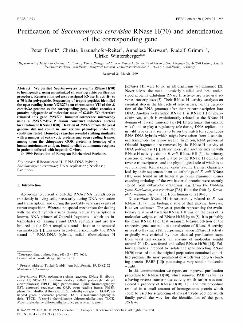
The newly worked out puri¢cation procedure, indicated inFig. 1A, and described in detail in Section 2, allows separationof PABP as well as of the reverse transcriptase activity fromRNase H(70) by using a hydrophobic interaction chromatog-raphy (HIC) on phenyl-Sepharose as second chromatographicstep. A further improvement compared to the original proce-dure is achieved by the employment of A¤-Gel Blue chroma-tography and chromatofocusing on a Mono P column (Table1). Even when starting with 120 g of yeast cells (wet weight)the amount of protein after this last chromatography was verylow and an overall puri¢cation factor could not be deter-mined. Analysis of the fractions from the Mono P columnby separation of proteins on SDS-PAGE and silver staining(Fig. 1B) revealed a prominent band around 70 kDa in thesame fractions which exhibited RNase H activity in the in siturenaturation gel assay (see Fig. 1B). Fraction 6 from theMono P column was further processed for isolation of RNase
H(70), peptide sequencing and identi¢cation of the corre-sponding gene.
3.2. Identi¢cation of the corresponding yeast geneTo obtain the 70 kDa polypeptide for protein sequencing,
CFig. 1. Puri¢cation of RNase H(70). A: Puri¢cation scheme; AS,ammonium sulfate. B: Protein and activity pro¢le of the ¢nal step,chromatofocusing on Mono P. Proteins from fractions 4^8 from theMono P column were separated by 12% SDS-PAGE and silverstained (left panel, the bands identical in all fractions result from astaining artifact), as well as analyzed by in situ renaturation gel as-say (right panel). Protein size markers are indicated between thetwo panels.
FEBS 21973 4-5-99
P. Frank et al./FEBS Letters 450 (1999) 251^256 253
the RNase H(70) preparation was processed as described inSection 2, including preparative SDS-PAGE, concentrationon Vandekerckhove gel, blotting onto nitrocellulose, andCoomassie brilliant blue R-250 staining. The stained spotwas cut out and digested overnight with trypsin. HPLC puri-
¢cation of the tryptic peptides and Edman sequencing yieldedthe following three peptide sequences: TSVQEDDHT,NTFNEISD, and CVEDDET. Using those sequences, we un-ambiguously identi¢ed ORF YGR276c as the gene for RNaseH(70) in the S. cerevisiae genome. We therefore renamed this
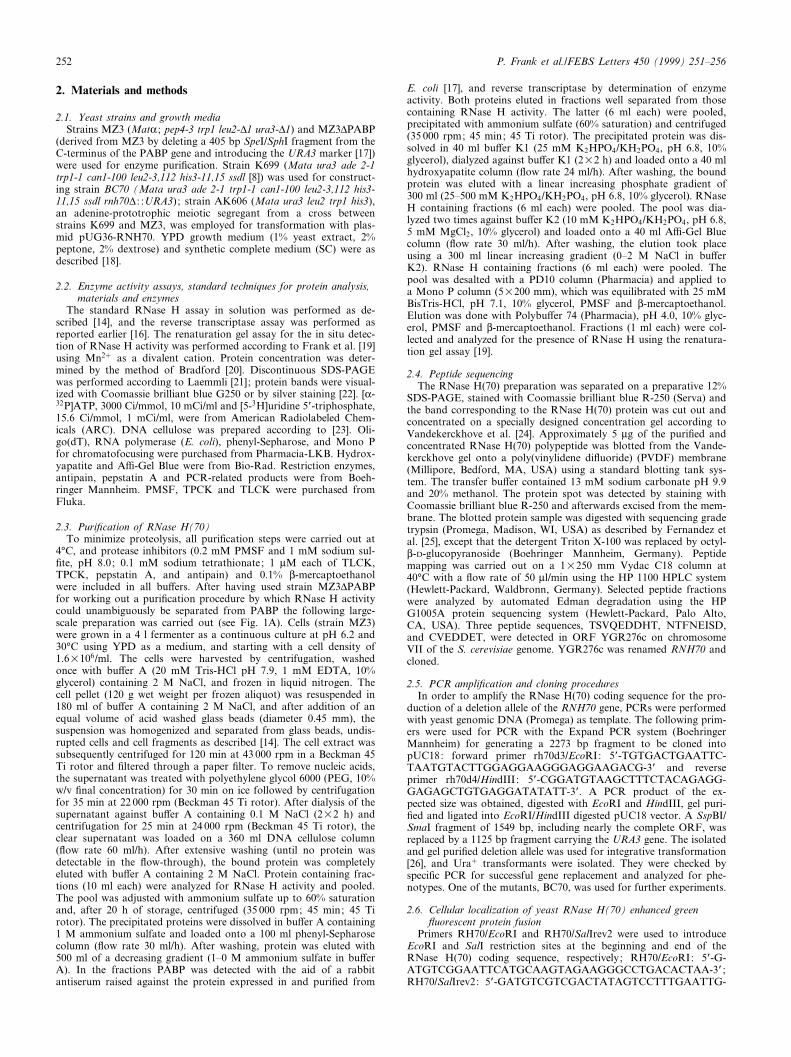
Fig. 2. Multiple sequence alignment of homologous parts from yeast RNase H(70), and corresponding eukaryotic open reading frames. Thealignment was generated, using the `multiple sequence alignment with hierarchical clustering' method of Corpet [27], and sequences from the S.cerevisiae ORF YGR276c (553 aa, marked RNase H(70), Swiss-Prot accession number P53331), the GOR antigen of the chimpanzee P. troglo-dytes (525 aa, partial sequence; EMBL accession number D10017), a C. elegans ORF (594 aa; marked caeno594; GenBank accession numberAF016430), the S. cerevisiae ORF YNL107w (130 aa; EMBL accession number Z73280), the Xenopus laevis XPMC2 protein (421 aa; GenBankaccession number U10185), the S. cerevisiae ORF YO080c (289 aa; GenBank accession number Z74822), a C. elegans ORF (271 aa; markedcaeno271; Swiss-Prot accession number Q101024), and the human ISG20 protein (179 aa; GenBank accession number X89773). Numbering ofamino acids above the alignment corresponds to the consecutive sequence of yeast RNase H(70) (only regions with existing consensus areshown). Amino acids identical in all sequences are marked in bold letters, and similar amino acids are underlined. In the consensus sequence(marked consensus), bold letters indicate identical and asterisks similar amino acids. In addition, identities between RNase H(70) and GOR aremarked # in the line `gcon' above the alignment. Parameters used for the alignment: Symbol comparison table: blosum62; gap weight: 12;gap length weight: 2. Five regions of clustered identical or similar amino acids are indicated as I, II, III, IV, and V below the consensus.
Table 1Puri¢cation of yeast RNase H(70)
Total protein(mg)
Protein(mg/ml)
Speci¢c activity(U/Wg)
Total activity(U)
Yield(%)
Puri¢cation factor(fold)
Extracta 6396 31.2 137 8.8U108 100 1DNA cellulose 280 1.6 1 052 2.9U108 34 8Phenyl-Sepharose 29 0.28 2 260 6.5U107 7.6 16Hydroxyapatite 2.88 0.16 20 270 5.8U107 6.8 148A¤-Gel Blue 0.13 0.003b 309 910 4.0U107 4.7 2 260Mono P n.d. n.d. n.d. n.d. n.d. n.d.
n.d.: not determined by activity test in solution.aCell extract after PEG precipitation.bEstimation by silver staining.
FEBS 21973 4-5-99
P. Frank et al./FEBS Letters 450 (1999) 251^256254
ORF, which encodes a potential protein of molecular mass62 849 and which is located on the right arm of chromosomeVII, RNH70. The obvious molecular mass di¡erence betweenthe putative protein encoded by ORF RNH70 and the isolatedprotein might be due to post-translational modi¢cation.
3.3. Sequence comparison uncovers a relationship between yeastRNase H(70) and other eukaryotic proteins
When searching the non-redundant protein database atNCBI using the BLAST program, we found highly signi¢canthomologies of the yeast RNase H(70) with the GOR antigenof the chimpanzee, Pan troglodytes, with a Caenorhabditiselegans ORF of 594 aa, with the S. cerevisiae ORFsYLR107w and YO080c, the Xenopus laevis XPMC2 protein,a C. elegans ORF of 271 aa, and the human ISG20 protein.An alignment of these sequences is shown in Fig. 2. Using the`multiple sequence alignment with hierarchical clustering'method of Corpet [27], we detected ¢ve highly conserved mo-tifs in all of the eight analyzed sequences. Another feature ofthe sequence becomes evident using the newly established po-sition-speci¢c iterated BLAST (PSI-Blast) program [28]. Itindicates that RNase H(70) is additionally related to prokary-otic DNA polymerases I and III and prokaryotic RNases T(data not shown).
3.4. Deletion of the gene RNH70 from the genome does notresult in a phenotype easily discernible under laboratoryconditions
Using integrative transformation [26] with the deletion con-struct described in Section 2, we obtained the viable haploidstrain BC70. A cell extract of this strain exhibited no signi¢-cant decrease in overall RNase H activity compared to anextract from the corresponding RNase H(70) pro¢cient strain(data not shown). RNase H activity was enriched from cell
extracts of both strains by removal of nucleic acids and DNAcellulose chromatography (see Section 2), and the resultingfractions were tested for RNase H activity using Mg2� asdivalent cation. Again no signi¢cant change in RNase H ac-tivity between the fraction derived from the deletion mutantand that derived from the strain with the intact RNH70 genewas found. Additional experiments were performed to lookfor a mutant phenotype of the deletion strain: there was nodi¡erence in the proliferation rates of the RNase H(70) pro-¢cient or de¢cient cells as determined at 16³C, 30³C and 37³C(data not shown).
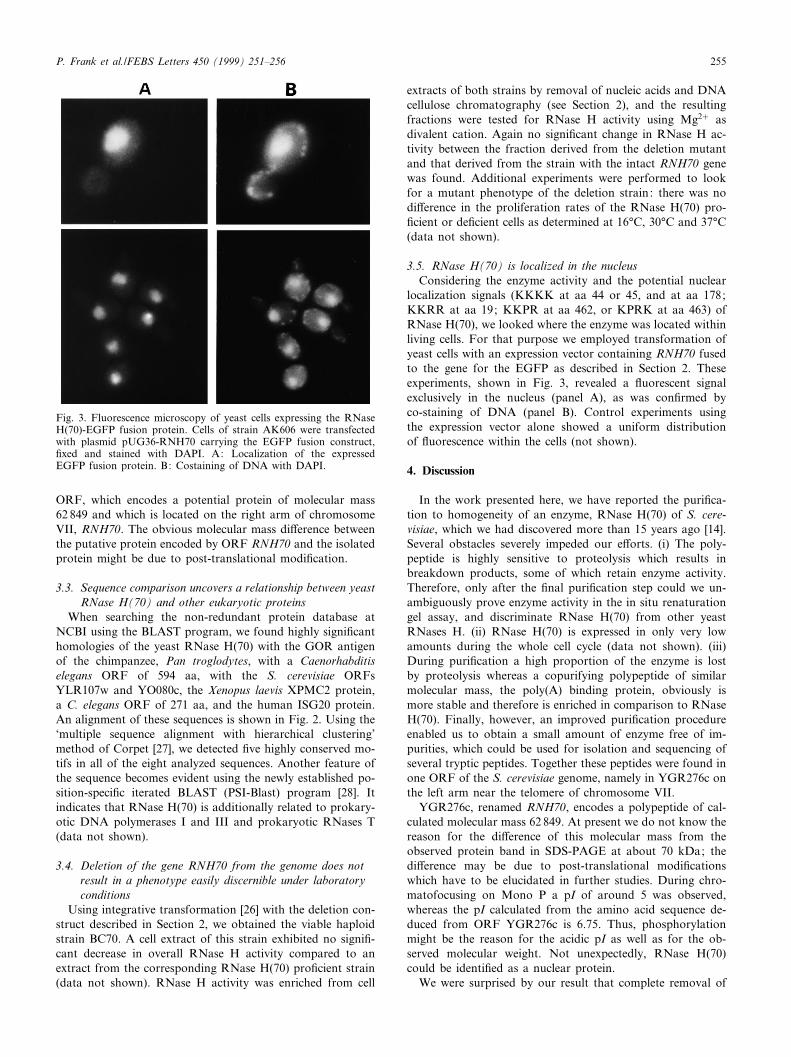
3.5. RNase H(70) is localized in the nucleusConsidering the enzyme activity and the potential nuclear
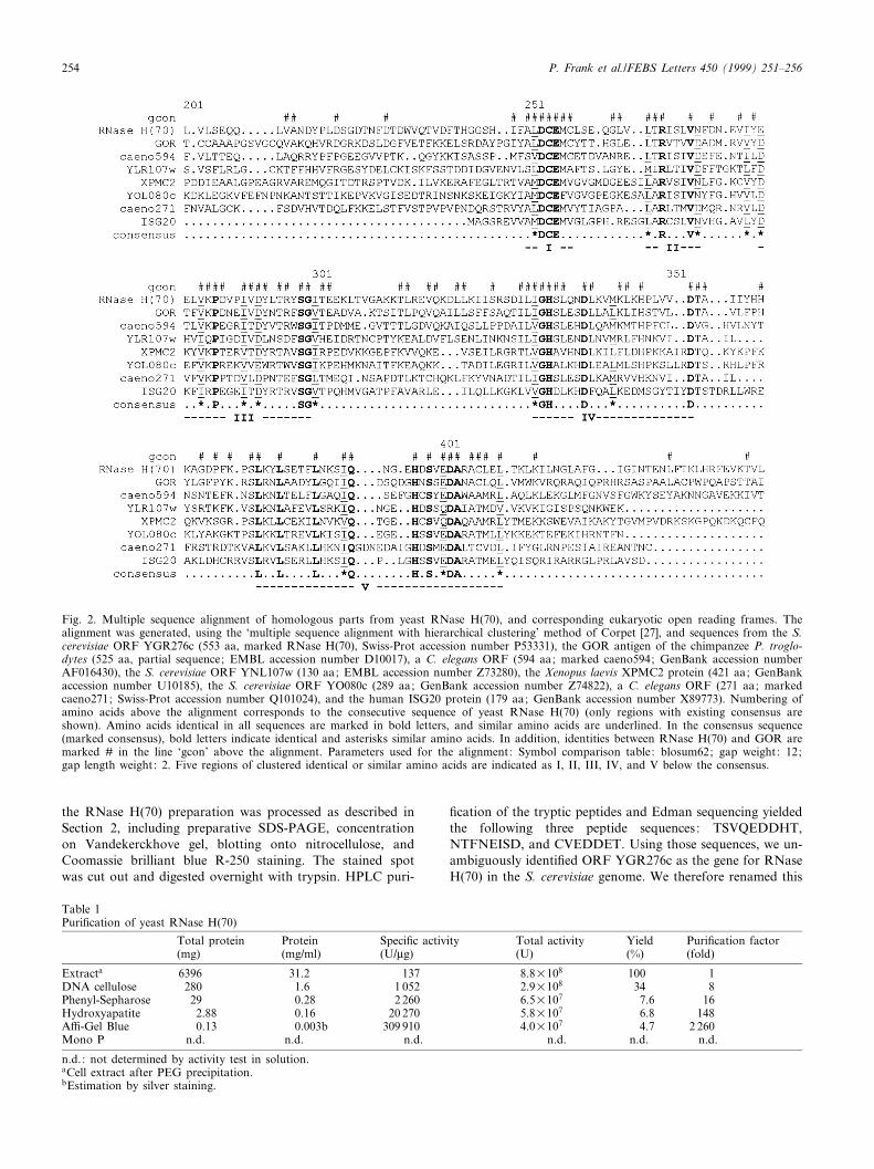
localization signals (KKKK at aa 44 or 45, and at aa 178;KKRR at aa 19; KKPR at aa 462, or KPRK at aa 463) ofRNase H(70), we looked where the enzyme was located withinliving cells. For that purpose we employed transformation ofyeast cells with an expression vector containing RNH70 fusedto the gene for the EGFP as described in Section 2. Theseexperiments, shown in Fig. 3, revealed a £uorescent signalexclusively in the nucleus (panel A), as was con¢rmed byco-staining of DNA (panel B). Control experiments usingthe expression vector alone showed a uniform distributionof £uorescence within the cells (not shown).
4. Discussion
In the work presented here, we have reported the puri¢ca-tion to homogeneity of an enzyme, RNase H(70) of S. cere-visiae, which we had discovered more than 15 years ago [14].Several obstacles severely impeded our e¡orts. (i) The poly-peptide is highly sensitive to proteolysis which results inbreakdown products, some of which retain enzyme activity.Therefore, only after the ¢nal puri¢cation step could we un-ambiguously prove enzyme activity in the in situ renaturationgel assay, and discriminate RNase H(70) from other yeastRNases H. (ii) RNase H(70) is expressed in only very lowamounts during the whole cell cycle (data not shown). (iii)During puri¢cation a high proportion of the enzyme is lostby proteolysis whereas a copurifying polypeptide of similarmolecular mass, the poly(A) binding protein, obviously ismore stable and therefore is enriched in comparison to RNaseH(70). Finally, however, an improved puri¢cation procedureenabled us to obtain a small amount of enzyme free of im-purities, which could be used for isolation and sequencing ofseveral tryptic peptides. Together these peptides were found inone ORF of the S. cerevisiae genome, namely in YGR276c onthe left arm near the telomere of chromosome VII.
YGR276c, renamed RNH70, encodes a polypeptide of cal-culated molecular mass 62 849. At present we do not know thereason for the di¡erence of this molecular mass from theobserved protein band in SDS-PAGE at about 70 kDa; thedi¡erence may be due to post-translational modi¢cationswhich have to be elucidated in further studies. During chro-matofocusing on Mono P a pI of around 5 was observed,whereas the pI calculated from the amino acid sequence de-duced from ORF YGR276c is 6.75. Thus, phosphorylationmight be the reason for the acidic pI as well as for the ob-served molecular weight. Not unexpectedly, RNase H(70)could be identi¢ed as a nuclear protein.
We were surprised by our result that complete removal of
Fig. 3. Fluorescence microscopy of yeast cells expressing the RNaseH(70)-EGFP fusion protein. Cells of strain AK606 were transfectedwith plasmid pUG36-RNH70 carrying the EGFP fusion construct,¢xed and stained with DAPI. A: Localization of the expressedEGFP fusion protein. B: Costaining of DNA with DAPI.
FEBS 21973 4-5-99
P. Frank et al./FEBS Letters 450 (1999) 251^256 255
gene RNH70 did not lead to an abnormal phenotype of thedeletion mutants, at least not under the conditions tested.Recently we had discovered another RNase H of yeast,RNase H(35) [8]. Deletion of the coding region of that enzymealso did not entail a severely pathological phenotype but asigni¢cant decrease of total RNase H activity in the cell ex-tract of the deletion mutant. Still another yeast gene encodinga protein with RNase H activity is known, RNH1 [7], thedeletion of which again does not have a dramatic impact onthe a¡ected cells. Strangely enough, even cells harboring com-bined deletions of the as yet known RNase H genes are viable(manuscript in preparation). Hence, di¡erent proteins possess-ing RNase H activity evidently are able to substitute for eachother, and additional such protein(s) must exist in yeast cells.The only candidate we are at present aware of is Rad27p/FEN1 [29].
Another remarkable ¢nding is the fact that the amino acidsequence of neither RNase H(35) nor RNase H(70) of S.cerevisiae is evolutionarily related to that of the `prototype'RNases H, the RNase HI of E. coli and the RNase H domainof retroviral reverse transcriptase. Unexpectedly, selective hy-drolysis of the RNA strand from a RNA-DNA hybrid can becarried out by polypeptides of completely unrelated primarystructures. A homology search with the yeast RNase H(70)amino acid sequence revealed the GOR protein as the closestrelative. This chimpanzee (and human) autoimmune antigenelicits an autoimmune response in a high proportion of hep-atitis C patients [30]. The currently incurable disease is causedby the single (+) stranded RNA hepatitis C virus, it has enor-mous medical impact, as it may ¢nally lead to cirrhosis and/orhepatocellular carcinoma. Although the complete sequence ofthe GOR protein is still unknown we suspect that it may bethe human homolog of S. cerevisiae RNase H(70). In thisconnection another vertebrate RNase H should be mentioned:about 10 years ago a bovine RNase H was described [31]which resembled yeast RNase H(70) in several respects butseemed to be di¡erent from bovine RNases HI and II. The78 kDa protein was, like yeast RNase H(70), shown to bestrongly inhibited by N-ethylmaleimide and to prefer Mg2�
over Mn2� as divalent cation. Furthermore it stimulated invitro enzyme activity of DNA polymerase K [31]. Whether thisbovine enzyme might be the homolog of yeast RNase H(70),or even of the so-called GOR protein, remains to be deter-mined. Another human relative of yeast RNase H(70) is theinterferon stimulated ISG20 protein. Together with PML(promyelocytic leukemia protein) and SP100 (an autoantigenof patients su¡ering from biliary cirrhosis), it is found innuclear matrix associated multiprotein complexes, the so-called PML nuclear bodies, which might play a role duringviral infections [32].
A remarkable homolog of RNase H(70) is also proteinXPMC2 of Xenopus laevis, which is able to suppress certainmutations leading to mitotic catastrophe in the ¢ssion yeastSchizosaccharomyces pombe [33]. Interestingly, the use of PSI-Blast homology searches uncovered even sequence parts ofprokaryotic DNA polymerases I and III, and RNases T asmore distant relatives of RNase H(70). We hypothesize thatthese related proteins (or protein domains) might comprise anew superfamily of evolutionarily conserved nucleases.
Acknowledgements: We thank Alexandra Bogusch for excellent tech-nical assistance, and Gaby Operenyi for help with the manuscript.Special thanks are due to Dr. Karl Bayer (University of AgriculturalSciences, Vienna) for helping us during large scale yeast cell cultiva-tion. This research was supported by the Fonds zur Fo«rderung derwissenschaftlichen Forschung in Oë sterreich (Grant S 5806-MOB) toU.W., the Anton Dreher Geda«chtnisschenkung fu«r medizinische For-schung (Grant 272/95) to P.F. and the Hochschuljubila«umsstiftungder Stadt Wien (Grant H-00097/96) to P.F.
References
[1] Kornberg, A. and Baker, T.A. (1992) DNA Replication, Free-man, New York.
[2] Wintersberger, U. (1990) Pharmacol. Ther. 48, 259^280.[3] Hughes, S.H., Arnold, E. and Hostomsky, Z. (1998) in: Ribonu-
cleases H (Crouch, R.J. and Toulme, J.J., Eds.), pp. 195^224,Editions INSERM, Paris.
[4] Johnson, M.S., McClure, M.A., Feng, D.F., Gray, J. and Doo-little, R.F. (1986) Proc. Natl. Acad. Sci. USA 83, 7648^7652.
[5] Kogoma, T. (1997) Microbiol. Mol. Biol. Rev. 61, 212^238.[6] Itaya, M. (1990) Proc. Natl. Acad. Sci. USA 87, 8587^8591.[7] Itaya, M., McKelvin, D., Chatterjie, S.K. and Crouch, R.J.
(1991) Mol. Gen. Genet. 227, 438^445.[8] Frank, P., Braunshofer-Reiter, C. and Wintersberger, U. (1998)
FEBS Lett. 421, 23^26.[9] Filippov, V., Filippova, M. and Gill, S.S. (1997) Biochem. Bio-
phys. Res. Commun. 240, 844^849.[10] Frank, P., Braunshofer-Reiter, C., Wintersberger, U., Grimm, R.
and Bu«sen, W. (1998) Proc. Natl. Acad. Sci. USA 95, 12872^12877.
[11] Wu, H., Lima, W.F. and Crooke, S.T. (1998) Antisense NucleicAcid Drug Dev. 8, 53^61.
[12] Cerritelli, S.M. and Crouch, R.J. (1998) Genomics 53, 300^307.[13] Frank, P., Braunshofer-Reiter, C., Po«ltl, A. and Holzmann, K.
(1998) Biol. Chem. 379, 1407^1412.[14] Karwan, R., Blutsch, H. and Wintersberger, U. (1983) Biochem-
istry 22, 5500^5502.[15] Sachs, A.B., Bond, M.W. and Kornberg, R.D. (1986) Cell 45,
827^835.[16] Karwan, R., Ku«hne, C. and Wintersberger, U. (1986) Proc. Natl.
Acad. Sci. USA 83, 5919^5923.[17] Valenta, B. (1993) PhD Thesis, University of Vienna.[18] Sherman, F. (1991) Methods Enzymol. 194, 3^21.[19] Frank, P., Cazenave, C., Albert, S. and Toulme, J.J. (1993) Bio-
chem. Biophys. Res. Commun. 196, 1552^1557.[20] Bradford, M.M. (1976) Anal. Biochem. 72, 248^254.[21] Laemmli, U.K. (1970) Nature 227, 680^685.[22] Merril, C.R., Goldman, D. and VanKeuren, M. (1984) Methods
Enzymol. 104, 441^447.[23] Litman, R. (1968) J. Biol. Chem. 243, 6222^6233.[24] Vandekerckhove, J. and Rasmussen, H.H. (1994) in: Cell Biol-
ogy: A Laboratory Handbook (Celis, J.E., Ed.), pp. 359^368,Academic Press, San Diego, CA.
[25] Fernandez, J., De Mott, M., Atherton, D. and Mische, S.M.(1992) Anal. Biochem. 201, 255^264.
[26] Rothstein, R. (1991) Methods Enzymol. 194, 281^301.[27] Corpet, F. (1988) Nucleic Acids Res. 16, 10881^10890.[28] Altschul, S.F., Madden, T.L., Scha¡er, A.A., Zhang, J., Zhang,
Z., Miller, W. and Lipman, D.J. (1997) Nucleic Acids Res. 25,3389^3402.
[29] Shen, B., Nolan, J.P., Sklar, L.A. and Park, M.S. (1997) NucleicAcids Res. 25, 3332^3338.
[30] Mishiro, S. et al. (1990) Lancet 336, 1400^1403.[31] Hagemeier, A. and Grosse, F. (1989) Eur. J. Biochem. 185, 621^
628.[32] Gongora, C., David, G., Pintard, L., Tissot, C., Hua, T.D., De-
jean, A. and Mechti, N. (1997) J. Biol. Chem. 272, 19457^19463.[33] Su, J.Y. and Maller, J.L. (1995) Mol. Gen. Genet. 246, 387^396.
FEBS 21973 4-5-99
P. Frank et al./FEBS Letters 450 (1999) 251^256256