Embed Size (px)
Citation preview
REVIEW
Putting it all together: intrinsic and extrinsic mechanisms
governing proteasome biogenesis Lauren A. Howell, Robert J. Tomko Jr., Andrew R. Kusmierczyk
Cite this article: Front. Biol. (2017) 12: 19-48. doi:10.1007/s11515-017-1439-1
View online: https://link.springer.com/article/10.1007/s11515-017-1439-1
You may also be interested in:
Haoyue Zhang, Kan Cao. Mechanisms of genome instability in Hutchinson-Gilford
progeria
J. K. Bailey, Dzwokai Ma. Cellular functions of MLL/SET-family histone H3 lysine 4
methyltransferase components
James M. Murphy, Hyeonsoo Park, Ssang-Taek Steve Lim. FAK and Pyk2 in disease
Christopher M. Olsen, Qing-Song Liu. Phosphodiesterase 4 inhibitors and drugs of
abuse: current knowledge and therapeutic opportunities
Pang-Kuo Lo, Benjamin Wolfson, Qun Zhou. Cellular, physiological and pathological
aspects of the long non-coding RNA NEAT1
Andrew Brandmaier, Sheng-Qi Hou, Sandra Demaria, Silvia C. Formenti, Wen H. Shen.
PTEN at the interface of immune tolerance and tumor suppression
Online submission via
https://mc.manuscriptcentral.com/fib
Available online at
https://link.springer.com/journal/11515;
http://journal.hep.com.cn/fib
Aims&Scope
Front. Biol. provides a forum for a broad
range of invited and unsolicited high
quality critical review papers, in English,
on the latest advances in all areas of
Biology in order to promote rapid
communication and the exchange of
ideas among biologists all around the
world. Front. Biol. also publishes, from
time to time, original scientific research,
short communications, selected topics,
timely reviews, mini-review covering the
latest aspects of genomics, proteomics,
neurobiology, genetics, cell and
developmental biology, biochemistry
and molecular biology, medical biology,
microbiology, immunology, and plant
sciences.
Jointly published by
Higher Education Press and Springer
offprint
offprint
REVIEW
Putting it all together: intrinsic and extrinsic mechanismsgoverning proteasome biogenesis
Lauren A. Howell1, Robert J. Tomko Jr. (✉)1, Andrew R. Kusmierczyk (✉)2
1 Department of Biomedical Sciences, Florida State University College of Medicine, Tallahassee, FL 32306, USA2 Department of Biology, Indiana University-Purdue University Indianapolis, Indianapolis, IN 46202, USA
© Higher Education Press and Springer-Verlag Berlin Heidelberg 2017
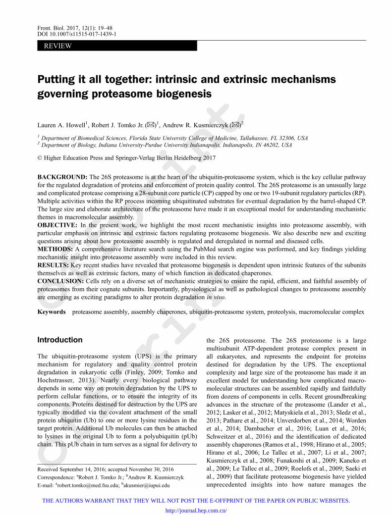
BACKGROUND: The 26S proteasome is at the heart of the ubiquitin-proteasome system, which is the key cellular pathwayfor the regulated degradation of proteins and enforcement of protein quality control. The 26S proteasome is an unusually largeand complicated protease comprising a 28-subunit core particle (CP) capped by one or two 19-subunit regulatory particles (RP).Multiple activities within the RP process incoming ubiquitinated substrates for eventual degradation by the barrel-shaped CP.The large size and elaborate architecture of the proteasome have made it an exceptional model for understanding mechanisticthemes in macromolecular assembly.OBJECTIVE: In the present work, we highlight the most recent mechanistic insights into proteasome assembly, withparticular emphasis on intrinsic and extrinsic factors regulating proteasome biogenesis. We also describe new and excitingquestions arising about how proteasome assembly is regulated and deregulated in normal and diseased cells.METHODS: A comprehensive literature search using the PubMed search engine was performed, and key findings yieldingmechanistic insight into proteasome assembly were included in this review.RESULTS: Key recent studies have revealed that proteasome biogenesis is dependent upon intrinsic features of the subunitsthemselves as well as extrinsic factors, many of which function as dedicated chaperones.CONCLUSION: Cells rely on a diverse set of mechanistic strategies to ensure the rapid, efficient, and faithful assembly ofproteasomes from their cognate subunits. Importantly, physiological as well as pathological changes to proteasome assemblyare emerging as exciting paradigms to alter protein degradation in vivo.
Keywords proteasome assembly, assembly chaperones, ubiquitin-proteasome system, proteolysis, macromolecular complex
Introduction
The ubiquitin-proteasome system (UPS) is the primarymechanism for regulatory and quality control proteindegradation in eukaryotic cells (Finley, 2009; Tomko andHochstrasser, 2013). Nearly every biological pathwaydepends in some way on protein degradation by the UPS toperform cellular functions, or to ensure the integrity of itscomponents. Proteins destined for destruction by the UPS aretypically modified via the covalent attachment of the smallprotein ubiquitin (Ub) to one or more lysine residues in thetarget protein. Additional Ub molecules can then be attachedto lysines in the original Ub to form a polyubiquitin (pUb)chain. This pUb chain in turn serves as a signal for delivery to
the 26S proteasome. The 26S proteasome is a largemultisubunit ATP-dependent protease complex present inall eukaryotes, and represents the endpoint for proteinsdestined for degradation by the UPS. The exceptionalcomplexity and large size of the proteasome has made it anexcellent model for understanding how complicated macro-molecular structures can be assembled rapidly and faithfullyfrom dozens of components in cells. Recent groundbreakingadvances in the structure of the proteasome (Lander et al.,2012; Lasker et al., 2012; Matyskiela et al., 2013; Sledz et al.,2013; Pathare et al., 2014; Unverdorben et al., 2014; Wordenet al., 2014; Dambacher et al., 2016; Luan et al., 2016;Schweitzer et al., 2016) and the identification of dedicatedassembly chaperones (Ramos et al., 1998; Hirano et al., 2005;Hirano et al., 2006; Le Tallec et al., 2007; Li et al., 2007;Kusmierczyk et al., 2008; Funakoshi et al., 2009; Kaneko etal., 2009; Le Tallec et al., 2009; Roelofs et al., 2009; Saeki etal., 2009) that facilitate proteasome biogenesis have yieldedunprecedented insights into how nature manages the
Received September 14, 2016; accepted November 30, 2016
Correspondence: aRobert J. Tomko Jr.; bAndrew R. Kusmierczyk
E-mail: [email protected]; [email protected]
Front. Biol. 2017, 12(1): 19–48DOI 10.1007/s11515-017-1439-1
THE AUTHORS WARRANT THAT THEY WILL NOT POST THE E-OFFPRINT OF THE PAPER ON PUBLIC WEBSITES.
http://journal.hep.com.cn/
offprint
offprint
challenges of macromolecular assembly, and has revealedimportant parallels to assembly of numerous other multi-subunit complexes. In this review, we discuss the basicmechanisms of proteasome assembly, with an emphasis onhow intrinsic features of subunits cooperate with extrinsicassembly chaperones to ensure efficient proteasome biogen-esis in vivo. Finally, we comment on arising questions in ourunderstanding of proteasome assembly in vivo, and their linksto human disease.
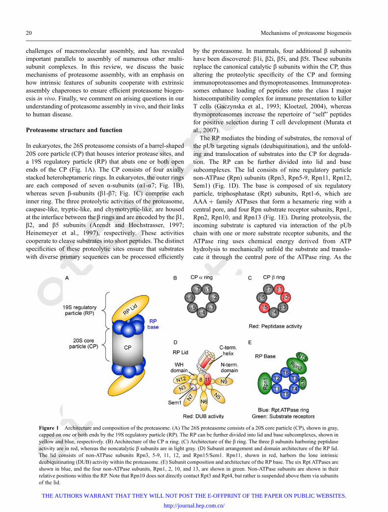
Proteasome structure and function
In eukaryotes, the 26S proteasome consists of a barrel-shaped20S core particle (CP) that houses interior protease sites, anda 19S regulatory particle (RP) that abuts one or both openends of the CP (Fig. 1A). The CP consists of four axiallystacked heteroheptameric rings. In eukaryotes, the outer ringsare each composed of seven α-subunits (α1-α7; Fig. 1B),whereas seven β-subunits (β1-β7; Fig. 1C) comprise eachinner ring. The three proteolytic activities of the proteasome,caspase-like, tryptic-like, and chymotryptic-like, are housedat the interface between the β rings and are encoded by the β1,β2, and β5 subunits (Arendt and Hochstrasser, 1997;Heinemeyer et al., 1997), respectively. These activitiescooperate to cleave substrates into short peptides. The distinctspecificities of these proteolytic sites ensure that substrateswith diverse primary sequences can be processed efficiently
by the proteasome. In mammals, four additional β subunitshave been discovered: β1i, β2i, β5i, and β5t. These subunitsreplace the canonical catalytic β subunits within the CP, thusaltering the proteolytic specificity of the CP and formingimmunoproteasomes and thymoproteasomes. Immunoprotea-somes enhance loading of peptides onto the class I majorhistocompatibility complex for immune presentation to killerT cells (Gaczynska et al., 1993; Kloetzel, 2004), whereasthymoproteasomes increase the repertoire of “self” peptidesfor positive selection during T cell development (Murata etal., 2007).
The RP mediates the binding of substrates, the removal ofthe pUb targeting signals (deubiquitination), and the unfold-ing and translocation of substrates into the CP for degrada-tion. The RP can be further divided into lid and basesubcomplexes. The lid consists of nine regulatory particlenon-ATPase (Rpn) subunits (Rpn3, Rpn5-9, Rpn11, Rpn12,Sem1) (Fig. 1D). The base is composed of six regulatoryparticle, triphosphatase (Rpt) subunits, Rpt1-6, which areAAA+ family ATPases that form a hexameric ring with acentral pore, and four Rpn substrate receptor subunits, Rpn1,Rpn2, Rpn10, and Rpn13 (Fig. 1E). During proteolysis, theincoming substrate is captured via interaction of the pUbchain with one or more substrate receptor subunits, and theATPase ring uses chemical energy derived from ATPhydrolysis to mechanically unfold the substrate and translo-cate it through the central pore of the ATPase ring. As the
Figure 1 Architecture and composition of the proteasome. (A) The 26S proteasome consists of a 20S core particle (CP), shown in gray,capped on one or both ends by the 19S regulatory particle (RP). The RP can be further divided into lid and base subcomplexes, shown inyellow and blue, respectively. (B) Architecture of the CP α ring. (C) Architecture of the β ring. The three β subunits harboring peptidaseactivity are in red, whereas the noncatalytic β subunits are in light gray. (D) Subunit arrangement and domain architecture of the RP lid.The lid consists of non-ATPase subunits Rpn3, 5-9, 11, 12, and Rpn15/Sem1. Rpn11, shown in red, harbors the lone intrinsicdeubiquitinating (DUB) activity within the proteasome. (E) Subunit composition and architecture of the RP base. The six Rpt ATPases areshown in blue, and the four non-ATPase subunits, Rpn1, 2, 10, and 13, are shown in green. Non-ATPase subunits are shown in theirrelative positions within the RP. Note that Rpn10 does not directly contact Rpt3 and Rpt4, but rather is suspended above them via subunitsof the lid.
20 Mechanisms of proteasome biogenesis
THE AUTHORS WARRANT THAT THEY WILL NOT POST THE E-OFFPRINT OF THE PAPER ON PUBLIC WEBSITES.
http://journal.hep.com.cn/
offprint
offprint
substrate is threaded through the pore of the ATPase ring,Rpn11 deubiquitinates the substrate so that ubiquitin can berecycled (Matyskiela et al., 2013; Sledz et al., 2013; Pathareet al., 2014; Worden et al., 2014).
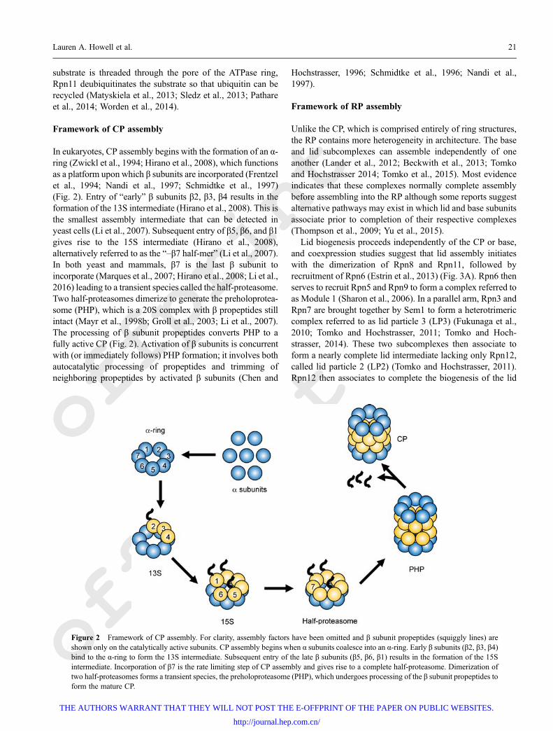
Framework of CP assembly
In eukaryotes, CP assembly begins with the formation of an α-ring (Zwickl et al., 1994; Hirano et al., 2008), which functionsas a platform upon which β subunits are incorporated (Frentzelet al., 1994; Nandi et al., 1997; Schmidtke et al., 1997)(Fig. 2). Entry of “early” β subunits β2, β3, β4 results in theformation of the 13S intermediate (Hirano et al., 2008). This isthe smallest assembly intermediate that can be detected inyeast cells (Li et al., 2007). Subsequent entry of β5, β6, and β1gives rise to the 15S intermediate (Hirano et al., 2008),alternatively referred to as the “–β7 half-mer” (Li et al., 2007).In both yeast and mammals, β7 is the last β subunit toincorporate (Marques et al., 2007; Hirano et al., 2008; Li et al.,2016) leading to a transient species called the half-proteasome.Two half-proteasomes dimerize to generate the preholoprotea-some (PHP), which is a 20S complex with β propeptides stillintact (Mayr et al., 1998b; Groll et al., 2003; Li et al., 2007).The processing of β subunit propeptides converts PHP to afully active CP (Fig. 2). Activation of β subunits is concurrentwith (or immediately follows) PHP formation; it involves bothautocatalytic processing of propeptides and trimming ofneighboring propeptides by activated β subunits (Chen and
Hochstrasser, 1996; Schmidtke et al., 1996; Nandi et al.,1997).
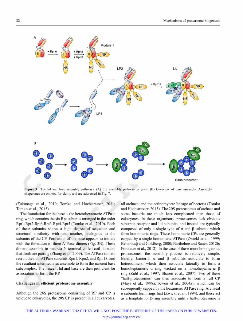
Framework of RP assembly
Unlike the CP, which is comprised entirely of ring structures,the RP contains more heterogeneity in architecture. The baseand lid subcomplexes can assemble independently of oneanother (Lander et al., 2012; Beckwith et al., 2013; Tomkoand Hochstrasser 2014; Tomko et al., 2015). Most evidenceindicates that these complexes normally complete assemblybefore assembling into the RP although some reports suggestalternative pathways may exist in which lid and base subunitsassociate prior to completion of their respective complexes(Thompson et al., 2009; Yu et al., 2015).
Lid biogenesis proceeds independently of the CP or base,and coexpression studies suggest that lid assembly initiateswith the dimerization of Rpn8 and Rpn11, followed byrecruitment of Rpn6 (Estrin et al., 2013) (Fig. 3A). Rpn6 thenserves to recruit Rpn5 and Rpn9 to form a complex referred toas Module 1 (Sharon et al., 2006). In a parallel arm, Rpn3 andRpn7 are brought together by Sem1 to form a heterotrimericcomplex referred to as lid particle 3 (LP3) (Fukunaga et al.,2010; Tomko and Hochstrasser, 2011; Tomko and Hoch-strasser, 2014). These two subcomplexes then associate toform a nearly complete lid intermediate lacking only Rpn12,called lid particle 2 (LP2) (Tomko and Hochstrasser, 2011).Rpn12 then associates to complete the biogenesis of the lid
Figure 2 Framework of CP assembly. For clarity, assembly factors have been omitted and β subunit propeptides (squiggly lines) areshown only on the catalytically active subunits. CP assembly begins when α subunits coalesce into an α-ring. Early β subunits (β2, β3, β4)bind to the α-ring to form the 13S intermediate. Subsequent entry of the late β subunits (β5, β6, β1) results in the formation of the 15Sintermediate. Incorporation of β7 is the rate limiting step of CP assembly and gives rise to a complete half-proteasome. Dimerization oftwo half-proteasomes forms a transient species, the preholoproteasome (PHP), which undergoes processing of the β subunit propeptides toform the mature CP.
Lauren A. Howell et al. 21
THE AUTHORS WARRANT THAT THEY WILL NOT POST THE E-OFFPRINT OF THE PAPER ON PUBLIC WEBSITES.
http://journal.hep.com.cn/
offprint
offprint
(Fukunaga et al., 2010; Tomko and Hochstrasser, 2011;Tomko et al., 2015).
The foundation for the base is the heterohexameric ATPasering, which contains the six Rpt subunits arranged in the orderRpt1-Rpt2-Rpt6-Rpt3-Rpt4-Rpt5 (Tomko et al., 2010). Eachof these subunits shares a high degree of sequence andstructural similarity with one another, analogous to thesubunits of the CP. Formation of the base appears to initiatewith the formation of three ATPase dimers (Fig. 3B). Thesedimers assemble in part via N-terminal coiled coil domainsthat facilitate pairing (Zhang et al., 2009). The ATPase dimersrecruit the non-ATPase subunits Rpn1, Rpn2, and Rpn13, andthe resultant intermediates assemble to form the nascent basesubcomplex. The nascent lid and base are then proficient forassociation to form the RP.
Challenges in efficient proteasome assembly
Although the 26S proteasome consisting of RP and CP isunique to eukaryotes, the 20S CP is present in all eukaryotes,
all archaea, and the actinomycete lineage of bacteria (Tomkoand Hochstrasser, 2013). The 20S proteasomes of archaea andsome bacteria are much less complicated than those ofeukaryotes. In these organisms, proteasomes lack obvioussubstrate receptor and lid subunits, and instead are typicallycomposed of only a single type of α and β subunit, whichform homomeric rings. These homomeric CPs are generallycapped by a single homomeric ATPase (Zwickl et al., 1999;Benaroudj and Goldberg, 2000; Barthelme and Sauer, 2012b;Forouzan et al., 2012). In the case of these more homogenousproteasomes, the assembly process is relatively simple.Briefly, bacterial α and β subunits associate to formheterodimers, which then associate laterally to form ahomoheptameric α ring stacked on a homoheptameric βring (Zuhl et al., 1997; Sharon et al., 2007). Two of these“half-proteasomes” can then associate to form a full CP(Mayr et al., 1998a; Kwon et al., 2004a), which can besubsequently capped by the hexameric ATPase ring. Archaealα subunits form rings first (Zwickl et al., 1994), and these actas a template for β-ring assembly until a half-proteasome is
Figure 3 The lid and base assembly pathways. (A) Lid assembly pathway in yeast. (B) Overview of base assembly. Assemblychaperones are omitted for clarity and are addressed in Fig. 7.
22 Mechanisms of proteasome biogenesis
THE AUTHORS WARRANT THAT THEY WILL NOT POST THE E-OFFPRINT OF THE PAPER ON PUBLIC WEBSITES.
http://journal.hep.com.cn/
offprint
offprint
formed, though a bacterial-like assembly pathway is alsopossible (Panfair et al., 2015). In support of such simple,autonomous assembly, heterologous expression of α, β, andATPase subunits typically results in formation of properlyassembled, active proteasomes. In the case of these protea-somes, only a few logistical issues must be addressed to formfunctional particles. Specifically, rings must form from theproper number of subunits, and the stacking of subunits toform rings must occur in a manner that does not interfere withcompletion of each ring. The size of the ring is probablypredetermined, because the curvature of the ring is controlledby the structure of the homomeric subunits that compose it.Also, whether a given ring forms completely prior toassociation with subunits of a neighboring ring is dictatedby the relative affinities of the subunits within that ring foreach other versus their affinities for subunits of theneighboring ring. Although the overall architecture ofproteasomes is retained in eukaryotes, the composition ismuch more complex due to diversification of α, β, andATPase subunits within the proteasome, as well as thepresence of substrate receptors and the lid. Such diversifica-tion yields many additional challenges to efficient and faithfulproteasome biogenesis. Subunit heterogeneity typicallyimposes specific positions for individual subunits within agiven ring, and it necessitates that rings associate with aproper register to one another. As the seven α, seven β, and sixATPase subunits evolved via diversification from a commonancestral α, β, or ATPase subunit (Wollenberg and Swaffield,2001; Gille et al., 2003), they share substantial sequence andstructural similarity with their orthologs, and are in somecases prone to misassembly (Gerards et al., 1997; Gerards etal., 1998; Yao et al., 1999; Takeuchi and Tamura 2004; Ishii etal., 2015). Thus, additional control mechanisms are necessaryto limit the formation of products that are nonproductive forproteasome biogenesis and could potentially even be toxic.Similarly, formation of assembly intermediates that stericallyocclude or otherwise interfere with incorporation of acomplete set of subunits must be avoided. Finally, as properenzymatic coupling of substrate binding, deubiquitination,unfolding, and proteolysis is necessary for proper function,the activities of the eukaryotic lid, base, and CP must besuppressed until the proteasome has fully assembled.
Despite these challenges, proteasome biogenesis occursvery rapidly and with near-perfect fidelity in normal cells. Asubstantial number of evolutionarily conserved regulatorymechanisms, mediated both by intrinsic subunit features andextrinsic assembly chaperones, cooperate to ensure such fastand faithful assembly in vivo. These mechanisms function tosculpt and guide the formation of a limited number ofassembly intermediates that, in many cases, then associate viadefined, hierarchical assembly pathways to yield mature,functional proteasomes. We review the best understoodexamples of these intrinsic and extrinsic regulatory mechan-isms herein focusing on the canonical eukaryotic 26Sproteasome.
Proteasome assembly chaperones andtheir mechanisms of action
In this section, we discuss what is known about the role ofassembly chaperones in the biogenesis of the proteasome,starting with the CP. Where appropriate, both yeast andmammalian terminology will be used. However, whenreferring to assembly in general, yeast terminology will beused to streamline the discussion.
CP chaperones
Five conserved eukaryotic proteins comprise three confirmeddedicated assembly chaperones in CP biogenesis (Pba1-Pba2;Pba3-Pba4; Ump1). A sixth protein (Blm10) may also play arole in CP assembly. In general, the assembly chaperonesfulfill both positive functions (i.e. actively promote desiredassembly events) and negative functions (i.e. preventundesired assembly events) during CP formation.
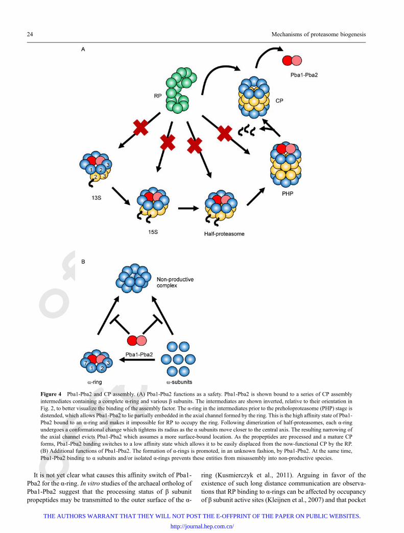
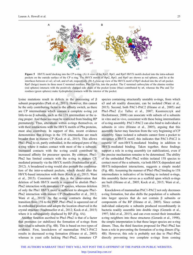
Pba1-Pba2/PAC1-PAC2This heterodimeric chaperone is involved at all stages of CPassembly and likely fulfils several roles. The easiest of theseto visualize is that of a “safety” that prevents prematureassociation of RP with CP (Fig. 4A) (Kusmierczyk et al.,2011; Wani et al., 2015). The binding of RP to CP is mediatedin part by highly conserved C-terminal HbYX motifs (Hb =hydrophobic; Y = tyrosine (or phenylalanine); X = any aminoacid) present on select Rpt subunits of the base (Smith et al.,2007; Gillette et al., 2008; Yu et al., 2010). These motifs insertinto pockets formed by two adjacent α subunits, one of whichcontributes a conserved lysine that forms a salt bridge withthe C-terminal carboxylate of the HbYX motif (Tian et al.,2011; Park et al., 2013) (Fig. 5 and also see below). Thus, RPhas the potential to interact with any species containing thesepockets, including mature CP, as well as any CP intermediateswith a full (or even potentially an incomplete) α-ring. Pba1and Pba2 also contain functional HbYX motifs (Kusmierczyket al., 2011) which allow them to interact with the same α-ringsurface as RP (Stadtmueller et al., 2012). The HbYX motif ofPba1 can insert into the pocket at the interface between α5 andα6 (forming a salt-bridge with the α6 pocket lysine) whereasthat of Pba2 inserts into the pocket between α6 and α7. Inmature 26S proteasomes, the HbYXmotif of Rpt5 inserts intothe same pocket as used by Pba1 (Beck et al., 2012;Schweitzer et al., 2016) though it can also occupy the pocketused by Pba2 (Tian et al., 2011). However, Pba1-Pba2 hasmuch higher affinity for α-rings present within CP inter-mediates than within those of mature CP, whereas theopposite is true for RP (Wani et al., 2015). This explainswhy Pba1-Pba2 is able to function as a safety in preventinginappropriate RP interaction with immature CP. Once matureCP has formed, the higher affinity of RP for CP effectivelyoutcompetes Pba1-Pba2 for the α-ring surface.
Lauren A. Howell et al. 23
THE AUTHORS WARRANT THAT THEY WILL NOT POST THE E-OFFPRINT OF THE PAPER ON PUBLIC WEBSITES.
http://journal.hep.com.cn/
offprint
offprint
It is not yet clear what causes this affinity switch of Pba1-Pba2 for the α-ring. In vitro studies of the archaeal ortholog ofPba1-Pba2 suggest that the processing status of β subunitpropeptides may be transmitted to the outer surface of the α-
ring (Kusmierczyk et al., 2011). Arguing in favor of theexistence of such long distance communication are observa-tions that RP binding to α-rings can be affected by occupancyof β subunit active sites (Kleijnen et al., 2007) and that pocket
Figure 4 Pba1-Pba2 and CP assembly. (A) Pba1-Pba2 functions as a safety. Pba1-Pba2 is shown bound to a series of CP assemblyintermediates containing a complete α-ring and various β subunits. The intermediates are shown inverted, relative to their orientation inFig. 2, to better visualize the binding of the assembly factor. The α-ring in the intermediates prior to the preholoproteasome (PHP) stage isdistended, which allows Pba1-Pba2 to lie partially embedded in the axial channel formed by the ring. This is the high affinity state of Pba1-Pba2 bound to an α-ring and makes it impossible for RP to occupy the ring. Following dimerization of half-proteasomes, each α-ringundergoes a conformational change which tightens its radius as the α subunits move closer to the central axis. The resulting narrowing ofthe axial channel evicts Pba1-Pba2 which assumes a more surface-bound location. As the propeptides are processed and a mature CPforms, Pba1-Pba2 binding switches to a low affinity state which allows it to be easily displaced from the now-functional CP by the RP.(B) Additional functions of Pba1-Pba2. The formation of α-rings is promoted, in an unknown fashion, by Pba1-Pba2. At the same time,Pba1-Pba2 binding to α subunits and/or isolated α-rings prevents these entities from misassembly into non-productive species.
24 Mechanisms of proteasome biogenesis
THE AUTHORS WARRANT THAT THEY WILL NOT POST THE E-OFFPRINT OF THE PAPER ON PUBLIC WEBSITES.
http://journal.hep.com.cn/
offprint
offprint
lysine mutations result in defects in the processing of βsubunit propeptides (Park et al., 2011). However, this cannotbe the only contributing factor to the affinity switch, as thereare CP intermediates which contain a complete α-ring yetlittle-to-no β subunits, such as the 13S intermediate or the α-ring proper. And these too must be restricted from binding RPprematurely. Thus, alterations within α-rings themselves, orwith their interactions with the HbYX motifs of Pba proteins,must also contribute. In support of this, recent evidencedemonstrates that α-rings in the 15S intermediate are muchbroader than in mature CP (Kock et al., 2015). This allowsPba1-Pba2 to sit, partly embedded, in the enlarged pore of theα-ring where it makes contact with most of the α subunits.Increased contacts with the α-ring could contribute toincreased affinity for precursor species. By contrast, Pba1-Pba2 has limited contacts with the α-ring in mature CP,mediated primarily via the HbYX motifs (Stadtmueller et al.,2012). A broadened α-ring would also perturb the conforma-tion of the inter-α-subunit pockets, which should alter theHbYX-based interaction with them (Kock et al., 2015; Waniet al., 2015). Consistent with this is the observation thatdeletion of both HbYX motifs is required to abolish Pba1-Pba2 interaction with immature CP species, whereas deletionof only the Pba1 HbYX motif is sufficient to abrogate Pba1-Pba2 interaction with mature CP (Stadtmueller et al., 2012;Wani et al., 2015). As the α-ring contracts during thetransition from 15S to the PHP, Pba1-Pba2 is squeezed out ofits embedded position and adopts the location observed in thecrystal structure (Stadtmueller et al., 2012; Kock et al., 2015)where it is subsequently displaced by RP (Fig. 4A).
Another function ascribed to Pba1-Pba2 is that of a factorthat promotes (or stabilizes) the formation of α-rings fromindividual α subunits (Fig. 4B). This is based on two types ofevidence. First, knockdown of mammalian PAC1-PAC2results in decreased α-ring formation (Hirano et al., 2005)whereas in yeast cells lacking Pba1-Pba2, immature CP
species containing structurally unstable α-rings, from whichα5 and α6 readily dissociate, can be isolated (Wani et al.,2015). Second, both PAC1-PAC2 (Hirano et al., 2005) andPba1-Pba2 (Le Tallec et al., 2007; Kusmierczyk andHochstrasser, 2008) can associate with subsets of α subunitsin vitro and in vivo, consistent with these being intermediatesof α-ring assembly. PAC1-PAC2 can also bind to individual αsubunits in vitro (Hirano et al., 2005), arguing that thisassembly factor may function from the very beginning of CPassembly. Since isolated α subunits cannot form a pocket torecognize a HbYX motif, this indicates that PAC1-PAC2 iscapable of non-HbYX-mediated binding in addition toHbYX-mediated binding. Taken together, these findingssupport a role in α-ring assembly (or stabilization), even ifthe precise mechanism remains to be determined. The abilityof the embedded Pba1-Pba2 within isolated 15S species tocontact most of the α subunits, via both HbYX-dependent andHbYX-independent interactions, suggests a simple model(Fig. 4B). Assuming the manner of Pba1-Pba2 binding to 15Sintermediates is indicative of its binding to isolated α-rings,this assembly factor serves as a scaffold upon which α-ringsare built (Hirano et al., 2005; Kock et al., 2015; Wani et al.,2015).
Knockdown of mammalian PAC1-PAC2 not only decreasesα-ring formation, but also shifts the population of α subunitsinto larger species that do not contain β subunits orcomponents of the RP (Hirano et al., 2005). Since certainindividual eukaryotic α subunits produced recombinantly inbacteria readily assemble into double rings (Gerards et al.,1997; Ishii et al., 2015), and can even recruit their immediateα-ring neighbors into these structures (Gerards et al., 1998),the simple interpretation is that these larger species are α-ringdimers. Thus, the third function proposed for Pba1-Pba2 hasbeen a role in preventing the formation of α-ring dimers (Fig.4B). However, this role is probably not due to Pba1-Pba2directly preventing two complete α-rings from coming
Figure 5 HbYX motif docking into the CP α-ring. (A) A view of the Rpt2, Rpt3, and Rpt5 HbYX motifs docked into the intra-subunitpockets on the outside surface of the CP α ring. The HbYX motifs of Rpt2, Rpt3, and Rpt5 are shown as red spheres, and lie at theinterfaces between α1-α2, α3-α4, and α5-α6, respectively. (B) A close-up view of the HbYX motif of Rpt5 docked into the α5-α6 pocket.Rpt5 (beige) inserts its three most C-terminal residues, Phe-Tyr-Ala, into the pocket. The C-terminal carboxylate of the alanine residue(red spheres) interacts with the positively charged side chain of the pocket lysine (blue) contributed by α6, whereas the Phe and Tyrresidues (green spheres) make hydrophobic contacts with the interior of the pocket.
Lauren A. Howell et al. 25
THE AUTHORS WARRANT THAT THEY WILL NOT POST THE E-OFFPRINT OF THE PAPER ON PUBLIC WEBSITES.
http://journal.hep.com.cn/
offprint
offprint
together. Double α-rings interact via a saw-toothed interfacemediated primarily by H1 helices (Panfair et al., 2015) akin tothe interaction between α- and β-rings in the half proteasome(Zwickl et al., 1994). However, Pba1-Pba2 binds to theopposite (i.e. outer) surface of the α-ring, where it would notproduce steric interference to ring dimerization. Perhaps Pba1-Pba2 binding results in an α-ring conformation that is notcapable of dimerizing; the distended α-ring found in the 15Scould be such a species (Kock et al., 2015). Or, perhaps Pba1-Pba2 binding alters the order of subunit association; this couldprevent pathways that lead to α-ring dimers (or any non-productive complex, for that matter) from becoming popu-lated. We favor this latter possibility precisely because it doesnot limit the identity of non-productive complexes, which canform in the absence of Pba1-Pba2, to α-ring dimers only.
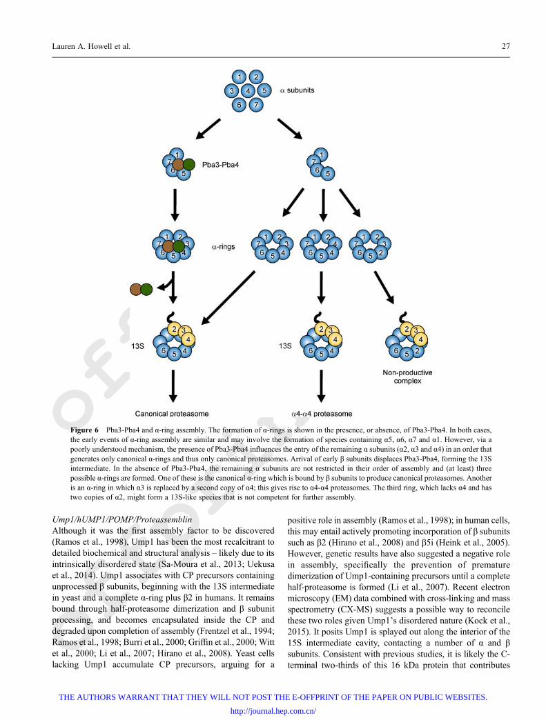
Pba3-Pba4/PAC3-PAC4This heterodimer functions early in assembly and isassociated with CP intermediates up until the 13S stage(Fig. 6) (Hirano et al., 2006; Le Tallec et al., 2007; Hirano etal., 2008; Yashiroda et al., 2008). Its early exit from theassembly pathway is explained by the manner in which itinteracts with a nascent CP. In vitro Pba3-Pba4 binds tightlyto α5 (Yashiroda et al., 2008) and subcomplexes of α subunitsthat contain α5 (Kusmierczyk et al., 2008). The binding ofPba3-Pba4 to helices H1 and H2 of α5, located on the αsubunit surface that faces β subunits, is not compatible withthe presence of β subunits, and it is displaced from the ring byincoming β4 (Hirano et al., 2008). Pba3-Pba4 has a uniquefunction among the assembly chaperones in that it ensures theformation of canonical 20S proteasomes in which eachsubunit is represented in its “proper” place (Kusmierczyk etal., 2008). The α3 subunit is not essential in Saccharomycescerevisiae and in its absence yeast synthesize an alternativeproteasome in which a second copy of α4 takes the place ofthe missing α3 (Velichutina et al., 2004). These “α4–α4proteasomes” also form in yeast when Pba3-Pba4 is absent,despite the continuing presence of α3 (Kusmierczyk et al.,2008). This argues that the efficient formation of normal α-rings requires Pba3-Pba4 function (Fig. 6).
As with Pba1-Pba2, the precise mechanism by whichPba3-Pba4 contributes to α-ring formation is not known. Thekey to understanding how Pba3-Pba4 functions begins withdetermining the significance of α4–α4 proteasome formation.Evidence for the physiologic relevance of α4–α4 proteasomestakes several forms. First, the non-essential nature ofα3 extends to other fungal species including Schizosacchar-omyces pombe (Kim et al., 2010a), Neurospora crassa (Colotet al., 2006), and probably Aspergillus nidulans, where anidentified α3 allele is likely to be assembly incompetent(Lee and Shaw, 2007). This argues that dispensability of α3 isnot a quirk of S. cerevisiae genetics. Second, the ability toform α4–α4 proteasomes has been demonstrated recently inmammalian cells, and the levels of these proteasomescorrelate inversely with levels of PAC3 in the cell, echoing
observations in yeast (Padmanabhan et al., 2016). Third, theability to form α4–α4 proteasomes correlates with resistanceto certain heavy metal stresses, a phenotype that is conservedfrom yeast to humans (Kusmierczyk et al., 2008; Padma-nabhan et al., 2016). Fourth, α4–α4 proteasome levels can bemodulated by altered levels of known oncogenes and tumorsuppressors, a condition representative of a number ofmalignancies (Padmanabhan et al., 2016). The conservedability of α subunits to assemble into canonical and α4–α4proteasomes, both of which are physiologically relevant,requires two types of α-ring to form. This implies that thepathway(s) to α-ring formation must diverge at some point toallow this to occur. Recently, a non-canonical complex wasisolated from yeast cells lacking Pba3-Pba4 (Takagi et al.,2014). It contained β2, β3, β4, and all α subunits except α4.Notably, α2 was present in twofold excess. This complex hasbeen proposed to resemble a 13S intermediate in which α2has taken the place of α4 in the α-ring (Fig. 6) (Takagi et al.,2014). Although this is likely a dead-end complex, if thiscomplex does contain an α-ring with a third arrangement ofsubunits, this supports a model in which formation of α ringsdiverges subsequent to formation of an α1, α5, α6, α7heterotetramer (Fig. 6).
The three rings formed in the absence of Pba3-Pba4 eachcontain α5, α6, α7, and α1 in their proper place, whereas α2,α3, and α4 can associate in a non-canonical manner. Thus,one can propose that one half of the ring (containing thecontiguous subunits α5 through α1) assembles the sameregardless of Pba3-Pba4 status. In analogy to β-ringformation, perhaps these are the “early” α subunits of α-ring assembly. The α2, α3, α4 subunits can complete the ringin several combinations, two of which are competent forfurther assembly, but the activity of Pba3-Pba4 ensures thecanonical placement of α2, α3, and α4 and promotes theformation of the canonical CP. In wild-type yeast, Pba3-Pba4activity is sufficient to ensure that this is essentially the onlytype of CP formed. In mammalian cells, PAC3-PAC4 levelsmay not be sufficient to result solely in canonical CP(Padmanabhan et al., 2016).
How the canonical placement of α2, α3, and α4 is favoredis not known, but when the co-crystal structure of Pba3-Pba4with α5 was modeled onto to the CP structure, it wasprojected to make substantial contacts with α4 and α6(Yashiroda et al., 2008). This observation, in combinationwith the formation of the aberrant 13S-like complex lackingα4 when Pba3-Pba4 is absent in yeast, suggests that Pba3-Pba4 promotes the interaction of α5 with α4 (Takagi et al.,2014). Others have shown little retention of α4 on an affinitycolumn in the presence of Pba3-Pba4 and α5 (Kusmierczyk etal., 2008), so it remains unclear if promoting α5-α4interaction is the sole mechanism by which Pba3-Pba4functions. Nevertheless, as with Pba1-Pba2, the presence ofthe Pba3-Pba4 assembly factor likely alters the order ofassociation for some of the α subunits, favoring the formationof the canonical α-ring over others.
26 Mechanisms of proteasome biogenesis
THE AUTHORS WARRANT THAT THEY WILL NOT POST THE E-OFFPRINT OF THE PAPER ON PUBLIC WEBSITES.
http://journal.hep.com.cn/
offprint
offprint
Ump1/hUMP1/POMP/ProteassemblinAlthough it was the first assembly factor to be discovered(Ramos et al., 1998), Ump1 has been the most recalcitrant todetailed biochemical and structural analysis – likely due to itsintrinsically disordered state (Sa-Moura et al., 2013; Uekusaet al., 2014). Ump1 associates with CP precursors containingunprocessed β subunits, beginning with the 13S intermediatein yeast and a complete α-ring plus β2 in humans. It remainsbound through half-proteasome dimerization and β subunitprocessing, and becomes encapsulated inside the CP anddegraded upon completion of assembly (Frentzel et al., 1994;Ramos et al., 1998; Burri et al., 2000; Griffin et al., 2000; Wittet al., 2000; Li et al., 2007; Hirano et al., 2008). Yeast cellslacking Ump1 accumulate CP precursors, arguing for a
positive role in assembly (Ramos et al., 1998); in human cells,this may entail actively promoting incorporation of β subunitssuch as β2 (Hirano et al., 2008) and β5i (Heink et al., 2005).However, genetic results have also suggested a negative rolein assembly, specifically the prevention of prematuredimerization of Ump1-containing precursors until a completehalf-proteasome is formed (Li et al., 2007). Recent electronmicroscopy (EM) data combined with cross-linking and massspectrometry (CX-MS) suggests a possible way to reconcilethese two roles given Ump1’s disordered nature (Kock et al.,2015). It posits Ump1 is splayed out along the interior of the15S intermediate cavity, contacting a number of α and βsubunits. Consistent with previous studies, it is likely the C-terminal two-thirds of this 16 kDa protein that contributes
Figure 6 Pba3-Pba4 and α-ring assembly. The formation of α-rings is shown in the presence, or absence, of Pba3-Pba4. In both cases,the early events of α-ring assembly are similar and may involve the formation of species containing α5, α6, α7 and α1. However, via apoorly understood mechanism, the presence of Pba3-Pba4 influences the entry of the remaining α subunits (α2, α3 and α4) in an order thatgenerates only canonical α-rings and thus only canonical proteasomes. Arrival of early β subunits displaces Pba3-Pba4, forming the 13Sintermediate. In the absence of Pba3-Pba4, the remaining α subunits are not restricted in their order of assembly and (at least) threepossible α-rings are formed. One of these is the canonical α-ring which is bound by β subunits to produce canonical proteasomes. Anotheris an α-ring in which α3 is replaced by a second copy of α4; this gives rise to α4-α4 proteasomes. The third ring, which lacks α4 and hastwo copies of α2, might form a 13S-like species that is not competent for further assembly.
Lauren A. Howell et al. 27
THE AUTHORS WARRANT THAT THEY WILL NOT POST THE E-OFFPRINT OF THE PAPER ON PUBLIC WEBSITES.
http://journal.hep.com.cn/
offprint
offprint
these important binding contacts (Burri et al., 2000). Thus theC terminus might facilitate the incorporation/stabilization of βsubunits. By contrast, the N-terminal third of Ump1, which isdispensable for CP binding (Burri et al., 2000), performs acheckpoint function. With CX-MS data placing it near β6,and potentially protruding out of the β-ring, the N terminus ofUmp1 could be ideally positioned to both block dimerizationand sense the arrival of β7 (Kock et al., 2015). If this isconfirmed, it would explain how Ump1 delays dimerizationuntil β7 incorporates, subsequently reorganizing and assum-ing a different orientation within the cavity, due to itsdisordered nature.
Blm10/PA200Yeast Blm10 and mammalian PA200 are known proteasomeactivators, capable of binding CP alone or as a hybrid with RP(Ustrell et al., 2002; Schmidt et al., 2005). This HEAT-repeatprotein forms a dome on the CP with an opening large enoughto fit unfolded substrates and/or peptides (Sadre-Bazzaz et al.,2010; Dange et al., 2011). Blm10 has been demonstrated topromote CP import into the nucleus (Weberruss et al., 2013).However, it also appears to function during CP assembly(Fehlker et al., 2003). Blm10 associates with yeast 13S, 15S,and PHP intermediates (Li et al., 2007), presumably via itsHbYX motif (Sadre-Bazzaz et al., 2010). Moreover, whendeletion of the β7 tail is combined with a deletion of theBLM10 gene, yeast exhibit a severe CP assembly defect(Marques et al., 2007). However, a precise function forBlm10 in CP assembly remains to be elucidated.
RP chaperones
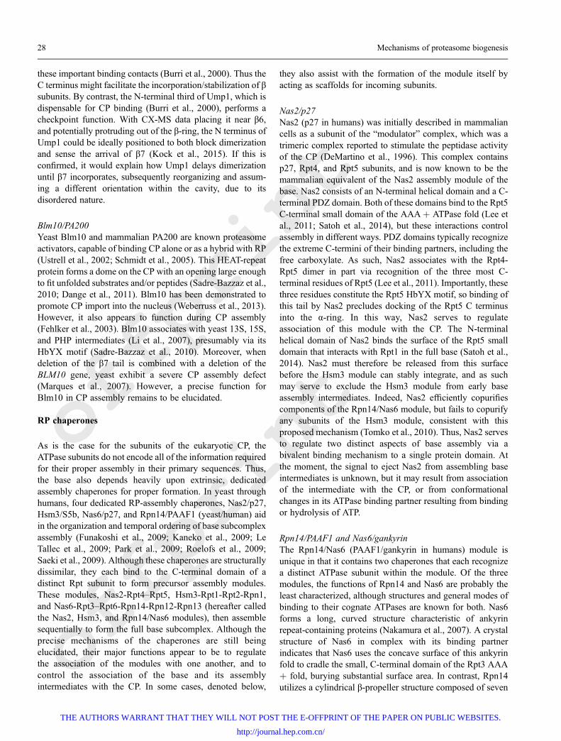
As is the case for the subunits of the eukaryotic CP, theATPase subunits do not encode all of the information requiredfor their proper assembly in their primary sequences. Thus,the base also depends heavily upon extrinsic, dedicatedassembly chaperones for proper formation. In yeast throughhumans, four dedicated RP-assembly chaperones, Nas2/p27,Hsm3/S5b, Nas6/p27, and Rpn14/PAAF1 (yeast/human) aidin the organization and temporal ordering of base subcomplexassembly (Funakoshi et al., 2009; Kaneko et al., 2009; LeTallec et al., 2009; Park et al., 2009; Roelofs et al., 2009;Saeki et al., 2009). Although these chaperones are structurallydissimilar, they each bind to the C-terminal domain of adistinct Rpt subunit to form precursor assembly modules.These modules, Nas2-Rpt4–Rpt5, Hsm3-Rpt1-Rpt2-Rpn1,and Nas6-Rpt3–Rpt6-Rpn14-Rpn12-Rpn13 (hereafter calledthe Nas2, Hsm3, and Rpn14/Nas6 modules), then assemblesequentially to form the full base subcomplex. Although theprecise mechanisms of the chaperones are still beingelucidated, their major functions appear to be to regulatethe association of the modules with one another, and tocontrol the association of the base and its assemblyintermediates with the CP. In some cases, denoted below,
they also assist with the formation of the module itself byacting as scaffolds for incoming subunits.
Nas2/p27Nas2 (p27 in humans) was initially described in mammaliancells as a subunit of the “modulator” complex, which was atrimeric complex reported to stimulate the peptidase activityof the CP (DeMartino et al., 1996). This complex containsp27, Rpt4, and Rpt5 subunits, and is now known to be themammalian equivalent of the Nas2 assembly module of thebase. Nas2 consists of an N-terminal helical domain and a C-terminal PDZ domain. Both of these domains bind to the Rpt5C-terminal small domain of the AAA+ ATPase fold (Lee etal., 2011; Satoh et al., 2014), but these interactions controlassembly in different ways. PDZ domains typically recognizethe extreme C-termini of their binding partners, including thefree carboxylate. As such, Nas2 associates with the Rpt4-Rpt5 dimer in part via recognition of the three most C-terminal residues of Rpt5 (Lee et al., 2011). Importantly, thesethree residues constitute the Rpt5 HbYX motif, so binding ofthis tail by Nas2 precludes docking of the Rpt5 C terminusinto the α-ring. In this way, Nas2 serves to regulateassociation of this module with the CP. The N-terminalhelical domain of Nas2 binds the surface of the Rpt5 smalldomain that interacts with Rpt1 in the full base (Satoh et al.,2014). Nas2 must therefore be released from this surfacebefore the Hsm3 module can stably integrate, and as suchmay serve to exclude the Hsm3 module from early baseassembly intermediates. Indeed, Nas2 efficiently copurifiescomponents of the Rpn14/Nas6 module, but fails to copurifyany subunits of the Hsm3 module, consistent with thisproposed mechanism (Tomko et al., 2010). Thus, Nas2 servesto regulate two distinct aspects of base assembly via abivalent binding mechanism to a single protein domain. Atthe moment, the signal to eject Nas2 from assembling baseintermediates is unknown, but it may result from associationof the intermediate with the CP, or from conformationalchanges in its ATPase binding partner resulting from bindingor hydrolysis of ATP.
Rpn14/PAAF1 and Nas6/gankyrinThe Rpn14/Nas6 (PAAF1/gankyrin in humans) module isunique in that it contains two chaperones that each recognizea distinct ATPase subunit within the module. Of the threemodules, the functions of Rpn14 and Nas6 are probably theleast characterized, although structures and general modes ofbinding to their cognate ATPases are known for both. Nas6forms a long, curved structure characteristic of ankyrinrepeat-containing proteins (Nakamura et al., 2007). A crystalstructure of Nas6 in complex with its binding partnerindicates that Nas6 uses the concave surface of this ankyrinfold to cradle the small, C-terminal domain of the Rpt3 AAA+ fold, burying substantial surface area. In contrast, Rpn14utilizes a cylindrical β-propeller structure composed of seven
28 Mechanisms of proteasome biogenesis
THE AUTHORS WARRANT THAT THEY WILL NOT POST THE E-OFFPRINT OF THE PAPER ON PUBLIC WEBSITES.
http://journal.hep.com.cn/
offprint
offprint
WD40 repeats to associate with the C-terminal domain ofRpt6 (Kim et al., 2010b). Although the structure of Rpn14 isknown, its binding interaction with Rpt6 has not beencharacterized at the atomic level. However, mutagenesisstudies support a binding mode that is similar to that of Nas6,in which the top of the cylindrical β-propeller makes criticalcontacts with the C-terminal domain of Rpt6 (Kim et al.,2010b).
How binding of Nas6 and Rpn14 to Rpt3 and Rpt6,respectively, facilitates proteasome assembly is largelyunknown. In yeast, the existence of the Rpn14/Nas6 modulehas been inferred solely on the basis of bimolecularinteractions and coimmunoprecipitation experiments, con-founding detailed architectural and mechanistic analysis.Thus, it remains unclear whether and exactly how the Rpn14/Nas6 module forms, and it is unknown if the chaperonesfacilitate pairing of Rpt3 and Rpt6 or the association of Rpn2and Rpn13. However, modeling of Nas6 onto the ATPase ringof the proteasome suggests that it would clash sterically withthe CP (Roelofs et al., 2009), at least under some conditions,and the same is likely true for Rpn14. Thus, these chaperonesmay restrict premature docking of this module onto the CP(Sokolova et al., 2015). A second possibility that is not yetexplored, is that one or more of these subunits may serve tostabilize Rpn2 and Rpn13 in the context of the assemblyintermediate. In recent structures of the 26S proteasome,Rpn13 contacts only Rpn2, and Rpn2 contacts the base onlyvia the very N-termini of Rpt3 and Rpt6 (da Fonseca et al.,2012; Lander et al., 2012; Lasker et al., 2012). Instead, Rpn2-Rpn13 depends almost entirely on contacts with lid subunitsfor stabilization within the RP. As it is believed that the lidand base form separately, these critical stabilizing contactswould be absent in the assembling base; Rpn2 and Rpn13may thus depend upon contacts with Rpn14 and/or Nas6 tostably associate with Rpt3 and Rpt6 during base biogenesis.
Hsm3/S5bHsm3 (S5b in humans) functions as both a chaperone and ascaffolding protein for the Hsm3 module. The proteinsequence of Hsm3 consists primarily of ARM/HEAT repeats,and the protein forms a concave fold that cradles the C-terminal domain of Rpt1 (Barrault et al., 2012; Takagi et al.,2012; Park et al., 2013). The structure of Hsm3 in complexwith the C-terminal domain of Rpt1 is highly reminiscent ofthe Nas6-Rpt6 C-terminal domain structure, in that it buriessubstantial surface of its concave face in Rpt1. This results intight interaction with Rpt1, but in the context of the module, italso positions the chaperone to make stabilizing contacts withboth Rpt2 and Rpn1 (Barrault et al., 2012). Hsm3 thus has adirect interaction with every subunit of this module.Mutations to any of these bridging contacts disrupts theformation of proteasomes in vivo, providing evidence thatHsm3 functions much as an assembly hub to recruit andstabilize each component of the complex (Barrault et al.,
2012). Similar to that observed for Nas6, modeling of theHsm3-Rpt1 C-terminal domain structure onto the fullproteasome indicates that it would clash substantially withthe CP (Park et al., 2013), suggesting that Hsm3 also serves tocontrol the association of this module with the CP.
Adc17Recently, an additional chaperone, Adc17, has been identifiedas a stress-inducible regulator of the Rpn14/Nas6 module inbudding yeast. Adc17 was identified as a high-copysuppressor of lethality in response to heat stress in cim3-1mutant yeast, which harbor a missense mutation in the RPT6coding sequence (Hanssum et al., 2014). Adc17 associateswith the N-terminal domain of Rpt6 and appears to promoteRpt3-Rpt6 dimerization, which in turn enhances proteasomeassembly to maintain protein homeostasis. When proteasomeactivity becomes limiting, expression of new proteasomesubunits is upregulated coincident with increased expressionof Adc17. Upregulation of the Rpt6 subunit in particularappears to be dependent on Adc17, as deletion of Adc17reduced protein levels of Rpt6. It is currently unclear why andhow this particular function becomes necessary in response tostress, but it may be required to limit an inherent tendency ofRpt3 and/or Rpt6 to mispair or misfold under conditions ofelevated expression. Many questions remain regarding thisnewly discovered chaperone, including how, if at all, itinfluences interaction between proteasomal subcomplexes, aswell as how it is released from the nascent proteasome. Noortholog of Adc17 has yet been identified in metazoans(Hanssum et al., 2014), raising the intriguing possibility oforganism-specific assembly chaperones.
Chaperone-dependent assembly of base precursor modulesOnce precursor assembly modules are formed, Nas2, Nas6,Hsm3, and Rpn14 coordinate the stepwise assembly of thebase subcomplex. There are currently two routes ofchaperone-mediated base assembly that have been proposed.In the first, the base assembles from the three precursormodules en vacuo (Fig. 7A), whereas in the second, assemblyof the base is templated by the CP (Fig. 7B). It is important tonote that these two proposed pathways are not necessarilymutually exclusive. In both models, the association ofmodules occurs in an ordered fashion, although the exactorder may differ between yeast and humans (Kaneko et al.,2009; Tomko et al., 2010), and in both cases, ejection of thechaperones is coupled to docking of the base (or a given basemodule) onto the CP.
Evidence for a template-independent model derives frominitial observations that chaperone-bound base subcomplex isreadily detectable in normal yeast (Funakoshi et al., 2009;Saeki et al., 2009). The observation that the full base containschaperones but not CP, coupled with the absence of thesechaperones in full proteasomes (Kriegenburg et al., 2008;Funakoshi et al., 2009; Le Tallec et al., 2009; Park et al.,
Lauren A. Howell et al. 29
THE AUTHORS WARRANT THAT THEY WILL NOT POST THE E-OFFPRINT OF THE PAPER ON PUBLIC WEBSITES.
http://journal.hep.com.cn/
offprint
offprint
2009; Roelofs et al., 2009; Saeki et al., 2009), led to an initialmodel in which the three chaperone-bound base modulesassemble and subsequently associate with the CP. In supportof such a model, studies in mammalian cells identifiedcomplexes very similar to the Hsm3, Nas2, and Rpn14/Nas6modules that, when mixed, would form a complex containingall subunits of the base, and that had ATPase activity
(Thompson et al., 2009). Similarly, immunoprecipitationexperiments in yeast demonstrated that Nas2 readilycopurified all components of the Nas2 and Rpn14/Nas6modules, but no components of the Hsm3 module, lid, or CP(Tomko et al., 2010). This implied an ordered association ofmodules, and suggested that exit of Nas2 was coupled toentrance of the Hsm3 module to complete base assembly
Figure 7 Overview of base assembly and chaperone eviction. Non-ATPase subunits are omitted for clarity. (A) and (B), Two non-exclusive pathways have been proposed for assembly of the RP base. In the first (A), the base forms independently of the CP. In the second(B), The CP acts as a template or scaffold for the incoming chaperone modules, and each chaperone is released as its respective moduledocks onto the CP. A gray dotted arrow with question mark indicates the as-yet untested possibility of crosstalk between these twoproposed pathways. (C) Proposed mechanism of coupling between ATP hydrolysis and chaperone eviction by the base. The base assumes“down” or “out” conformations according to the nature of the nucleotide bound (ADP-bound vs. ATP-bound). In the ADP-bound, “down”state, the AAA+ small domains (shown as small circles) that are bound by the chaperones point downward, generating steric clash (T-bars) between the chaperones and the CP. In the ATP-bound state, the chaperones are positioned outward, relieving steric hindrance andallowing formation of a metastable chaperone-base-CP complex. Subsequent ATP hydrolysis forcefully repositions the small domains tothe down position, which shears the chaperones from the small domains (eviction). Although a full base is shown in this model, the sameconcept could in principle allow for shearing of chaperones from ATPase-active intermediates as they dock on the CP (B).
30 Mechanisms of proteasome biogenesis
THE AUTHORS WARRANT THAT THEY WILL NOT POST THE E-OFFPRINT OF THE PAPER ON PUBLIC WEBSITES.
http://journal.hep.com.cn/
offprint
offprint
prior to CP and lid binding. An analogous stepwiseincorporation was inferred in mammalian cells on the basisof intermediates that accumulated upon RNAi knockdown ofbase assembly chaperones (Kaneko et al., 2009). However, inthis model the Nas2 module, rather than the Hsm3 module,was the last to enter the assembling base. Regardless, thestepwise nature of base assembly and the absence of the CP inall reported base assembly intermediates was otherwiseconsistent with findings in yeast. Later, the yeast basesubcomplex, complete with assembly chaperones, wassuccessfully produced in E. coli by coexpression of the ninebase subunits and the four constitutive base assemblychaperones (Beckwith et al., 2013). As E. coli is devoid ofproteasomes and associated proteins, this effort served todefine the minimal chaperone requirement for base assemblyand provided unequivocal evidence that the base canassemble independent of the CP (or lid).
In the CP-templated model of base assembly, base modulesare delivered to the CP, and completion of the base occurs onthe surface of the CP α ring. Such a role was first proposedbased on the observation that base assembly intermediatesaccumulated when the surface of the CP α-ring was alteredvia deletion of the α3 subunit or Pba3-4 chaperone, whichyields proteasomes containing a second α4 subunit in theplace of the α3 subunit (Kusmierczyk et al., 2008) (Fig. 6). Asecond critical clue pointing toward a role for the CP camefrom studies in which the most C-terminal residue of eachATPase was systematically deleted (Park et al., 2009).Truncation of the tails of Rpt4 and Rpt6 unexpectedlyresulted in strong base assembly defects, whereas theassembly of the CP and lid were unaffected. Truncation ofthese tails led to accumulation of the Hsm3 module, andbased on these observations the authors proposed thatdocking of the Rpt4 and Rpt6 tails into the CP is a majordriving force in base assembly in vivo. In support of such arole, the C-terminal tails of chaperone-bound ATPases wereshown to be critical for release of the chaperones from theassembled base upon association with the CP—altering thelength of the tail by only a few amino acids promoted theretention of assembly chaperones on the base, even in thepresence of the CP (Park et al., 2009). The authors proposedthat, in this way, the association of the base and CP serves as aregulatory mechanism to eject the chaperones once theircognate assembly intermediate has stably docked onto the CP.
Both models are in agreement that chaperones mustdissociate from the RP after base assembly to properly dockto and activate the CP, as none of the chaperones have everbeen identified in complex with normal, mature 26Sproteasomes. The eviction of chaperones appears to be tightlycoupled to association with the CP and with ATP hydrolysisby the base. In cryo-electron microscopy (cryo-EM) struc-tures of the proteasome prepared in the presence of the non-hydrolyzable ATP analog ATPgS (which mimics an ATP-bound state), the C-terminal domains recognized by thechaperones assume an “out” position in which they are
extended radially from the center of the ring (Fig. 7C, ATP-bound) (Sledz et al., 2013). In contrast, these domains areanticipated to adopt a “down” position upon ATP hydrolysis(Fig. 7C, ADP-bound) based on an analogous structureprepared in the presence of ATP, which is readily hydrolyzed,yielding a proteasome with ADP (and potentially some ATP)bound (Lasker et al., 2012; Unverdorben et al., 2014). In thedown position, molecular modeling of the chaperone-boundATPase ring indicates substantial steric clash with the CP(Roelofs et al., 2009; Park et al., 2013). However, in the ATP-bound state, the repositioning of the C-terminal domains ofthe ATPase subunits to the outward position would likelyallow docking of the chaperone-bound base onto the CP.Subsequent ATP hydrolysis would then be anticipated toshear the chaperones from the ATPase ring upon transition tothe down state (Fig. 7C, Eviction). Indeed, provision of thenonhydrolyzable ATP analog ATPgS allows the stableformation of a ternary chaperone-base-CP complex (Park etal., 2013), and studies on intermediates of the base purifiedfrom mammals indicated that base intermediates are incap-able of hydrolyzing ATP (Thompson et al., 2009), andimportantly, once they are reconstituted to form the base,ATPase activity is stimulated. Thus, the base appears to utilizeATP-dependent conformational changes to drive eviction ofthe chaperones and subsequently allow stable associationwith the CP.
Intrinsic mechanisms guiding proteasomeassembly
Although chaperones play an integral part in the efficient andfaithful assembly of the proteasome from its cognate subunits,the subunits themselves also govern their own assembly, andin several cases, transiently function to drive assemblyforward. Intriguingly, the majority of these intrinsic regula-tory features seem to be unique to eukaryotic proteasomes,consistent with an increased requirement for mechanisms tocontrol biogenesis in a compositionally and architecturallymore complicated structure. In many cases, these intrinsicregulatory features take the form of flexible and/or disorderedappendages of subunits, which either make critical contactswith their neighbors during assembly, or serve to shieldcritical activities during the assembly process.
Propeptides of β subunits
The three proteolytically active β subunits (β1, β2, β5) andtwo non-active β subunits (β6, β7) are expressed as cleavableproproteins; a recent study provides exquisite detail on theactual mechanism of autocatalytic propeptide processing(Huber et al., 2016). These propeptides also fulfill severalroles during assembly. They protect the proteolytic subunitsfrom N-terminal acetylation on the catalytic threonines of thecatalytic subunits, which would inactivate them (Arendt and
Lauren A. Howell et al. 31
THE AUTHORS WARRANT THAT THEY WILL NOT POST THE E-OFFPRINT OF THE PAPER ON PUBLIC WEBSITES.
http://journal.hep.com.cn/
offprint
offprint
Hochstrasser, 1999; Groll et al., 1999; Jager et al., 1999). Thepropeptides of β5 are essential for viability, and their deletionimpairs CP assembly at similar points in yeast and mammals(Chen and Hochstrasser, 1996; Hirano et al., 2008). In theformer, β5 lacking its propeptide (β5Dpro) fails to incorporateinto the 13S whereas in the latter, β5Dpro incorporates intothe 13S but fails to recruit the next subunit, β6. The β5propeptides are large enough (75 amino acids in yeast) toform independently functioning units; this is evidenced bysuppression of lethality due to deletion of the propeptidewhen it is expressed in trans as a separate polypeptide (Chenand Hochstrasser, 1996; Jager et al., 1999). It is not clear if theβ5 propeptide adopts any defined structure, although it hasbeen suggested that part of the β5 propeptide may protrudeout of the β-ring in a 15S intermediate (Kock et al., 2015; Li etal., 2016); this would be consistent with its postulated role ofhelping two half-proteasomes to dimerize (Li et al., 2007).Deletion of UMP1 in yeast can also suppress the lethality ofthe β5 propeptide deletion (Ramos et al., 1998). This suggeststhat Ump1and β5 propeptide functions are linked, perhapsantagonistically. Consistent with this, human Ump1 can bindto the propeptide of β5 directly (Heink et al., 2005), thoughsuch direct interaction has not been demonstrated in yeast.One suggested possibility, at least in yeast, is that the β5propeptide overcomes the inhibitory (i.e. checkpoint) func-tion of Ump1 on the dimerization of two half-proteasomes (Liet al., 2007; Kock et al., 2015).
The β2 propeptide (29 amino acids in yeast) is not requiredfor viability in yeast, although deleting it results in very stronggrowth defects under heat stress (Arendt and Hochstrasser,1999; Jager et al., 1999). However, it is essential in amammalian cell model, where it is required for β3incorporation (Hirano et al., 2008). The yeast β2 propeptidehas some ability to function in trans and its deletion results inprocessing defects of the β5 and β7 subunit propeptides (Chenand Hochstrasser, 1996; Jager et al., 1999). The β1 propeptide(19 amino acids in yeast) is dispensable for viability in yeastand mammalian cells, although like β2, it also contributes toβ5 processing (Chen and Hochstrasser, 1996; Jager et al.,1999; Hirano et al., 2008). Synergistic defects when thepropeptides of β1 and β2 are deleted simultaneously (Arendtand Hochstrasser, 1999) argue for concerted roles inassembly. The β7 propeptide (41 amino acids in yeast) isnot essential (Jager et al., 1999) and is partially processedduring assembly by β2, leaving an eight amino acid extension(i.e. a segment upstream of the Gly-1/Thr1 cleavage sitewhich normally exposes the catalytic threonine) (Groll et al.,1999). Its role in assembly is not known. The β6 propeptide(28 amino acids in yeast) is also partially processed duringassembly by β2, leaving a nine amino acid extension (Groll etal., 1999). However, this propeptide does have a role to playin assembly. A partial deletion of the propeptide, up until thenine amino acid extension, has no effect on viability but itdoes suppress the lethality of β5Dpro (Li et al., 2007).Complete deletion of the propeptide is lethal, but can be
rescued by deletion of UMP1 (Li et al., 2007). This isreminiscent of the effect of UMP1 deletion on β5Dpro andsuggests that the β6 propeptide, like the β5 propeptide, mayhelp to overcome an inhibitory effect of Ump1.
Tails of β subunits
The C-termini of certain β subunits play key roles duringassembly. The β2 subunit has a long tail (~30 amino acids)that wraps around β3, its neighbor in the β ring (Groll et al.,1997; Unno et al., 2002). This tail is essential in yeast andmammalian cells; its absence results in the failure toincorporate β3 (Ramos et al., 2004; Hirano et al., 2008). Asit wraps around β3, the β2 tail also makes contacts with thenext β subunit, β4. This likely contributes to the stability of,and ability to isolate, the 13S intermediate which containsthese three β subunits bound to a complete α-ring (Li et al.,2007). The β7 subunit has a ~19 amino acid tail that insertsbetween the β1 and β2 subunits of the opposite β ring (Groll etal., 1997; Unno et al., 2002). Consequently, it is required forprocessing of the β1 propeptide and for β1 catalytic activity(Ramos et al., 2004). Moreover, it serves as a brace that helpshold two β-rings together in the CP. Its absence results inaccumulation of the 15S intermediate, meaning that it is alsoimportant for β7 insertion – the rate limiting step of CPformation (Ramos et al., 2004; Li et al., 2007; Marques et al.,2007; Hirano et al., 2008). The β7 tail likely functions inconcert with the β5 propeptide in helping to bring two half-proteasomes together; β7 is a high-copy suppressor of thelethality of β5Dpro, but only if its tail is present (Li et al.,2007; Li et al., 2016).
Features of α subunits
With a few differences, the tertiary folds of α subunits and βsubunits are essentially superimposable (Lowe et al., 1995;Groll et al., 1997). This conservation reflects their commonevolutionary origin (Volker and Lupas, 2002). The mostnotable difference occurs at the N terminus. Whereas βsubunits contain propeptides of varying length and relativelypoor sequence conservation, all α subunits contain N-terminalextensions. These extensions of ~35 amino acids include ahighly conserved H0 helix which is important for α subunitassembly (Zwickl et al., 1994). The presence of the H0 helixhelps explain why α subunits, but not β subunits, canassemble into rings. Striking examples of this can be foundwhen certain eukaryotic α subunits, expressed in bacteria,assemble into single, double, and higher order rings (Gerardset al., 1997; Gerards et al., 1998;Yao et al., 1999; Ishii et al.,2015). Since not all α subunits form rings on their own, H0are not the only determinants that contribute to α subunitassembly. The available binding energy resulting from theconsiderable buried surface area between eukaryotic αsubunit pairs within a ring (>2500 Å2) also contributes to αring formation (Kwon et al., 2004b), as do stabilizing salt
32 Mechanisms of proteasome biogenesis
THE AUTHORS WARRANT THAT THEY WILL NOT POST THE E-OFFPRINT OF THE PAPER ON PUBLIC WEBSITES.
http://journal.hep.com.cn/
offprint
offprint
bridges (Panfair et al., 2015). What is not known is how all ofthese features combined contribute to the order in which αsubunits assemble to form an α-ring.
Intrinsic features regulating assembly and incorporationof the lid
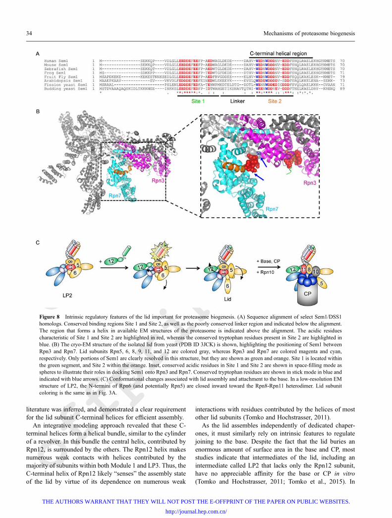
Sem1 as a molecular tether during lid assemblySimilar to the CP, the lid relies on unstructured proteindomains to serve as stabilizing factors during assembly. Thishas been best documented for the lid subunit Sem1/Rpn15(DSS1 in humans), which serves as a molecular tether tostabilize an otherwise unstable assembly intermediate until itcan be efficiently incorporated into the assembling lid(Tomko and Hochstrasser, 2014). Sem1 is an unusualproteasome subunit, with an exceptionally small size (~10kDa) and a near-complete lack of secondary and tertiaryprotein structure (Kragelund et al., 2016). Aside from a C-terminal α-helix, Sem1 contains no well-defined protein fold,and consists of two highly conserved binding domainsseparated by an unstructured linker sequence (Fig. 8A). Thetwo binding domains are rich in acidic residues, which areimportant for recognition of their binding partners. The firstbinding site, constituting residues 29-45 in yeast, recognizes apositively charged crevice in the proteasomal Rpn3 lidsubunit (Fig. 8B) (Wei et al., 2008; Dambacher et al., 2016).Sem1 is able to bind Rpn3 in the absence of any otherproteins, and may serve as its folding or stabilizing chaperone(Tomko and Hochstrasser, 2014). In addition to conservedacidic residues, the second site in Sem1 contains twoconserved tryptophan residues that dock Sem1 into hydro-phobic pockets in the lid. These tryptophan residues makecritical contacts with Rpn3 and Rpn7 (Fig. 8B, inset), and arenecessary for stable binding to Rpn3 or Rpn7 (Tomko andHochstrasser, 2014; Dambacher et al., 2016).
Although Rpn3 and Rpn7 interact extensively in the fullyassembled proteasome and in the isolated lid, these proteinsdisplay poor affinity for one another in isolation, implyingthat their stable association relies on a remodeling of theirinteraction surfaces during assembly, or that they arestabilized by interactions between additional subunits in thecontext of larger assembly products (Tomko and Hochstras-ser, 2014). During lid biogenesis, Sem1 binds these twosubunits and stabilizes their otherwise weak interaction toyield the trimeric lid assembly intermediate LP3 (Fig. 3A).This tethering role was supported by experiments demon-strating that separation of the two binding sites, viaexpression of Sem1 as two fragments split through the linkerregion, failed to promote LP3 formation. Similarly, a mutantSem1 harboring an extended linker sequence readilypromoted LP3 formation, whereas a mutant form with ashortened linker region did not, indicating a minimal reachbetween Sites 1 and 2 is required for its function. The flexiblelinker region in Sem1 between sites 1 and 2 is both disordered
and poorly conserved, which allowed engineering of aprotease cleavage site into it without disrupting the assemblyfunction of Sem1. Using this cleavable form of Sem1, thetethering function of Sem1 was assessed in a variety ofproteasomal assembly intermediates (Tomko and Hochstras-ser, 2014). Importantly, cleavage of Sem1 in the context ofLP3 resulted in dissociation of Rpn3 and Rpn7, consistentwith the proposed tethering role, but once these subunits hadincorporated into larger complexes, Sem1 could be cleavedwith no apparent loss of interaction between Rpn3 and Rpn7.Together, this indicated that the tethering role of Sem1 wasimportant only during the initial stages of lid assembly, andthat this role becomes dispensable once Rpn3 and Rpn7 areincorporated into higher order intermediates, consistent with amodel where their interface is stabilized via remodeling and/or interactions with other subunits within the lid.
Sem1 is somewhat promiscuous among proteasomesubunits—it has been shown to be an integral component ofother multisubunit protein complexes, including the TREX-2mRNA export complex, and a complex containing theBRCA-2 tumor suppressor (reviewed in (Kragelund et al.,2016)). The binding sites on Sem1 that associate with theproteasome overlap substantially with those used to associatewith these complexes, which suggests first that Sem1 canlikely associate with only one of these structures at a time, butalso, that Sem1 may be reversibly recruited from one complexto another to control assembly or function of these respectivecomplexes. Whether Sem1 serves as an assembly chaperonefor these other complexes has not been studied in great detail,but it is known that DSS1 functions to stabilize BRCA-2 akinto Sem1 with Rpn3 (Li et al., 2006). Sem1 has recently beenreported to bind ubiquitin using binding site 2 (Paraskevo-poulos et al., 2014), although this role has been disputed (Shiet al., 2016). Nonetheless, under some circumstances,occupation of this site by ubiquitin may also serve to fine-tune the assembly or function of the proteasome indirectly viaSem1.
Lid subunit C-terminal helicesThe use of avidity via multiple binding interactions is alsoutilized more broadly within the proteasomal lid to driveassembly. Each lid subunit, save for Sem1, contributes C-terminal α-helices to an unusual helical bundle (Estrin et al.,2013). In the context of lid assembly, this helical bundle alsoserves to drive the stepwise assembly of the lid from itscognate subunits by generating avid binding surfaces. Thesesurfaces recruit subsequent subunits to the nascent complex.An elegant study using heterologously expressed lid subunitsin E. coli demonstrated that this helical bundle is a criticaldeterminant of the lid subunit assembly sequence (Estrin etal., 2013). By systematically coexpressing a truncated lidsubunit lacking its C-terminal helix with the other eightsubunits and assessing lid assembly by gel filtration, atentative assembly sequence congruent with the available
Lauren A. Howell et al. 33
THE AUTHORS WARRANT THAT THEY WILL NOT POST THE E-OFFPRINT OF THE PAPER ON PUBLIC WEBSITES.
http://journal.hep.com.cn/
offprint
offprint
literature was inferred, and demonstrated a clear requirementfor the lid subunit C-terminal helices for efficient assembly.
An integrative modeling approach revealed that these C-terminal helices form a helical bundle, similar to the cylinderof a revolver. In this bundle the central helix, contributed byRpn12, is surrounded by the others. The Rpn12 helix makesnumerous weak contacts with helices contributed by themajority of subunits within both Module 1 and LP3. Thus, theC-terminal helix of Rpn12 likely “senses” the assembly stateof the lid by virtue of its dependence on numerous weak
interactions with residues contributed by the helices of mostother lid subunits (Tomko and Hochstrasser, 2011).
As the lid assembles independently of dedicated chaper-ones, it must similarly rely on intrinsic features to regulatejoining to the base. Despite the fact that the lid buries anenormous amount of surface area in the base and CP, moststudies indicate that intermediates of the lid, including anintermediate called LP2 that lacks only the Rpn12 subunit,have no appreciable affinity for the base or CP in vitro(Tomko and Hochstrasser, 2011; Tomko et al., 2015). In
Figure 8 Intrinsic regulatory features of the lid important for proteasome biogenesis. (A) Sequence alignment of select Sem1/DSS1homologs. Conserved binding regions Site 1 and Site 2, as well as the poorly conserved linker region and indicated below the alignment.The region that forms a helix in available EM structures of the proteasome is indicated above the alignment. The acidic residuescharacteristic of Site 1 and Site 2 are highlighted in red, whereas the conserved tryptophan residues present in Site 2 are highlighted inblue. (B) The cryo-EM structure of the isolated lid from yeast (PDB ID 3JCK) is shown, highlighting the positioning of Sem1 betweenRpn3 and Rpn7. Lid subunits Rpn5, 6, 8, 9, 11, and 12 are colored gray, whereas Rpn3 and Rpn7 are colored magenta and cyan,respectively. Only portions of Sem1 are clearly resolved in this structure, but they are shown as green and orange. Site 1 is located withinthe green segment, and Site 2 within the orange. Inset, conserved acidic residues in Site 1 and Site 2 are shown in space-filling mode asspheres to illustrate their roles in docking Sem1 onto Rpn3 and Rpn7. Conserved tryptophan residues are shown in stick mode in blue andindicated with blue arrows. (C) Conformational changes associated with lid assembly and attachment to the base. In a low-resolution EMstructure of LP2, the N-termini of Rpn6 (and potentially Rpn5) are closed inward toward the Rpn8-Rpn11 heterodimer. Lid subunitcoloring is the same as in Fig. 3A.
34 Mechanisms of proteasome biogenesis
THE AUTHORS WARRANT THAT THEY WILL NOT POST THE E-OFFPRINT OF THE PAPER ON PUBLIC WEBSITES.
http://journal.hep.com.cn/
offprint
offprint
agreement, blockade of lid assembly via genetic means inyeast leads to the accumulation of lid intermediates, all ofwhich are devoid of base or CP (Fukunaga et al., 2010;Tomko and Hochstrasser 2011). A major unansweredquestion thus has been: what mechanism restrains lidattachment until completion of lid assembly? Recentinvestigations into the structures of the isolated lid and lidassembly intermediates have implicated several conforma-tional changes as pivotal maturation events that permitcompletion of RP assembly.
Recently, a combination of quantitative crosslinking-massspectrometry (QCL-MS) and negative stain EM reported thatthe LP2 intermediate undergoes substantial conformationalrearrangement upon incorporation of Rpn12 that in turnpermits its efficient assembly into the proteasome holoen-zyme (Tomko et al., 2015). In contrast to the structure of theproteasome-associated lid, LP2 adopts a more compact statein which the N-termini of several subunits appear to moveinward toward the PCI horseshoe and the Rpn8-11 hetero-dimer, similar to a closed fist (Fig. 8C). Importantly, provisionof the conserved C-terminal helix of Rpn12 alone wassufficient to drive this conformational reorganization and RPformation (Fig. 8C), implicating engagement of the lid helicalbundle as the critical determinant of this large scaleconformational shift.
A second layer of control lies in a conformational change inthe position of the Rpn8-Rpn11 deubiquitinating modulewithin the lid (Dambacher et al., 2016). The cryo-EMstructure of the isolated lid unexpectedly revealed that theRpn8-Rpn11 module is positioned approximately perpendi-cular to the orientation observed in the full proteasome. In thisposition, it is collapsed inward toward the core of the lid, andis cradled by contacts with the neighboring lid subunits Rpn5and Rpn9. This conformation is highly reminiscent of thatobserved in the low-resolution EM structure of LP2 and thusis likely sterically incompatible with the base (Tomko et al.,2015). Thus, at least two critical conformational changes arenecessary for lid-base association—a repositioning of Rpn5and Rpn6 N-termini that are folded inward toward the core ofthe lid, occluding the base, and rotation of the Rpn8-Rpn11module into an extended conformation. Further mechanisticstudies will be essential to clarify how these importantstructural transitions occur during RP biogenesis.
Maturation of RP enzymatic activities
Within the proteasome holoenzyme, substrate binding,unfolding, deubiquitination, and proteolysis are tightlycoupled. Decoupling of these activities would result in thecounterproductive return of deubiquitinated or unfoldedprotein substrates to the cellular milieu without degradation,which could disrupt cellular processes or initiate formation oftoxic protein inclusions. Because enzymatic coupling ofproteasomal activities is dependent on the proper engagementand communication between proteasomal subcomplexes, it is
imperative that the activities of isolated subcomplexes orintermediates be suppressed during biogenesis, and that theymature successfully upon complete assembly of the holoen-zyme. The processing of the β subunit propeptides (describedabove) is one example whereby a catalytic activity of theproteasome is restrained until it is safely contained within theproteolytic chamber at the interior of the CP.
In the past two years, much progress has been made inunderstanding maturation of the lid’s Rpn11-dependentdeubiquitinating activity. At least two mechanisms restrictRpn11 activity until incorporation of the lid into theproteasome. The first is an autoinhibitory mechanism presentin the closed conformation observed in the isolated lid and,potentially, intermediates (Tomko et al., 2015; Dambacher etal., 2016). In this conformation where the Rpn8-Rpn11 iscollapsed inward, the Rpn11 active site is shielded from theenvironment. Importantly, the side chain of Rpn5-Asn274intrudes into the Rpn11 active site where it displaces a watermolecule necessary for catalysis. The repositioning of theRpn8-Rpn11 module upon incorporation of the lid into theproteasome exposes the Rpn11 active site and frees it frominhibition by Rpn5, permitting catalysis.
A second mechanism restricting Rpn11 activity prior tocompletion of proteasome assembly relies upon physicalseparation of Rpn11 from its substrates, and exploits theunique nature of Rpn11 among deubiquitinating enzymes toaccomplish this. Whereas most DUBs either trim polyubi-quitin chains from the distal end or cleave between Ubmoieties, Rpn11 cleaves the isopeptide bond between themost proximal ubiquitin and the substrate itself, releasingpolyubiquitin chains en bloc (Verma et al., 2002; Yao andCohen, 2002). Rpn11 must therefore tolerate substantialvariability in the substrate sequence surrounding the scissileUb isopeptide bond, and does so by making little to no contactwith the substrate (Pathare et al., 2014; Worden et al., 2014).Although this yields very poor substrate affinity in isolation,this is compensated for in the proteasome via high affinitybinding of the substrate’s polyubiquitin chain by proteasomalubiquitin receptors contained within the base, and the pullingof the isopeptide bond into the active site of Rpn11 by thebase ATPases during substrate unfolding and translocation.Thus, Rpn11 activity is restrained during assembly by virtueof its physical separation from its major substrate recruitmentmechanisms. We posit that this may be particularly importantearly during the assembly of the lid, in the context ofintermediates that do not yet contain Rpn5 to bind Rpn11 andexclude the catalytic water as observed in the isolated lidstructure.
As mentioned above, the ability of the proteasomalATPases to hydrolyze ATP appears to be inhibited in thecontext of assembly intermediates, and is activated uponcompletion of base assembly. The mechanism by whichATPase activity is restrained until completion of baseassembly is unknown, but likely depends upon criticalinteractions of the ATPase subunits with neighboring
Lauren A. Howell et al. 35
THE AUTHORS WARRANT THAT THEY WILL NOT POST THE E-OFFPRINT OF THE PAPER ON PUBLIC WEBSITES.
http://journal.hep.com.cn/
offprint
offprint
ATPases to orient nucleotide properly for catalysis (Kim etal., 2015), which would serve to couple completion of theATPase ring to maturation of ATPase activity. The fullyassembled base contains ubiquitin receptors and unfoldingactivity, even in the absence of the lid and CP. Thus, it can inprinciple recruit and unfold substrates in isolation, decouplingunfolding from deubiquitination or proteolysis. Unfolding ofsubstrates without deubiquitination or degradation isrepressed by the activity of the Ubp6 deubiquitinase (Sakataet al., 2011), which is found associated with the earliest Rpt1-and Rpt2-containing base assembly intermediates and ispresent on the base and the full proteasome. Ubp6 abuts theATPase ring (Aufderheide et al., 2015; Bashore et al., 2015),and can deubiquitinate substrates that are recruited to baseassembly intermediates (Sakata et al., 2011). Althoughdeubiquitination of proteasome substrates without degrada-tion may be unproductive, it may be less likely to promotetoxicity than spurious unfolding and release of unfoldedproteins that could then aggregate and cause toxicity. Thelevel of free base subcomplexes in the cell is very low basedon estimates from yeast cell extracts (Funakoshi et al., 2009);it could be that the cell can tolerate a small amount of spurioussubstrate deubiquitination at the expense of sparing proteinaggregation and the resultant toxicity. Alternatively, the basehas been reported to have refolding activity, at least in thecontext of the 26S proteasome or base-CP complexes (Braunet al., 1999). It is possible that this chaperone-like function isactive in the context of assembly intermediates to guardagainst spurious unfolding and subsequent release of anysubstrates that engage the assembling RP.
Emerging themes in proteasome assembly
It remains important to continue expanding our knowledge ofintrinsic features of proteasome components, and the extrinsicfactors that act upon them. However, it is also clear thatproteasome assembly is more than the sum of these parts.Below we present some of the frontiers that will give rise tothe next chapters in our understanding of the biogenesis ofthis essential molecular machine.
Quality control of failed assembly products
The degradation of individual polypeptides by the UPS iswell understood, whereas the mechanisms mediating degra-dation of the proteasome itself or proteasomal subcomplexesare only recently coming to light (Marshall et al., 2015; Peterset al., 2015; Marshall et al., 2016). Studies of proteasomeassembly in various organisms have revealed that formationof non-native or off-pathway assembly products can and doesoccur. But, the nature and fate of those assembly products hasnot yet been addressed in great detail. A clearance pathway isalmost certainly necessary for such species, and perhaps
compounding the difficulty of this task, these species may notdisplay obvious features that demarcate them as “damaged”in the same way a misfolded protein does, necessitatingdistinct mechanisms to recognize and clear them.
Recent investigations of proteasome quality controlmechanisms have revealed a novel pathway of Atg8-mediated proteasome autophagy, termed proteaphagy (Mar-shall et al., 2015), which has since been demonstrated inseveral species (Cohen-Kaplan et al., 2016; Waite et al.,2016). Proteaphagy was first observed in Arabidopsis asaccumulation of proteasome subunits in autophagy (atg)mutants with compromised ubiquitin-like protein Atg8lipidation or autophagic induction by Atg1 kinase. This wasshown to be due to autophagic turnover of proteasomes.Proteaphagy could be stimulated by nitrogen starvation or bytreatment of cells with the noncovalent proteasome inhibitorMG132. The pathways mediating proteasome turnover inresponse to these two stimuli appeared to be distinct—theAtg1 kinase responsible for nutrient sensing was essentialonly in proteaphagy induced through nitrogen starvation,whereas Atg8 was required for proteaphagy induced by bothnitrogen starvation and MG132. The association of ubiquitinreceptor RPN10 was found to increase concomitantly withproteasome inhibition, accompanied by an increase inproteasome-associated ubiquitination. RPN10, which bindsboth Atg8 and ubiquitin, was identified as the tether betweenubiquitinated proteasomes and Atg8 to facilitate the targetingof proteasomes to autophagic membranes and subsequentvacuolar degradation. However, Atg8-RPN10 interactionrequires a ubiquitin-interacting motif within RPN10 that isabsent in some species. The ubiquitin receptor protein Cue5was identified as the functional equivalent of RPN10 in yeast,binding both Atg8 and the ubiquitinated proteasome complex(Marshall et al., 2016). Together, this data identified the firstpathway capable of destroying fully assembled proteasomes.
Although the observation that treatment with proteasomeinhibitors stimulates proteasome turnover is clear, it remainsunclear whether this pathway specifically serves a trueproteasome quality control function. The inhibitor utilized,MG132, is a noncovalent and reversible inhibitor, indicatingthat irreversible inactivation is not necessary to stimulateproteaphagy. Also, the induction of proteaphagy is greatlydelayed compared to the rapid proteasome inhibition byMG132 (Marshall et al., 2016). Rather, we suggest that thisturnover may instead represent “collateral damage” toproteasomes resulting from enhanced nonselective macro-autophagy in response to proteasome inhibition. Indeed,numerous studies have observed upregulation of the basalautophagy rate to compensate for proteasome inhibition(Iwata et al., 2005; Ding et al., 2007; Pandey et al., 2007;Hoang et al., 2009; Zhu et al., 2010). Nonetheless, thispathway remains a highly attractive mechanism to dispose ofirreversibly damaged or misassembled complexes containingproteasome subunits.
36 Mechanisms of proteasome biogenesis
THE AUTHORS WARRANT THAT THEY WILL NOT POST THE E-OFFPRINT OF THE PAPER ON PUBLIC WEBSITES.
http://journal.hep.com.cn/
offprint
offprint
Recently, misfolded proteasome subunits have beenreported to accumulate in insoluble protein deposits(IPODs), which are thought to serve as intermediatecompartments for these subunits. In some cases, normalsubunits were also recruited, raising the possibility thatpartially assembled, defective complexes were delivered tothese IPODs. Small heat shock protein Hsp42, an importantfactor in IPOD assembly, appears to function as an essentialchaperone for IPOD delivery, as hsp42D precludes protea-some subunit localization in IPODs and abolishes autophagicclearance of proteasome subunits (Marshall et al., 2015;Peters et al., 2015). Proteasomes do not colocalize withIPODs under normal conditions, therefore suggesting therelationship between IPODs and proteasomes is a qualitycontrol mechanism (Kaganovich et al., 2008). It has beensuggested that Hsp42-mediated sequestration of proteasomesubunits to IPODs is an alternative to a more favored pathwayinvolving the UPS-mediated degradation of its own mis-assembled subunits.
An additional mechanism that may control proteasomequality is via binding and inactivation of structurallydefective proteasomes by Ecm29. Ecm29 is an evolutionarilyconserved protein consisting primarily of HEAT repeats thatassociates only with proteasomes containing both RP and CPcomplexes (Leggett et al., 2002). Ecm29 appears to beselectively recruited to proteasomes that harbor a defect at theinterface between the CP and the RP (Lee et al., 2011; Park etal., 2011; De La Mota-Peynado et al., 2013). In yeast,mutations to the HbYX motif of Rpt5, or to the pocket lysineresidues in α subunits, has been shown to enhance theassociation of Ecm29. Importantly, lysate-mixing experi-ments clearly demonstrated that Ecm29 preferentially asso-ciated with structurally defective proteasomes over normalproteasomes (Lehmann et al., 2010).
Ecm29 appears to contact both the CP and the RP, based onEM, crosslinking, and biochemical studies (Leggett et al.,2002; Lehmann et al., 2010; De La Mota-Peynado et al.,2013; Wani et al., 2016), and recent enzymatic experimentsdemonstrated clearly that it functions in part to suppress thecatalytic activity of the ATPase ring (De La Mota-Peynado etal., 2013). In this manner, Ecm29 may serve to suppress theactivity of functionally defective proteasomes that mayotherwise interfere with degradation of proteasome sub-strates. Recently, it was shown that phosphorylation of the α7subunit of the CP serves as a major recruiting signal forEcm29 (Wani et al., 2016). This represents the first exampleof phosphorylation-dependent recruitment of a proteasome-interacting protein to the proteasome, and points toward apotential signaling mechanism for marking defective protea-somes. Two important questions persist regarding thisfinding: first, the kinase responsible for this phosphorylationevent has not yet been determined. Second, whether suchphosphorylation is constitutively present on proteasomes isunknown. Understanding the latter question would serve toclarify whether the structural defect serves to recruit the
kinase for phosphorylation, or if instead the phosphorylationsites are only accessible to Ecm29 in proteasomes harboring astructural defect.
The role of subunit expression stoichiometry in assemblyefficiency and fidelity
As alluded to earlier, subunit heterogeneity in eukaryotesbrings with it additional complications to efficient macro-molecular assembly. Two of these are order of assembly and,given the structural similarities between subunits, thepotential for subunit mispairing. These two can influenceeach other and are thus not mutually exclusive. We’vediscussed above how assembly factors and intrinsic featuresof subunits can mitigate these difficulties. A third level ofcomplexity is the stoichiometry of the individual components.The levels at which proteasome subunits are expressed canimpact the assembly pathway followed, as well as thecomposition and abundance of the assembly products.Control of proteasome levels is best understood in yeast.
In yeast, proteasome levels are regulated at the transcrip-tional level by Rpn4. This C2H2 zinc finger motif proteinrecognizes conserved PACE (proteasome-associated controlelement) sequences in the promoters of proteasome genes(Mannhaupt et al., 1999; Xie and Varshavsky, 2001), thoughsome deviation from the canonical 9 base pair PACEconsensus is observed (Shirozu et al., 2015). Rpn4 is turnedover rapidly by the proteasome via ubiquitin-dependent and-independent mechanisms (Xie and Varshavsky 2001; Ju andXie, 2004). When proteasome activity is compromised, Rpn4levels rise leading to increased production of proteasomesubunits. This is followed by increased assembly ofproteasomes which, in turn, resume the rapid Rpn4 turnover,leading to a downregulation of further subunit synthesis. Inmammals, there is no Rpn4 homolog but proteasome subunitlevels are also coordinately regulated (Meiners et al., 2003),in an analogous regulatory loop, by the transcription factorNrf1 (Radhakrishnan et al., 2010). This protein is an ER-targeted glycoprotein (Wang and Chan, 2006; Zhang et al.,2007) that is constitutively turned over by the proteasome in aCdc48-dependent fashion (Radhakrishnan et al., 2014). Whenproteasome function is compromised, Nrf1 levels accumulateand the protein is proteolytically cleaved by the proteasomeitself (Sha and Goldberg, 2014), or by an aspartyl protease(Koizumi et al., 2016; Lehrbach and Ruvkun, 2016), prior toactivating proteasome gene expression.
Though not the only means by which proteasome subunitlevels are regulated, these elegant feedback mechanisms helpmaintain proteasome subunits at approximately stoichio-metric levels, which in yeast have been variously estimated tobe between 10000 to 30000 copies per cell for differentsubunits (Russell et al., 1999; Ghaemmaghami et al., 2003;Kulak et al., 2014). However, there are important futureconsiderations hiding behind this apparent stoichiometry.First, these numbers refer to total subunit amounts, the vast
Lauren A. Howell et al. 37
THE AUTHORS WARRANT THAT THEY WILL NOT POST THE E-OFFPRINT OF THE PAPER ON PUBLIC WEBSITES.
http://journal.hep.com.cn/
offprint
offprint
majority of which exist in fully assembled proteasomes. Forunderstanding assembly, it is the level of unincorporatedsubunits that will be relevant, but much more difficult todetermine. Second, suppose we assume that the two-to-threefold variation in total subunit levels is reflected in thevariation (i.e. two-to-threefold) of unincorporated subunitlevels, these small differences may be more than enough toinfluence the order of subunit assembly (and misassembly).An interesting analysis of complex haploinsufficiency inter-actions (CHI) in yeast found associations between hetero-zygous deletion of the yeast actin gene ACT1 andheterozygous deletions of essential proteasome genes (Haareret al., 2011). The obvious implication is that the proteasomeregulates actin dynamics. But beyond this simple interpreta-tion is the intriguing observation that not all proteasomegenes exhibited CHI with actin. In the CP, only α6, α7, α1, β2,β4, and β5 exhibited CHI with actin. In the RP base, onlyRpt2 did so, whereas all of the RP lid subunits (except Sem1)exhibited CHI with actin. The authors hypothesized that thiswas due to assembly defects caused when the levels of someof the subunits became limiting (Haarer et al., 2011). Thisexample highlights the need for experiments aimed atinvestigating the relationship between subunit levels, stoi-chiometry, and assembly efficiency/fidelity.
Localization in assembly
Proteasomes are present in the nucleus and cytoplasm ofmammalian cells in roughly comparable proportions (Reits etal., 1997). In yeast, proteasomes are concentrated in thenucleus (and nuclear periphery) (Enenkel et al., 1998; Russellet al., 1999). The concentration of 26S proteasomes isestimated to be just under 1 mM in the yeast nucleus and about5-fold less in the cytoplasm (Pack et al., 2014). Remarkablysimilar values for the mean cytoplasmic concentration (~190nM) of 26S proteasomes were found in mammalian neurons(Asano et al., 2015). The question of where assembly takesplace has generated many suggestions, but no single agreedupon model. Certain CP and RP subunits have nuclearlocalization sequences (NLS) (Tanaka et al., 1990; Nederlofet al., 1995; Wendler et al., 2004); hence any assemblyintermediates that contain them should, in theory, betransportable. Consistent with this, isolated base, lid, andCP have all been reported to be capable of import into thenucleus independently of each other (Wang et al., 1997; Mayret al., 1999; Wendler et al., 2004; Isono et al., 2007). Somehave reported import of CP occurring with Rpn1, Rpn2,Hsp90, and importin β in a Xenopus extract assay (Savulescuet al., 2011), while others implicate a role for Blm10 inhelping import CP (Weberruss et al., 2013). It has even beenargued that CP precursors are imported into the yeast nucleus,where assembly is completed (Lehmann et al., 2002), andalternatively that full 26S proteasomes are imported into theyeast nucleus, thereby arguing that assembly can proceed tocompletion in the cytoplasm (Pack et al., 2014). The
distribution of the various CP and RP assembly factorsthroughout the cell does not help in resolving the question ofassembly location, though Ump1 appears to be primarilynuclear (Lehmann et al., 2002; Huh et al., 2003; Hoefer et al.,2006; Le Tallec et al., 2007; Saeki et al., 2009).
An interesting observation placed human Ump1 directly incontact with membranes and serving to recruit CP assemblyintermediates to the ER (Fricke et al., 2007). Moreover, CPassembly defects were observed when TRC40 or Bag6, twoproteins involved in the pathway which inserts tail-anchoredproteins into the mammalian ER membrane, were knockeddown (Akahane et al., 2013). Specifically, β subunits werepoorly incorporated and this correlated with poor recruitmentof CP assembly intermediates to the ER membrane. Stronggenetic interactions between proteasome assembly factorsand yeast components of the tail-anchoring pathway werealso observed, suggesting a conserved mechanism involvingCP assembly at ER membranes might be at play (Akahane etal., 2013). The same study also reported that Bag6 wasrequired for the stability of the Nas2 module as Bag6knockdowns resulted in accumulation of RP-like specieslacking Rpt4 and Rpt5 (Akahane et al., 2013), suggestingBag6 might have multiple roles in proteasome assembly.However, the question of where individual steps of assemblymight occur awaits a firm conclusion.
Assembly as a regulatory mechanism – making protea-somes for the job at hand
The ability to modify proteasomes for different functions isnot a new concept. One simple way to envision modificationis through subunit substitution. The discovery and character-ization of mammalian immunoproteasomes, containinginducible paralogs (β1i, β2i, β5i) in place of the constitutivecatalytically-active subunits (β1, β2, β5), was an importantpart of the foundation upon which the field of proteasomebiogenesis was built ((Driscoll et al., 1993; Gaczynska et al.,1993) and reviewed in (Basler et al., 2013)). For example, theidentification of the first proteasome assembly intermediates(Frentzel et al., 1994; Nandi et al., 1997; Schmidtke et al.,1997) and some key studies on the roles of β subunitpropeptides were carried out on immunoproteasomes (Griffinet al., 1998; Kingsbury et al., 2000; De et al., 2003).Immunoproteasomes also provide an excellent example ofhow assembly can be used as a regulatory mechanism togenerate alternate versions of a multi-protein complex suitedfor different tasks. For a recent review of immunoproteasomestructure and function, we direct the readers here (Basler etal., 2013). Below, we will briefly discuss how assemblyproceeds when multiple paralogs are present in the same cell.In the case of the immune β subunits, there is a different orderof β subunit assembly due to cooperative recruitment amongthe inducible subunits (Griffin et al., 1998). The β1i subunitenters the α-ring early, versus β1 which is a “late” β subunit,and recruits β2i (Groettrup et al., 1997; Hirano et al., 2008).
38 Mechanisms of proteasome biogenesis
THE AUTHORS WARRANT THAT THEY WILL NOT POST THE E-OFFPRINT OF THE PAPER ON PUBLIC WEBSITES.
http://journal.hep.com.cn/
offprint
offprint
This is followed by β3, β4, and β5i (Bai et al., 2014). The keyis that the presence of β1i and β2i facilitates the recruitment ofβ5i (Kingsbury et al., 2000), which can occur even after β3, inthe absence of β4 (Bai et al., 2014). Moreover, thepropeptides of β2i and β5i specifically direct entry of thesesubunits into immunoproteasomes; this specificity is trans-plantable, as demonstrated by elegant peptide swap experi-ments (Kingsbury et al., 2000; De et al., 2003). Thus intrinsicfeatures of paralogous subunits can influence assembly. Thisis further confirmed by the third mammalian β5 paralog, thethymus specific β5t that gives rise to thymoproteasomes(Murata et al., 2007), whose propeptide endows it with anability to outcompete the constitutive β5 subunit as well (Baiet al., 2014). However, extrinsic factors are also important, ashuman Ump1 preferentially binds to β5i, over β5, and directsits assembly into immunoproteasomes (Heink et al., 2005).
In addition to paralogy, there are other means of usingassembly as a regulatory mechanism for functional speciali-zation. One was introduced earlier in the form of alternativeα4–α4 proteasomes which are found conserved between yeastand humans (Velichutina et al., 2004; Kusmierczyk et al.,2008; Padmanabhan et al., 2016). It is still not understoodhow these proteasomes are assembled, or what regulates theprocess. In human cells, phosphorylation of the α4 subunitprevents its ubiquitin-dependent degradation leading toincreased levels of this subunit (Li et al., 2015); as α4 levelsrise, increased levels of α4–α4 proteasomes form (Padma-nabhan et al., 2016). Thus mass action, in the form of anincreased α4/α3 ratio (and/or an increased α4/PAC3-PAC4ratio), may be sufficient to allow formation of α4–α4proteasomes (Padmanabhan et al., 2016). Notably, in yeastthe only CP assembly factor that lacks either the canonicalPACE elements for the Rpn4 transcription factor, or theminimum Rpn4-responsive elements (PACE-core), is Pba3-Pba4 (Shirozu et al., 2015). Thus, conditions that stabilizeRpn4, and lead to upregulation of proteasome subunits,should not significantly impact Pba3-Pba4 levels. Thisalso sets up a potential mass-action scenario whereby anincreased α4/Pba3-Pba4 ratio could lead to increased α4–α4proteasome formation. Experiments are underway to test thishypothesis.
The functional significance of α4–α4 proteasomes is notyet known. However, generation of α4–α4 proteasomescreates a CP lacking the major gating subunit and thus shouldbe constitutively open (Groll et al., 2000); it also gives rise toa different α-ring that could impact the binding of RP or otheractivators (see below). Either, or both, of these features couldcontribute to the role of α4–α4 proteasomes in the cell.Whether replacement of α3 with α4 is the only example ofnon-paralogous subunit substitution within the proteasomeremains to be seen, though this is easier to envision occurringwith structurally homologous subunits (such as subunits ofthe CP) or when the substituted subunit is not essential, likeα3.
Another means of using assembly to regulate proteasome
function is via the use of alternative regulators that bind theCP. This review is focused on the canonical 26S proteasome,in which a CP is bound by one or more RP complexes.However, it bears mention that CP can interact with a host ofother protein complexes, most with demonstrated ability tostimulate the catalytic activity of the CP. These include theaforementioned Blm10/PA200, which is broadly conservedacross the eukarya (Ustrell et al., 2002; Schmidt et al., 2005),and members of the 11S family of activators (also calledPA28, or REG) found primarily in higher eukaryotes. Thelatter includes the heteroheptameric PA28ab (or REGab)complex and the homoheptameric PA28g (or REGg). Thefunctions of the 11S complexes are not well understood (forrecent reviews, readers are directed here (Mao et al., 2008;Stadtmueller and Hill, 2011; Cascio, 2014)) but they can capCP independently, or as hybrid complexes with RP. Thoughthey lack HbYX motifs, the C-termini of 11S complexesinsert into the same inter α-subunit pockets used by otheractivators (Whitby et al., 2000; Forster et al., 2005). Inaddition, PI31, originally identified as an inhibitor of CP invitro (Chu-Ping et al., 1992), represents a fourth class of CPregulator conserved from yeast to mammals (Zaiss et al.,2002; Bader et al., 2011; Hatanaka et al., 2011; Li et al., 2014;Yashiroda et al., 2015). Phylogenomic analysis suggests thatall four classes of regulator (RP, Blm10/PA200, 11S, andPI31) were present in the last eukaryotic common ancestor,despite the subsequent loss of some these proteins in manydescendant lineages (Fort et al., 2015). Finally, there isevidence that the archaeal ortholog of Cdc48 forms degrada-tion-competent complexes with archaeal CP, though if thesame is true of eukaryotic Cdc48 remains to be conclusivelydemonstrated (Barthelme and Sauer, 2012a; Barthelme andSauer, 2013; Barthelme et al., 2014; Barthelme et al., 2015).
Regardless of which combination of regulators is present ina given eukaryotic cell, the fact that they all likely share aconserved binding mode with CP on the outer α-ring surfacemeans that they must compete for this surface. The existenceof two such α-ring surfaces on each CP also makes hybridcomplexes possible, as mentioned above. Consequently,questions about the function of these CP-regulator complexes(especially the various possible hybrids) are also ultimatelyquestions of assembly and the regulation thereof. To illustratethis point, recent work by Welk and colleagues (2016)demonstrated differential recruitment of various regulators, inresponse to proteasome inhibition, thereby generating alter-natively-capped proteasomes (Welk et al., 2016).
Disruptions to proteasome assembly in human disease
Although numerous studies have tied changes in proteasomeactivity to human disease, the exact molecular mechanismsunderpinning breakdowns in proteasomal proteolysis remainpoorly studied. Recent advances in DNA sequencing haveyielded a wealth of information about genetic variations in thegenomes of diseased tissues. These approaches have
Lauren A. Howell et al. 39
THE AUTHORS WARRANT THAT THEY WILL NOT POST THE E-OFFPRINT OF THE PAPER ON PUBLIC WEBSITES.
http://journal.hep.com.cn/
offprint
offprint
uncovered a substantial number of genetic changes and singlenucleotide polymorphisms within proteasome subunit orassembly chaperone genes (reviewed in (Gomes, 2013)). Inthe majority of cases, the potential effects of thesepolymorphisms are unknown. However, several recentstudies have provided evidence that disruption of proteasomeassembly results from polymorphisms in proteasome subunitsor assembly chaperones, and may represent an underlyingcause of several human diseases. We highlight threeparticularly compelling examples here.
Nakajo-Nishimura syndrome is a rare autosomal recessiveinflammatory disorder characterized by periodic fever, skinrashes, joint contractures, and lipomuscular atrophy. A recentsequencing effort unveiled a single nucleotide transversion(c.602G>T) within the coding sequence of the PSMB8 gene(Arima et al., 2011). Strikingly, this variant was present in allpatients tested, immediately implicating it in diseasepathology. PSMB8 encodes the β5i subunit of the immuno-proteasome, which has important roles in the innate andadaptive immune systems, and this mutation results in acoding sequence mutation of Gly201 to valine. This glycineresidue is highly conserved, and its mutation to valinedisrupts immunoproteasome assembly. The resultant loss ofproteasome activity due to reduced assembly of immunopro-teasomes was associated with increased inflammatorycytokine production in peripheral lymphocytes, providingstrong evidence that this variant is responsible, or at least amajor contributing factor, to the disease pathology. Intrigu-ingly, a second variant in PSMB8 observed in a relatedinflammatory disease, JMP syndrome (Agarwal et al., 2010),reduces chymotryptic activity without disturbing proteasomeassembly. This suggests that disruption of immunoprotea-some function via mutations to PSMB8 may define a relatedclass of autoinflammatory diseases, and indicates thatdisruption of immunoproteasome assembly can be aspathogenic as mutations directly impacting proteolysis.
A second example derives from studies of an Italian familywith a documented history of type II diabetes (T2D).Sequencing a chromosomal region identified via classicalgenetic mapping as being linked to T2D led to theidentification of a haplotype containing two intronic variantsand a coding variant of the PSMD9 gene (Gragnoli andCronsell, 2007), which encodes the human ortholog of theNas2 base assembly chaperone. This coding variant results ina missense mutation of Glu197 to glycine. Modeling of thisresidue position onto the available crystal structures of Nas2from yeast (Singh et al., 2014) indicates this residue lies in theterminal strand of a β-sheet that makes up the core of the PDZdomain. Glycine is known to favor disorder, furthersuggesting that this mutation may disrupt the structure, andin turn the function, of human Nas2, causing predisposition ofcarriers to T2D. In support of a role for alterations inproteasome assembly in T2D etiology, a second disease-associated variant in the immediate upstream sequence ofPSMA6, which encodes the α1 subunit of the CP, is associated
with T2D in the Chinese Dongxiang and Han populations(Liu et al., 2012). This variant, -8C>G, is within the Kozakconsensus sequence of the PSMA6 gene, strongly implying itresults in an alteration in subunit translation. The effects of α1subunit depletion, as well as the effect of this mutation on CPassembly in vivo, have not been empirically tested, but thiscircumstantial evidence again supports a link between defectsin proteasome assembly and human disease that awaitsevaluation.
A study of European patients with Keratosis lineariz withichthyosis congenita and sclerosing keratoderma syndrome(KLICK) syndrome provides a compelling third example.KLICK syndrome is a rare autosomal recessive disease,characterized by ichthyosis, palmoplantar keratoderma withconstricting bands around fingers, flexural deformities offingers, and keratotic papules in a linear distribution on theflexural side of large joints (Dahlqvist et al., 2010). Genome-wide SNP analysis revealed a single-nucleotide deletion atposition c. - 95 in the proteasome maturation protein (POMP)gene, the human ortholog of Ump1, present in all patientstested. This deletion was accompanied by a redistribution ofPOMP, as well as proteasomal subunits α7 and β5. Theredistribution occurs during the formation of the horny layerof the epidermis, whose thickening is a hallmark pathology ofKLICK syndrome, suggesting that the disease is caused byproteasome insufficiency at a specific stage of epidermaldifferentiation.
Together, these pioneering examples provide strongevidence that disruption of proteasome assembly couldyield pathological outcomes as severe as direct interferencewith proteasome function. Considering the large number ofgenomic variations in proteasome subunits and assemblychaperones that have been recently reported (Gomes 2013;Lek et al., 2016), it will be important in the future to test howthese variations affect proteasome assembly and resultantcellular proteolytic capacity. The recent publication of atomicresolution structures of the human proteasome (Huang et al.,2016; Schweitzer et al., 2016) will serve as powerful tools toguide in silico and in vivo attempts to identify variants mostlikely to impact proteasome biogenesis in vivo.
Perspectives
Now that a basic understanding of the factors and possibleproteasome assembly pathways has emerged, it has becomeclear that proteasome biogenesis depends critically on acombination of extrinsic factors and intrinsic features forefficient formation in vivo. Emerging paradigms include rolesfor the dedicated assembly chaperones in restricting thepossible arrangements of subunits within ring-based struc-tures, as well as roles for intrinsically disordered regions, inthe forms of tails, propeptides, or in some cases, entiresubunits, to reinforce metastable, transient intermediates.With this newfound understanding, many new questions
40 Mechanisms of proteasome biogenesis
THE AUTHORS WARRANT THAT THEY WILL NOT POST THE E-OFFPRINT OF THE PAPER ON PUBLIC WEBSITES.
http://journal.hep.com.cn/
offprint
offprint
arise. Major uncertainties that are coming to the forefrontinclude the relative contributions of redundant assemblypathways in vivo, as well as how their use may change underconditions of increased proteasome biogenesis, in whichsome assembly chaperones may become limiting while othersaccumulate. Similarly, it will be important to understand howassembly may be compromised in human diseases character-ized by breakdown in protein quality control, such asAlzheimer’s disease, Parkinson’s disease, or type II diabetes.We posit that the advent of next-generation sequencing oflarge collections of disease-associated tissues will continue toreveal mutations predicted to disrupt the proteasomeassembly network, and could point toward new targets forintervention in diseases impacted by changes to proteasomebiogenesis or function.
Acknowledgements
The authors apologize to their colleagues whose work could not bediscussed due to space limitations. This work was supported in part bystart-up funds from the Florida State University College of Medicine (R.J.T.Jr.) and by a Research Support Funds Grant from Indiana University-Purdue University Indianapolis (A.R.K.).
Compliance with ethics guidelines
Lauren A. Howell, Robert J. Tomko Jr. and Andrew R. Kusmierczykdeclare that they have no conflict of interest. This article does not containany studies with human or animal subjects performed by any of theauthors.
References
Agarwal A K, Xing C, DeMartino G N, Mizrachi D, Hernandez M D,Sousa A B, Martínez de Villarreal L, dos Santos H G, Garg A (2010).PSMB8 encoding the beta5i proteasome subunit is mutated in jointcontractures, muscle atrophy, microcytic anemia, and panniculitis-induced lipodystrophy syndrome. Am J Hum Genet, 87(6): 866–872
Akahane T, Sahara K, Yashiroda H, Tanaka K, Murata S (2013).Involvement of Bag6 and the TRC pathway in proteasome assembly.Nat Commun, 4: 2234
Arendt C S, Hochstrasser M (1997). Identification of the yeast 20Sproteasome catalytic centers and subunit interactions required foractive-site formation. Proc Natl Acad Sci USA, 94(14): 7156–7161
Arendt C S, Hochstrasser M (1999). Eukaryotic 20S proteasomecatalytic subunit propeptides prevent active site inactivation by N-terminal acetylation and promote particle assembly. EMBO J, 18(13):3575–3585
Arima K, Kinoshita A, Mishima H, Kanazawa N, Kaneko T, MizushimaT, Ichinose K, Nakamura H, Tsujino A, Kawakami A, Matsunaka M,
Kasagi S, Kawano S, Kumagai S, Ohmura K, Mimori T, Hirano M,Ueno S, Tanaka K, Tanaka M, Toyoshima I, Sugino H, Yamakawa A,Tanaka K, Niikawa N, Furukawa F, Murata S, Eguchi K, Ida H,Yoshiura K (2011). Proteasome assembly defect due to a proteasomesubunit beta type 8 (PSMB8) mutation causes the autoinflammatory
disorder, Nakajo-Nishimura syndrome. Proc Natl Acad Sci USA, 108(36): 14914–14919
Asano S, Fukuda Y, Beck F, Aufderheide A, Forster F, Danev R,Baumeister W (2015). Proteasomes. A molecular census of 26Sproteasomes in intact neurons. Science, 347(6220): 439–442
Aufderheide A, Beck F, Stengel F, Hartwig M, Schweitzer A, Pfeifer G,Goldberg A L, Sakata E, Baumeister W, Förster F (2015). Structural
characterization of the interaction of Ubp6 with the 26S proteasome.Proc Natl Acad Sci USA, 112(28): 8626–8631
Bader M, Benjamin S, Wapinski O L, Smith D M, Goldberg A L, StellerH (2011). A conserved f box regulatory complex controls proteasomeactivity in Drosophila. Cell, 145(3): 371–382
Bai M, Zhao X, Sahara K, Ohte Y, Hirano Y, Kaneko T, Yashiroda H,
Murata S (2014). Assembly mechanisms of specialized core particlesof the proteasome. Biomolecules, 4(3): 662–677
Barrault M B, Richet N, Godard C, Murciano B, Le Tallec B, RousseauE, Legrand P, Charbonnier J B, Le Du M H, Guerois R, OchsenbeinF, Peyroche A (2012). Dual functions of the Hsm3 protein inchaperoning and scaffolding regulatory particle subunits during theproteasome assembly. Proc Natl Acad Sci USA, 109(17): E1001–E1010
Barthelme D, Chen J Z, Grabenstatter J, Baker T A, Sauer R T (2014).Architecture and assembly of the archaeal Cdc48�20S proteasome.Proc Natl Acad Sci USA, 111(17): E1687–E1694
Barthelme D, Jauregui R, Sauer RT (2015). An ALS disease mutation inCdc48/p97 impairs 20S proteasome binding and proteolyticcommunication. Protein Sci, 24:1521–1527
Barthelme D, Sauer R T (2012a). Identification of the Cdc48�20Sproteasome as an ancient AAA+ proteolytic machine. Science, 337(6096): 843–846
Barthelme D, Sauer RT (2012b). Identification of the Cdc48�20Sproteasome as an ancient AAA+ proteolytic machine. Science, 337(6096): 843–846
Barthelme D, Sauer R T (2013). Bipartite determinants mediate anevolutionarily conserved interaction between Cdc48 and the 20Speptidase. Proc Natl Acad Sci USA, 110(9): 3327–3332
Bashore C, Dambacher CM, Goodall E A, Matyskiela M E, Lander G C,Martin A (2015). Ubp6 deubiquitinase controls conformationaldynamics and substrate degradation of the 26S proteasome. NatStruct Mol Biol, 22(9): 712–719
Basler M, Kirk C J, Groettrup M (2013). The immunoproteasome inantigen processing and other immunological functions. Curr OpinImmunol, 25(1): 74–80
Beck F, Unverdorben P, Bohn S, Schweitzer A, Pfeifer G, Sakata E,Nickell S, Plitzko J M, Villa E, Baumeister W, Forster F (2012).Near-atomic resolution structural model of the yeast 26S proteasome.
Proc Natl Acad Sci USA, 109(37): 14870–14875
Beckwith R, Estrin E, Worden E J, Martin A (2013). Reconstitution ofthe 26S proteasome reveals functional asymmetries in its AAA+
unfoldase. Nat Struct Mol Biol, 20(10): 1164–1172
Benaroudj N, Goldberg A L (2000). PAN, the proteasome-activatingnucleotidase from archaebacteria, is a protein-unfolding molecular
chaperone. Nat Cell Biol, 2(11): 833–839
Braun B C, Glickman M, Kraft R, Dahlmann B, Kloetzel P M, Finley D,Schmidt M (1999). The base of the proteasome regulatory particleexhibits chaperone-like activity. Nat Cell Biol, 1(4): 221–226
Burri L, Hockendorff J, Boehm U, Klamp T, Dohmen R J, Levy F
Lauren A. Howell et al. 41
THE AUTHORS WARRANT THAT THEY WILL NOT POST THE E-OFFPRINT OF THE PAPER ON PUBLIC WEBSITES.
http://journal.hep.com.cn/
offprint
offprint
(2000). Identification and characterization of a mammalian proteininteracting with 20S proteasome precursors. Proc Natl Acad SciUSA, 97(19): 10348–10353
Cascio P (2014). PA28alphabeta: the enigmatic magic ring of theproteasome? Biomolecules, 4(2): 566–584
Chen P, Hochstrasser M (1996). Autocatalytic subunit processingcouples active site formation in the 20S proteasome to completion of
assembly. Cell, 86(6): 961–972
Chu-Ping M, Slaughter C A, DeMartino G N (1992). Purification andcharacterization of a protein inhibitor of the 20S proteasome(macropain). Biochim Biophys Acta, 1119(3): 303–311
Cohen-Kaplan V, Livneh I, Avni N, Fabre B, Ziv T, Kwon Y T,Ciechanover A (2016). p62- and ubiquitin-dependent stress-induced
autophagy of the mammalian 26S proteasome. Proc Natl Acad SciUSA, 113(47): E7490–E7499
Colot H V, Park G, Turner G E, Ringelberg C, Crew C M, Litvinkova L,Weiss R L, Borkovich K A, Dunlap J C (2006). A high-throughputgene knockout procedure for Neurospora reveals functions formultiple transcription factors. Proc Natl Acad Sci USA, 103(27):10352–10357
da Fonseca P C, He J, Morris E P (2012). Molecular Model of the Human26S Proteasome. Mol Cell, 46(1): 54–66
Dahlqvist J, Klar J, Tiwari N, Schuster J, Törmä H, Badhai J, Pujol R,van Steensel M AM, Brinkhuizen T, Gijezen L, Chaves A, Tadini G,Vahlquist A, Dahl N (2010). A single-nucleotide deletion in thePOMP 5′ UTR causes a transcriptional switch and altered epidermalproteasome distribution in KLICK genodermatosis. Am J Hum
Genet, 86(4): 596–603
Dambacher C M, Worden E J, Herzik M A, Martin A, Lander G C(2016). Atomic structure of the 26S proteasome lid reveals themechanism of deubiquitinase inhibition. eLife, 5: e13027
Dange T, Smith D, Noy T, Rommel P C, Jurzitza L, Cordero R J B,Legendre A, Finley D, Goldberg A L, Schmidt M (2011). Blm10
protein promotes proteasomal substrate turnover by an active gatingmechanism. J Biol Chem, 286(50): 42830–42839
De M, Jayarapu K, Elenich L, Monaco J J, Colbert R A, Griffin T A(2003). Beta 2 subunit propeptides influence cooperative proteasomeassembly. J Biol Chem, 278(8): 6153–6159
De La Mota-Peynado A, Lee S Y, Pierce B M, Wani P, Singh C R,Roelofs J (2013). The proteasome-associated protein Ecm29 inhibits
proteasomal ATPase activity and in vivo protein degradation by theproteasome. J Biol Chem, 288(41): 29467–29481
DeMartino G N, Proske R J, Moomaw C R, Strong A A, Song X,Hisamatsu H, Tanaka K, Slaughter C A (1996). Identification,purification, and characterization of a PA700-dependent activator ofthe proteasome. J Biol Chem, 271(6): 3112–3118
Ding W X, Ni H M, Gao W, Yoshimori T, Stolz D B, Ron D, Yin X M(2007). Linking of autophagy to ubiquitin-proteasome system isimportant for the regulation of endoplasmic reticulum stress and cellviability. Am J Pathol, 171(2): 513–524
Driscoll J, Brown M G, Finley D, Monaco J J (1993). MHC-linked LMPgene products specifically alter peptidase activities of the proteasome.Nature, 365(6443): 262–264
Enenkel C, Lehmann A, Kloetzel P M (1998). Subcellular distribution ofproteasomes implicates a major location of protein degradation in thenuclear envelope-ER network in yeast. EMBO J, 17(21): 6144–6154
Estrin E, Lopez-Blanco J R, Chacon P, Martin A (2013). Formation of an
Intricate Helical Bundle Dictates the Assembly of the 26SProteasome Lid. Structure, 21(9): 1624–1635
Fehlker M, Wendler P, Lehmann A, Enenkel C (2003). Blm3 is part ofnascent proteasomes and is involved in a late stage of nuclear
proteasome assembly. EMBO Rep, 4(10): 959–963
Finley D (2009). Recognition and processing of ubiquitin-proteinconjugates by the proteasome. Annu Rev Biochem, 78(1): 477–513
Forouzan D, Ammelburg M, Hobel C F, Stroh L J, Sessler N, Martin J,Lupas A N (2012). The archaeal proteasome is regulated by anetwork of AAA ATPases. J Biol Chem, 287(46): 39254–39262
Forster A, Masters E I, Whitby F G, Robinson H, Hill C P (2005). The1.9 A structure of a proteasome-11S activator complex andimplications for proteasome-PAN/PA700 interactions. Mol Cell, 18
(5): 589–599
Fort P, Kajava A V, Delsuc F, Coux O (2015). Evolution of proteasomeregulators in eukaryotes. Genome Biol Evol, 7(5): 1363–1379
Frentzel S, Pesold-Hurt B, Seelig A, Kloetzel P M (1994). 20 Sproteasomes are assembled via distinct precursor complexes.Processing of LMP2 and LMP7 proproteins takes place in 13–16 S
preproteasome complexes. J Mol Biol, 236(4): 975–981
Fricke B, Heink S, Steffen J, Kloetzel P M, Kruger E (2007). Theproteasome maturation protein POMP facilitates major steps of 20Sproteasome formation at the endoplasmic reticulum. EMBO Rep, 8(12): 1170–1175
Fukunaga K, Kudo T, Toh-e A, Tanaka K, Saeki Y (2010). Dissection of
the assembly pathway of the proteasome lid in Saccharomycescerevisiae. Biochem Biophys Res Commun, 396(4): 1048–1053
Funakoshi M, Tomko R J Jr, Kobayashi H, Hochstrasser M (2009).Multiple assembly chaperones govern biogenesis of the proteasomeregulatory particle base. Cell, 137(5): 887–899
Gaczynska M, Rock K L, Goldberg A L (1993). Gamma-interferon and
expression of MHC genes regulate peptide hydrolysis by protea-somes. Nature, 365(6443): 264–267
Gerards W L, Enzlin J, Häner M, Hendriks I LA M, Aebi U ,Bloemendal H, Boelens W (1997). The human alpha-type proteaso-mal subunit HsC8 forms a double ringlike structure, but does notassemble into proteasome-like particles with the beta-type subunitsHsDelta or HsBPROS26. J Biol Chem, 272(15): 10080–10086
Gerards W L, de Jong W W, Bloemendal H, Boelens W (1998). Thehuman proteasomal subunit HsC8 induces ring formation of otheralpha-type subunits. J Mol Biol, 275(1): 113–121
Ghaemmaghami S, Huh W K, Bower K, Howson R W, Belle A,Dephoure N, O’Shea E K, Weissman J S (2003). Global analysis ofprotein expression in yeast. Nature, 425(6959): 737–741
Gille C, Goede A, Schlöetelburg C, Preißner R, Kloetzel P M, Göbel UB, Frömmel C (2003). A comprehensive view on proteasomalsequences: implications for the evolution of the proteasome. J MolBiol, 326(5): 1437–1448
Gillette T G, Kumar B, Thompson D, Slaughter C A, DeMartino G N(2008). Differential roles of the COOH termini of AAA subunits ofPA700 (19 S regulator) in asymmetric assembly and activation of the26 S proteasome. J Biol Chem, 283(46): 31813–31822
Gomes A V (2013). Genetics of proteasome diseases. Scientifica (Cairo),2013: 637629
Gragnoli C, Cronsell J (2007). PSMD9 gene variants within NIDDM2may rarely contribute to type 2 diabetes. J Cell Physiol, 212(3): 568–571
42 Mechanisms of proteasome biogenesis
THE AUTHORS WARRANT THAT THEY WILL NOT POST THE E-OFFPRINT OF THE PAPER ON PUBLIC WEBSITES.
http://journal.hep.com.cn/
offprint
offprint
Griffin T A, Nandi D, Cruz M, Fehling H J, Kaer L V, Monaco J J,Colbert R A (1998). Immunoproteasome assembly: cooperativeincorporation of interferon gamma (IFN-gamma)-inducible subunits.J Exp Med, 187(1): 97–104
Griffin T A, Slack J P, McCluskey T S, Monaco J J, Colbert R A (2000).Identification of proteassemblin, a mammalian homologue of theyeast protein, Ump1p, that is required for normal proteasome
assembly. Mol Cell Biol Res Commun, 3(4): 212–217
Groettrup M, Standera S, Stohwasser R, Kloetzel P M (1997). Thesubunits MECL-1 and LMP2 are mutually required for incorporationinto the 20S proteasome. Proc Natl Acad Sci USA, 94(17): 8970–8975
Groll M, Brandstetter H, Bartunik H, Bourenkow G, Huber R (2003).
Investigations on the maturation and regulation of archaebacterialproteasomes. J Mol Biol, 327(1): 75–83
Groll M, Ditzel L, Löwe J, Stock D, Bochtler M, Bartunik H D, Huber R(1997). Structure of 20S proteasome from yeast at 2.4 A resolution.Nature, 386(6624): 463–471
Groll M, Glickman M H, Finley D, Bajorek M, Köhler A, Moroder L,
Rubin D M, Huber R (2000). A gated channel into the proteasomecore particle. Nat Struct Biol, 7(11): 1062–1067
Groll M, Heinemeyer W, Jager S, Ullrich T, Bochtler M, Wolf D H,Huber R (1999). The catalytic sites of 20S proteasomes and their rolein subunit maturation: a mutational and crystallographic study. ProcNatl Acad Sci USA, 96(20): 10976–10983
Haarer B, Aggeli D, Viggiano S, Burke D J, Amberg D C (2011). Novelinteractions between actin and the proteasome revealed by complexhaploinsufficiency. PLoS Genet, 7(9): e1002288
Hanssum A, Zhong Z, Rousseau A, Krzyzosiak A, Sigurdardottir A,Bertolotti A (2014). An inducible chaperone adapts proteasomeassembly to stress. Mol Cell, 55(4): 566–577
Hatanaka A, Chen B, Sun J Q, Mano Y, Funakoshi M, Kobayashi H, JuY, Mizutani T, Shinmyozu K, Nakayama J, Miyamoto K, Uchida H,Oki M (2011). Fub1p, a novel protein isolated by boundaryscreening, binds the proteasome complex. Genes Genet Syst, 86(5):305–314
Heinemeyer W, Fischer M, Krimmer T, Stachon U, Wolf D H (1997).The active sites of the eukaryotic 20 S proteasome and theirinvolvement in subunit precursor processing. J Biol Chem, 272(40):25200–25209
Heink S, Ludwig D, Kloetzel P M, Kruger E (2005). IFN-gamma-induced immune adaptation of the proteasome system is anaccelerated and transient response. Proc Natl Acad Sci USA, 102(26): 9241–9246
Hirano Y, Hayashi H, Iemura S, Hendil K B, Niwa S, Kishimoto T,Kasahara M, Natsume T, Tanaka K, Murata S (2006). Cooperation of
multiple chaperones required for the assembly of mammalian 20Sproteasomes. Mol Cell, 24(6): 977–984
Hirano Y, Hendil K B, Yashiroda H, Iemura S, Nagane R, Hioki Y,Natsume T, Tanaka K, Murata S (2005). A heterodimeric complexthat promotes the assembly of mammalian 20S proteasomes. Nature,437(7063): 1381–1385
Hirano Y, Kaneko T, Okamoto K, Bai M, Yashiroda H, Furuyama K,Kato K, Tanaka K, Murata S (2008). Dissecting beta-ring assemblypathway of the mammalian 20S proteasome. EMBO J, 27(16): 2204–2213
Hoang B, Benavides A, Shi Y, Frost P, Lichtenstein A (2009). Effect of
autophagy on multiple myeloma cell viability. Mol Cancer Ther, 8(7): 1974–1984
Hoefer M M, Boneberg E M, Grotegut S, Kusch J, Illges H (2006).Possible tetramerisation of the proteasome maturation factor POMP/proteassemblin/hUmp1 and its subcellular localisation. Int J BiolMacromol, 38(3-5): 259–267
Huang X, Luan B, Wu J, Shi Y (2016). An atomic structure of the human
26S proteasome. Nat Struct Mol Biol, 23(9): 778–785
Huber E M, Heinemeyer W, Li X, Arendt C S, Hochstrasser M, Groll M(2016). A unified mechanism for proteolysis and autocatalyticactivation in the 20S proteasome. Nat Commun, 7: 10900
Huh WK, Falvo J V, Gerke L C, Carroll A S, Howson RW, Weissman JS, O’Shea E K (2003). Global analysis of protein localization in
budding yeast. Nature, 425(6959): 686–691
Ishii K, Noda M, Yagi H, Thammaporn R, Seetaha S, Satoh T, Kato K,Uchiyama S (2015). Disassembly of the self-assembled, double-ringstructure of proteasome alpha7 homo-tetradecamer by alpha6. SciRep, 5: 18167
Isono E, Nishihara K, Saeki Y, Yashiroda H, Kamata N, Ge L, Ueda T,
Kikuchi Y, Tanaka K, Nakano A, Toh-e A (2007). The assembly
pathway of the 19S regulatory particle of the yeast 26S proteasome.Mol Biol Cell, 18(2): 569–580
Iwata A, Riley B E, Johnston J A, Kopito R R (2005). HDAC6 andmicrotubules are required for autophagic degradation of aggregatedhuntingtin. J Biol Chem, 280(48): 40282–40292
Jager S, Groll M, Huber R, Wolf D H, Heinemeyer W (1999).Proteasome beta-type subunits: unequal roles of propeptides in coreparticle maturation and a hierarchy of active site function. J Mol Biol,291(4): 997–1013
Ju D, Xie Y (2004). Proteasomal degradation of RPN4 via two distinctmechanisms, ubiquitin-dependent and-independent. J Biol Chem,279(23): 23851–23854
Kaganovich D, Kopito R, Frydman J (2008). Misfolded proteinspartition between two distinct quality control compartments. Nature,454(7208): 1088–1095
Kaneko T, Hamazaki J, Iemura S, Sasaki K, Furuyama K, Natsume T,Tanaka K, Murata S (2009). Assembly pathway of the Mammalianproteasome base subcomplex is mediated by multiple specificchaperones. Cell, 137(5): 914–925
Kim D U, Hayles J, Kim D, Wood V, Park H O, WonM, Yoo H S, DuhigT, Nam M, Palmer G, Han S, Jeffery L, Baek S T, Lee H, Shim Y S,Lee M, Kim L, Heo K S, Noh E J, Lee A R, Jang Y J, Chung K S,Choi S J, Park J Y, Park Y, Kim H M, Park S K, Park H J, Kang E J,Kim H B, Kang H S, Park H M, Kim K, Song K, Song K B, Nurse P,Hoe K L (2010a). Analysis of a genome-wide set of gene deletions inthe fission yeast Schizosaccharomyces pombe. Nat Biotechnol, 28(6):617–623
Kim S, Saeki Y, Fukunaga K, Suzuki A, Takagi K, Yamane T, Tanaka K,Mizushima T, Kato K (2010b). Crystal structure of yeast rpn14, achaperone of the 19 S regulatory particle of the proteasome. J BiolChem, 285(20): 15159–15166
Kim Y C, Snoberger A, Schupp J, Smith D M (2015). ATP binding toneighbouring subunits and intersubunit allosteric coupling underlieproteasomal ATPase function. Nat Commun, 6(8520):1
Kingsbury D J, Griffin T A, Colbert R A (2000). Novel propeptidefunction in 20 S proteasome assembly influences beta subunitcomposition. J Biol Chem, 275(31): 24156–24162
Lauren A. Howell et al. 43
THE AUTHORS WARRANT THAT THEY WILL NOT POST THE E-OFFPRINT OF THE PAPER ON PUBLIC WEBSITES.
http://journal.hep.com.cn/
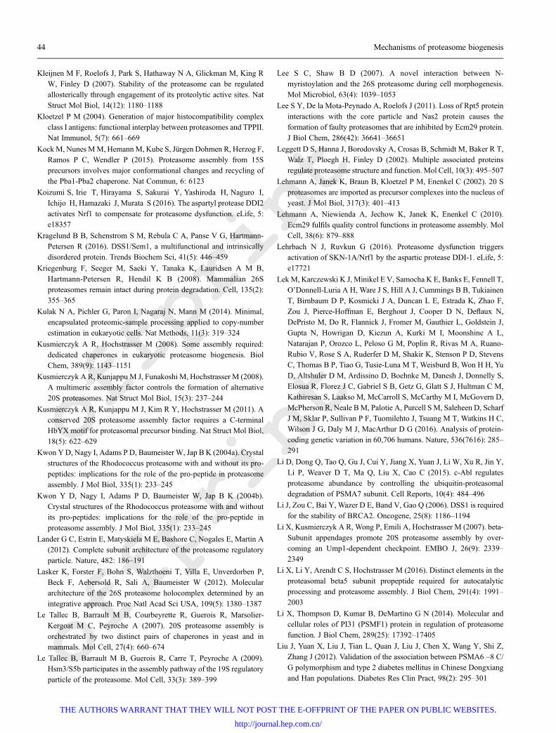
offprint
offprint
Kleijnen M F, Roelofs J, Park S, Hathaway N A, Glickman M, King RW, Finley D (2007). Stability of the proteasome can be regulatedallosterically through engagement of its proteolytic active sites. NatStruct Mol Biol, 14(12): 1180–1188
Kloetzel P M (2004). Generation of major histocompatibility complexclass I antigens: functional interplay between proteasomes and TPPII.Nat Immunol, 5(7): 661–669
KockM, Nunes MM, HemannM, Kube S, Jürgen Dohmen R, Herzog F,Ramos P C, Wendler P (2015). Proteasome assembly from 15Sprecursors involves major conformational changes and recycling ofthe Pba1-Pba2 chaperone. Nat Commun, 6: 6123
Koizumi S, Irie T, Hirayama S, Sakurai Y, Yashiroda H, Naguro I,Ichijo H, Hamazaki J, Murata S (2016). The aspartyl protease DDI2
activates Nrf1 to compensate for proteasome dysfunction. eLife, 5:e18357
Kragelund B B, Schenstrom S M, Rebula C A, Panse V G, Hartmann-Petersen R (2016). DSS1/Sem1, a multifunctional and intrinsicallydisordered protein. Trends Biochem Sci, 41(5): 446–459
Kriegenburg F, Seeger M, Saeki Y, Tanaka K, Lauridsen A M B,
Hartmann-Petersen R, Hendil K B (2008). Mammalian 26Sproteasomes remain intact during protein degradation. Cell, 135(2):355–365
Kulak N A, Pichler G, Paron I, Nagaraj N, Mann M (2014). Minimal,encapsulated proteomic-sample processing applied to copy-numberestimation in eukaryotic cells. Nat Methods, 11(3): 319–324
Kusmierczyk A R, Hochstrasser M (2008). Some assembly required:dedicated chaperones in eukaryotic proteasome biogenesis. BiolChem, 389(9): 1143–1151
Kusmierczyk A R, Kunjappu M J, Funakoshi M, Hochstrasser M (2008).A multimeric assembly factor controls the formation of alternative20S proteasomes. Nat Struct Mol Biol, 15(3): 237–244
Kusmierczyk A R, Kunjappu M J, Kim R Y, Hochstrasser M (2011). Aconserved 20S proteasome assembly factor requires a C-terminalHbYX motif for proteasomal precursor binding. Nat Struct Mol Biol,18(5): 622–629
Kwon YD, Nagy I, Adams P D, Baumeister W, Jap B K (2004a). Crystalstructures of the Rhodococcus proteasome with and without its pro-peptides: implications for the role of the pro-peptide in proteasomeassembly. J Mol Biol, 335(1): 233–245
Kwon Y D, Nagy I, Adams P D, Baumeister W, Jap B K (2004b).Crystal structures of the Rhodococcus proteasome with and withoutits pro-peptides: implications for the role of the pro-peptide inproteasome assembly. J Mol Biol, 335(1): 233–245
Lander G C, Estrin E, Matyskiela M E, Bashore C, Nogales E, Martin A(2012). Complete subunit architecture of the proteasome regulatoryparticle. Nature, 482: 186–191
Lasker K, Forster F, Bohn S, Walzthoeni T, Villa E, Unverdorben P,Beck F, Aebersold R, Sali A, Baumeister W (2012). Moleculararchitecture of the 26S proteasome holocomplex determined by anintegrative approach. Proc Natl Acad Sci USA, 109(5): 1380–1387
Le Tallec B, Barrault M B, Courbeyrette R, Guerois R, Marsolier-Kergoat M C, Peyroche A (2007). 20S proteasome assembly is
orchestrated by two distinct pairs of chaperones in yeast and inmammals. Mol Cell, 27(4): 660–674
Le Tallec B, Barrault M B, Guerois R, Carre T, Peyroche A (2009).Hsm3/S5b participates in the assembly pathway of the 19S regulatoryparticle of the proteasome. Mol Cell, 33(3): 389–399
Lee S C, Shaw B D (2007). A novel interaction between N-myristoylation and the 26S proteasome during cell morphogenesis.Mol Microbiol, 63(4): 1039–1053
Lee S Y, De la Mota-Peynado A, Roelofs J (2011). Loss of Rpt5 proteininteractions with the core particle and Nas2 protein causes theformation of faulty proteasomes that are inhibited by Ecm29 protein.J Biol Chem, 286(42): 36641–36651
Leggett D S, Hanna J, Borodovsky A, Crosas B, Schmidt M, Baker R T,Walz T, Ploegh H, Finley D (2002). Multiple associated proteinsregulate proteasome structure and function. Mol Cell, 10(3): 495–507
Lehmann A, Janek K, Braun B, Kloetzel P M, Enenkel C (2002). 20 Sproteasomes are imported as precursor complexes into the nucleus ofyeast. J Mol Biol, 317(3): 401–413
Lehmann A, Niewienda A, Jechow K, Janek K, Enenkel C (2010).Ecm29 fulfils quality control functions in proteasome assembly. MolCell, 38(6): 879–888
Lehrbach N J, Ruvkun G (2016). Proteasome dysfunction triggersactivation of SKN-1A/Nrf1 by the aspartic protease DDI-1. eLife, 5:e17721
LekM, Karczewski K J, Minikel E V, Samocha K E, Banks E, Fennell T,O’Donnell-Luria A H, Ware J S, Hill A J, Cummings B B, TukiainenT, Birnbaum D P, Kosmicki J A, Duncan L E, Estrada K, Zhao F,Zou J, Pierce-Hoffman E, Berghout J, Cooper D N, Deflaux N,DePristo M, Do R, Flannick J, Fromer M, Gauthier L, Goldstein J,Gupta N, Howrigan D, Kiezun A, Kurki M I, Moonshine A L,Natarajan P, Orozco L, Peloso G M, Poplin R, Rivas M A, Ruano-Rubio V, Rose S A, Ruderfer D M, Shakir K, Stenson P D, StevensC, Thomas B P, Tiao G, Tusie-Luna M T, Weisburd B, Won H H, Yu
D, Altshuler D M, Ardissino D, Boehnke M, Danesh J, Donnelly S,Elosua R, Florez J C, Gabriel S B, Getz G, Glatt S J, Hultman C M,Kathiresan S, Laakso M, McCarroll S, McCarthy M I, McGovern D,McPherson R, Neale BM, Palotie A, Purcell S M, Saleheen D, ScharfJ M, Sklar P, Sullivan P F, Tuomilehto J, Tsuang M T, Watkins H C,Wilson J G, Daly M J, MacArthur D G (2016). Analysis of protein-coding genetic variation in 60,706 humans. Nature, 536(7616): 285–
291
Li D, Dong Q, Tao Q, Gu J, Cui Y, Jiang X, Yuan J, Li W, Xu R, Jin Y,Li P, Weaver D T, Ma Q, Liu X, Cao C (2015). c-Abl regulatesproteasome abundance by controlling the ubiquitin-proteasomaldegradation of PSMA7 subunit. Cell Reports, 10(4): 484–496
Li J, Zou C, Bai Y, Wazer D E, Band V, Gao Q (2006). DSS1 is requiredfor the stability of BRCA2. Oncogene, 25(8): 1186–1194
Li X, Kusmierczyk A R, Wong P, Emili A, Hochstrasser M (2007). beta-Subunit appendages promote 20S proteasome assembly by over-coming an Ump1-dependent checkpoint. EMBO J, 26(9): 2339–2349
Li X, Li Y, Arendt C S, Hochstrasser M (2016). Distinct elements in theproteasomal beta5 subunit propeptide required for autocatalyticprocessing and proteasome assembly. J Biol Chem, 291(4): 1991–2003
Li X, Thompson D, Kumar B, DeMartino G N (2014). Molecular andcellular roles of PI31 (PSMF1) protein in regulation of proteasomefunction. J Biol Chem, 289(25): 17392–17405
Liu J, Yuan X, Liu J, Tian L, Quan J, Liu J, Chen X, Wang Y, Shi Z,Zhang J (2012). Validation of the association between PSMA6 -8 C/G polymorphism and type 2 diabetes mellitus in Chinese Dongxiangand Han populations. Diabetes Res Clin Pract, 98(2): 295–301
44 Mechanisms of proteasome biogenesis
THE AUTHORS WARRANT THAT THEY WILL NOT POST THE E-OFFPRINT OF THE PAPER ON PUBLIC WEBSITES.
http://journal.hep.com.cn/
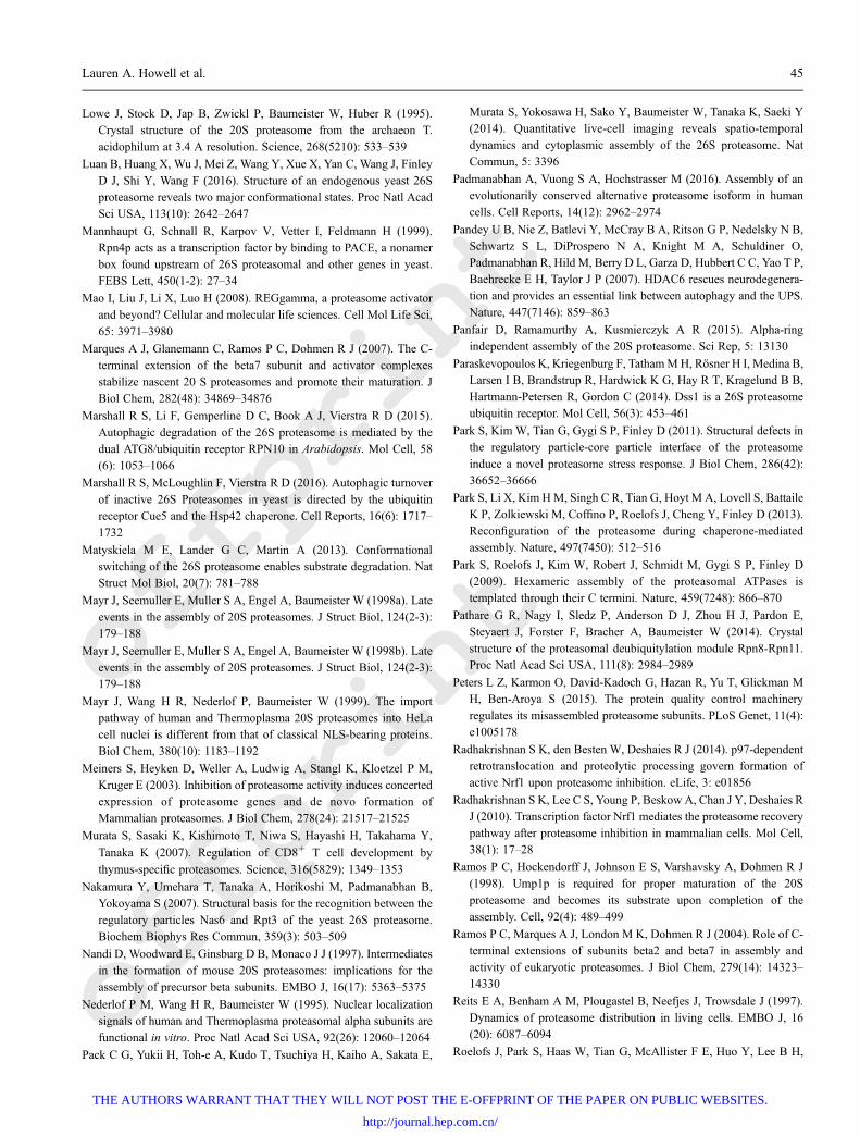
offprint
offprint
Lowe J, Stock D, Jap B, Zwickl P, Baumeister W, Huber R (1995).Crystal structure of the 20S proteasome from the archaeon T.acidophilum at 3.4 A resolution. Science, 268(5210): 533–539
Luan B, Huang X, Wu J, Mei Z, Wang Y, Xue X, Yan C, Wang J, FinleyD J, Shi Y, Wang F (2016). Structure of an endogenous yeast 26Sproteasome reveals two major conformational states. Proc Natl AcadSci USA, 113(10): 2642–2647
Mannhaupt G, Schnall R, Karpov V, Vetter I, Feldmann H (1999).Rpn4p acts as a transcription factor by binding to PACE, a nonamerbox found upstream of 26S proteasomal and other genes in yeast.FEBS Lett, 450(1-2): 27–34
Mao I, Liu J, Li X, Luo H (2008). REGgamma, a proteasome activatorand beyond? Cellular and molecular life sciences. Cell Mol Life Sci,
65: 3971–3980
Marques A J, Glanemann C, Ramos P C, Dohmen R J (2007). The C-terminal extension of the beta7 subunit and activator complexesstabilize nascent 20 S proteasomes and promote their maturation. JBiol Chem, 282(48): 34869–34876
Marshall R S, Li F, Gemperline D C, Book A J, Vierstra R D (2015).
Autophagic degradation of the 26S proteasome is mediated by thedual ATG8/ubiquitin receptor RPN10 in Arabidopsis. Mol Cell, 58(6): 1053–1066
Marshall R S, McLoughlin F, Vierstra R D (2016). Autophagic turnoverof inactive 26S Proteasomes in yeast is directed by the ubiquitinreceptor Cue5 and the Hsp42 chaperone. Cell Reports, 16(6): 1717–1732
Matyskiela M E, Lander G C, Martin A (2013). Conformationalswitching of the 26S proteasome enables substrate degradation. NatStruct Mol Biol, 20(7): 781–788
Mayr J, Seemuller E, Muller S A, Engel A, Baumeister W (1998a). Lateevents in the assembly of 20S proteasomes. J Struct Biol, 124(2-3):179–188
Mayr J, Seemuller E, Muller S A, Engel A, Baumeister W (1998b). Lateevents in the assembly of 20S proteasomes. J Struct Biol, 124(2-3):179–188
Mayr J, Wang H R, Nederlof P, Baumeister W (1999). The importpathway of human and Thermoplasma 20S proteasomes into HeLacell nuclei is different from that of classical NLS-bearing proteins.Biol Chem, 380(10): 1183–1192
Meiners S, Heyken D, Weller A, Ludwig A, Stangl K, Kloetzel P M,Kruger E (2003). Inhibition of proteasome activity induces concertedexpression of proteasome genes and de novo formation ofMammalian proteasomes. J Biol Chem, 278(24): 21517–21525
Murata S, Sasaki K, Kishimoto T, Niwa S, Hayashi H, Takahama Y,
Tanaka K (2007). Regulation of CD8+ T cell development by
thymus-specific proteasomes. Science, 316(5829): 1349–1353
Nakamura Y, Umehara T, Tanaka A, Horikoshi M, Padmanabhan B,Yokoyama S (2007). Structural basis for the recognition between theregulatory particles Nas6 and Rpt3 of the yeast 26S proteasome.Biochem Biophys Res Commun, 359(3): 503–509
Nandi D, Woodward E, Ginsburg D B, Monaco J J (1997). Intermediatesin the formation of mouse 20S proteasomes: implications for theassembly of precursor beta subunits. EMBO J, 16(17): 5363–5375
Nederlof P M, Wang H R, Baumeister W (1995). Nuclear localizationsignals of human and Thermoplasma proteasomal alpha subunits arefunctional in vitro. Proc Natl Acad Sci USA, 92(26): 12060–12064
Pack C G, Yukii H, Toh-e A, Kudo T, Tsuchiya H, Kaiho A, Sakata E,
Murata S, Yokosawa H, Sako Y, Baumeister W, Tanaka K, Saeki Y(2014). Quantitative live-cell imaging reveals spatio-temporaldynamics and cytoplasmic assembly of the 26S proteasome. NatCommun, 5: 3396
Padmanabhan A, Vuong S A, Hochstrasser M (2016). Assembly of anevolutionarily conserved alternative proteasome isoform in humancells. Cell Reports, 14(12): 2962–2974
Pandey U B, Nie Z, Batlevi Y, McCray B A, Ritson G P, Nedelsky N B,Schwartz S L, DiProspero N A, Knight M A, Schuldiner O,Padmanabhan R, Hild M, Berry D L, Garza D, Hubbert C C, Yao T P,Baehrecke E H, Taylor J P (2007). HDAC6 rescues neurodegenera-tion and provides an essential link between autophagy and the UPS.Nature, 447(7146): 859–863
Panfair D, Ramamurthy A, Kusmierczyk A R (2015). Alpha-ringindependent assembly of the 20S proteasome. Sci Rep, 5: 13130
Paraskevopoulos K, Kriegenburg F, TathamMH, Rösner H I, Medina B,Larsen I B, Brandstrup R, Hardwick K G, Hay R T, Kragelund B B,Hartmann-Petersen R, Gordon C (2014). Dss1 is a 26S proteasomeubiquitin receptor. Mol Cell, 56(3): 453–461
Park S, Kim W, Tian G, Gygi S P, Finley D (2011). Structural defects inthe regulatory particle-core particle interface of the proteasome
induce a novel proteasome stress response. J Biol Chem, 286(42):36652–36666
Park S, Li X, Kim HM, Singh C R, Tian G, Hoyt M A, Lovell S, BattaileK P, Zolkiewski M, Coffino P, Roelofs J, Cheng Y, Finley D (2013).Reconfiguration of the proteasome during chaperone-mediatedassembly. Nature, 497(7450): 512–516
Park S, Roelofs J, Kim W, Robert J, Schmidt M, Gygi S P, Finley D(2009). Hexameric assembly of the proteasomal ATPases istemplated through their C termini. Nature, 459(7248): 866–870
Pathare G R, Nagy I, Sledz P, Anderson D J, Zhou H J, Pardon E,Steyaert J, Forster F, Bracher A, Baumeister W (2014). Crystalstructure of the proteasomal deubiquitylation module Rpn8-Rpn11.Proc Natl Acad Sci USA, 111(8): 2984–2989
Peters L Z, Karmon O, David-Kadoch G, Hazan R, Yu T, Glickman MH, Ben-Aroya S (2015). The protein quality control machineryregulates its misassembled proteasome subunits. PLoS Genet, 11(4):e1005178
Radhakrishnan S K, den Besten W, Deshaies R J (2014). p97-dependentretrotranslocation and proteolytic processing govern formation ofactive Nrf1 upon proteasome inhibition. eLife, 3: e01856
Radhakrishnan S K, Lee C S, Young P, Beskow A, Chan J Y, Deshaies RJ (2010). Transcription factor Nrf1 mediates the proteasome recoverypathway after proteasome inhibition in mammalian cells. Mol Cell,38(1): 17–28
Ramos P C, Hockendorff J, Johnson E S, Varshavsky A, Dohmen R J(1998). Ump1p is required for proper maturation of the 20Sproteasome and becomes its substrate upon completion of the
assembly. Cell, 92(4): 489–499
Ramos P C, Marques A J, London M K, Dohmen R J (2004). Role of C-terminal extensions of subunits beta2 and beta7 in assembly andactivity of eukaryotic proteasomes. J Biol Chem, 279(14): 14323–14330
Reits E A, Benham A M, Plougastel B, Neefjes J, Trowsdale J (1997).
Dynamics of proteasome distribution in living cells. EMBO J, 16(20): 6087–6094
Roelofs J, Park S, Haas W, Tian G, McAllister F E, Huo Y, Lee B H,
Lauren A. Howell et al. 45
THE AUTHORS WARRANT THAT THEY WILL NOT POST THE E-OFFPRINT OF THE PAPER ON PUBLIC WEBSITES.
http://journal.hep.com.cn/
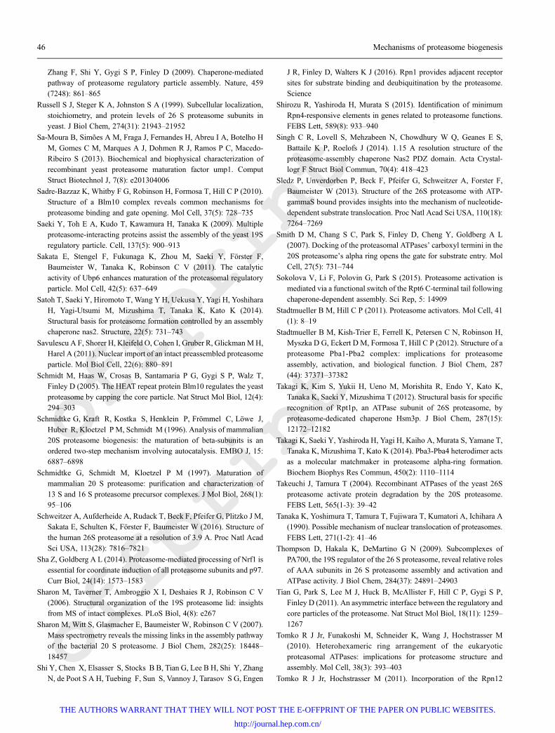
offprint
offprint
Zhang F, Shi Y, Gygi S P, Finley D (2009). Chaperone-mediatedpathway of proteasome regulatory particle assembly. Nature, 459(7248): 861–865
Russell S J, Steger K A, Johnston S A (1999). Subcellular localization,stoichiometry, and protein levels of 26 S proteasome subunits inyeast. J Biol Chem, 274(31): 21943–21952
Sa-Moura B, Simões A M, Fraga J, Fernandes H, Abreu I A, Botelho H
M, Gomes C M, Marques A J, Dohmen R J, Ramos P C, Macedo-Ribeiro S (2013). Biochemical and biophysical characterization ofrecombinant yeast proteasome maturation factor ump1. ComputStruct Biotechnol J, 7(8): e201304006
Sadre-Bazzaz K, Whitby F G, Robinson H, Formosa T, Hill C P (2010).Structure of a Blm10 complex reveals common mechanisms forproteasome binding and gate opening. Mol Cell, 37(5): 728–735
Saeki Y, Toh E A, Kudo T, Kawamura H, Tanaka K (2009). Multipleproteasome-interacting proteins assist the assembly of the yeast 19Sregulatory particle. Cell, 137(5): 900–913
Sakata E, Stengel F, Fukunaga K, Zhou M, Saeki Y, Förster F,Baumeister W, Tanaka K, Robinson C V (2011). The catalyticactivity of Ubp6 enhances maturation of the proteasomal regulatoryparticle. Mol Cell, 42(5): 637–649
Satoh T, Saeki Y, Hiromoto T, Wang Y H, Uekusa Y, Yagi H, YoshiharaH, Yagi-Utsumi M, Mizushima T, Tanaka K, Kato K (2014).Structural basis for proteasome formation controlled by an assemblychaperone nas2. Structure, 22(5): 731–743
Savulescu A F, Shorer H, Kleifeld O, Cohen I, Gruber R, GlickmanMH,Harel A (2011). Nuclear import of an intact preassembled proteasome
particle. Mol Biol Cell, 22(6): 880–891
Schmidt M, Haas W, Crosas B, Santamaria P G, Gygi S P, Walz T,Finley D (2005). The HEAT repeat protein Blm10 regulates the yeastproteasome by capping the core particle. Nat Struct Mol Biol, 12(4):294–303
Schmidtke G, Kraft R, Kostka S, Henklein P, Frömmel C, Löwe J,
Huber R, Kloetzel P M, Schmidt M (1996). Analysis of mammalian20S proteasome biogenesis: the maturation of beta-subunits is anordered two-step mechanism involving autocatalysis. EMBO J, 15:6887–6898
Schmidtke G, Schmidt M, Kloetzel P M (1997). Maturation ofmammalian 20 S proteasome: purification and characterization of13 S and 16 S proteasome precursor complexes. J Mol Biol, 268(1):95–106
Schweitzer A, Aufderheide A, Rudack T, Beck F, Pfeifer G, Plitzko J M,Sakata E, Schulten K, Förster F, Baumeister W (2016). Structure ofthe human 26S proteasome at a resolution of 3.9 A. Proc Natl AcadSci USA, 113(28): 7816–7821
Sha Z, Goldberg A L (2014). Proteasome-mediated processing of Nrf1 isessential for coordinate induction of all proteasome subunits and p97.Curr Biol, 24(14): 1573–1583
Sharon M, Taverner T, Ambroggio X I, Deshaies R J, Robinson C V(2006). Structural organization of the 19S proteasome lid: insightsfrom MS of intact complexes. PLoS Biol, 4(8): e267
Sharon M, Witt S, Glasmacher E, Baumeister W, Robinson C V (2007).Mass spectrometry reveals the missing links in the assembly pathwayof the bacterial 20 S proteasome. J Biol Chem, 282(25): 18448–
18457
Shi Y, Chen X, Elsasser S, Stocks B B, Tian G, Lee B H, Shi Y, ZhangN, de Poot S A H, Tuebing F, Sun S, Vannoy J, Tarasov S G, Engen
J R, Finley D, Walters K J (2016). Rpn1 provides adjacent receptorsites for substrate binding and deubiquitination by the proteasome.Science
Shirozu R, Yashiroda H, Murata S (2015). Identification of minimumRpn4-responsive elements in genes related to proteasome functions.FEBS Lett, 589(8): 933–940
Singh C R, Lovell S, Mehzabeen N, Chowdhury W Q, Geanes E S,
Battaile K P, Roelofs J (2014). 1.15 A resolution structure of theproteasome-assembly chaperone Nas2 PDZ domain. Acta Crystal-logr F Struct Biol Commun, 70(4): 418–423
Sledz P, Unverdorben P, Beck F, Pfeifer G, Schweitzer A, Forster F,Baumeister W (2013). Structure of the 26S proteasome with ATP-gammaS bound provides insights into the mechanism of nucleotide-dependent substrate translocation. Proc Natl Acad Sci USA, 110(18):7264–7269
Smith D M, Chang S C, Park S, Finley D, Cheng Y, Goldberg A L(2007). Docking of the proteasomal ATPases’ carboxyl termini in the20S proteasome’s alpha ring opens the gate for substrate entry. MolCell, 27(5): 731–744
Sokolova V, Li F, Polovin G, Park S (2015). Proteasome activation ismediated via a functional switch of the Rpt6 C-terminal tail following
chaperone-dependent assembly. Sci Rep, 5: 14909
Stadtmueller B M, Hill C P (2011). Proteasome activators. Mol Cell, 41(1): 8–19
Stadtmueller B M, Kish-Trier E, Ferrell K, Petersen C N, Robinson H,Myszka D G, Eckert D M, Formosa T, Hill C P (2012). Structure of aproteasome Pba1-Pba2 complex: implications for proteasome
assembly, activation, and biological function. J Biol Chem, 287
(44): 37371–37382
Takagi K, Kim S, Yukii H, Ueno M, Morishita R, Endo Y, Kato K,Tanaka K, Saeki Y, Mizushima T (2012). Structural basis for specificrecognition of Rpt1p, an ATPase subunit of 26S proteasome, byproteasome-dedicated chaperone Hsm3p. J Biol Chem, 287(15):12172–12182
Takagi K, Saeki Y, Yashiroda H, Yagi H, Kaiho A, Murata S, Yamane T,Tanaka K, Mizushima T, Kato K (2014). Pba3-Pba4 heterodimer actsas a molecular matchmaker in proteasome alpha-ring formation.Biochem Biophys Res Commun, 450(2): 1110–1114
Takeuchi J, Tamura T (2004). Recombinant ATPases of the yeast 26Sproteasome activate protein degradation by the 20S proteasome.FEBS Lett, 565(1-3): 39–42
Tanaka K, Yoshimura T, Tamura T, Fujiwara T, Kumatori A, Ichihara A(1990). Possible mechanism of nuclear translocation of proteasomes.FEBS Lett, 271(1-2): 41–46
Thompson D, Hakala K, DeMartino G N (2009). Subcomplexes ofPA700, the 19S regulator of the 26 S proteasome, reveal relative rolesof AAA subunits in 26 S proteasome assembly and activation andATPase activity. J Biol Chem, 284(37): 24891–24903
Tian G, Park S, Lee M J, Huck B, McAllister F, Hill C P, Gygi S P,Finley D (2011). An asymmetric interface between the regulatory andcore particles of the proteasome. Nat Struct Mol Biol, 18(11): 1259–1267
Tomko R J Jr, Funakoshi M, Schneider K, Wang J, Hochstrasser M(2010). Heterohexameric ring arrangement of the eukaryotic
proteasomal ATPases: implications for proteasome structure andassembly. Mol Cell, 38(3): 393–403
Tomko R J Jr, Hochstrasser M (2011). Incorporation of the Rpn12
46 Mechanisms of proteasome biogenesis
THE AUTHORS WARRANT THAT THEY WILL NOT POST THE E-OFFPRINT OF THE PAPER ON PUBLIC WEBSITES.
http://journal.hep.com.cn/
offprint
offprint
subunit couples completion of proteasome regulatory particle lidassembly to lid-base joining. Mol Cell, 44(6): 907–917
Tomko R J Jr, Hochstrasser M (2013). Molecular architecture andassembly of the eukaryotic proteasome. Annu Rev Biochem, 82(1):415–445
Tomko R J Jr, Hochstrasser M (2014). The intrinsically disordered Sem1protein functions as a molecular tether during proteasome lid
biogenesis. Mol Cell, 53(3): 433–443
Tomko R J Jr, Taylor D W, Chen Z A, Wang H W, Rappsilber J,Hochstrasser M (2015). A Single alpha helix drives extensiveremodeling of the proteasome lid and completion of regulatoryparticle assembly. Cell, 163(2): 432–444
Uekusa Y, Okawa K, Yagi-Utsumi M, Serve O, Nakagawa Y,
Mizushima T, Yagi H, Saeki Y, Tanaka K, Kato K (2014). Backbone(1)H, (1)(3)C and (1)(5)N assignments of yeast Ump1, anintrinsically disordered protein that functions as a proteasomeassembly chaperone. Biomol NMR Assign, 8(2): 383–386
Unno M, Mizushima T, Morimoto Y, Tomisugi Y, Tanaka K, YasuokaN, Tsukihara T (2002). The structure of the mammalian 20Sproteasome at 2.75 A resolution. Structure, 10(5): 609–618
Unverdorben P, Beck F, led P, Schweitzer A, Pfeifer G, Plitzko J M,Baumeister W, Forster F (2014). Deep classification of a large cryo-EM dataset defines the conformational landscape of the 26Sproteasome. Proc Natl Acad Sci USA, 111(15): 5544–5549
Ustrell V, Hoffman L, Pratt G, Rechsteiner M (2002). PA200, a nuclearproteasome activator involved in DNA repair. EMBO J, 21(13):3516–3525
Velichutina I, Connerly P L, Arendt C S, Li X, Hochstrasser M (2004).Plasticity in eucaryotic 20S proteasome ring assembly revealed by asubunit deletion in yeast. EMBO J, 23(3): 500–510
Verma R, et al (2002). Role of Rpn11 metalloprotease in deubiquitina-tion and degradation by the 26S proteasome. Science, 298(5593):611–615
Volker C, Lupas A N (2002). Molecular evolution of proteasomes. CurrTop Microbiol Immunol, 268: 1–22
Waite K A, De-La Mota-Peynado A, Vontz G, Roelofs J (2016).Starvation induces proteasome autophagy with different pathways forcore and regulatory particles. J Biol Chem, 291(7): 3239–3253
Wang H R, Kania M, Baumeister W, Nederlof P M (1997). Import ofhuman and Thermoplasma 20S proteasomes into nuclei of HeLa cellsrequires functional NLS sequences. Eur J Cell Biol, 73: 105–113
WangW, Chan J Y (2006). Nrf1 is targeted to the endoplasmic reticulummembrane by an N-terminal transmembrane domain. Inhibition ofnuclear translocation and transacting function. J Biol Chem, 281(28):19676–19687
Wani P S, Rowland M A, Ondracek A, Deeds E J, Roelofs J (2015).Maturation of the proteasome core particle induces an affinity switchthat controls regulatory particle association. Nat Commun, 6: 6384
Wani P S, Suppahia A, Capalla X, Ondracek A, Roelofs J (2016).Phosphorylation of the C-terminal tail of proteasome subunit alpha7is required for binding of the proteasome quality control factorEcm29. Sci Rep, 6: 27873
Weberruss M H, Savulescu A F, Jando J, Bissinger T, Harel A, GlickmanM H, Enenkel C (2013). Blm10 facilitates nuclear import ofproteasome core particles. EMBO J, 32(20): 2697–2707
Wei S J, Williams J G, Dang H, Darden T A, Betz B L, Humble M M,Chang F M, Trempus C S, Johnson K, Cannon R E, Tennant R W
(2008). Identification of a specific motif of the DSS1 protein requiredfor proteasome interaction and p53 protein degradation. J Mol Biol,383(3): 693–712
Welk V, Coux O, Kleene V, Abeza C, Trümbach D, Eickelberg O,Meiners S (2016). Inhibition of proteasome activity inducesformation of alternative proteasome complexes. J Biol Chem, 291(25): 13147–13159
Wendler P, Lehmann A, Janek K, Baumgart S, Enenkel C (2004). Thebipartite nuclear localization sequence of Rpn2 is required for nuclearimport of proteasomal base complexes via karyopherin alphabeta andproteasome functions. J Biol Chem, 279(36): 37751–37762
Whitby F G, Masters E I, Kramer L, Knowlton J R, Yao Y, Wang C C,Hill C P (2000). Structural basis for the activation of 20Sproteasomes by 11S regulators. Nature, 408(6808): 115–120
Witt E, Zantopf D, Schmidt M, Kraft R, Kloetzel P M, Kruger E (2000).Characterisation of the newly identified human Ump1 homologuePOMP and analysis of LMP7(beta 5i) incorporation into 20 Sproteasomes. J Mol Biol, 301(1): 1–9
Wollenberg K, Swaffield J C (2001). Evolution of proteasomal ATPases.Mol Biol Evol, 18(6): 962–974
Worden E J, Padovani C, Martin A (2014). Structure of the Rpn11-Rpn8dimer reveals mechanisms of substrate deubiquitination duringproteasomal degradation. Nat Struct Mol Biol, 21(3): 220–227
Xie Y, Varshavsky A (2001). RPN4 is a ligand, substrate, andtranscriptional regulator of the 26S proteasome: a negative feedbackcircuit. Proc Natl Acad Sci USA, 98(6): 3056–3061
Yao T, Cohen R E (2002). A cryptic protease couples deubiquitinationand degradation by the proteasome. Nature, 419(6905): 403–407
Yao Y, Toth C R, Huang L, Wong M L, Dias P, Burlingame A L,Coffino P, Wang C C (1999). alpha5 subunit in Trypanosoma bruceiproteasome can self-assemble to form a cylinder of four stackedheptamer rings. Biochem J, 344(Pt 2): 349–358
Yashiroda H, Mizushima T, Okamoto K, Kameyama T, Hayashi H,Kishimoto T, Niwa S, Kasahara M, Kurimoto E, Sakata E, Takagi K,Suzuki A, Hirano Y, Murata S, Kato K, Yamane T, Tanaka K (2008).Crystal structure of a chaperone complex that contributes to theassembly of yeast 20S proteasomes. Nat Struct Mol Biol, 15(3): 228–236
Yashiroda H, Toda Y, Otsu S, Takagi K, Mizushima T, Murata S (2015).N-terminal alpha7 deletion of the proteasome 20S core particle
substitutes for yeast PI31 function. Mol Cell Biol, 35(1): 141–152
Yu Y, Smith D M, Kim H M, Rodriguez V, Goldberg A L, Cheng Y(2010). Interactions of PAN’s C-termini with archaeal 20S protea-some and implications for the eukaryotic proteasome-ATPaseinteractions. EMBO J, 29(3): 692–702
Yu Z, Livnat-Levanon N, Kleifeld O, Mansour W, Nakasone M A,
Castaneda C A, Dixon E K, Fushman D, Reis N, Pick E, Glickman MH (2015). Base-CP proteasome can serve as a platform for stepwiselid formation. Biosci Rep, 35(3): e00194
Zaiss D M, Standera S, Kloetzel P M, Sijts A J (2002). PI31 is amodulator of proteasome formation and antigen processing. ProcNatl Acad Sci USA, 99(22): 14344–14349
Zhang F, Hu M, Tian G, Zhang P, Finley D, Jeffrey P D, Shi Y (2009).Structural insights into the regulatory particle of the proteasome fromMethanocaldococcus jannaschii. Mol Cell, 34(4): 473–484
Zhang Y, Lucocq J M, Yamamoto M, Hayes J D (2007). The NHB1 (N-terminal homology box 1) sequence in transcription factor Nrf1 is
Lauren A. Howell et al. 47
THE AUTHORS WARRANT THAT THEY WILL NOT POST THE E-OFFPRINT OF THE PAPER ON PUBLIC WEBSITES.
http://journal.hep.com.cn/
offprint
offprint
required to anchor it to the endoplasmic reticulum and also to enableits asparagine-glycosylation. Biochem J, 408(2): 161–172
Zhu K, Dunner K Jr, McConkey D J (2010). Proteasome inhibitorsactivate autophagy as a cytoprotective response in human prostatecancer cells. Oncogene, 29(3): 451–462
Zuhl F, Seemuller E, Golbik R, Baumeister W (1997). Dissecting theassembly pathway of the 20S proteasome. FEBS Lett, 418(1-2): 189–
194
Zwickl P, Kleinz J, Baumeister W (1994). Critical elements inproteasome assembly. Nat Struct Biol, 1(11): 765–770
Zwickl P, Ng D, Woo K M, Klenk H P, Goldberg A L (1999). Anarchaebacterial ATPase, homologous to ATPases in the eukaryotic26S proteasome, activates protein breakdown by 20 S proteasomes. JBiol Chem, 274(37): 26008–26014
48 Mechanisms of proteasome biogenesis
THE AUTHORS WARRANT THAT THEY WILL NOT POST THE E-OFFPRINT OF THE PAPER ON PUBLIC WEBSITES.
http://journal.hep.com.cn/