Embed Size (px)
Citation preview

Quantified abundance of magnetofossils at thePaleocene–Eocene boundary from synchrotron-basedtransmission X-ray microscopyHuapei Wanga, Jun Wangb, Yu-chen Karen Chen-Wiegartb, and Dennis V. Kentc,d,1
aEarth, Atmospheric and Planetary Sciences, Massachusetts Institute of Technology, Cambridge, MA 02139; bPhoton Sciences Directorate, BrookhavenNational Laboratory, Upton, NY 11973; cEarth and Planetary Sciences, Rutgers University, Piscataway, NJ 08854; and dLamont–Doherty Earth Observatory,Columbia University, Palisades, NY 10964
Contributed by Dennis V. Kent, September 2, 2015 (sent for review June 19, 2015; reviewed by Michael Winklhofer)
The Paleocene–Eocene boundary (∼55.8 million years ago) ismarked by an abrupt negative carbon isotope excursion (CIE)that coincides with an oxygen isotope decrease interpreted asthe Paleocene–Eocene thermal maximum. Biogenic magnetite (Fe3O4)in the form of giant (micron-sized) spearhead-like and spindle-likemagnetofossils, as well as nano-sized magnetotactic bacteria mag-netosome chains, have been reported in clay-rich sediments inthe New Jersey Atlantic Coastal Plain and were thought to accountfor the distinctive single-domain magnetic properties of these sed-iments. Uncalibrated strong field magnet extraction techniqueshave been typically used to provide material for scanning andtransmission electron microscopic imaging of these magnetic par-ticles, whose concentration in the natural sediment is thus difficultto quantify. In this study, we use a recently developed ultrahigh-resolution, synchrotron-based, full-field transmission X-ray micro-scope to study the iron-rich minerals within the clay sediment intheir bulk state. We are able to estimate the total magnetizationconcentration of the giant biogenic magnetofossils to be only∼10% of whole sediment. Along with previous rock magneticstudies on the CIE clay, we suggest that most of the magnetitein the clay occurs as isolated, near-equidimensional nanoparticles,a suggestion that points to a nonbiogenic origin, such as cometimpact plume condensates in what may be very rapidly depositedCIE clays.
Paleocene–Eocene thermal maximum | New Jersey Atlantic Coastal Plain |impact plume condensate | Marlboro Clay | Ocean Drilling ProgramLeg 174AX
The Paleocene–Eocene boundary is marked by an abruptglobal negative carbon isotope excursion (CIE) in both marine
and continental carbon reservoirs (1, 2) that coincides withan oxygen isotope decrease that is interpreted as a rapidglobal warming event at ∼55.8 million years ago, the Paleocene–Eocene thermal maximum (PETM) (3). The thick zone ofanomalously high magnetization coincident with the CIE atthe base of the Manasquan Formation (now known as theMarlboro Clay) was initially discovered in a cored section atAncora (Ocean Drilling Program Leg 174AX) on the AtlanticCoastal Plain of New Jersey (4). Magnetic hysteresis mea-surements on the bulk sediment indicated that the anomaloushigh magnetization corresponds to an increased abundance offine-grained magnetite with single-domain (SD)-like magneticproperties. Two other drill cores (Clayton and Bass River)show a similar association of high concentration of SD mag-netite in a kaolinite-rich interval with minimum carbon isotopevalues and, with the Ancora site, form a transect across theNew Jersey Atlantic Coastal Plain (5). Transmission electronmicroscopic (TEM) imaging on a redeposited thin layer of bulkclay from the CIE interval in the Clayton site resulted infinding only a handful of isolated nanoscale (∼50 to ∼70 nm)magnetite grains (5) because of the grains’ low concentration,with distances between grains estimated to be ∼20 times larger
than their lengths in three-dimensional (3D) space (6). Thisunique magnetite nanoparticle-rich interval associated withthe CIE on the New Jersey Atlantic Coastal Plain was sug-gested to have originated from impact plume condensates (5)in what now appears may have been very rapidly deposited(∼1–3 cm/y) clays (7), providing circumstantial evidence for amajor cometary impact at the onset of the CIE, which likely trig-gered the PETM (5, 8).Subsequent studies have confirmed the anomalously high
concentrations of SD-like material from the CIE clay from theAtlantic Coastal Plain, making these CIE sections the thickestdominated by SD magnetite recognized thus far in the strati-graphic record (9, 10). In these follow-up studies, scanningelectron microscopic (SEM) and TEM observations on mag-netically extracted materials revealed the presence of chains ofmagnetite crystals that strongly resembled magnetosomes ofmagnetotactic bacteria (MTB); this finding led to the conclusionthat the SD-like magnetic properties of the CIE clays werepredominantly of biogenic origin. However, quantitative analy-sis of the magnetic extraction procedure suggested that theextracted material accounts for only ∼5% of the total magneti-zation (6). To further complicate the problem, SEM and TEMstudies on the magnetic extracts also found giant micron-sized magnetofossils, including spearhead-like and spindle-like ones, inside the CIE clay across the New Jersey AtlanticCoastal Plain (11–13), and these magnetofossils were foundto have SD-like magnetic properties, by electron holography,attributable to their distinct shapes (13). Using mainly SEM
Significance
The Paleocene–Eocene thermal maximum (PETM) is an abruptglobal warming event that occurred at about 55.8 Ma and isclosely linked to a large carbon isotope excursion. What causedthe PETM is unresolved. An unusual abundance of single-domainmagnetite particles in PETM sediments on the Atlantic CoastalPlain might represent condensates from a comet impact. Alter-natively, the magnetic nanoparticles may be of biogenic origin.We are now able to quantify the concentration of those magneticgrains that are distinctly of biogenic origin using synchrotron-based transmission X-ray microscopy. These and related findingsallow us to exclude magnetofossils as a significant source ofmagnetization of the PETM sediments and point to an impactcondensate origin of the magnetite particles.
Author contributions: H.W. and D.V.K. designed research; H.W., J.W., and Y.-c.K.C.-W.performed research; H.W. and D.V.K. analyzed data; and H.W., J.W., and D.V.K. wrotethe paper.
Reviewers included: M.W., Ludwig Maximilians University of Munich.
The authors declare no conflict of interest.1To whom correspondence should be addressed. Email: [email protected].
This article contains supporting information online at www.pnas.org/lookup/suppl/doi:10.1073/pnas.1517475112/-/DCSupplemental.
12598–12603 | PNAS | October 13, 2015 | vol. 112 | no. 41 www.pnas.org/cgi/doi/10.1073/pnas.1517475112

or TEM with their limited fields of view (∼micron) andshallow penetration depth (∼100 nm), the absolute concen-tration of these biogenic magnetites could not be quantified,
and thus the anomalously high magnetization of the CIE claycould not be ascribed to either mainly biogenic or nonbiogenicorigins.
Fig. 1. (A) TXM mosaic scan of the front view of a submillimeter bulk CIE clay dust AN560.1-X1 in fast mode. (B) Slow mosaic mode scan of the boxed area inA. (C) Zoomed-in view of the boxed area in B.
Wang et al. PNAS | October 13, 2015 | vol. 112 | no. 41 | 12599
EART
H,ATM
OSP
HERIC,
ANDPL
ANET
ARY
SCIENCE
S

ResultsIn this study, we used a recently developed full-field hard X-rayultrahigh-resolution (∼30–50 nm without binning pixels) syn-chrotron-based transmission X-ray microscope (TXM) (14) witha field of view of 20 μm × 20 μm to study magnetite and otheriron-rich minerals in bulk CIE clay (AN560.1 from 165.5 mdepth of the Ancora core) from just above the onset of the CIEand the coincident high-magnetization layer (5, 6). In the frontand side views of the TXM image of a submillimeter bulk CIEclay dust (AN560.1-X1) mounted on a tungsten carbide (WC)pin (Fig. 1 and Fig. S1), we can identify a variety of detrital ironminerals, as well as pyrite framboids of ∼10-μm sizes and theirclusters, a large ∼5-μm nonbiogenic magnetite with its distinctoctahedral crystal shape (15), a large ∼10-μm spindle-like crystalthat is likely to be an extraordinarily large giant magnetite mag-netofossil, and also a cluster of spindle-like crystals of ∼5–10 μm insize, which likely captures the general configuration of thecreature that produced these giant magnetite crystals. Mosaicscans of four extra bulk clay dusts from AN560.1 (Fig. S2)have similar iron minerals as shown in AN560.1-X1. For several
20-μm-size cylindrical volumes that contain iron minerals withinthe bulk clay, we also conducted nanotomography to study themicrostructure of the particles in three dimensions (Fig. S3 andMovies S1–S7), which helped us to further characterize the ironminerals using their crystal shapes.We also scanned the magnetically extracted materials from the
CIE clay sample (AN560.1) obtained in a previous study (6),which showed an intense increase of the concentration of iron-rich minerals (Fig. S4), whereas the TXM scan of the magneti-cally extracted residue of the CIE clay showed a certain decreaseof iron mineral concentration (Fig. S5). To test the capability ofthe TXM to identify magnetite magnetofossils of ∼1–2 μm orless, we conducted mosaic scans on a redeposited thin layer ofthe magnetically extracted materials (Fig. 2A). In this 100 μm ×100 μm area, we can identify many ∼1–2-μm-size giant magneto-fossils (e.g., Fig. 2 B–F) and estimate that there are many tens ofspearhead-like and spindle-like biogenic magnetites. This is muchmore than that we can clearly identify from the TXM scans of thebulk submillimeter sized CIE clay dusts because the ∼100- to
Fig. 2. (A) TXM mosaic scan of a thin layer of redeposited magnetically extracted materials from CIE clay sample AN560.1. (B–F) Examples of clearlyidentifiable ∼1–2-μm-sized giant magnetofossils.
12600 | www.pnas.org/cgi/doi/10.1073/pnas.1517475112 Wang et al.

∼200-μm thicknesses of the bulk clay dusts significantly reducethe contrast and clarity of the X-ray images.Although the spatial resolution of the TXM is ∼30–50 nm
without binning pixels for a 20-μm × 20-μm field of view (14),the X-ray absorption from an individual ∼50- to ∼100-nm-sizednanoparticle is still too weak to produce a clear contrast to showthe magnetosome chains produced by MTB or nonbiogenic iso-lated magnetite nanoparticles (Fig. 2). For the same reason, themagnetosomes in samples of a freeze-dried culture of MTB strainMV-1 (6, 16) are also not clearly observed by TXM (Fig. S6).To clearly identify the giant magnetofossils and better estimate
their concentration within the CIE clay, we redeposited knownamount of the bulk CIE clay in a thin layer with an estimatedaverage thickness of 4 μm on a 50-nm-thick silicon nitride (SN)window for ultrahigh-resolution TXM scans. After scanning atotal area of 0.55 mm2 by taking 1,370 TXM images, we identified∼50 giant magnetofossils, including spearhead-like and spindle-like ones (Fig. 3). Our X-ray images also provide opportunities tocharacterize their morphology. Fig. 4 shows the size and shapedistribution of the giant magnetofossils identified by TXM in thisstudy, as well as the magnetofossils (including MTB magneto-somes) identified by SEM and TEM in previous studies (9, 13).Because of the weak X-ray absorption from our sample and thelimitation of spatial resolution of the TXM, we do not confi-dently observe the elongated prismatic magnetites or the MTB
magnetosomes. Nevertheless, our TXM identified that spear-head-like and spindle-like giant magnetofossils inside the bulkclay generally have similar sizes and shapes as the ones pre-viously identified by SEM or TEM. However, the magnetitesobserved by SEM or TEM are from magnetic extracts of theclay, which account for only 5% of its total magnetization (6),and thus not representing the entire magnetic assemblage of thebulk clay. This finding may be the reason that we find slightlylarger scatter of the size and shape distribution of the giantmagnetofossils in the bulk clay from our TXM study.By knowing the exact amount of the bulk CIE clay that we
scanned (2.2 × 10⁻³ mm3 or 3.5 μg), we are able to calculate thesaturation magnetization (Ms) contribution of the giant magneto-fossils that we observed [based on magnetite Ms = 92 Am2/kg (17)],which turns out to be 1.6 × 10⁻³ Am2/kg or only about 10% ofthe Ms of the bulk CIE clay [1.5 × 10⁻² Am2/kg (5)].
DiscussionOur quantitative results show that the spearhead-like andspindle-like giant magnetofossils that are clearly identified inour TXM scans constitute only ∼10% of the magnetic particles(by weight or volume) of the entire magnetization assemblageof the CIE clay. This would leave ∼90% of the magnetizationassemblage to be elongated prismatic magnetite crystals, mag-netosome chains produced by MTB, and isolated nonbiogenic
Fig. 3. TXM images of identifiable giant magnetofossils. (A–C) Spearhead-like magnetofossils. (D–F) Spearhead-like magnetofossils with buds. (G–J) Bullet-shaped spearhead-like magnetofossils. (K–M) Spindle-like magnetofossils.
Wang et al. PNAS | October 13, 2015 | vol. 112 | no. 41 | 12601
EART
H,ATM
OSP
HERIC,
ANDPL
ANET
ARY
SCIENCE
S

magnetite nanoparticles. However, previous rock magneticstudies, including thermal fluctuation tomography (shades in Fig. 4),first-order reversal curves, and ferromagnetic resonance spec-tra on the bulk CIE clay strongly suggest that most of the SDmagnetite grains are isolated near-equant nanoparticles (6),rather than elongated prismatic crystals or magnetosome chainswith significant shape anisotropy as hallmarks of biogenic ori-gin. This is consistent with the only published TEM study on abulk CIE clay sample (redeposited to a thin layer of ∼100-nmthickness), which found only a few isolated ∼50–70-nm magne-tite grains but no shapes or chains ascribable to biogenic (5),because biogenic magnetite nanoparticles are known to be verydifficult to disaggregate from their original chains (18).In conclusion, along with previous rock magnetic and TEM
studies, our quantitative constraints using ultrahigh-resolutionsynchrotron-based full-field X-ray microscopy suggest that theanomalously high SD magnetization of bulk CIE clay is domi-nated by isolated near-equidimensional magnetite nanoparticles,which are likely formed as vapor condensates inside an impactplume. Similar nanophase iron particles have been produced bypulse-laser irradiation of San Carlos Olivine with ∼9 wt% wustite(FeO) in the laboratory to simulate space weathering vapor rede-position by micrometeorite impacts (19). Iron-rich nanoparticles
have also been detected with Mössbauer spectroscopy at severalCretaceous–Paleogene (K-Pg) boundary sites and are ascribed tocondensates from an impact ejecta plume of the K-Pg asteroid(20). Our analogous results for the nonbiogenic SD magnetites inthe CIE clay support the comet impact hypothesis as the triggerof the global environmental changes across the Paleocene–Eocene boundary.
MethodsThe TXM used in this study is located at beamline X8C at the NationalSynchrotron Light Source (NSLS) at Brookhaven National Laboratory (BNL).We used the high-resolution imagingmodewith a field-of-view of 20 μm × 20μm. Large mosaic TXM images are stitched from multiple field-of-view im-ages. To study the iron minerals within the clay, we used X-ray energy of 7.2keV, just above the iron absorption K-edge (7.112 keV) (21), to allow max-imum X-ray absorption. We used epoxy (almost X-ray–transparent) to securethe submillimeter bulk clay dusts on WC pins with tips of ∼50 μm. For eachsample, its WC pin was mounted on a kinematic sample holder to be placedon a stage with motion of x, y, and z translations and rotation.
We first scanned submillimeter-sized CIE bulk clay dusts using a fast mosaicmode with 8 × 8 binning pixels (each pixel size is 10 nm) to cover the entireclay dust (Fig. 1A). Then, we used a slower but higher-resolution mosaicmode with 2 × 2 binning pixels to scan a smaller area of interest (Fig. 1B). For3D tomographic studies, we took 721–1,081 images for each tomographywith 20-μm target area in high-resolution mode (∼30–50 nm) from anglesbetween 0° and 180°.
For the magnetically extracted materials from the CIE clay and the extractresidues, we mounted them in clusters on steel pins for overall mosaic scans(Figs. S4 and S5). We then redeposited a thin layer of magnetically extractedmaterials on a Kapton tape (almost X-ray–transparent) by using an alcoholsolution and mounted the tape on the sample stage perpendicular to theX-ray beam using a steel plate with openings and clips for further mosaicscans in a high-resolution mode with 2 × 2 binning pixels (Fig. 2).
For the freeze-dried MTB strain MV-1, we redeposited the samples in analcohol solution on an SN window of 50-nm thickness (nearly X-ray–trans-parent) to allow minimum and homogeneous background X-ray absorptionfrom the window, which introduced minimum background-imaging noise tothe sample. We mounted the SN window perpendicular to the X-ray beamand used the ultrahigh-resolution mode without binning pixels for a reso-lution of one pixel size of 10 nm (Fig. S6). Only ∼micron-sized particles areidentified, which may be derived from the ferric quinate used in the cultureor hydrous ferric oxides that are precursors to magnetite precipitation (16).
For the redeposited bulk CIE clay, we suspended 60 mg of clay in 9 g (or∼11.5 mL) of pure isopropyl alcohol. After shaking them well, we depositeda drop of the clay suspension (∼0.03 mL) on a 5-mm × 5-mm SN window.Based on the estimated clay density of 1.6 g/cm3, we calculated that theredeposited and naturally dried clay layer has an average thickness of 4 μm.We took 1,370 X-ray images in the ultrahigh-resolution mode of single-pixelresolution and 20-μm × 20-μm field of view, which added up to a totalscanned area of 0.55 mm2 (volume of 2.2 × 10⁻³ mm3 or weight of 3.5 μg ofbulk clay).
ACKNOWLEDGMENTS. We thank Bruce M. Moskowitz (Institute for RockMagnetism, University of Minnesota) for the MV-1 sample and Joseph L.Kirschvink (California Institute of Technology) for discussions on giant magne-tofossils and MTB. We also thank Michael Winklhofer (Ludwig MaximiliansUniversity of Munich) and one anonymous reviewer for constructive com-ments. Use of the NSLS at BNL is supported by the United States Depart-ment of Energy, Basic Energy Sciences program, under Contract DE-AC02-98CH10886. Additional funding for this work was provided by the RutgersBoard of Governors Professor Research Fund. This is Contribution 7932 fromthe Lamont–Doherty Earth Observatory.
1. Kennett JP, Stott LD (1991) Abrupt deep-sea warming, palaeoceanographic changesand benthic extinctions at the end of the Palaeocene. Nature 353(6341):225–229.
2. Koch PL, Zachos JC, Gingerich PD (1992) Correlation between isotope records inmarine and continental carbon reservoirs near the Paleocene Eocene boundary.Nature 358(6384):319–322.
3. Zachos JC, Lohmann KC, Walker JCG, Wise SW (1993) Abrupt climate change andtransient climates during the Paleogene: A marine perspective. J Geol 101(2):191–213.
4. Lanci L, Kent DV, Miller KG (2002) Detection of Late Cretaceous and Cenozoic se-quence boundaries on the Atlantic coastal plain using core log integration of mag-netic susceptibility and natural gamma ray measurements at Ancora, New Jersey.J Geophys Res 107(B10):2216.
5. Kent DV, et al. (2003) A case for a comet impact trigger for the Paleocene/Eocenethermal maximum and carbon isotope excursion. Earth Planet Sci Lett 211(1-2):13–26.
6. Wang H, Kent DV, Jackson MJ (2013) Evidence for abundant isolated magnetic nano-particles at the Paleocene-Eocene boundary. Proc Natl Acad Sci USA 110(2):425–430.
7. Wright JD, Schaller MF (2013) Evidence for a rapid release of carbon at the Paleocene-Eocene thermal maximum. Proc Natl Acad Sci USA 110(40):15908–15913.
8. Cramer BS, Kent DV (2005) Bolide summer: The Paleocene/Eocene thermal maximumas a response to an extraterrestrial trigger. Palaeogeogr Palaeocl 224(1-3):144–166.
9. Kopp RE, et al. (2007) Magnetofossil spike during the Paleocene-Eocene thermalmaximum: Ferromagnetic resonance, rock magnetic, and electron microscopy evi-dence from Ancora, New Jersey, United States. Paleoceanography 22(4):PA4103.
Fig. 4. Size-shape diagram of the giant magnetofossils and MTB magne-tosomes. Solid and empty symbols represent data from this TXM study andprevious SEM and TEM studies (9, 13), respectively. Triangles, rhombuses,and squares are spearhead-like, spindle-like, and elongated prismatic giantmagnetofossils, respectively. Diamonds are magnetosomes produced byMTB. Lines define the size–shape ranges of superparamagnetic (SP), SD, andmultidomain (MD) states for magnetite. Gray shades show the size–shapedistribution of magnetites within the bulk CIE clay determined by thermalfluctuation tomography, with star indicating the peak concentration (6).
12602 | www.pnas.org/cgi/doi/10.1073/pnas.1517475112 Wang et al.

10. Lippert PC, Zachos JC (2007) A biogenic origin for anomalous fine-grained mag-netic material at the Paleocene-Eocene boundary at Wilson Lake, New Jersey.Paleoceanography 22(4):PA4104.
11. Chang L, et al. (2012) Giant magnetofossils and hyperthermal events. Earth Planet SciLett 351:258–269.
12. Kopp RE, et al. (2009) An Appalachian Amazon? Magnetofossil evidence for thedevelopment of a tropical river-like system in the mid-Atlantic United States duringthe Paleocene-Eocene thermal maximum. Paleoceanography 24(4):PA4211.
13. Schumann D, et al. (2008) Gigantism in unique biogenic magnetite at the Paleocene-Eocene Thermal Maximum. Proc Natl Acad Sci USA 105(46):17648–17653.
14. Wang J, et al. (2012) Automated markerless full field hard x-ray microscopic to-mography at sub-50 nm 3-dimension spatial resolution. Appl Phys Lett 100:143107.
15. Witt A, Fabian K, Bleil U (2005) Three-dimensional micromagnetic calculations fornaturally shaped magnetite: Octahedra and magnetosomes. Earth Planet Sci Lett233(3-4):311–324.
16. Bazylinski DA, Frankel RB, Jannasch HW (1988) Anaerobic magnetite production by amarine, magnetotactic bacterium. Nature 334(6182):518–519.
17. Dunlop DJ, Özdemir Ö (2001) Rock Magnetism: Fundamentals and Frontiers (Cam-bridge Univ Press, Cambridge, UK).
18. Mann S, Sparks NHC, Walker MM, Kirschvink JL (1988) Ultrastructure, morphologyand organization of biogenic magnetite from sockeye salmon, Oncorhynchus nerka:Implications for magnetoreception. J Exp Biol 140:35–49.
19. Sasaki S, Nakamura K, Hamabe Y, Kurahashi E, Hiroi T (2001) Production of ironnanoparticles by laser irradiation in a simulation of lunar-like space weathering.Nature 410(6828):555–557.
20. Wdowiak TJ, et al. (2001) Presence of an iron-rich nanophase material in the upperlayer of the Cretaceous-Tertiary boundary clay. Meteorit Planet Sci 36(1):123–133.
21. Baker DR, et al. (2012) An introduction to the application of X-ray microtomographyto the three-dimensional study of igneous rocks. Lithos 148:262–276.
Wang et al. PNAS | October 13, 2015 | vol. 112 | no. 41 | 12603
EART
H,ATM
OSP
HERIC,
ANDPL
ANET
ARY
SCIENCE
S
Supporting InformationWang et al. 10.1073/pnas.1517475112
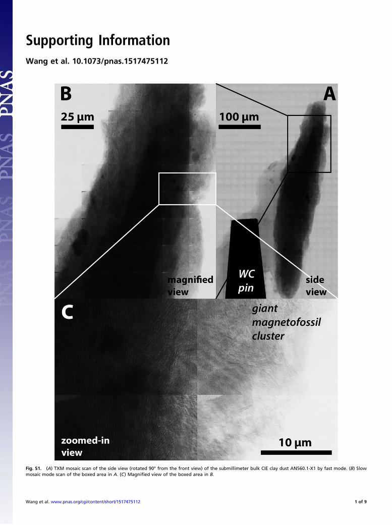
Fig. S1. (A) TXM mosaic scan of the side view (rotated 90° from the front view) of the submillimeter bulk CIE clay dust AN560.1-X1 by fast mode. (B) Slowmosaic mode scan of the boxed area in A. (C) Magnified view of the boxed area in B.
Wang et al. www.pnas.org/cgi/content/short/1517475112 1 of 9

Fig. S2. (A–O) TXM mosaic scans of the front and side view of four extra submillimeter bulk CIE clay dusts from AN560.1 by fast mode with slow mosaic modescans showing the areas in the boxes. Similar iron minerals can be identified as in AN560.1-X1 (Fig. 1 and Fig. S1).
Wang et al. www.pnas.org/cgi/content/short/1517475112 2 of 9

Fig. S3. Three-dimensional TXM tomographic reconstructions viewed from different angles as indicated on each image. Brightness is inverted in 3Dreconstructions with brighter meaning greater X-ray absorption and higher iron concentration. (A–H) Several areas containing detrital iron mineral grains.(I–L) Pyrite framboids. (M–P) A possible magnetofossil cluster of the creature that produced spindle-like magnetite crystals in its original state. Corre-sponding 3D structures viewed from 0° to 360° are shown in Movies S1–S7.
Wang et al. www.pnas.org/cgi/content/short/1517475112 3 of 9

Fig. S4. TXM mosaic scans of cluster-1 (A and B) and cluster-2 (C and D) of magnetically extracted materials from AN560.1.
Wang et al. www.pnas.org/cgi/content/short/1517475112 4 of 9

Fig. S5. TXM mosaic scans of a magnetic extract residue of AN560.1. (A) Side view. (B) Front view.
Fig. S6. (A and B) TXM images of two clusters of freeze-dried cultured MTB strain MV-1.
Wang et al. www.pnas.org/cgi/content/short/1517475112 5 of 9
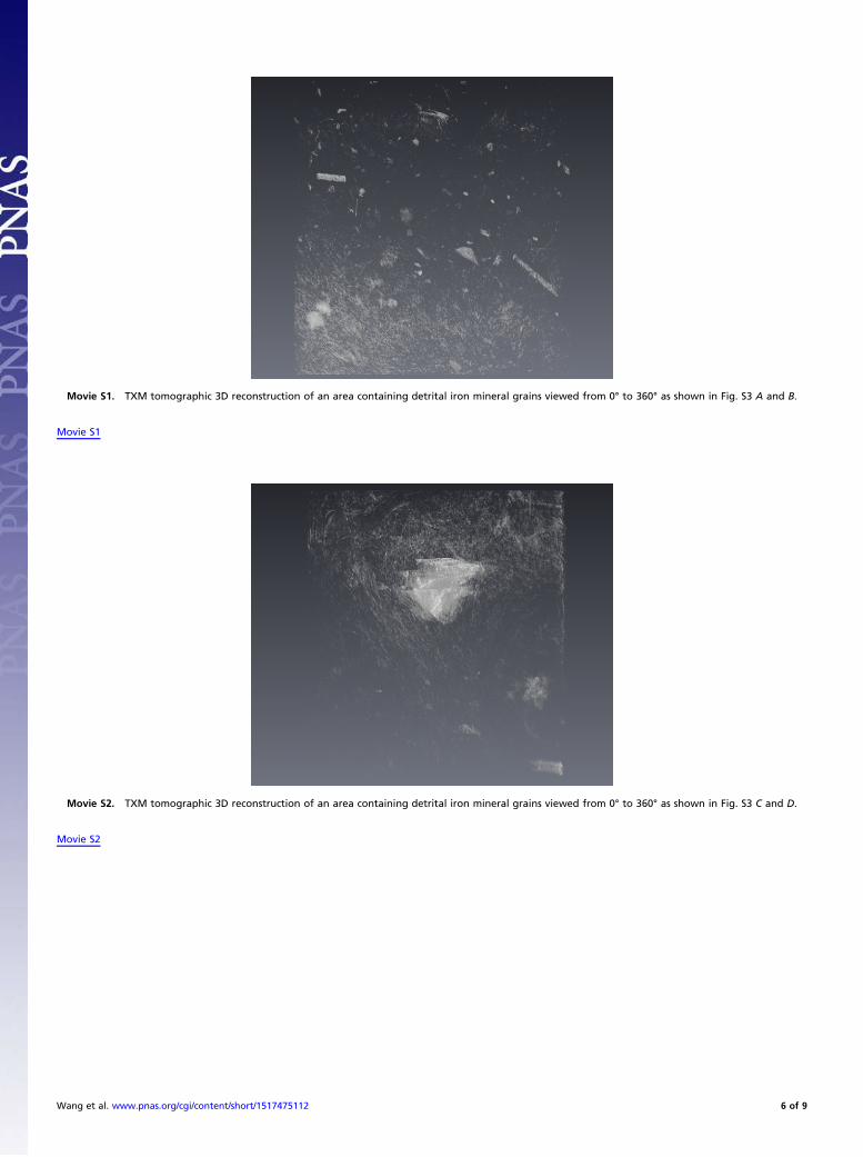
Movie S1. TXM tomographic 3D reconstruction of an area containing detrital iron mineral grains viewed from 0° to 360° as shown in Fig. S3 A and B.
Movie S1
Movie S2. TXM tomographic 3D reconstruction of an area containing detrital iron mineral grains viewed from 0° to 360° as shown in Fig. S3 C and D.
Movie S2
Wang et al. www.pnas.org/cgi/content/short/1517475112 6 of 9

Movie S3. TXM tomographic 3D reconstruction of an area containing detrital iron mineral grains viewed from 0° to 360° as shown in Fig. S3 E and F.
Movie S3
Movie S4. TXM tomographic 3D reconstruction of an area containing detrital iron mineral grains viewed from 0° to 360° as shown in Fig. S3 G and H.
Movie S4
Wang et al. www.pnas.org/cgi/content/short/1517475112 7 of 9

Movie S5. TXM tomographic 3D reconstruction of a pyrite framboid viewed from 0° to 360° as shown in Fig. S3 I and J.
Movie S5
Movie S6. TXM tomographic 3D reconstruction of a pyrite framboid viewed from 0° to 360° as shown in Fig. S3 K and L.
Movie S6
Wang et al. www.pnas.org/cgi/content/short/1517475112 8 of 9

Movie S7. TXM tomographic 3D reconstruction of a possible magnetofossil cluster of the creature that produced spindle-like magnetite crystals in its originalstate viewed from 0° to 360° as shown in Fig. S3 M to P.
Movie S7
Wang et al. www.pnas.org/cgi/content/short/1517475112 9 of 9