Embed Size (px)
Citation preview
Quantifying the energetics of highly conserved water molecules in carbohydrate-binding proteins.
Elisa FaddaComputational Glycoscience Lab, School of Chemistry, NUI Galway
Design of Drugs and Chemicals that Influence Biology, IPAM, UCLA, Apr 4 th- 8th 2011

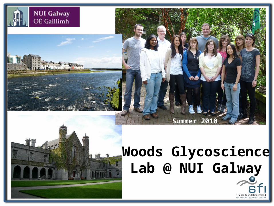
Woods Glycoscience Lab @ NUI Galway
Summer 2010

• Enzyme Re-engineering • Inhibitors (Glycomimetics) Design
“In house” Approach to Glycoscience @ NUIG
Computational Predictions
Biological Assays
Virtual Glycan Array Screening CFG Screening
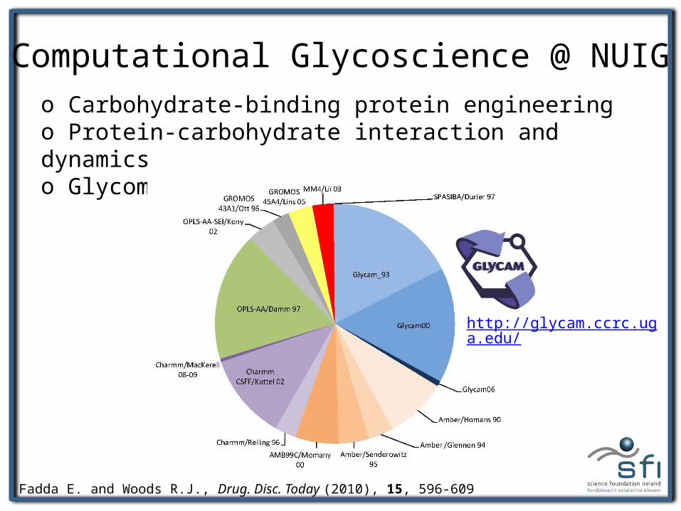
Computational Glycoscience @ NUIGo Carbohydrate-binding protein engineeringo Protein-carbohydrate interaction and dynamicso Glycomimetics
Fadda E. and Woods R.J., Drug. Disc. Today (2010), 15, 596-609
http://glycam.ccrc.uga.edu/
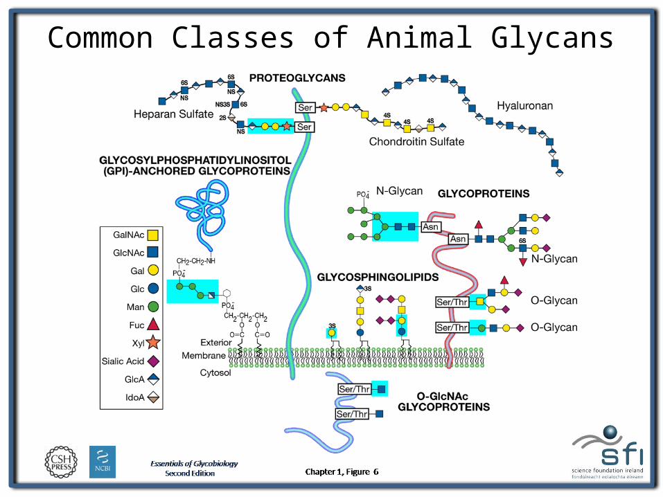
Common Classes of Animal Glycans
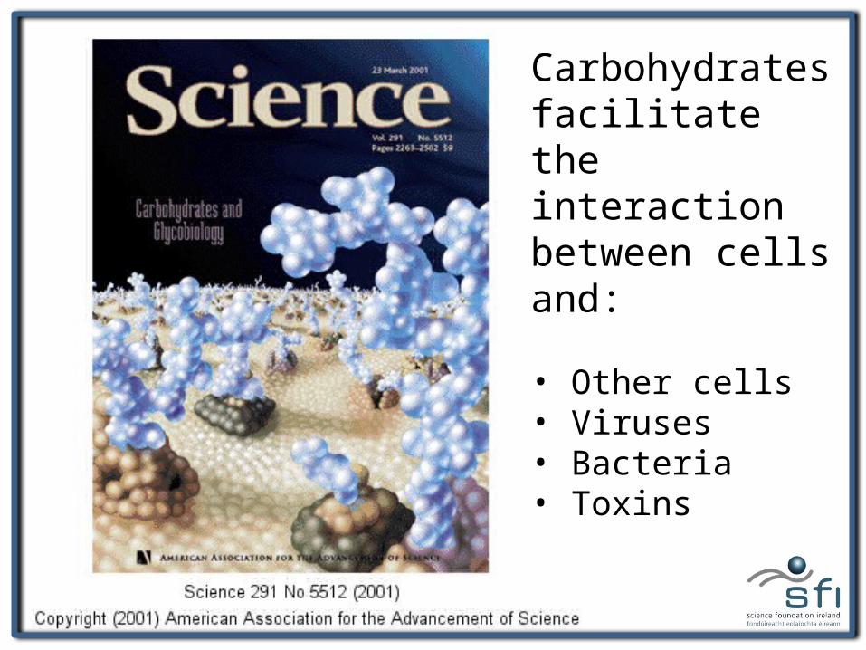
Carbohydrates facilitate the interaction between cells and:
• Other cells• Viruses• Bacteria• Toxins
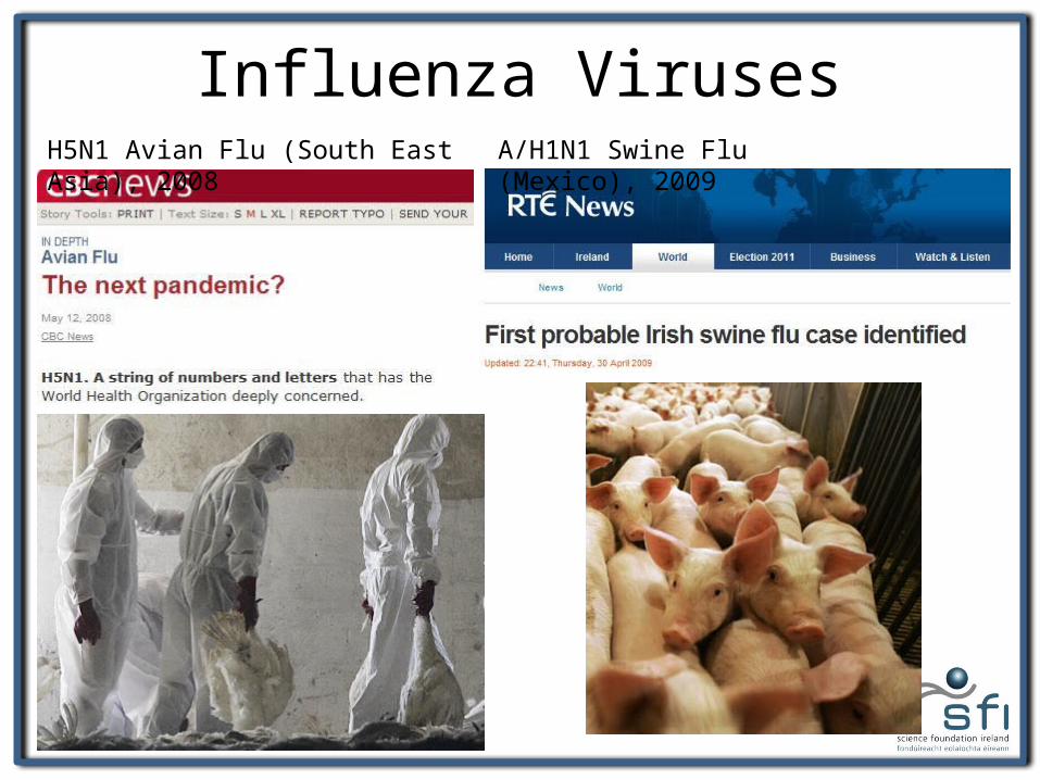
Influenza VirusesH5N1 Avian Flu (South East Asia), 2008 A/H1N1 Swine Flu (Mexico), 2009
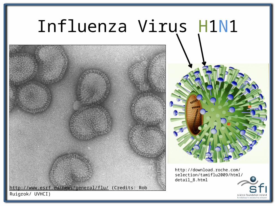
Influenza Virus H1N1
http://www.esrf.eu/news/general/flu/ (Credits: Rob Ruigrok/ UVHCI)
http://download.roche.com/selection/tamiflu2009/html/detail_8.html
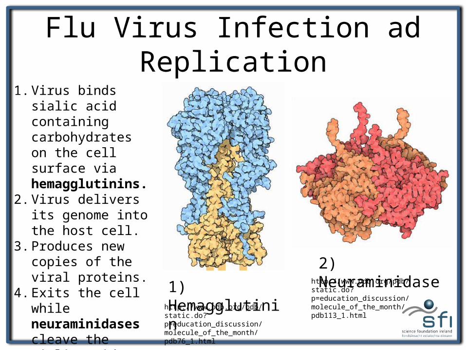
Flu Virus Infection ad Replication
http://www.pdb.org/pdb/static.do?p=education_discussion/molecule_of_the_month/pdb76_1.html
1) Hemagglutinin2) Neuraminidase
http://www.pdb.org/pdb/static.do?p=education_discussion/molecule_of_the_month/pdb113_1.html
1. Virus binds sialic acid containing carbohydrates on the cell surface via hemagglutinins.
2. Virus delivers its genome into the host cell.
3. Produces new copies of the viral proteins.
4. Exits the cell while neuraminidases cleave the sialic acid from the glycans on the cell surface.
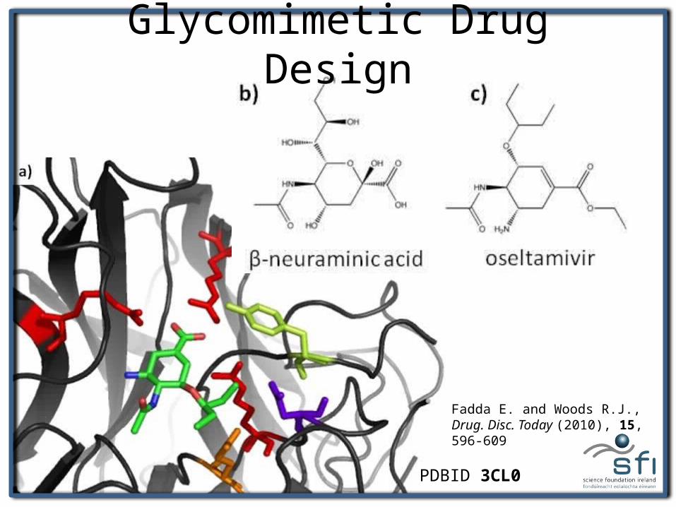
Glycomimetic Drug Design
PDBID 3CL0
Fadda E. and Woods R.J., Drug. Disc. Today (2010), 15, 596-609
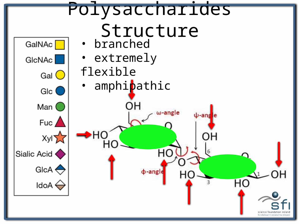
Polysaccharides Structure• branched• extremely flexible• amphipathic
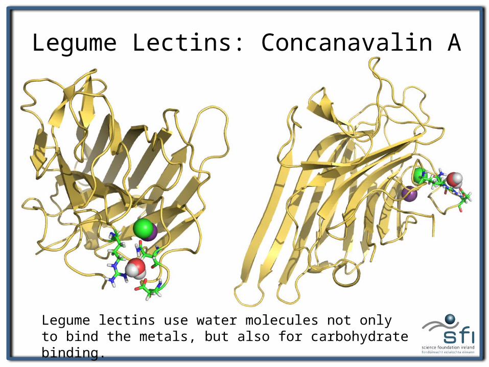
Legume Lectins: Concanavalin A
Legume lectins use water molecules not only to bind the metals, but also for carbohydrate binding.
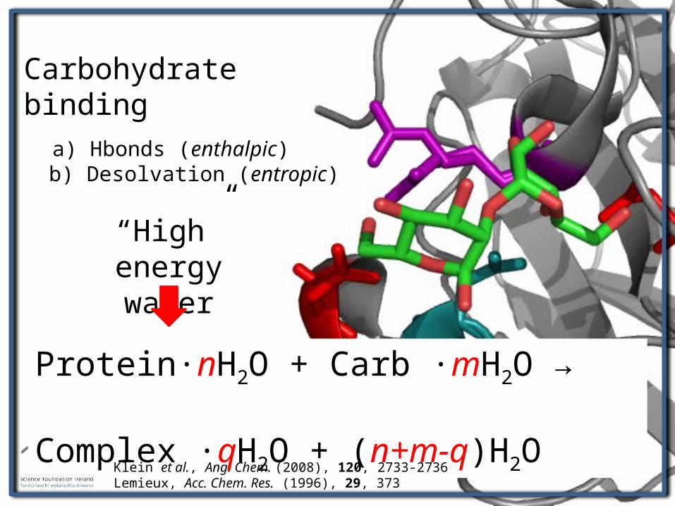
Carbohydrate binding a) Hbonds (enthalpic) b) Desolvation (entropic)
Protein∙nH2O + Carb ∙mH2O → Complex ∙qH2O + (n+m-q)H2O
“High” energy water
Klein et al., Ang. Chem. (2008), 120, 2733-2736Lemieux, Acc. Chem. Res. (1996), 29, 373
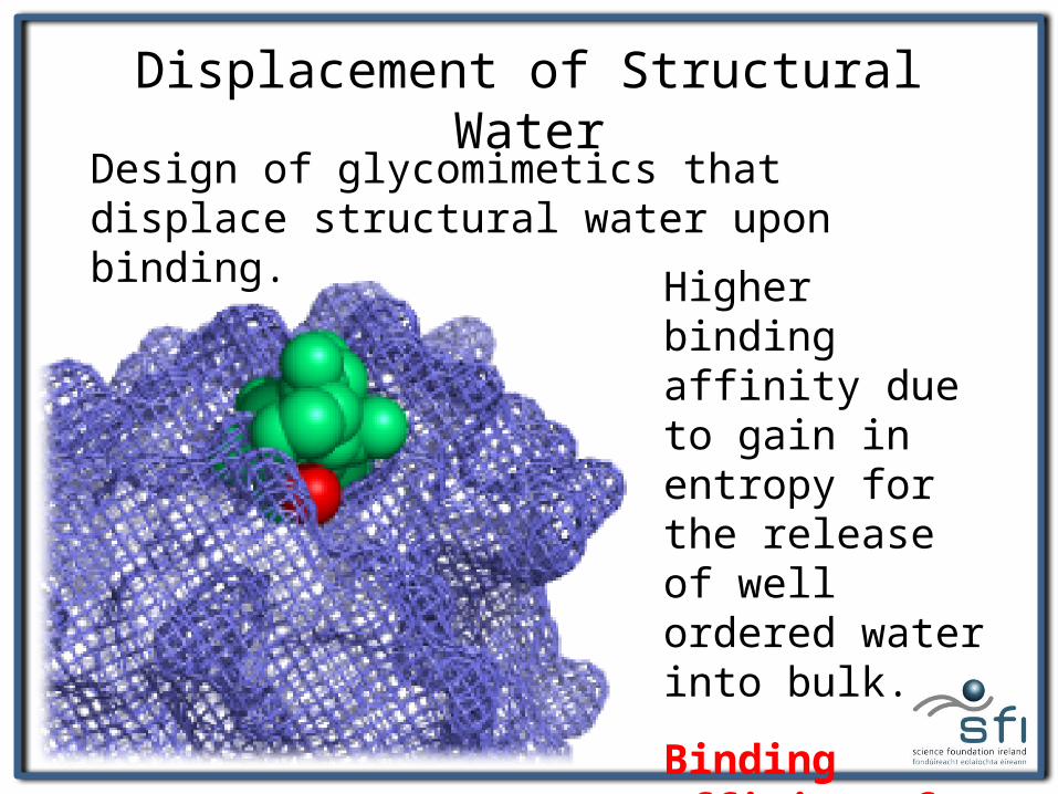
Displacement of Structural WaterDesign of glycomimetics that displace structural water upon binding.
Higher binding affinity due to gain in entropy for the release of well ordered water into bulk.
Binding affinity of structural water.
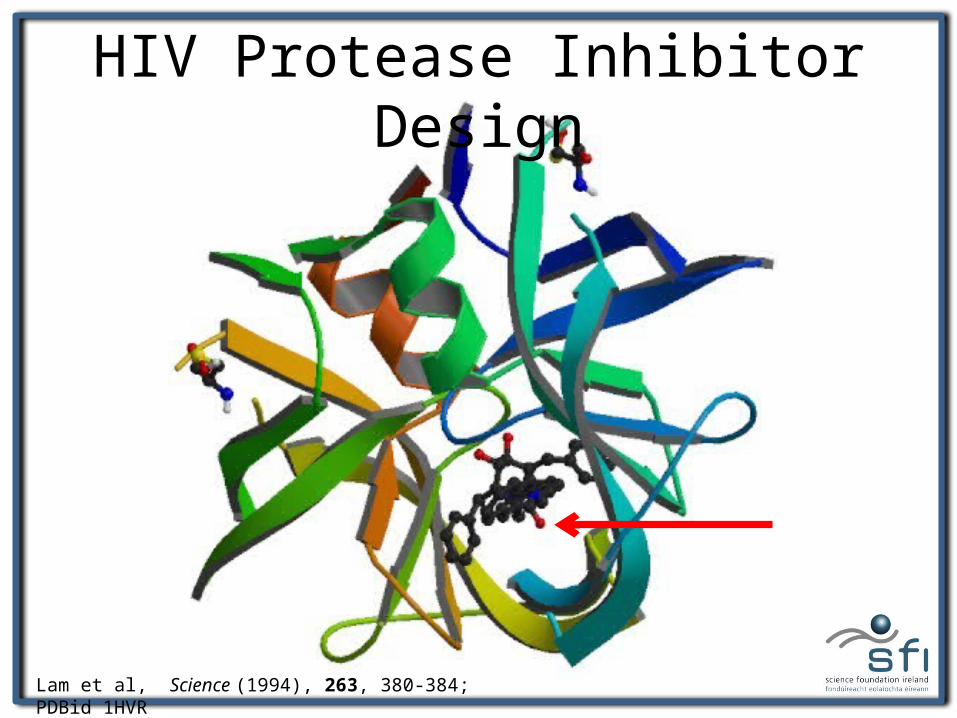
HIV Protease Inhibitor Design
Lam et al, Science (1994), 263, 380-384; PDBid 1HVR
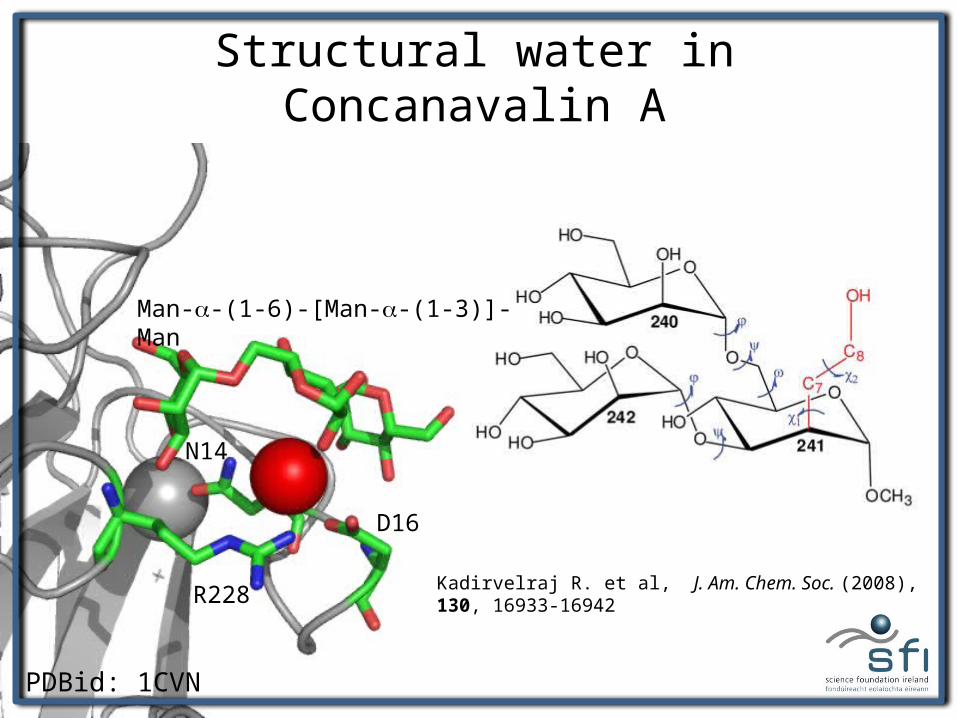
Structural water in Concanavalin A
PDBid: 1CVN
Kadirvelraj R. et al, J. Am. Chem. Soc. (2008), 130, 16933-16942
Man-a-(1-6)-[Man-a-(1-3)]-Man
R228
D16
N14
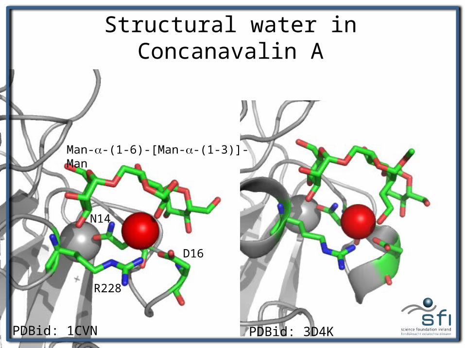
Structural water in Concanavalin A
PDBid: 1CVN
Man-a-(1-6)-[Man-a-(1-3)]-Man
R228
D16
N14
PDBid: 3D4K
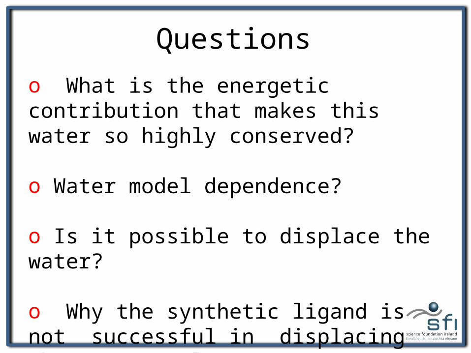
Questionso What is the energetic contribution that makes this water so highly conserved?
o Water model dependence?
o Is it possible to displace the water?
o Why the synthetic ligand is not successful in displacing the structural water?
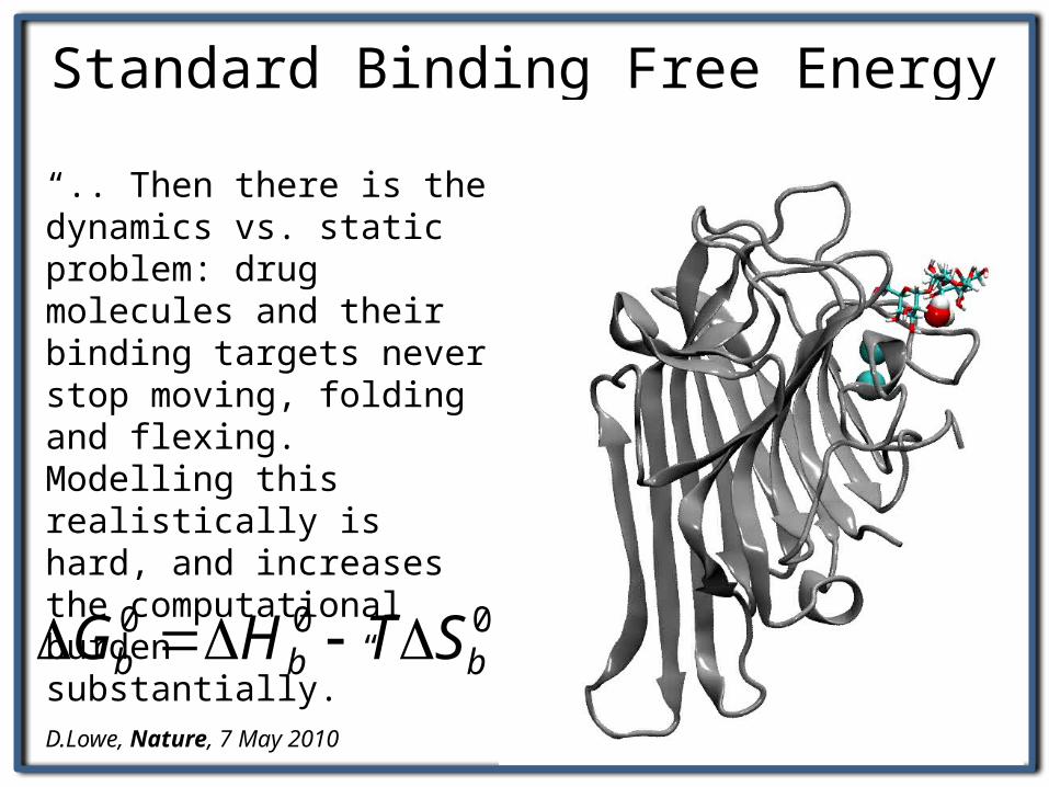
Standard Binding Free Energy
000bbb STHG
“.. Then there is the dynamics vs. static problem: drug molecules and their binding targets never stop moving, folding and flexing. Modelling this realistically is hard, and increases the computational burden substantially.”D.Lowe, Nature, 7 May 2010
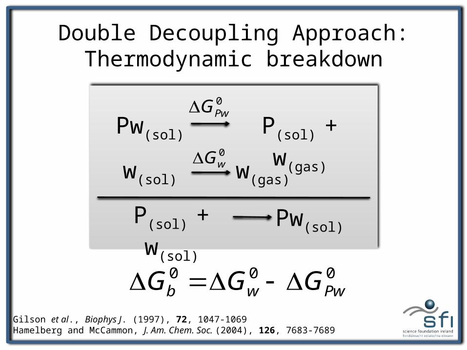
Double Decoupling Approach: Thermodynamic breakdown
Pw(sol) P(sol) + w(gas)
w(sol) w(gas)
P(sol) + w(sol) Pw(sol)
0PwG
0wG
000Pwwb GGG
Gilson et al., Biophys J. (1997), 72, 1047-1069Hamelberg and McCammon, J. Am. Chem. Soc. (2004), 126, 7683-7689
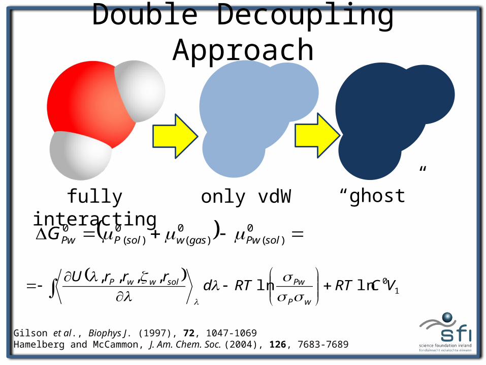
Double Decoupling Approach
Gilson et al., Biophys J. (1997), 72, 1047-1069Hamelberg and McCammon, J. Am. Chem. Soc. (2004), 126, 7683-7689
1
0lnln,,,,
VCRTRTdrrrU
wP
PwsolwwP
0)(
0)(
0)(
0solPwgaswsolPPwG
fully interacting only vdW “ghost”
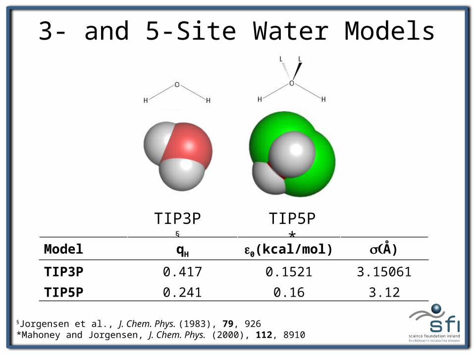
3- and 5-Site Water Models
TIP3P§ TIP5P*
Model qH e0(kcal/mol) (s Å)
TIP3P 0.417 0.1521 3.15061TIP5P 0.241 0.16 3.12
§Jorgensen et al., J. Chem. Phys. (1983), 79, 926*Mahoney and Jorgensen, J. Chem. Phys. (2000), 112, 8910
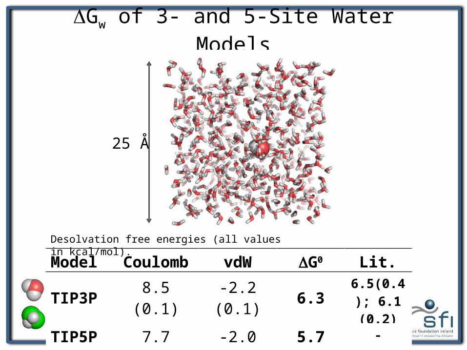
DGw of 3- and 5-Site Water Models
25 Å
Model Coulomb vdW DG0 Lit.
TIP3P 8.5 (0.1) -2.2 (0.1) 6.3 6.5(0.4); 6.1 (0.2)
TIP5P 7.7 (0.1) -2.0 (0.1) 5.7 -
Desolvation free energies (all values in kcal/mol).
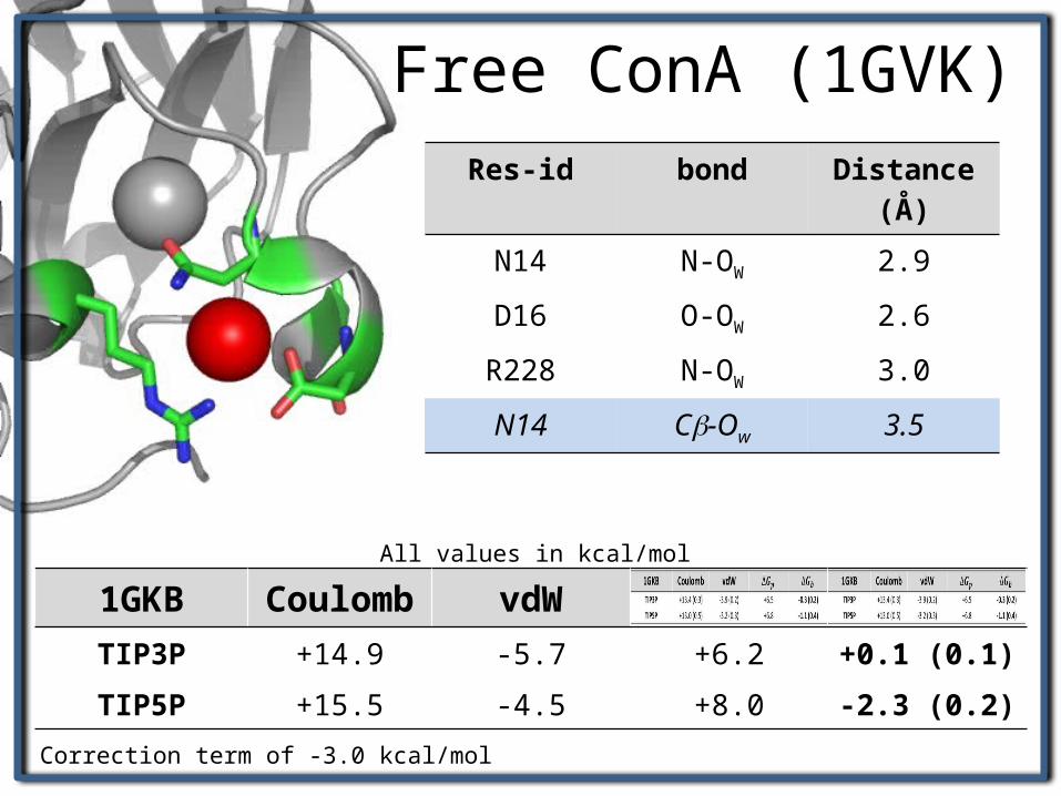
Free ConA (1GVK)
1GKB Coulomb vdWTIP3P +14.9 -5.7 +6.2 +0.1 (0.1)TIP5P +15.5 -4.5 +8.0 -2.3 (0.2)
Res-id bond Distance (Å)N14 N-OW 2.9D16 O-OW 2.6R228 N-OW 3.0N14 Cb-Ow 3.5
All values in kcal/mol
Correction term of -3.0 kcal/mol
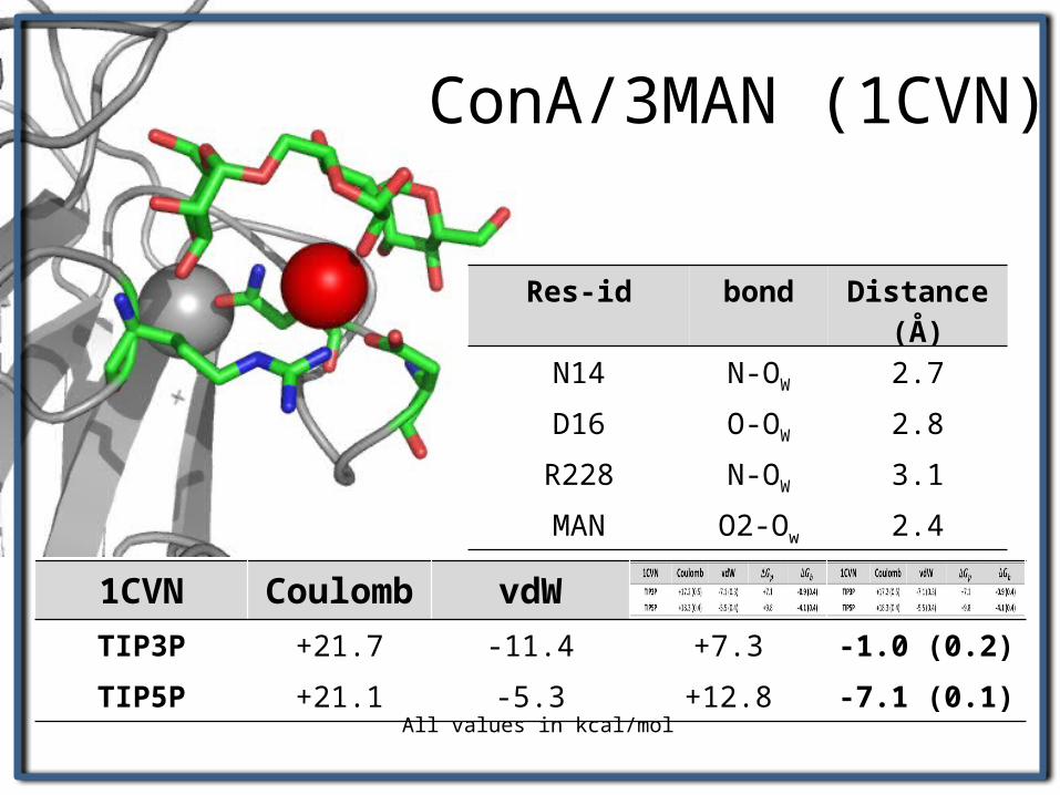
ConA/3MAN (1CVN)
1CVN Coulomb vdWTIP3P +21.7 -11.4 +7.3 -1.0 (0.2)TIP5P +21.1 -5.3 +12.8 -7.1 (0.1)
All values in kcal/mol
Res-id bond Distance (Å)
N14 N-OW 2.7
D16 O-OW 2.8
R228 N-OW 3.1
MAN O2-Ow 2.4
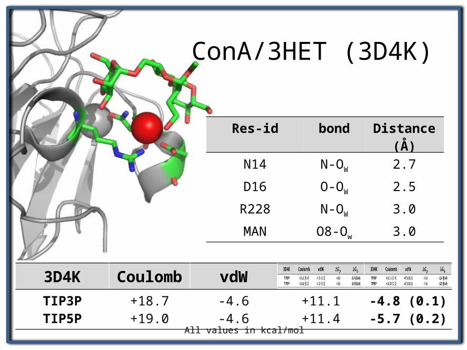
ConA/3HET (3D4K)
3D4K Coulomb vdWTIP3PTIP5P
+18.7+19.0
-4.6-4.6
+11.1+11.4
-4.8 (0.1)-5.7 (0.2)
All values in kcal/mol
Res-id bond Distance (Å)
N14 N-OW 2.7
D16 O-OW 2.5
R228 N-OW 3.0
MAN O8-Ow 3.0
ConA/3HETConA/3MAN
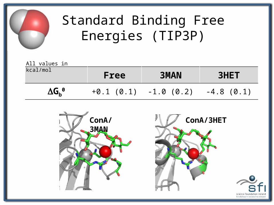
Standard Binding Free Energies (TIP3P)
Free 3MAN 3HET
DGb0 +0.1 (0.1) -1.0 (0.2) -4.8 (0.1)
All values in kcal/mol
ConA/3HETConA/3MAN
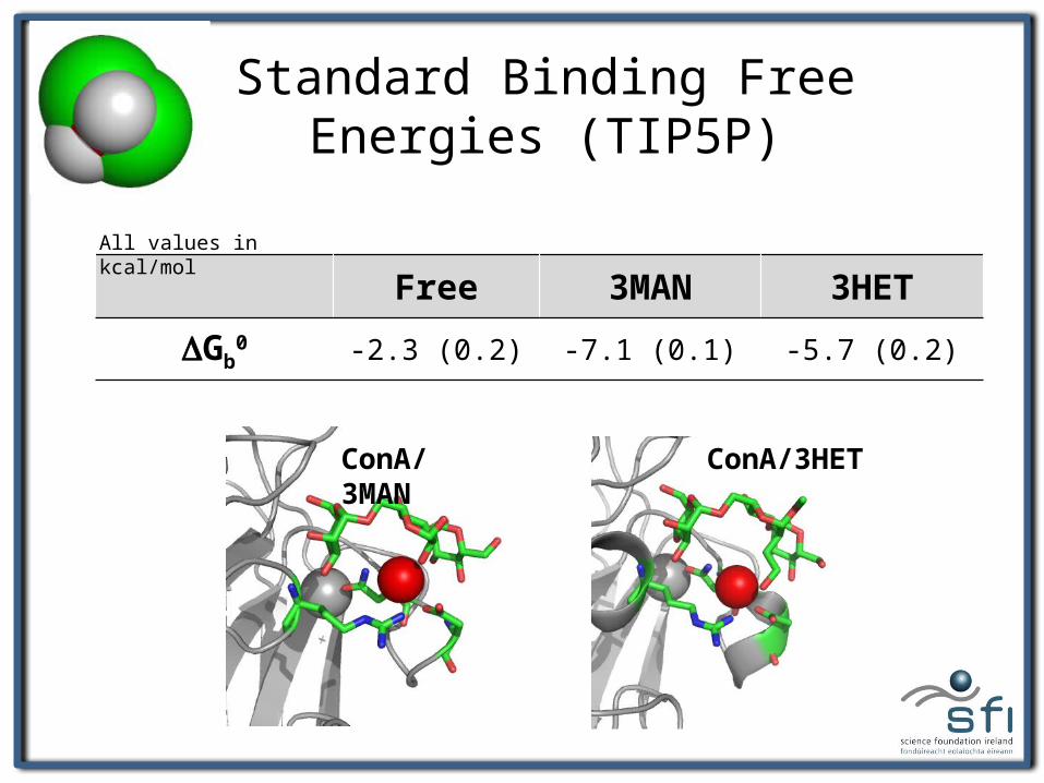
Standard Binding Free Energies (TIP5P)
Free 3MAN 3HET
DGb0 -2.3 (0.2) -7.1 (0.1) -5.7 (0.2)
All values in kcal/mol
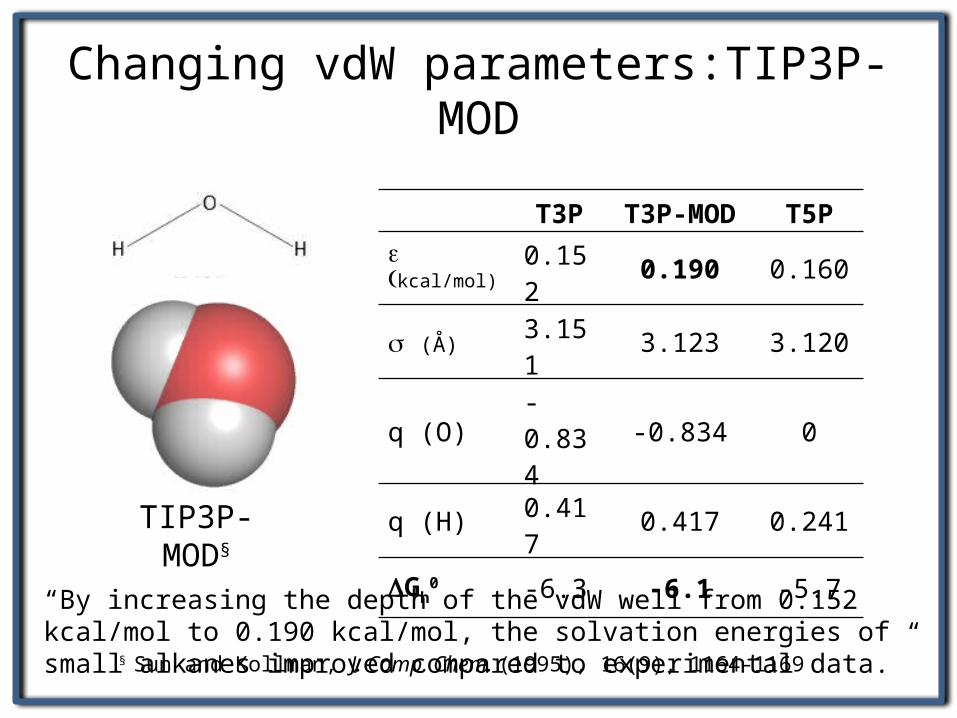
Changing vdW parameters:TIP3P-MOD
TIP3P-MOD§
§ Sun and Kollman, J. Comp. Chem. (1995), 16(9), 1164-1169
T3P T3P-MOD T5P
e (kcal/mol) 0.152 0.190 0.160
s (Å) 3.151 3.123 3.120
q (O) -0.834 -0.834 0
q (H) 0.417 0.417 0.241
DGh0 -6.3 -6.1 -5.7
“By increasing the depth of the vdW well from 0.152 kcal/mol to 0.190 kcal/mol, the solvation energies of small alkanes improved compared to experimental data.”
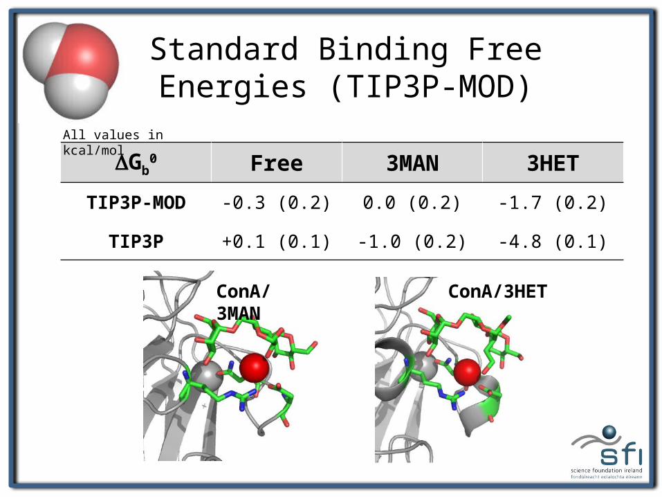
ConA/3HETConA/3MAN
Standard Binding Free Energies (TIP3P-MOD)
DGb0 Free 3MAN 3HET
TIP3P-MOD -0.3 (0.2) 0.0 (0.2) -1.7 (0.2)
TIP3P +0.1 (0.1) -1.0 (0.2) -4.8 (0.1)
All values in kcal/mol
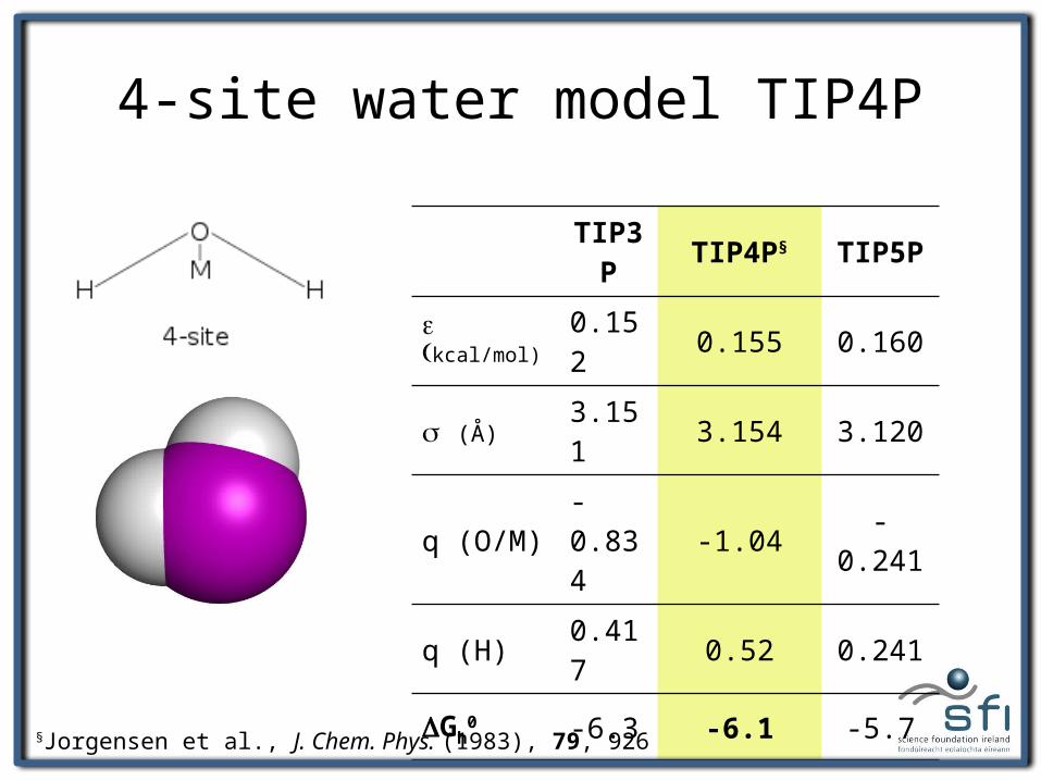
4-site water model TIP4P
TIP3P TIP4P§ TIP5P
e (kcal/mol) 0.152 0.155 0.160
s (Å) 3.151 3.154 3.120
q (O/M) -0.834 -1.04 -0.241
q (H) 0.417 0.52 0.241
DGh0 -6.3 -6.1 -5.7
§Jorgensen et al., J. Chem. Phys. (1983), 79, 926
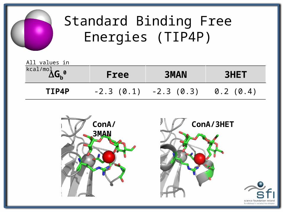
ConA/3HETConA/3MAN
Standard Binding Free Energies (TIP4P)
DGb0 Free 3MAN 3HET
TIP4P -2.3 (0.1) -2.3 (0.3) 0.2 (0.4)
All values in kcal/mol
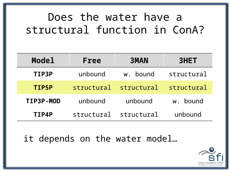
Does the water have a structural function in ConA?
Model Free 3MAN 3HET
TIP3P unbound w. bound structural
TIP5P structural structural structural
TIP3P-MOD unbound unbound w. bound
TIP4P structural structural unbound
it depends on the water model…
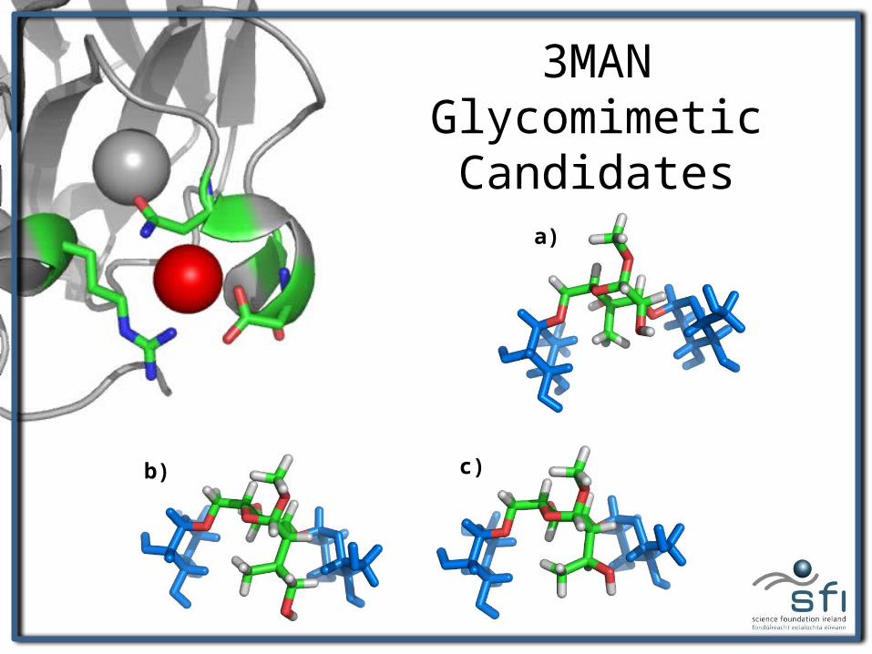
a)
b) c)
a)
3MAN Glycomimetic Candidates
Conclusions• The choice of water model has a significant impact on the
assessment and interpretation of standard binding free energies.
• Within the context of non-polarizable force fields, TIP5P 5-site model seems to be a step in the right direction.
• The water is not displaced by the synthetic ligand because it is able to preserve its tetrahedral coordination.
• A bulkier synthetic ligand (e.g. hydroxypropyl) might be able to form favourable vdW contacts with N14 Cb, with the OH replacing the water in the binding site.
AcknowledgementsProf. Rob WoodsOliver GrantJoanne Martin Hannah Smith Niall Walshe
Dr. Nina WeisserDr. Lori YangDr. Jen Hendel Dr. Marleen RendersValerie Murphy
@ Sickkids:Dr. Régis PomèsChris Neale